Function of CD44(Pgp-1) homing receptor
in human T cell precursors
Antonio de la Hera, Agustin Acevedo1, Wendy Marston, and
Francisco Sanchez-Madrid2
The Basel Institute for Immunology, Basel, Switzerland
Departments of 1Pathology and immunology, Hospital de la Princesa, Madrid, Spain
Key words: CD34+ stem cells, gp90 homing receptors, lymphocyte migration, T cell development,
thymic stroma
Abstract
T cell precursors migrate from extrathymlc hematopoietic tissues and differentiate after
encountering the thymic mlcroenvlronment. We asked whether human T cell precursors express the CD44(Pgp-1/gp90HR) class of homing receptors that have been implicated in the traffic of hematopoietic cells, such as lymphocyte entry to peripheral lymphoid organs. Flow cytometry and immunopreclpltatlon studies demonstrate that CD7+34+, CD1 - 2 - 3 - 4 - 8 - 1 4 - 1 6 - 2 0 - cells In bone marrow and thymus, which have been shown to exhibit features of T cell precursors, bear CD44. Immunohlstologlcal studies show that clusters of thymocytes in the subcapsular and the Inner cortex and most medullary thymocytes are clearly CD44 + , whereas the expression of CD44 is selectively downregulated in CD3- and CD3low functionally Incompetent cortical thymocytes.
The expression of CD44 Is not restricted to T cell precursors but also occurs In thymic stroma, which bear a different molecular species of CD44. CD44-specific antibodies exert stimulatory effects on T cell precursors, a process that is dependent on stromal cells. We postulate that CD44 might be an adhesion molecule for precursor homing to thymus and that it participates In cell-to-cell interactions within the thymic environment.
Introduction
T cell development occurs within a defined microenvironment, the thymus Prethymic T cell progenitors develop in the fetal liver or postnatal bone marrow, circulate through the blood, and eventually home into the thymus A few functionally competent thymocytes ( — 1%) emigrate to the periphery and recirculate through peripheral lymphoid organs (reviewed in refs 1 - 3 ) .
Two families of gp90 homing receptors (hereafter referred to as gp90HRs) have been described (4-7). gp90HRKa"T«> is indistinguishable from CD44(Pgp-1) antigen ( 8 - 1 6 ; this report) Although human gp90HRHerm8S shares MEL-14 antibody epitope with mouse gp90HRMEL14 they are structurally distinct (7,17-20). Both gp90HRs appear to mediate specific binding for endothelium and have therefore been implicated in T and B lymphocyte traffic (6,7) It is generally assumed that gp90HRs are acquired during B and T cell development in bone marrow and thymus respectively (6), and that CD44 in normal human thymus is associated with thymocyte functional maturation and location in the medulla (14)
In spite of this knowledge, very little is known about the role
of such receptors (and their ligands) in T cell precursor migration, commitment to T cell lineage, and intrathymic cell traffic. Here we entertain the hypothesis that acquisition of the gp90HR, as recognized by CD44 antibodies, precedes T cell differentiation in the thymus, and might play a role in early T cell development. We show that CD44 monoclonal antibodies (mAbs) bind to isolated pre- and intrathymic T cell precursors as well as thymic stromal cells, that CD44 is differentially expressed by mature and immature thymocytes, and that CD44 mAbs may exert stimulatory effects on T cell precursor and mature T cells.
Methods
Isolation of cells
Thymic lobes were obtained during corrective surgery and single cell suspensions were prepared. Bone marrow cells were from donor samples remaining after bone marrow transplantation Pathology studies showed that all tissues used were normal
Correspondence to Antonio de la Hera, Basel Institute for Immunology, Grenzacherstrasse 487, Basel 4005, Switzerland
Peripheral blood was from Kantonspital (Basel) Wood bank donors.
Thymocytes and bone marrow cells expressing CD1, CD2, CD3, CD4, or CD8 T cell differentiation antigens, CD14 + myeloid cells, CD16+ natural killer cells, and CD20-bearing B cells were depleted by incubation with cytotoxic mAbs and rabbit complement, as described elsewhere (21). Viable cells not coated with mAbs were isolated, after incubation with M-450 magnetic beads armed with anti-mouse Ig antibodies, by Ficoll-Hypaque density centrifugation. The CD7 + cells were further separated from non-T lineage hematopoietic cells and stromal components using M-450 beads coupled with anti-CD7 mAbs and a sintered magnetic alloy. Cells were detached from the beads by incubation at 37°C. We followed the manufacturer's recommendation throughout the method. FACS analysis showed that 98% of the selected cells had the CD7 + , C D 1 2 3 -4 - 8 - 1 -4 - 1 6 - 2 0 - phenotype. Double-negative thymocytes were prepared by incubation with CD4 and CD8 mAbs and complement The CD7-depleted population of thymocytes is a population highly enriched in thymic stromal cells, as determined with TE3A and TE4 mAbs specific for thymic epithelia. Thymic stroma was cultured in serum-free media supplemented with epidermal growth factor for 3 weeks to prevent the outgrowth of fibroblasts. After this time the cells (TE3/4 + 7-) were maintained in complete medium supplemented with 10% serum
Homing receptors in T cell precursors 599 Monoclonal antibodies
When possible, we will use the cluster of differentiation (CD) nomenclature proposed at the Oxford Workshop (8). CD1, Na1/34 (22); CD2, Leu-5 (Becton-Dickinson, Mountain View, CA); CD3, OKT3 (23; American Type Culture Collection, ATCC, Bethesda, MA); CD4, HP2/6 (24); CD7, 3A1 (ATCC), and RFT2 (25); CD8, B9.4 (26); CD14, CoulterCloneMo2 (Coulter Elec-tronics, Hialeath, FL); CD16, Leu11b; CD19, CoulterCloneB4; CD20, CoulterCloneBI, and BC-1 (8); CD34, HPCA (Becton-Dickinson). OKT9 (transfernn receptor, Ortho Pharmaceutical Co., Rantan, NJ), W6/32, TE3A, TE4, and TE7 (Serotec, Bicester, UK); Ki67 (27); and MEL-14 (5; ATCC).
The CD44 mAbs F-10-44-2 (9), 50B4 (10), and (Pgp-1) I42/5 (13) were generously provided by the authors (see Acknowledgements); A3D8 (15) was purchased from Serotec. The hybridoma clone HP2/9 (24) was generated by the fusion of P3X63 myeloma cells with spleen cells obtained from a mouse immunized with JM thymoma line (32). It produces an IgGi mAb. Because the nomenclature for the CD44 mAbs is quite fragmentary in the literature we shall briefly review it here. CD44 was defined by the 'cluster' of mAbs F-10-44-2, J-173, and 106-4D5 in the Third Workshop on Leukocyte Differentiation Antigens (8), but several other mAbs, such as those used above, can be formally considered as CD44-specific based on the results of Letarte (10). This author established that the gp85 molecular
Q O 0 O O
b
1 • i 1 ' ' • \ iA
CO QO
CD44
S
QO
o6
o
Q5
O
CO QO
o
O
CD44
" i i"Control
Log
)0Green Fluorescence
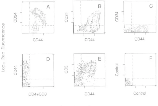
Fig. 1. CD44 is expressed in early T cell precursors and down-modulated during intrathymic development. Early T cell precursors from bone marrow (A), thymus (B), thymic epithelial cells (C), double-negative thymocytes (D), and total thymocytes (E) were isolated as described in Methods and stained with antibodies specific for the indicated antigens for two-color flow cytometry analyses. Antibodies were CD34 (HPCA, 7,) and CD44 (50B4,
y2) m the top panels, and CD44 (HP2/9, 7,) and either CD4+CD8 (HP2/6 + B9.4, y2) or CD3 (OKT3, y2) m t h e bottom panels. Samples were
species exhibit at least three epitopes defined by the prototype mAbs F-10-44-2/A3D8/etc, 50B4/etc , and A1G3. Therefore, the brain-granulocyte T lymphocyte (9), p80 on mature thymocyte (14), and In(Lu) on erythrocyte (15) molecules all are recognized by CD44-specific mAbs Moreover, anti-Pgp-1 mAbs have been independently shown to recognize the CD44 antigen (11,12). Also, in spite of their apparent relationship (9), CD44 and sialophorin are different molecular entities, the latter now termed CD43 (8,28)
Immunohistochemical staining of lympoid organs
Acetone-fixed, 4-/tm-thick sections were sequentially incubated with mAb culture supernatants and peroxidase-conjugated rabbit anti-mouse Ig (Dakopatts, Copenhagen, Denmark); each incubation was followed by three washes. The reaction was
1 2 3456789
A
T
1 2 3 4 5 6 7 8 9 0 A B C D
B
Fig. 2. Immunoprecipitation of CD44(Pgp-1/gp90HR) antigens from
early thymocytes. Human early thymocytes, isolated from a pediatnc thymus (2.5 years old) as described in Methods (21), were 125I
surface-labeled and the lysates immunoprecipitated with the following mAbs (A) Lane 1, CD2 + CD3 + CD4 + CD8 T cell mAbs), lane 2, CD20 (anti-Bcell mAb); lane 3, CD14 + CD16 (anti-myelcHd/NK cell mAbs); lane 4, CD7 (early thymocyte); lane 5,142/5 (anti-Pgp-1); lane 6, F-10-44-2 (COM); lane 7, 50B4 (CD44); lane 8, HP2/9, and lane 9, MEL-14 (gp90HR). (B) Lane 1, HP2/9 precipitate after preclearing/immunodepletion of the lysate with F-10-44-2; lane 2, HP2/9 undepleted; lane 3, CD7 undeleted; lane 4, MEL-14 precleared with HP2/9, lane 5, MEL-14 precleared with F-10-44-2, lane 6, CD7 precleared with HP2/9; lane 7, MEL-14 undepleted; lane 8, MEL-14 precleared with CD7, lane 9, MEL-14 precleared with anti-Pgp-1; lane 0, anti-Pgp-1 undepleted, lane A, F-10-44-2 undepleted; lane B, 50B4 undepleted; lane C, F-10-44-2 precleared with CD7; and lane D, F-10-44-2 precleared with HP2/9 Samples were subjected to electrophoresis and autoradiography for 16 and 12 days respectively
developed with Graham - Karnovsky medium containing 0.5 mg/ ml of 3,3-diaminobenzimidine tetrahydrochlonde (DAB) and hydrogen peroxide (29). Sections were mounted in buffered gelatin for microscopic examination.
Quantitative flow cytometry
The procedure for indirect immunofluorescence surface staining of cells has been described (21) For two-color analyses, IgG-y, and lgG72 antibodies were used and labeled with either fluorescem isothiocyanate (FITC, green) or phycoerythnn-conjugated (PE, orange-red), second-layer, isotype-specific antibodies (Southern Biotechnology, Birmingham, AL). Quantitation of the staining of 10" (one-color) or 5 x 10⁴ (two-color) viable cells was performed with either a Coulter EPICS-C cell-sorter or a FACscan instrument, as detailed elsewhere (21).
Immunoprecipitation and electrophoresis
Cells were radioiodinated with chloroglycoluril (lodogen, Pierce, Rockford, IL), and lysed in phosphate-buffered saline, pH 7.4, containing 1 % Triton X-100, 1 % hemoglobin, and 1 mM phenyl-methylsulfonylfluonde. For immunoprecipitation, 125Habeled cell lysates were incubated with 5 ^g of the indicated mAbs followed by rabbit anti-mouse or anti-rat Igs (Jackson Labs, ME) coupled to protein A-Sepharose (1 mg/ml). For immuno-depletion, immunoprecipitation with the preclearing antibody was repeated twice before precipitation with the relevant antibody. Immunoprecipitates were processed and samples subjected to SDS-7-15% PAGE and autoradiography with enhancing screens, as described elsewhere (29).
Cell cultures
Cultures were maintained in RPM11640 medium supplemented with 2 mM glutamine, 10 mM HEPES, and 10% human serum (complement-depleted and pooled male AB donor), hereafter referred to as complete medium. For proliferative assays, 1.6 x 10s cells/well were cultured in 96-well microtiter plates in
1 2 3 4 5 6 7
Fig. 3. Thymic epithelial cells express a different class of CD44
molecules Peripheral Wood mononudear cells (1,3), lymph node cells (4), thymocytes (5), and thymic epithelial cells (2,6,7) were 12SI
surface-labeled and the lysates precipitated with lane 1, 50B4, lane 2, 50B4 precleared with F-10-44-2, lanes 3, 4, 5, 7, F-10-44-2, and lane 6, F-10-44-2 precleared with 50B4 Samples were subjected to electrophoresis and autoradiography for 14 days. Filled arrowheads indicate the 'common' p85 band, open arrowheads show the multiple high-molecular-weight bands unique to thymic epithelial in the CD44 antibody precipitates from different sources
Homing receptors in T cell precursors 601
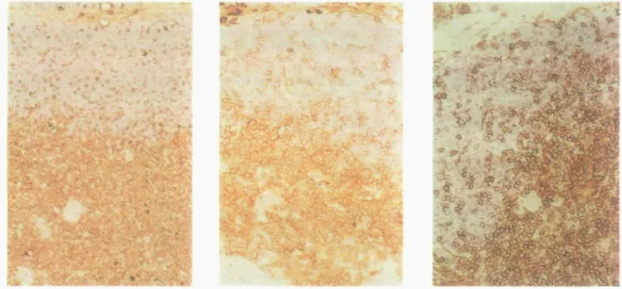
Fig. 4. Immunohistochemical staining shows binding of HP2/9 mAb to clusters of cortical thymocytes, most medullary thymocytes and thymtc stromal
components. Pediatric thymus sections were incubated with HP2/9 mAb and the staining was revealed by a DAB-peroxidase reaction as a brown precipitate, as described in Methods (29) Note that the thymic medulla is placed at the bottom while the outer cortex and the capsula are at the top in each panel The results are indicated with three different thymus sections at increasing magnification from left to right.
complete medium supplemented with antibodies, 10ng/ml phorbol mynstate acetate (PMA), an ionophore (A23187,1 /tM), or phytohemagglutimn (PHA, 1 /*g/ml), in the indicated combinations The mAbs were directly bound to the plastic surface of the wells (1 ng, 500 ng, 100 ng, and 10 ng/well), as descnbed elsewhere (30). In some experiments early thymocytes were supplemented with autologous thymic stroma at a 1 80 ratio of thymic stroma to early thymocytes. Fresh stroma cells irradiated (2500 rad) were used in those complementation experiments. Limiting dilution analyses to determine the frequency of proliferating cells were performed after stimulation with PHA (1 ftg/ml) in the presence of 50 U/ml of mterleukin-2 (IL2) and irradiated filler cells, as described by Lopez-Botet and Moretta (31). For IL2 production, 2 x 10⁵ cells were cultured in the presence of PMA plus ionophore for 24 h. Supernatants were assayed for IL2 activity as described (30), using the IL2-dependent mouse CTLL2 cells. Human rll_2 (Hoffmann-LaRoche, Basel) was used as an internal control.
Results
CD44(Pgp- 1/gp90HR) is expressed by early T cell precursors and thymic stroma
The expression of CD44 on T cell precursors was assessed by using immunofluorescence and flow cytometry Normally, there are low numbers of T cell precursors in fetal liver, adult bone marrow, or the pediatric thymus (1,2). Early T cell precursors in humans have been characterized by both their unique phenotype (CD7 + CD45 + , CD1 - 2 - 3 - 4 - 8 - ) (33,34) and by their capacity to differentiate into either a/S or yd TCR/CD3+ mature T cells in the presence of thymus stroma cells and IL2 in vitro (21,35). We isolated CD7 + , C D 1 - 2 - 3 - 4 - 8 - 1 4 - 1 6 - 2 0 - cells from bone
Table 1 . COM - thymocytes are functionally unresponsive cells
Frequency of proliferating cells IL2 production (U/ml) Peripheral blood mononuclear cells Thymocytes Unseparated CD44h|oh QP44intermec*ale CD44-1/1 47 1/18 73 1/4.15 1/40 82 <1/150 21 3 12 1 <0.1
The indicated cell populations were analyzed for the frequency of proliferating cells in limiting dilution assays in the presence of optimal amounts of PHA, IL2, and irradiated peripheral blood mononuclear cells (31). The IL2 production was measured in 24 h supernatants, after stimulation with PMA plus an ionophore, using an IL2-dependent mouse line (30) Thymocytes were separated into CD44(H2/9) high (the same level as peripheral Wood lymphocytes), negative, and intermediate subsets using a fluorescence-activated cell sorter (31). Since the distinction between high and intermediate levels of CD44 expression is arbitrary (41), we cannot determine whether the few proliferating cells in the CD44W e r m e*"e isolate were actually CD44k)w cells or whether they
represent contaminating CD44hist1 cells
marrow and thymus and found that most of these cells express the CD44 molecule (Fig. 1A and B). Moreover, a sizeable fraction of them (113 in thymus and 2/3 in bone marrow) co-express CD34, a putative marker for hematopoietic stem cells (36; Fig. 1A and B). We further explored the expression of the CD44(Pgp-1/ gp90HR) antigens in early thymocytes using sequential immunoprecipitation studies with a panel of mAb that recognized four different distinct epitopes in the human CD44(Pgp-1/ gp90HR) (5,10,13). As shown in Fig. 2(A), the material immunoprecipitated by all these mAb from lysates of
Table 2. CD44/gp90HR transduce co-mitogenic activation signals
Peripheral Wood mononudear cells Early thymocytes Proliferation (cpm x PMA 6 1 PMA ION 174 148 10-3/mm) PMA CD7 163 96 induced by PMA CD44 92 9 PMA CD44 + AC 96 79 PMA Pgp-1 + AC 88 62 PMA + AC 6 13 CD44 + AC 4 11 PMA HLA-A.B + AC 3 5
1 6 x 10s cells were cultured in the presence of PMA, lonophore, solid-phase bound antibodies, and irradiated thymic accessory cells (AC) in
the indicated combinations F-10-44-2 (COM), I42/5 (CD44/Pgp-1), 3A1 (CD7), and HLA-A.B (W6/32) antibodies were at 500 ng/well, PMA at 10 ng/ml, lONophore at 1 pM A23187, and the irradiated autotogous thyme stroma at 2000 AC cells/well, which were found to be optimal in preliminary experiments (30; unpublished results). Proliferation was measured after 3 days by adding 1 /iCi [methyl-3H]thymidine (Radiochemical Centre, Arnersham) per
well for the last 12 h of culture Results represent the mean [3H]thymidine uptake in triplicate cultures of an experiment representative of three.
surface-labeled human early thymocytes migrated on SDS-PAGE with a near-idnetical mobility at —85 kd Also, CD7 mAb but not mAbs specific for T-, B-, and myeloid-differentiation antigens immunoprecipitated labeled material at —40 kd, consistent with the early thymocyte phenotype of the population used (21,33,34) We confirmed that HP2/9 (32) is a CD44 mAb as shown by reciprocal precleanng in immunodepletion experiments Moreover, HP2/9 mAb specifically immunodepletes the precipitates of the prototype gp90HR mAb, MEL-14, as do CD44 and Pgp-1 mAbs (Fig. 2B) These results are consistent with the recently established co-identity of CD44, gp90HRHermes, and Pgp-1 (11,12,16). We conclude that human early thymocytes express CD44(Pgp-1/gp90HR) homing receptors as assessed by both quantitative flow cytometry and biochemical methods. Interestingly, we found that thymic stromal cells express CD44 Freshly isolated thymic stromal cells were CD44+ but lacked the CD34 molecule (Fig. 1C). These cells remained CD44+ in culture, which allowed us to characterize biochemically the CD44 molecule they expressed. As shown in Fig. 3, the nature of the CD44 molecules expressed on thymocytes, tonsil lymphocytes, and peripheral blood mononudear cells was roughly identical, but strikingly different from CD44 expressed on thymic stroma. In the latter cells, additional high-molecular-weight bands besides the 90 kd species found on T cell precursors are apparent. All these bands were immunodepleted by two CD44 mAb specific for different epitopes (10), indicating that either the additional bands carry the two CD44 determinants, or they are non-covalently associated to the 8 0 - 9 0 kd band.
CD44 expression is downregulated during intrathymic development in functionally incompetent cells
The level of CD44 surface expression is regulated during thymic ontogeny. For instance, only a portion of the C D 4 8 -'double-negative' thymocytes express CD44 (Fig. 1D). Based on expression of CD3 and CD44, four major subsets of thymocytes were evident: CD3-CD44-, CD3**CD44-, CD3)°1'€D44'°«, and CD3f«ohCD44h>otl (Fig. 1E). Minor subsets of CD3-CD44+ and CD3lowCD44hlBh were also detected, which comprise some double-negative thymocytes and the T cell precursors (Fig. 1A and B; see above). We also studied reactivity of CD44 mAb on tissue sections of thymus using immunohistochemical staining Most cortical thymocytes are unstained (Fig. 4, left panel). Most medullary thymocytes as well as some scattered thymocytes in the subcapsular and inner cortex are positive in thymus sections
The CD44t»oti cortical thymocytes are aggregated in several independent clusters of 2 - 9 cells (Fig. 4, right panel).
The fact that CD44- thymocytes are localized in the cortex and lack CD3 suggested that they could be immature thymocytes. By using limiting dilution analyses of the frequency of proliferating cells, and measuring the production of IL2 in bulk cultures, we found that CD44- thymocytes were indeed inert cells while the CD44hiah population contained functional thymocytes. The CD44inl9rm«lia>e population contained some fewer functional cells (Table 1).
CD44 could participate in T cell precursor activation
We have studied whether CD44 might be an immunoadhesin involved in the activation of T lymphocytes CD44 mAbs by themselves were not mitogenic for peripheral blood lymphocytes. Activation of T cells with mAbs directed to accessory molecules such as CD2, CD4, and CD8 usually require co-stimulatory signals (30,37). PMA along with mAbs against different surface antigens can elicit T cell proliferation (30) We found that the CD44(Pgp-1)-specific mAbs F-10-44-2 and I42/5 together with PMA were able to stimulate peripheral Wood lymphocytes to proliferate (Table 2). The effect of CD44 mAbs was specific and dependent on the amount of antibody added. We were also interested in studying the effect of CD44-specific mAb on human early thymocytes. The results depicted in Table 2 show that early thymocytes proliferate in response to PMA + A23187 stimulation. The CD44-spectfic mAbs can substitute for the effect of ionophore in the presence but not in the absence of thymic stroma 'accessory cell' (AC) The effect was observed with CD7-specific mAb which did not require accessory cells to promote proliferation (30), whereas anti-HLA A,B,C mAb did not exhibit such activity. At present it is not clear whether the effect of CD44-specific mAbs is mediated directly at the level of the T cell precursors, via thymic stromal cells, or both.
Discussion
We have demonstrated that CD44 is expressed on CD7 + , C D 1 - 2 - 3 - 4 - 8 - 1 4 - 1 6 - 2 0 - cells in the thymus and bone marrow and that a sizeable portion of these cells also bears CD34 CD34 is an antigen present on 1 - 2% bone marrow cells that include the pluripotent hematopoietc stem cells (36), hence these results suggest that CD44 may be already expressed by the pluripotent stem cells or prethymic T cell precursor stage. We
also show that clusters of thymocytes in the cortex and most medullary thymocytes are also CD44+ and that the expression of CD44 is significantly decreased or switched-off on the CD3low-to-neoafcve cortical thymocytes Since early thymocytes and peripheral T cells bear CD44, we conclude that CD44 expression is down-regulated during mtrathymic development in functionally inert thymocytes. This interpretation would imply that CD44 is not acquired late in the T cell ontogeny by medullary and functionally mature thymocytes (14,33). CD44-related homing receptors do not therefore show a different distribution in human and other species: (i) mouse plunpotent stem cells and pro-thymocytes express Pgp-1 (13); (ii) fetal pro-thymocytes from mouse express MEL-14 and both mouse and sheep express Pgp-1 (11, 38,39); and (in) subsets of immature and mature adult mouse thymocytes were found to be Pgp-1 + or MEL-14+ (11,40,41). We have shown that thymic stromal cells express several different molecular forms of CD44, all of which can be immunodepleted by CD44 mAbs defining two distinct epitopes Our results agree with the findings that CD44 may be either a multigene family or present in multiple exons being expressed in two distinct patterns in epithelial and hematopoietic cells (33; M. Letarte, personal communication). The above results uncover a homing function for CD44(Pgp-1) receptors. As endothelial cells also bear CD44, we can postulate that CD44 is expressed not only on the migrating cell (6,7) but also at the surface of cells that may direct homing processes (i.e. endotheha or thymic stroma).
We also found here that CD44-specific antibodies exert co-stimulatory effects on the proliferation of T cell precursors, a phenomenon that is dependent on stromal cells. Since both T cell precursors and thymic stroma express CD44, we could not delineate the mechanisms underlying the effect of the anti-CD44 mAbs. The mechanism by which accessory cells participate in the CD44 co-mitogenic effects on T cells needs to be studied, especially as thymic stroma is normally required for T cell differentiation (13,35). CD7 + , CD1 2 3 4 8 1 4 1 6 2 0 -early thymocytes can be expanded using polyclonal activators in the absence of thymic stroma without differentiation. These cells retain CD44 expression and CD44-specific antibodies interfere in their binding with thymic stroma (unpublished results). Cell-to-cell interaction through different adhesion molecules may be a source of information, along with growth factors and other environmental signals, that a cell precursor receives and translates into the appropriate growth movement, and differentiation response (42) In this view, the possibility that COM could be an adhesion (homing) receptor does not preclude the possibility that CD44 molecules could also be implicated in T cell differentiation. Both cell-adhesion and movement functions of CD44 are well documented in other cell types (7,43). Also, as shown here for CD44, other surface receptors are involved in cell adhesion and cell activation, such as CD2 and its ligand LFA3, or LFA-1 (30,37). In the B cell lineage, the LFA-1 integnn may be involved in the differentiation process of PRO-B cells promoted by bone marrow stroma cells (44). Along these lines, we postulate that COM could play a role as an adhesion molecule for T cell precursor homing to the thymus and might perhaps participate in some precursor - stromal cell interactions occurring during early T cell activation for proliferation and differentiation.
Homing receptors in T cell precursors 603
Acknowledgements
We thank Beat Imhof, Charles Mackay, P Matzinger, and R. Palacios for critical reading of the manuscript, the Thoracic Surgery Unit, Kantonspital, Base), and the Paediatnc Cardiosurgery Unit, CERyC, Madrid, for the thymus samples; Dr Alvarez-Mon for the bone marrow samples, Drs R. Dalchau, G. Jannossy, M. Letarte, C. Mawas, T. Springer, I Trowbridge, and J. Vives for the antibodies; Dr F. Sinigaglia for the rlL2; B Pfeifler and H. Spalmger for the artwork; and D. Thorpe for technical assistance The Basel Institute was founded and is fully supported by Hoffmann-LaRoche & Co Ltd, Basel, Switzerland This work was partially supported by CICyT and FISS A.H. is on leave of absence from the Centra de Investigaciones Biologicas, CSIC, Madrid.
Abbreviations CD DBA FITC gp90HR IL2 PE PHA PMA TCR References
cluster of antibodies directed to differentiation antigens
diaminobenzimidine tetrahydrochlonde fluorescein isothiocyanate
a class of 90 kd homing receptors interieukin-2
phycoerythrin phytohemagglutinin phorbol mynstate acetate T cell receptor
1 Tonbio, M. L , Alonso, J M., Barcena, A., Gutierrez, J C , de la Hera, A , Marcos, M. A. R., Marquez, C , and Martinez-A , C 1988 Human T-cell precursors' involvement of the interieukin 2 pathway in the generation of mature T cells Immunol. Rev 104:55 2 Palacios, R and Pelkonen, J. 1988. Prethymic and mtrathymic T-cell
progenitors. Immunol Rev 104:5
3 MacDonald, H. R , Howe, R. C, Pedrazini, T., Lees, R K, Budd, R. C, Schneider, R , Liao, N S., Zinkernagel, R. M., Louis, J A , Raulet, D H , Hengartner, H., and Miescher, G 1988 T cell lineages, repertoire selection and tolerance induction. Immunol
Rev 104157
4 Jalkanen, S T, Bargatze, R F , Herron, L. R., and Butcher, E. C. 1986 A lymphoid cell surface glycoprotein involved in endothelial cell recognition and lymphocyte homing in man Eur Immunol. 16.1195.
5 Gallatin, M , Weissman, I. L , and Butcher, E C. 1983. A cell-surface molecule involved in organ-specific homing of lymphocytes. Nature 304 30
6 Gallatin, M , St. John, T. P., Siegelman, M., Reichert, R, But-cher, E C , and Weissman, I L. 1986 Lymphocyte homing recep-tors Cell 44 673.
7 Butcher, E C 1986. The regulation of lymphocyte traffic. Curr. Top
Microbiol. Immunol 128 85.
8 McMichael, A J 1987. In Leukocyte Typing III Oxford University Press, Oxford
9 Dalchau, R , Kirtey, J., and Fabre, J W. 1980. Monoclonal antibody to a human brain-granulocyte-T lymphocyte antigen probably homologous to the W3/13 antigen of the rat Eur J Immunol. 10:745 10 Letarte, M 1986 Human p85 glycoprotein bears three distinct
epitopes defined by several monoclonal antibodies Mol Immunol 23.639.
11 Mackay, C. R., Maddox, J. F , Wijffels, G L , Mackay, I R , and Walker, I. D 1988. Characterization of a 95,000 molecule on sheep leucocytes homologous to munne Pgp-1 and human CD44.
Immunology 65.93
12 Omary, M. B., Trowbridge, I. S., Letarte, M , Kagnoff, M F, and Isake, C. M 1988 Structural heterogeneity of human Pgp-1 and its relationship with p85 Immunogenetics 27460.
13 Trowbridge, I S., Lesley, J , Schulte, R., Hyman, R , and Trotter, J 1983. Biochemical characterization and cellular distribution of a
polymorphic, munne cell-surface glycoproiein expressed on lymphoid tissues. Immunogenetics 15:299.
14 Haynes, B. F , Harden, E A., Telen, M. J , Hemler, M E., Strominger, J E., Palker, T J., Scearce, R. M., and Eisenbarth, G S 1983 Differentiation of human thymocytes I. Acquisition of a novel human cell surface protein (p80) during normal intrathymic T cell maturation. J Immunol 131.1195
15 Telen, M. J , Eisenbarth, G. S , and Haynes, B. F 1983. Human erythrocyte antigens. Regulation of expression of a novel erythrocyte surface antigen by the inhibitor Lutheran ln(Lu) gene J Clin. Invest 71.1878.
16 Picker, L J., de los Toyos, J , Telen, M., Haynes, B F., and Butcher, E. C 1989 Monoclonal antibodies against the CD44 [ln(Lu)-related p80], and Pgp-1 antigens recognize the hermes class of lymphocyte homing receptors. J Immunol 142 2046
17 Stamenkovic, I , Amiot, M , Pesando, J M , and Seed, B 1989 A lymphocyte molecule implicated in lymph node homing is a member of cartilage link protein family. Cell 55 1057
18 Goldstein, L, Zhou, D F H , Picker, L. J , Mmty, C N., Bargatze, R F., Ding, J. F., and Butcher, E. C 1989 A human lymphocyte homing receptor, the Hermes antigen, is related to cartilage proteoglycan and link proteins. Cell 55'1063.
19 Siegelman, M H , van de Rijn, M., and Weissman, I L 1989 Mouse lymph node homing receptor cDNA clone encodes a glycoprotem revealing tandem interaction domains Science 243 1165. 20 Lasky, L A , Singer, M S., Yednock, T. A., Dowbenko, D., Fennie, C ,
Rodriguez, H , Nguyen, T , Stachel, S , and Rosen, S D 1989 Cloning of a lymphocyte homing receptor reveals a lectin domain. Cell 551045
21 Tofibo, M L , de la Hera, A , Borst, J , Marcos, M A. R., Marquez, C , Alonso, J M., Barcena, A , and Martinez-A , C 1988. Involvement of the interleukin 2 pathway in the rearrangement and expression of both a/3 and y& T cell receptor genes in human T cell precursors.
J. Exp Med. 1682231
22 McMichael, A J., Pitch, J. R , Galfre, G., Masson, D Y , Fabre, J W., and Milstein, C 1979. A human thymocyte antigen defined by a hybrid myeloma monoclonal antibody Eur J Immunol 9:205
23 Reinherz, E. L and Schlossman, S. F. 1980. Discrete stages of human intrathyme differentiation analysis of normal thymocytes and leukemia lymphoblasts of T cell lineage. Cell 19 821
24 Sanchez-Madrid, F , O. de Landazun, M , Morago, G , Cebnan, M., Acevedo, A., and Bernabeu, C 1986 VLA-31 a novel polypeptide
associated within the VLA molecular complex cell distribution and molecular characterization. Eur J Immunol. 16.1343.
25 Campana, D and Janossy, G. 1986 Leukemia diagnosis and testing of complement fixing antibodies for bone marrow purging in ALL
Blood 68 1264
26 Malissen, 8., Rebai, N., Liabeuf, A., and Mawas, C 1982 Human cytotoxic T cell structures associated with the expression of cytolysis
Eur J Immunol 15:88.
27 Gerdes, J , Lemke, H , Baisch, H , Wacker, H. M , Schwab, V , and Stem, H 1984 Cell cycle analysis of a cell proliferation-associated human nuclear antigen defined by the monoclonal antibody Ki-67.
J. Immunol. 133-1710.
28 Borche, L, Lozano, F., Vilella, R. and Vives, J. 1987. CD43 monoclonal antibodies recognize the large aaloglycoprotein of human leukocytes. Eur. J Immunol. 17 1523.
29 Pulido, R., Cebnan, M., Acevedo, A , O de Landazun, M , and Sanchez-Madrid, F. 1988. Comparative biochemical and tissue distribution study of four distinct CD45 antigen specificities. J Immunol 140 3851
30 Carrera, A C, Rincon, M , Sanchez-Madrid, F , Lopez-Botet, M , and O. de Landazun, M 1988. Tnggering of co-mitogenic signals in T cell proliferation by anti-LFA-1 (CD18, CD11a), LFA-3, and CD7 monoclonal antibodies J Immunol. 141 1919
31 Lopez-Botet, M. and Moretta, A. 1985 Functional analysis of human thymocytes. J. Immunol 134.2299
32 de la Hera, A , Alvarez-Mon, M., Sanchez-Madrid, F., Martinez-A., C , and Durantez, A. 1988 Co-expression of Mac-1 and p150,95 on CD5+ B cells. Eur J Immunol 18 1131
33 Lobach, D F. and Haynes, B. F 1987. Ontogeny of the human thymus during fetal development J. Clin Immunol 7 81
34 Campana, D , Janossy, G , Coustan-Smith, E , Amlot, P L , Tian.W -T , Ip, S., and Wong, L 1989 Expressxxi of the T cell receptor associated proteins during T cell ontogeny in man. J Immunol. 14257 35 de la Hera, A , Marston, W., Aranda, C , Toribio, M. L , and
Martinez-A , C. 1989 Thymic stroma is required for the development of human T cell lineages in vitro. J. Immunol. 1 in press
36 Berenson, R. J , Andrews, R G , Besinger, W. I , Kalamasz, D , Knitter, G , Buckner, C D, and Bernstein, I D 1988 Antigen CD34+ marrow cells engraft lethally irradiated baboons. J Clin
Invest 81 951
37 Meuer, S C , Hussey, R E , Fabb, M , Fox, D A., Acuto, O , Fitzgerald, K. A , Hodgdon, R C , Protentis, J P., Schlossman, S F , and Reinherz, E. L. 1984 An alternative pathway of T-cell activation a functional rde for the 50KD T11 sheep erythrocyte receptor protein
Cell 36-897.
38 Lynch, F. and Ceredig, R. 1988 Ly-24(Pgp-1) expression by thymocytes and peripheral T cells Immunol Today 9:7
39 Reichert, R A , Weissman, I. L , and Butcher, E C. 1986 Phenotypic analysis of thymocytes that express homing receptors for peripheral lymph nodes J. Immunol. 138343.
40 Budd, R H , Cerottini, J. C , Horvath, C , Bron, C , Pedrazzim, T , Howe, R. C , and MacDonald, H. R 1987 Distinction of virgin and memory T lymphocytes. J Immunol 138 3120.
41 Shortman, K, Wilson, A , van Ewijk, W , and Scollay, R 1987. Phenotype and localization of thymocytes expressing the homing receptor-associated antigen MEL-14 arguments for the view that most mature thymocytes are located in the medulla. J. Immunol. 138.342 42 Barnstable, C J 1986 Clues about glues in development. Nature
321-731.
43 Jacobson, K , O'Dell, D , Holifield, B , Murphy, T. L , and August, J T 1984. Redistribution of a major cell surface glycoprotem during cell movement J Cell Biol 99 1613
44 Kinashi, T, Inaba, K., Tsubata, T., Tashiro, K , Palacios, R , and Honjo, T 1988. Differentiation of an interleukin 3-dependent precursor B-cell clone into immunoglobulin-producing cells in vitro Proc Natl