IL-2 receptor a chain (CD25JAC)
expression defines a crucial stage in pre-B
cell development
Antonlus Rollnk, Ulf Grawunder, Thomas H. Winkler, Hajlme Karasuyama and
Fritz Melchere
Basel Institute for Immunology, Grenzacherstrasse 487, CH-4005 Basel, Switzerland
Key words: B cell differentiation, IL-2Ro-chain (CD25.TAC) expression, pre-B-l/pre-B-ll transition, selection
of in-frame VDJ-rearrangements, surrogate light chain
Abstract
The analysis of the expression of the a chain of the IL-2 receptor (CD25.TAC) on the surface of B lineage cells In mouse bone marrow reveals that It Is a useful marker to distinguish pre-B-l from pre-B-ll cells. CD25 Is not expressed on CD45R(B220)+ c-*/f+ CD43+ TdT+ X.+ cM~ slg- IgH
chain locus DJH-rearranged pre-B-l cells of mouse bone marrow. It Is expressed on large cycling CD45R(B220)+ c-Wf- CD43+ TdT- Xg+ cM+ slg- and on small resting CO45R(B220)+ c - W f CO43~
TdT~ \f- CM" slg- IgH chain locus VHDJH-rearranged pre-B-ll cells. Therefore, the transition
from pre-B-l to large pre-B-ll cells Is marked by the downregulatlon of c-ldt and terminal
deoxynucleotldyl transferase (TdT), and by the upregulatton of CD25. SCID, RAG-2T, pMT and X,T mutant mice do have normal, If not elevated numbers of pre-B-l cells but lack all CD25+ pre-B-ll cells in their bone marrow. The expression of a transgenlc H chain under control of the >tH chain enhancer in RAG-2T bone marrow B lineage precursors allows the development of large and small CD25+ pre-B-ll cells. The results suggest that the differentiation of pre-B-l to pre-B-ll cells In mouse bone marrow requires the expression of /iH chains and surrogate L chains In
membranes, probably on the surface of precursor B cells. Introduction
B cell development in mouse bone marrow has been dissected into different stages of cycling, large and non-cycling, small cell populations which differentially express a variety of cell surface and intracellular markers. Furthermore these different cell populations either proliferate on stromal cells in the presence of IL-7, on IL-7 alone or not at all, and can or cannot populate B cell lineage compartments in severe combined immunodeftcient (SCID) mice. Osmond (1) defined earty, intermediate and late pro-B cells, and large and small pre-B cells by the differential expression of the common leukocyte antigen CD45R(B220) and IgM on the surface, and by the expression of terminal deoxy-nucteotidyl transferase (TdT) and /iH chain in the cytoplasm (c/i). Measurements of the incidence, the size, proliferation capacity, turnover and production rates of these precursor B cell compartments complemented the analyses. Hardy era/. (2) distinguished fractions A - F by the differential expression of cell surface CD45R(B220), CD43, heat stable antigen, BP-1, IgM and IgD. These subpopulations were further characterized by their responsiveness to IL-7 and stromal cells in vitro, previously
defined as differential properties of precursor B cells by Nishikawa
et a/. (3,4). Furthermore, the DNA of these subpopulations was
analyzed by polymerase chain reactions (PCR) for the rearrange-ment status of H and L chain genes. These analyses were completed in a second publication (5) by reverse transcription polymerase chain reaction (RT-PCR) analyses of the expression levels of the genes encoding TdT, myosin-like light chain (6), Xs and Vp^e (7,8), as well as RAG-1 and RAG-2 (9,10).
We have previously distinguished stromal cell/IL-7-reactive, transplantable pre-B-l cells (11) which express c-kit (12) and surrogate L chain on the surface (13), from pre-B-ll cells which have lost the capacity for long-term proliferation on stromal cells in the presence of IL-7 and which no longer express c-kit and surrogate L chain on the surface. Approximately 20% of all pre-B-ll cells are cycling, while the remaining cells are resting and a very large proportion of them express c^ (14,15). A part of the cycling pre-B-ll cells, but none of the resting pre-B-ll cells, express surrogate L chain in the cytoplasm.
Mutant mice with defects in B lymphocyte development have
Correspondence to: A. Rolink
been found [SCID/SCID (16)], or have been generated by targeted disruption of the RAG-1 or the RAG-2 gene (17,18), the transmembrane portion of the /JH chain gene (19) or the Xj gene (20). Previous analyses have shown that all these mutant mice have normal, if not elevated numbers of pre-B-l cells but contain <2.5% of the normal number of pre-B-ll cells in their bone marrow (18,19,21 -24). As a consequence of all these analyses, two pathways of B cell development have been proposed (22,24). The decisive point of transition into the major pathway of development, defective in the mutant mice, is proposed to occur at the boundary of pre-B-l to pre-B-ll cells.
In this paper we first analyze the pre-B-l and pre-B-ll cell populations of normal bone marrow for the expression of the markers used by Osmond, by Hardy et al. and by ourselves. We then employ a monoclonal antibody specific for the a chain of the mouse IL-2 receptor [CD25(TAC)] to distinguish pre-B-l cells [which do not express CD25(TAC)] from large and small pre-B-ll cells (all of which express it). Rnally, the analysis for CD25(TAC) expression of B lineage cells in bone marrow of the mutant mice shows that < 1 % of all precursors express CD25(TAC) and, thus, defines the defect in B cell development in the four types of mutants as an inability of precursors to undergo transition from the pre-B-l to the large, cycling pre-B-ll compartment. The results are discussed in view of possible rdes of surrogate L chains in association either with a complex or early proteins or with pH chains.
Methods
Mice
Female BALB/c, DBA/2J, C3H/HeJ, CBA/J and C57BI/6J mice ( 4 - 5 weeks old) were purchased from Biological Research Laboratories (Fullinsdorf, Switzerland). CB17 SCID mice were originally obtained from M. Bosma (Institute for Cancer Research, Fox Chase, Philadelphia, PA), RAG-2T mice were originally obtained from F. Alt (The Children's Hospital, Boston, MA), /tMT mice were originally obtained from K. Rajewsky (Institute fur Genetik, Cologne, Germany) and X5 T mice were bred in our own facilities. Mice carrying the Sp6 /*H transgene (25) were kindly provided by A. Iglesias (Max-Planck-lnstitCit fur Psychiatrie, Martinsried, Germany). These transgenic mice were crossed with RAG-2T mice. Mice expressing the pH transgene were then backcrossed to RAG-2T mice to obtain homozygous RAG-2T mice expressing a ?H transgene.
Antibodies
The FTTC-, phycoerythrin (PE)- and allophycocyanin-conjugated mAb RA3 6B2 (antw:D45R,B220), the FITC- and bJotin-conjugated mAb 7D4 [antt-CD25(TAC)] and the FITC-conjugated mAb 6C3 (anti-BP-1) were all obtained from Pharmingen (San Diego, CA). The mAbs ACK, (antks-W/) (26), S7 (anti-CD43) (2) and LM34 (anti-Xfi) (13) were purified from culture supernatants on Protein G-Sepharose columns (Pharmacia, Uppsala, Sweden) as recommended by the supplier. Purified mAbs were conjugated with FITC, PE or biotin according to standard protocols. FITC-conjugated goat anti-mouse IgM was purchased from Southern Biotechnology Associates (Birmingham, AL) and FITC-conjugated goat anti-rabbit IgG was obtained from Jackson ImmunoResearch Laboratories (Milan Analytica AG, La Roche, Switzerland). The
rabbit anti-mouse TdT antiserum was a kind gift of D. Mathis (CNRS, I'lNSERM Institut de Chimie Biologique, Strasbourg, France). Streptavidin - PE was obtained from Southern Bio-technology Associates, streptavidin -Tri-Color was obtained from Caltag (San Francisco, CA) and streptavidin-RED 613 was obtained from Gibco BRL (Gaithersburg, MD).
Cell surface staining and flow cytometric analysis
Bone marrow cells were depleted of slgM+ B cells by the use of sheep anti-mouse Ig-conjugated Dynabeads (Dynal AS, Skeryen, Norway) as recommended by the supplier. Depletion was tested by flow cytometric analysis and revealed always < 1 % contamination of slgM+ B cells in the bone marrow cell preparations.
Cell surface staining was performed as described (23). Cell sorting was performed using the FACStar-plus (Becton-Dickinson, Mountain view, CA). Gates for sorting were set by forward and side scatter on either total, small or large nucleated bone marrow cells.
Intracellular staining
For testing cj» and cXj protein expression cells were fixed for 10 min in 4 % paraformaldehyde in PBS on ice and then permeabilized with 0.2% Tween-20 in PBS for 20 min at 37°C. For detecting c^ expression, cells were then incubated with FITC-conjugated goat anti-mouse IgM (Southern Biotechnology Associates) while 0X5 expression was visualized by incubating the cells first with biotin-conjugated LM34 followed by strept-avidin - PE or streptstrept-avidin - Tri-Cokx. Stained cells were analyzed by FACScan. For determining TdT expression cells were fixed for 5 min on ice with 1%paraforrrakJehyde are! then permeabiBzed with 70% methanol on ice for 30 min. Cells were then incubated with rabbit anti-mouse TdT followed by incubation with FITC-conjugated goat anti-rabbit IgG. Stained cells were analyzed by FACScan.
Cell cycle analysis
Sorted bone marrow cells were fixed in 70% ethanol overnight at 4°C. Fixed cells were treated with 0.5 mg/ml RNase (Boehringer Mannheim, Mannheim, Germany) for 30 min at 37°C and then with 0.5 mg/ml Pepsin (Sigma, St Louis, MO) for 15 min at 37°C to prepare nuclei. Nuclei were subsequently incubated with 10 nglm\ ethidium bromide in 0.1 M Tris (pH 8.0) contain-ing 0.25% (w/v) BSA for 15 min at room temperature. DNA content was analyzed using a FACScan instrument.
Results
Expression of c-kit, CD43 and CD25(TAC) by CD45R(B220)+
slgM~ B lineage precursors of normal and mutant mouse bone marrow
Previously we have shown that pre-B-l cells which had DJH rearranged IgH chain loci and which possess the long-term capacity to proliferate on stromal cells in the presence of IL-7 express CD45R(B220), c-kit and CD43, but not CD25 on their surface (27). Upon in vitro differentiation of these pre-B-l cells into more mature B cells by omission of IL-7 they lose c-kit and CD43 expression but gain the expression of CD25 on the surface. Therefore, CD25 might be a useful marker to distinguish DJH
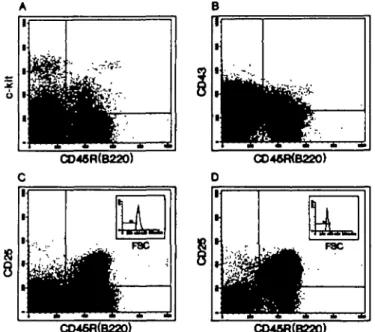
rearranged pre-B-l cells from VHDJH rearranged pre-B-ll cells. Here we analyzed the expression of c-kit, CD43 and CD25 on the surface of CD45R(B220)+ slgM- B lineage precursors of normal and mutant mouse bone marrow. In Fig. 1 the two-color FACS analysis of bone marrow depleted of slgM+ B cells of 4 - 5 week old BALB/c mice is shown. For the CD45R(B220)/
c-kit and the CD45R(B220)/CD43 expression all nucleated bone
marrow cells were analyzed, while CD45R(B220)/CD25 expres-sion was analyzed on large cells (Fig. 1C, see inset) and on small cells (Fig. 1D, see inset). Table 1 summarizes the quantitation of these analyses in five normal inbred strains of mice and five
CD46R(B220)
1
ii
HI
C046R(B220) 1 1 • Fac CO46R(B220) I t * :n
F8C z E CO48fl(B220) Fig. 1 . Dual labeling analysts of slgM depleted bone marrow cells of4 week old BALB/c mice for CD45R(B220)/c-*/r (A), CO45R(B220)/CD43 (B) and CD45R(B220)/CD25{TAC) (C and D). For the expression of CD45R{B220)/c-Wf and CD45R{B220yCD43, total nucleated bone marrow cells were analyzed while CD45R(B220)CD25(TAC) expression was analyzed by forward and side scatter gating (see insets) on large (C) and on small cells (D). Percentages of double positive cells are summarized in Table 1.
mutant strains of mice for total bone marrow. In the bone marrow of 4 - 5 week old normal mice the percentages of CD45R(B220)+/c-WT and CD45R(B220)+/CD43+ range from 3 to 5 and from 5 to 8% respectively. Thus, the CD45R(B220)+/CD43+ population is 1.5-2 times larger than the CD45R(B220)+/c->c/f+ population. Between 5 and 7% of the bone marrow cells of these mice are large CD45R (B220)+/CD25+ cells and between 15 and 20% are small CD45R(B220)+/CD25+ cells.
In the bone marrow of age matched SCID, XsT, ^Ml and RAG-2, the percentages of CD45R(B220)+/c-Wr+ and of CD45R(B220)+/CD43+ cells are sfightly elevated when compared with the percentages found in the bone marrow of normal mice (see Table 1). In marked contrast, CD45R(B220)+/CD25+ large as well as small cells in the bone marrow of these mutant mice are below the FACS detection level. This indicates that both the Xs protein and a membrane-bound n protein are required for the formation of a CD45R(B220)+/CD25+ precursor B cell population. To test this hypothesis RAG-2T mice which are rearrangement incompetent but express normal levels of Xs (13,18) were supplemented with a transgenic membrane-bound M chain. Analysis of the bone marrow of these mice revealed a relatively normal number of CD45R(B220)+/c-tof+ and CD45R(B220)+/ CD43+ cells when compared with normal inbred strains of mice, but a 2- to 3-fold lower number when compared with normal RAG-2T mice (Table 1). About 3 - 4 % of the bone marrow cells of the RAG-2T mice expressing a transgenic membrane-bound
fi chain co-express CD45R(B220) and CD25, and are large, and
1 3 - 1 4 % co-express these markers and are small. Thus, the formation of a CD45R(B220)+/c-M+/CD43+ precursor B cell compartment in the bone marrow is independent of rearrange-ment and the expression of X5, while the formation of a CD45R(B220)+/CD25+ compartment is dependent on the expression of the X5 protein and a membrane-bound /» chain.
Expression of c-kit, CD43, CD25, BP-1, TdT, c/t and cXs protein
by CD45R(B220)+/c-kit+, CD45R(B220)+/CD43* and
CD45R(B220)*/CD25* precursor B cells of normal mouse bone marrow
CD45R(B220)+ slgM- precursor B cells from bone marrow of 4 - 5 week old BALB/c mice were separated by preparative
two-Table 1 . Phenotypic stages of development of progenitor and precursor B cells in normal and mutant mouse bone marrow"
Mice BALB/c DBA/2 C3H/HeJ CBA/J C57BI/6 SCID RAG-2T RAG-2T + tgj< Phenotypic markers CD45R(B220)+ c-kit* 3.2 3.7 3.7 5.5 3.3 4.5 6.2 5.0 6.0 2.0 (percentage of nucleated CD45R(B220) CD43+ 5.5 6.4 5.9 8.5 6.3 8.5 9.5 8.0 8.4 3.4
bone marrow cells)
CD45R(B220)+ CD25* (large) 6.7 5.7 5.9 5.4 5.0 0.1 0.5 0.2 0.6 3.5 CD45R(B220)+ CD25+ (small) 19.8 18.2 14.8 15.0 22.0 0.6 0.7 0.2 0.2 13.5 •The data are values derived from studies on the bone marrow of a pool of three or four 4 - 5 week old mice.
CO45R(B220)*C-Wt* CD45fi(B220)+CD43+ CO45R(B220)+CO25+ CO4«R(B220)+e-W«* CD4M(B220)+CD43* CD45fl<B220)*CO2S+ C (Uugeceto) 0-910*) (Me*> CO26
h •
71
C026V
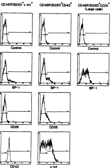
CO 43 c-HItRg. 2. FACS analysis of cell surface expression of BP-1, CD25 and COW on sorted CD45R(B220)+/c-fc/f\ of BP-1 CD25 and c-kit on sorted CD45R(B220)+/CD43+ and BP-1 on sorted large CD45R (B220)+/CD25+ bone marrow derived precursor B cells from 4 week otd BALB/c mice. The percentages of positive cells are summarized in Table 2.
color FACS into CD45R(B220)+/c-M + , CD45R(B220)+/CD43\ CD45R(B220)+/CD25+ large and CD45R(B220)+/CD25+ small cells (Fig. 1). The separated cells were subsequently analyzed for the expression of BP-1, CD25, c-kit and CD43 on the surface (Fig. 2) as well as for the intraceBular expression of TdT, Xs protein and 11H chains (Fig. 3). Table 2 summarizes the quantitation of these analyses.
About half of the CD43+ precursor B cells express c-kit, while none of the CD25+ precursors do. Almost all of the c-kit+ precursor B cells express CD43, while only 20% of the large and practically none of the small CD25+ precursor B cells do. BP-1 expression in variable numbers was found on all subpopulations of bone marrow derived precursor B cells. One third of the CD45R(B220)+/CD43 + precursors do express CD25. TdT expression was observed in the vast majority of c-kit+ precursors and in about half of the CD43+ precursors. No TdT is detectable in CD25+ precursor B cells. The 0X5 protein is detectable in the vast majority of CD45R(B220)+/c-ttT and of CD45R(B220)+/ CD43+ cells. Only 25% of the CD25+ large precursor cells
Central Control Cental Control
TUT TdT TdT TdT
cX5
Fig. 3. FACS analysis of intracellular expression of TdT, Xj and p in sorted CD45R(B220)+/c-/tf+ , CD45R(B220)+/CD43\ large CD45R (B220)+/CD25+ and small CD45R(B220)+/CD25+ precursor B cells derived from 4 week old BALB/c mice bone marrow. The percentages are summarized in Table 2.
express X5 protein in the cytoplasm, while none of the CD25+ small precursors do. C/i expression is almost absent in CD45R
(B220)+/c-kit+ precursors, whereas one third of CD43+ precursors express c/i. The vast majority of CD25+ B cell precursors express C/t. These analyses indicate that pre-B-l cells are almost all CD45R(B220)+/c-M+/CD43+/CD25-/TdT+/ cX5+/c/»-. A part of them express BP-1.
Pre-B-ll cells can be divided into large and small. All pre-B-ll cells are c-kit- /TdT- /CD25+7c/i+. About 20% of the large pre-B-ll cells are CD43+, as well as cXs+, the other 80% are CD43" and 0X5". Half of them express BP-1. Within the small pre-B-ll cells practically all cells are CD43-/cXs-. A part of them is BP-1 + . Thus, c-kit expression is restricted to pre-B-l cells, whereas CD25 expression is restricted to pre-B-ll cells. Precursor B cells that express CD43 consist of at least a mixture of pre-B-l and pre-B-ll cells.
Cell cycle analysis of CD45R(B220)+Ic-kil*, CD45R(B220)+/
CD43+ and CD45R(B220)+/CD25+ precursor B cells of normal
mouse bone marrow
CD45R(B220)+ slgM" precursor B cells from bone marrow of 4 - 5 week old BALB/c mice were separated by preparative two-color FACS into CD45R(B220)+/c-M + , CD45R(B220)+/CD43\ CD45R(B220)+/CD25+ large, and CD45R(B220)+/CD25+ small cells (Fig. 1). From the separated cells nuclei were prepared and their DNA content was analyzed as described in Methods. 01
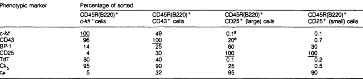
Table 2. Phenotype marker analysis of sorted populations of progenitor and precursor B cells of normal mouse bone marrow
Phenotypic marker Percentage of sorted CD45R(B220)+ c-kit* cells CD45R(B220)+ CD43+ cells CD45R(B220)+ CD25+ (large) cells CD45R(B220)+ CD25+ (small) cells c-kit CD43 BP-1 CD25 TdT
cx
5 100 96 14 4 80 95 5 49 100 25 30 40 90 32 0.1" 20» 60 100 0.1 25 95 0.1 0.7 30 100 0.2 0.5 90"These numbers are derived by calculation from staining done with CD45R(B220)+/c-tor+ and CD45R(B220)'lXD43+ sorting experiments (Rg. 2).
the CD45R(B220)+/c-Wf+ precursors 45% are in S and phases of the cell cycle (Fig. 4A). Of the CD45R(B220)+/CD43+ population - 2 0 % is in S and Gj/M phase of the cell cycle (Fig. 4B), whereas - 7 0 % and < 5 % of large (Rg. 4C) respectively small (Fig. 4D) cells of the CD45R(B220)+/CD25+ population are in the S and G2/M phase of the cell cycle. Thus, CD45R (B22O)+/c-tof+ pre-B-l cells and even more the CD45R(B220)+/ CD25+ large pre-B-ll cells are actively cycling while small CD45R(B220)+/CD25+ pre-B-ll cells do not cycle. The marker analyses described in the previous sections showed that CD45R(B220)+/CD43+ precursors consist of at least a mixture of pre-B-l cells and large pre-B-ll cells. The finding that far less of the CD45R(B220)+/CD43+ cells are in S and G-./M phases of the cell cycle than those of the pre-B-l compartment (20 versus 45%) and those of the large pre-B-ll compartment (20 versus 70%) indirectly indicates that the CD45R(B220)+/CD43+ compartment must contain a third population of cells which does not cycle. The possible nature of this third population will be discussed below.
Discussion
We have analyzed the precursor B cell compartments of normal and mutant mouse bone marrow. The results which are summarized in Table 1 are derived from analysis performed on 4 - 5 week old mice. Here it is important to note that with age, although the relative ratios between the different subpopulations stay constant their absolute numbers decrease (23).
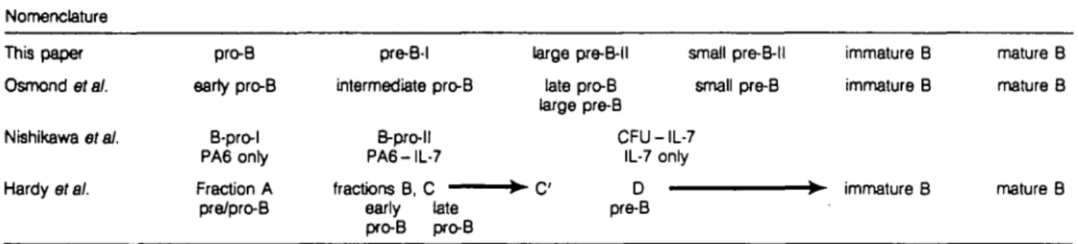
Previous work by Osmond etal. (1,28,29), Hardy etal. (2,5), Nishikawa etal. (4), Rajewsky etal. (19-21,30,31) and ourselves (14,24,32) has defined subpopulattons of cells in the development of mature B cells from pluripotent stem cells and B-lineage-committed progenitors in mouse bone marrow. In this paper we add the analysis of the expression of the IL-2 receptor o chain (CD25.TAC) on the surface of defined stages of B eel development It is a useful marker to distinguish pre-B-l from pre-B-ll cells. Figure 5 shows a model of B cell development in mouse bone marrow based on our analyses, and Table 3 correlates this model and the corresponding nomenclature to those used by different laboratories. Generally, the different subpopulations can be correlated well with each other. Only a few discrepancies and uncertainties remain.
Osmond 's late pro-B cells and large pre-B cells, defined as Cfi~ and cn+ CD45R(B220)+/ slgM~, TdT" respectively is one example of a discrepancy. Osmond's late pro-B cells are different
DMA content
Fig. 4. DNA content analysts in nuclei prepared from sorted CD45R (B220)+/c-*/r + (A), CD45R(B220)+/CD43+ (B), large CD45R(B220)+/
CD25+ (C) and small CD45R(B220)+/CD25+ (D) precursor B cells
derived from slgM depleted bone marrow of 4 week old BALB/c mice. The marker indicates the percentage of cells in S and Gj/M phases of the cell cycle. These percentages of cells in S and Gj/M phases are A, 45%; B, 20%; C, 70%; D, 3%.
from our large pre-B-ll cells, since the latter are > 8 5 % C/»+ (Table 2). It might well be possible that Osmond's c>r late pro-B cells, detected in situ in bone marrow sections, are out-of-frame rearranged large pre-B-ll cells (Fig. 5), which are on their way to die by apoptosis. By the time we have taken them out of bone marrow, stained them as single cell suspensions and analyzed them by FACS, they might already be dead. Alternatively, the level of expression of n in the cytoplasm might differ in the two populations, so that by in situ fluorescence analyses under the microscope half of the cells might score as negative, while FACS detects them as positive.
Another area of uncertain correlations is the transition from Hardy's pro-B to pre-B cells, i.e. the transitions from fractions B to C, to C , to D. Both Hardy's (5) and Rajewsky's (21) laboratories find cells with V^D^-rearranged H chain gene loci in fractions C, C and D, while practically all of our pre-B-l cells are DJH-rearranged and the vast majority of pre-B-ll cells have undergone productive VHDJH rearrangement. The transition from pre-B-l to large, and later small pre-B-ll cells is most
Table 3. B lymphopoiesis in mouse bone marrow (correlation and comparison of nomenclature used in different laboratories) Nomenclature This paper Osmond era/. Nishlkawa et at. Hardy etal. pro-B earty pro-B B-pro-l PA6 only Fraction A pre/pro-B pre-B-l intermediate pro-B B-pro-ll PA6-IL-7 fractions B, C early late pro-B pro-B large pre-B-l I late pro-B large pre-B small pre-B-ll small pre-B CFU - IL-7 IL-7 only D pre-B immature B immature B immature B mature B mature B mature B
o
CD2S(TAC)?•ctive cyding cells
c-kB CO43" XS'/VpnB TdT
COM
(TAC)-proB / preB I large preB I
oa
•igD
mature B
Rg. 5. A model of B cell development in normal mouse bone marrow based on the analysis described in this paper. This model describes the phenotypes of the vast majority of all B lineage cells in bone marrow. We expect minor populations to express other phenotypes in transition from one to another major compartment, and cannot rule out other, minor pathways of B cell development. Large circles, active cycling cells; small circles, resting cells; 'irregular' circles, cells bound to die.
clearly defined by the change of expression of c-kit from positive to negative, of TdT from positive to negative and of CD25 from negative to positive. It underlines the usefulness of CD25 since it is so far the only surface marker which is upregulated during this transition.
Approximately twice as many CD45R(B220)+ cells express CD43 than do c-kit (Table 1). Practically all the c-kit* cells appear to co-express CD43 (Table 2) and most of them are cycling. Some of the remaining CD43+, c-kit - cells are large, cycling cells and express cp, 0X5 and CD25, but not TdT. They are expected to be a subpopulation of large pre-B-ll cells in transit from pre-B-l to small pre-B-ll cells. However, the relationship of pre-B-l to large pre-B-ll cells, which are either CD43+ or CD43", to small pre-B-ll cells has to be investigated in future experiments. CD43 appears to be down-regulated later than c-kit and TdT, and also later than CD25 is up-regulated, and maybe at the same time as the expression of surrogate L chain is down-regulated.
Another part of the CD43 + , c-kit ~ population appears to be less actively cycling and is c/t". They might be pre-B-ll cells with non-productively VHDJH rearranged H chain gene loci on their way to apoptosis (Fig. 5). Alternatively, they could be pro-B cells before the expression of c-kit and TdT, maybe related to fraction A of Hardy etal. (2,5). In the latter case, they should have all their H chain gene loci in germline cofiguration. Future experiments are aimed at testing this.
While CD25 on the surface of pre-B-ll cells is a useful marker to distinguish them from pre-B-l cells and from immature B cells, its function in B cell development at these early stages remains unknown. IL-2 has no effect on either proliferation or differentiation of pre-B cells in tissue culture (unpublished observations). Moreover, mice with a targeted deletion of the IL-2 gene have no obvious B cell deficiency (33). Therefore it remains to be investigated whether the other, i.e. the 0 and 7 chains, of the IL-2 receptor are expressed at this earty stage or whether
the o chain is connected to other polypeptides, possibly with other functions.
B lineage committed, CD45R(B220)+ precursor B cells in
RAG-2T, /iMT, X5T and SCID mice lack CD25+ cells almost
completely, while they are present in almost normal numbers in RAG-2T mice expressing a transgenic /iH chain gene under the control of the heavy chain enhancer (E^) (Table 1). This indicates that the expression of the pH chain and surrogate L chain in membranes (and probably on the surface) of precursor B cells allows their entry into the pre-B-ll compartments and their proliferative expansion. Since the composition of cells at the transition point from pre-B-l to large pre-B-ll cefe is indistinguishable in RAG-2T, SCID, MMT and X J mice, and since no large CD25* cells can be found in the bone marrow of any of these mutant mice the block in the differentiation appears to be at the same stage of B cell development for all these mutants.
Pre-B-l cells express surrogate L chain together with a complex of polypeptides (p130, p65) on their surface (13), while large, cycling c-Wr, CD43+ pre-B-ll cells co-express the /iH chain and surrogate L chain, and are likely to assemble them in an Ig-like complex, in membranes, probably on the surface. The role of the complex of p130/p65 with the surrogate L chain is unclear. One possible role could be to signal pre-B-l cells to enter the large pre-B-ll compartment, while the complex of /tH chain with surrogate L chain in or on the large pre-B-ll cells might signal the cells to expand by proliferation. Such a scenario would have predicted that SCID, RAG-2T and tiMl mice should have some CD25+ large pre-B-ll cells (since they are potentially capable of
expressing the p150/p65 surrogate L chain complex) while X5T mice would not (since they cannot express this early complex of proteins on the surface). This, however, appears not to be the case, since bone marrow of all these mutant mice lack practically all of the CD25+ large pre-B-ll cells. Consequently, the only role of surrogate L chain in B cell development identified so far is to expand by proliferation those pre-B-ll cells which express membrane-bound pH chains.
Acknowledgements
We thank Dr Antonio Iglesias for the gift of the Sp6 pH transgenic mice, and Drs Jan Anderson, Klaus Karlajainen and Hans-Reimer Rodewakj for critical reading of this manuscript. The technical assistance of Ms Andrea Groenewegen is gratefuBy acknowledged. The Basel Institute for Immunology was founded and is supported by F. Hoffmann-La Roche Ltd, Basel, Switzerland. Abbreviations PE TdT References cytoplasmic \$ cytoptasmic MH chain phycoerythrin
terminal deoxynucleotidyl transferase
1 Osmond, D. G. 1991. Proliferation kinetics and the lifespan of B cells in central and peripheral lymphoid organs. Curr. Opin. Immunol. 3:179.
2 Hardy, R. R., Carmack, C. E., Shinton, S. A., Kemp, J. D. and Hayakawa, K. 1991. Resolution and characterization of proB and pre-pro B cell stages in normal mouse bone marrow. J. Exp. Med. 173:1213.
3 Nishikawa, S., Ogawa, M., Nishikawa, S., Kunisada. T. and Kodama,
H. 1988. B lymphopoiesis on stromal cell clone: stromal cell clones acting on different stages of B cell differentiation. Eur. J. Immunol. 18:1767.
4 Era, T., Ogawa, M., Nishkawa, S., Okamoto, M., Honjo. T., Akagi, K., Mlyazakl, J. and Yamamura, K. 1991. Differentiation of growth signal requirement of B lymphocyte precursor is directed by expression of immunoglobulin. EMBO J. 10:337.
5 Li, Y. S., Hayakawa, K. and Hardy, R. R. 1993. The regulated expression of B lineage associated genes during B ceO differentiation in bone marrow and fetal Over. J. Exp. Med. 178:951.
6 Oltz, E. M., Yancopoulos, G. D., Morrow, M. A., Rollnk. A., Lee, G., Wong, F., Kaplan, K., Gillis, S., Melchers, F. and Alt, F. W. 1992. A novel regulatory myosin light chain gene distinguishes pre-B cell subsets and is IL-7 indudble. EMBO J. 11:2759.
7 Sakaguchi, N. and Melchers, F. 1986. Xj, a new llght-chaln-related locus selectively expressed in preB lymphocytes. Nature 324:579. 8 Kudo, A. and Melchers, F. 1987. A second gene, V ^ in the Xj
locus of the mouse, which appears to be selectively expressed in preB lymphocytes. EMBO J. 6:2267.
9 Schatz, D. G., OetBnger, M. A. and Baltimore, D. 1989. The V(D)J recombination activating gene, RAG-1. CeB 59:1035.
10 Oettinger, M. A., Schatz, D. G., Gorka, C and Baltimore, D. 1990. RAG-1 and RAG-2, adjacent genes that synergtstically activate V(D)J recombination. Science 248:1517.
11 Rolink, A., Kudo, A., Karasuyama, H., Kikuchi, Y. and Melchers, F. 1991. Long-term proliferation early preB cell lines and clones with the potential to develop to surface-lg positive mrtogen reactive B cells 'in vitro1 and 'in vivo'. EMBO J. 10:327.
12 RoOnk A., Streb, M., Nishikawa, S. I. and Melchers, F. 1991. The c-kit encoded tyrosine kinase regulates the proliferation of early preB celts. Eur. J. Immunol. 21:2609.
13 Karasuyama, H., Melchers, F. and Rollnk, A. 1993. A complex of glycoproteins is associated with Vp^e/Xj surrogate Hght chain on the surface of p heavy chain-negative earty precursor B cell ines. J. Exp. Med. 178:469.
14 Rolink, A. and Melchers, F. 1993. Generation and regeneration of cells of the B-)ymphocyte lineage. Curr. Opin. Immunol. 5:207. 15 Karasuyama, H., Rolink, A., Shinkai, Y., Young, F., Alt, F. W. and
Melchers, F. 1994. The expression of Vp^e/Xj surrogate light chain in early bone marrow precursor B cells of normal and B-cell deficient mutant mice. Cell in press.
16 Bosma, G. C , Custer, R. P. and Bosma, M. J. 1983. A severe combined immunodeficiency mutation in the mouse. Nature 301:527. 17 Mombaerts, P., lacomlni, J., Johnson, R. S., Herrup, K., Tonegawa, S.
and Papatoannou, V. E. 1992. RAG-1-deficient mice have no mature B and T lymphocytes. Cell 68:869.
18 Shinkai, Y., Rathbun, G., Lam, K. P., Oltz, E. M., Stewart, V., Mendelsohn, M., Charron, J., Datta, M., Young, F., Stall, A. M. and Alt, F. W. 1992. RAG-2-deficient mice lack mature lymphocytes owing to inability to Initiate V(D)J rearrangement. Cell 68:855.
19 Kitamura, D., Roes, J., KOhn, R. and Rajewsky, K. 1991. A B cell-deficient mouse by targeted disruption of the membrane exon of the immunoglobulin p chain gene. Nature 350:423.
20 Kitamura, D., Kudo, A., Schaal, S., MOIIer, W., Melchers, F. and Rajewsky, K. 1992. A critical role of Xs in B cell development. CeH 69:823.
21 Ehlich, A., Schaal, S., Gu, H., Kitamura, D., MOIIer, W. and Rajewsky, K. 1993. Immunoglobulin heavy and light chain genes rearrange independently at earty stages of B ceO development. CeB 72:695. 22 Rolink, A., Karasuyama.H., Grawunder, U., Haasner, D., Kudo, A. and Melchers, F. 1993. B cell development in mice with a defective X5 gene. Eur. J. Immunol. 23:1284.
23 Rofink, A., Haasner, D., Ntshikawa, S. I. and Melchers, F. 1993. Changes in frequencies of clonable preB cells during life in different lymphoid organs of mice. Blood 81:2290.
24 RoHnk, A. Karasuyama, H., Haasner, D., Grawunder, U., Martensson, I. L, Kudo, A. and Melchers, F. 1994. Two pathways of B lymphocyte development in mouse bone marrow and the roles of surrogate L chain in this development Immunol. Rev. in press.
25 Iglesias, A., Nichogiannopoulou, A., Williams, G. S., Flaswinkel, H. and Kohler, G. 1993. Early B cell development requires p signaling. Eur. J. Immunol. 23:2622.