Functional expression of a human TCR/3
gene in transgenic mice
Joachim Rothe, Stefan Ryser, Urs Mueller1, Michael Steinmetz2, and
Horst Bluethmann
Biology Department, Pharmaceutical Research New Technologies, F.Hoffmann-La Roche Ltd, 4002 Basel, Switzerland
1 Basel Institute of Immunology, 4002 Basel, Switzerland
2Present address: Department of Biotechnology Research, F. Hoffmann-La Roche Ltd, Nutley, NJ 07110, USA
Key words: allelic exclusion, human TCR/3 gene, thymocyte development, transgenic mice
Abstract
A functionally rearranged TCR/3 (Tcrb) gene was isolated from a cloned human T helper cell recognizing the CS.T3 epitope of Plasmodium falciparum with HLA-DR2. Transgenic mice were generated by co-injection of the human gene together with the mouse Tcrb enhancer. Analysis of transgenic mice shows that the functional Tcrb gene of xenogenic, i.e. human, origin exerts allelic exclusion of endogenous Tcrb genes. Cytofluorometric analysis revealed expression of the human TCR/3 chain on virtually all thymocytes and peripheral T cells together with endogenous TCR/3 chains and CD3 components. No surface expression of mouse TCR/3 chain or
rearrangement of endogenous Tcr genes was detectable. Expression of the hybrid receptor causes a reduction in the number of thymocytes and a bias for CD4+CD8" T cells in the thymus as compared with non-transgenic littermates. Peripheral transgenic T cells mount a normal proliferative response against allogeneic targets in mixed lymphocyte reactions. These results show that a hybrid mouse/human TCR is able to pass positive and negative selection in the thymus, and is functional in transgenic mice
Introduction
T cells recognize foreign antigens presented by self MHC class I or class II molecules with the help of highly diverse, but clonally distributed TCRs (for review see 1). TCRs are composed of variable a and /3 chains associated with non-variant polypeptides of the CD3 complex. The diversity of the TCRa and /3 chains is generated somatically in T cells by rearrangement of distinct germ line gene segments. During maturation of thymocytes the T cell repertoire is subjected to positive and negative selection in the thymus to render it functional and self-tolerant (2,3).
Transfection experiments have previously demonstrated that a TCRa/3-CD3 complex containing xenogeneic polypeptide chains can be expressed on the surface of human cells and mediate T cell activation (4,5). In particular, human Jurkat cell variants with inactive Tcra or b genes were transfected with functionally rearranged mouse Tcra or b genes to replace the inactivated subunit and shown to express hybrid mouse a - h u m a n /3 and human a-mouse /3 heterodimers associated with the human CD3 complex on the surface. Following ligand binding, the hybrid receptors could mediate transmembrane
signals as measured by lymphokine secretion. More recently in transgenic mice, a hybrid TCR-CD3 complex containing a human CD3e chains was shown to be expressed on mouse T cells and to be able to transduce activation signals (6).
Transgenic mice with functionally rearranged Tcr genes have proven to be an excellent model to study T cell maturation and repertoire selection (7,8). Introduction of functionally rearranged mouse Tcrb genes into the germline of mice was shown to lead to suppression of endogenous variable (Ve) gene segment rearrangement (9-12). Whereas allelic exclusion was found to strictly apply to the Tcrb locus, it seems that rearrangement of the Tcra locus is controlled in a different way. Several T cell clones have been described which contain two productively rearranged Tcra alleles (13-17).
Here we demonstrate that a functionally rearranged human Tcrb gene can be expressed on T cells of transgenic mice and is able to suppress endogenous V0 gene segment
rearrange-ment. T cells bearing hybrid TCRs, consisting of the transgenic human /3 chain associated with mouse a chains and the CD3
Correspondence to: H.Bluethmann
components, can mature in transgenic mice and form a T cell repertoire with an apparently normal frequency of alloreactive cells.
R E
Methods
Generation of transgenic mice
Constructs were injected into zygotes of (C57BL/6xDBA/2)F, mice as described (18,19). Founders were backcrossed with strain C57BL/6J to establish transgenic lines B6-Tg58 and B6-Tg62. Screening for transmission of the transgene in the offspring was performed either by Southern blot analysis using a 32P-labelled fragment of the human V05.1 segment as a probe or by the polymerase chain reaction (PCR), essentially as described elsewhere (20). The 5' primer used to detect the transgene was specific for the 3' end of the human V^S.I segment (CCAGTAAAGGCTGGAGTCAC) and the 3' primer was specific for the human J31.2 region (TACAACGGTTAACCT-GGTCC).
PCR
Oligonucleotides specific for sequences of the murine Ve segments 2,6, 8.3 and 14 were used as 5' primers together with an oligonucleotide recognizing a sequence 3' of ^ 2 . 6 as a 3' primer at final concentrations of 1 mM each (21). Depending on the primers used, the annealing temperature varied from 50 to 65°C and the magnesium concentration from 1 to 2 mM. PCR was routinely performed with 25 cycles using 2 U Taq-Poly-merase (Cetus) and 100 ng genomic DNA isolated from relevant organs or cells before separating the amplified fragments on agarose gels and transferring them to a nylon membrane (Zeta-Probe, Bio-Rad Laboratories, Richmond, CA) by vacuum blotting. DNA probes
For detection of the transgene we used a 300 bp EcoRV - HincW fragment of the human V05.1 gene segment. A 1.4 kb Pst\ fragment specific for mouse DS1 was used to analyze D3- J0 rearrangements, while a 1.2 kb EcoRI - C/al fragment covering the mouse J02 cluster was applied to detect V0- D3J0 rearrangements after PCR amplification (22). All probes were labelled to high specific activity with [32P]dCTP by random primer extension.
Cytofluorometric analysis
Preparation of T cells and analysis by flow cytometry was essentially as described (19). Briefly, thymus and lymph nodes were homogenized, and the cells recovered and washed twice in PBS/5% FCS/0.1% NaN3. Samples of 106 cells were stained in the same buffer at 4°C with optimal concentrations of antibodies. After two further washes, 105 cells were analyzed using a FACScan cytometer (Becton-Dickinson, Mountain View, CA). Data were processed with the FACScan research software. In order to make surface antigens accessible to the antibody /3F1, cells were fixed prior to staining with 10 volumes of a freshly prepared 1 °/o solution of formaldehyde in PBS at room tempera-ture. After 15 min incubation, cells were washed twice with PBS.
1 Kb
Fig. 1. Functionally rearranged Tcrb gene from the T helper cell clone
MG-30. Schematic representation of the Tcrb construct used for the generation of transgenic mouse lines Tg58 and Tg62. The black box indicates the human V^5.1 segment functionally rearranged to D^1 and J01.2 gene segments. The striped box represents the human C^1 segment. L: leader; "location of a non-functional leader sequence. H: H/ndlll; R: EcoRV; E: EcoRI.
Jp2.1
Jp2.2
Jp2.3
JP2.4
JP2.5
Jp2.6
-kb
-
1.4
-
1.1
-
0.9
_
0.6
-
0.3
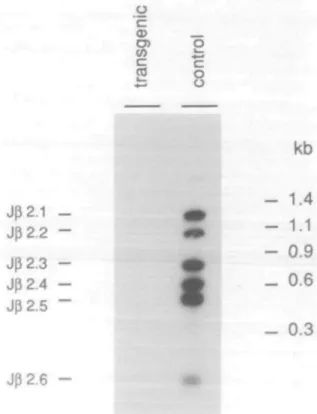
Fig. 2. Suppression of rearrangements of endogenous V^8.3 gene
segments in transgenic mice. Southern hybridization of PCR amplified fragments using DNA from transgenic and non-transgenic thymocytes from control littermates. The 5' primer was specific for V^8.3 and the 3' primer specific for a sequence 3' of Jfl2. The probe for hybridization was specific for Jg2. There is no detectable signal with DNA from transgenic thymocytes, while V^8.3 rearrangements to all six JB2 segments are
clearly visible in the non-transgenic control.
Antibodies
Cells were stained with the following antibodies: anti-human pan-TCR/3 (J8F1, T-Cell sciences, Cambridge, MA), anti-mouse CD4-phycoerythrin(PE; L3T4, Becton-Dickinson), anti-mouse CD8-FITC (Lyt-2, Becton-Dickinson), anti-mouse CD3e-biotinylated (145-2C11, (23)), anti-mouse pan-TCR/3 (H 57-597, (24)), anti-mouse Ve2 (B20.6.5, (25)), anti-mouse Vp3 (KJ 25, (26)), anti-mouse V,j6 (44-22-1, (27)), and anti-mouse V ^ (F23.1,
a) Thymus Lymph node b) Thymus
control
transgenic
[3F1 (human) panp (mouse)
Fig. 3. (a) Expression of the human TCR/3 chain on thymocytes and lymph node T cells. Cytofluorometric analysis of fixed thymocytes (left) and
lymph node cells (right) from transgenic and non-transgenic mice. All thymocytes and peripheral T cells from transgenic mice were stained with the pan-human TCR/3-specific antibody /3F1. The brightly stained population in the right panels are B cells which were directly stained by the second reagent (anti-lg - FITC). (b) Expression of endogenous TCR/3 chains on thymocytes. Staining of transgenic and non-transgenic thymocytes with the pan-mouse TCR/3 chain-specific antibody H57-597. No mouse TCR/3 chains are expressed on thymocytes transgenic for the human TCR/3 chain.
(28)). Secondary reagents were either FITC-labeled anti-mouse Ig (Silenus Laboratories, Victoria, Australia) or PE-labeled streptavidin (Becton-Dickinson).
Mixed lymphocyte reaction (MLR)
Purified spleen and lymph node cells (2.5x10⁵/well) from transgenic B6-Tg58 or B6-Tg62 animals and non-transgenic controls were co-cultured in triplicate with 106 irradiated syngeneic or allogeneic spleen cells as stimulators for 24, 48, or 72 h. Addition of 1 pC\ of [3H]thymidine (Amersham, Arlington Heights, IL) to each well was 20 h before harvesting and measuring the [3H]thymidine uptake by liquid scintillation counting.
ed human Tcrb gene together with a 5.5 kb fragment carrying the mouse Tcrb enhancer (29). The mouse enhancer was co-injected to ensure expression of the human transgene in T cells at a high level. Both fragments were injected in equimolar amounts into zygotes from (C57BL/6 x DBA/2JF, mice. Of the four founder mice obtained, two did not transmit the transgenes to their offspring. Of the others, transgenic lines B6-Tg58 and B6-Tg62 were established by backcrossing with C57BL/6 mice. Southern blot analysis revealed that the transgenes had in-tegrated in 1 - 2 and - 1 0 copies respectively. The human Tcrb gene was found to co-segregate with the enhancer transgene, meaning that both had integrated into the same chromosomal site.
Results
Isolation of a functionally rearranged human Tcrb gene We used the cloned human T helper cell MG-30, recognizing the CS.T3 epitope (amino acids 378-398) of the circumsporo-zoite protein of Plasmodium falciparum in association with the HLA class II molecule DR2, to isolate a human Tcrb gene. Screening of EMBL3 genomic libraries made from this clone us-ing Cg probes yielded full length clones containing the
functionally rearranged Tcrb gene. A 10.5 kb EcoRI genomic fragment which contains a functionally rearranged V05.1 gene segment joined to Dfi1 and J01.2 gene segments was sub-cloned into pUC 19 (Fig. 1). About 500 bp upstream of the N/,,5.1 leader exon, a second leader sequence was identified, which is non-functional due to a mutation of the splice donor site. Generation of transgenic mice
Transgenic mice were generated by microinjection of the 10.5 kb genomic EcoRI fragment containing the functionally
rearrang-The human Tcrb transgene exerts allelic exclusion of endogenous mouse Tcrb genes
Endogenous D0- Je rearrangements were assayed in thymocytes of transgenic mice by Southern blot analysis. Similarly to a functional murine Tcrb transgene (9), the human Tcrb transgene did not inhibit endogenous D0- Jg rearrangements (data not shown).
Endogenous Vp-DgJ3 rearrangements were analyzed by PCR with primers specific for different V3 gene segments (see Methods). As shown in Fig. 2, six fragments were amplified when DNA from non-transgenic thymocytes and a primer pair specific for Vg8.3 and 3' J^2 sequences were used. These six fragments correspond to rearrangements of V^8.3 gene segments of one of each of the six Jff2 segments. No Vfl8.3 rearrangements,
however, were detectable in thymocytes from transgenic mice. Analogous results were obtained with primers specific for V02,
V36, and V^14 regions (not shown). This shows that the human transgene is able to completely suppress endogenous \le
transgenic untreated transgenic trypsinized non-transgenic control PF1 (human)
Fig. 4. Surface expression of the human TCR/3 chain. Thymocytes from transgenic mice were fixed and stained with the antibody 0F1 before or after treatment with trypsin for 15 min. Thymocytes from non-transgenic mice were used as control. The decrease in signal intensity after trypsin treatment is clearly visible and even more pronounced after longer incubation times.
The human Tcrb transgene is transcribed in a cell-type specific way
Cell-type specific transcription of the human transgene was analyzed by Northern blot hybridization (data not shown). In both transgenic mouse lines, mature transcripts of 1.3 kb in size were detected in transgenic thymus and to a lesser extent also in spleen, but not in brain, heart, lung, kidney or bone marrow. A low level of unspecific transcription was found in testes.
The human TCR/3 chain is expressed on the surface of transgenic T cells
Attempts to immunoprecipitate the human 0 chain from the surface of transgenic T cells using the antibody /3F1, specific for a common determinant on human TCR/3 chains, were unsuccessful. Cytofluorometric analysis, however, demonstrated that the human /3 chain is expressed on virtually all T cells in transgenic mice.
T cells were fixed with formaldehyde to allow the 0F1 antibody to recognize its determinant on the human j3 chain (30). As shown in Fig. 3(A), essentially all thymocytes and peripheral T cells from transgenic mice were stained with /3F1 demonstrating expression of the human 0 chain. To ensure that the determinant seen by /3F1 is indeed located on the cell surface and not in the cytoplasm exclusively, cells were gently trypsinized for different time intervals prior to formaldehyde fixation and staining. Prolonged trypsiniza-tion led to decreasing signals as revealed by 0F1 and reached control levels after 30 min of trypsinization, proving that the human /3 chain is indeed expressed on the cell surface (Fig. 4).
Thymocytes from transgenic mice were also analyzed for the
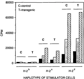
80000 60000 £ 40000
o
20000 • C=control T=transgenic C T H-2L H-2" H-21-HAPLOTYPE OF STIMULATOR CELLS Fig. 5. Kinetics of MLR. Transgenic (T) and control (C) responder cells were co-cultured with irradiated syngeneic (H^13) and allogeneic (H-2S
and H-2d) spleen cells. [3H]thymidine incorporation was measured after
24 (black), 48 (grey) and 72 (striped) h.
expression of endogenous mouse TCR/3 chains with the antibody H57-597 which detects a common determinant on mouse /3 chains. Whereas in normal mice ~3O°/o of thymocytes were stained with this antibody, no staining above background levels were obtained for transgenic thymocytes (Fig. 3B). In addition, antibodies specific for different mouse V,, regions (V^2, Vg3, V ^ , and V^)—covering together more than one third of the normal mouse y0 repertoire—did not stain transgenic thymocytes (not
shown).
Transgenic thymocytes and peripheral T cells were also stained with the antibody 145-2C11 specific for the e subunit of the murine CD3 complex. No difference with respect to the number of CD3+ T cells as well as the level of CD3 surface expression was found in transgenic mice when compared with normal littermates (not shown). Taken together, these results demonstrate that the human TCR/3 chain is expressed on transgenic T cells in the complete absence of mouse /3 chains.
The human transgenic 0 chain participates in the formation of a functional TCR
Since the human /3 transgene exerts allelic exclusion of endogenous mouse /3 genes and leads to the expression of a human 0 chain on the surface of transgenic T cells in the absence of detectable mouse /3 chains, it is reasonable to assume that the human /3 chain is associated with mouse TCRa chains and CD3 polypeptides. Are these hybrid receptors able to recognize foreign antigens and to trigger T cell responses?
The function of transgenic T cells was assayed by their ability to respond to allogeneic targets in mixed lymphocyte reactions (MLRs). Splenocytes from SJL (H-2S, Mls-1b, Mls-23) and DBA/2
(H-2d, Mls-1a, Mls-2a) mice were used as stimulators. As seen
in Fig. 5, T cells from transgenic as well as control littermates (H-2b, Mls-1b, Mls-2b) were indistinguishable in their ability to
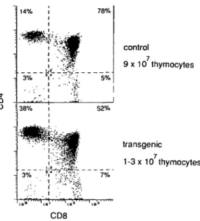
i Q O control 9 x 10 thymocytes transgenic 1 -3 x 10 thymocytes CD8
Fig. 6. CD4/CD8 subpopulations of thymocytes. Two-color
cytofluoro-metric analysis of thymocytes for non-transgenic (upper panel) and transgenic mice (lower panel). Numbers of quadrants give the relative percentage of thymocyte subpopulations.
These results reveal that the frequency of alloreactive T cells is as high in transgenic as in control animals. Thus the invariant human TCR/3 chain together with a variable mouse TCRa chain and murine CD3 molecules can form functional TCR in transgenic mice.
Expression of the human transgene in H-2b mice affects
thymocyte differentiation
The accumulation of functional T cells in the periphery of transgenic mice, expressing a mouse-human hybrid TCR receptor, indicates that these receptors can pass positive and negative selection in the thymus. The expression of the human TCR/3 chain in transgenic mice, however, has a strong impact on the number and phenotype of T cells in the thymus (Fig. 6). Whereas normal littermates contain ~ 9 x 1 07 thymocytes, this number is reduced to ~ 1 - 3 x 1 07 in transgenic mice. Furthermore, a relative increase of CD4+ single-positive and a decrease of CD4+CD8+ double-positive T cells were found. The relative size of the CD4-CD8" and CD4~CD8+ subpopulations remained unchanged.
In contrast to what was observed in the thymus, the periphery of transgenic mice contained normal numbers of T cells and CD4+ or CD8+ subpopulations. Also the number of B cells is as in control littermates.
Discussion
Experiments using Tcrb transgenes in mice have shown that allelic exclusion is a regulated rather than a stochastic event (9-12). Here we demonstrate that a functional Tcrb transgene of human origin is able to completely suppress V0-DgJe
rearrangements of the endogenous Tcrb loci in the mouse. Thus, the mechanisms regulating allelic exclusion seem to be conserved between mouse and man. Experiments with a mutant
Tcrb transgene showed that expression of the C region domain is sufficient to induce allelic exclusion in transgenic mice (31). The transcript by itself, however, had no effect. Analyses of mice double transgenic for either a Tcrb minilocus and a functional Tcrb gene (21), or two functional Tcrb transgenes (32), revealed that allelic exclusion may operate at a level after gene rearrangement.
Surface expression of the human TCR/3 chain was detected with the pan-specific mAb /3F1 after fixation of T cells, which is necessary to render the determinant accessible to the antibody. The gradual elimination of the determinant by prolonged digestion with trypsin clearly demonstrates that the human TCR/3 chain is expressed on the cell surface. No antibody specific for the variable region of the human transgene was available. The antibody 42 1C1 (33), raised against the human V35.3 domain, did not detect the V ^ . I region encoded by our transgene.
By immunoprecipitation we could not give direct evidence for a hybrid formation of the human TCR/3 chain with various mouse TCRa chains, because the pan-human TCR/3 antibody /3F1 detects a hidden determinant of the TCR - CD3 complex on the cell surface. This determinant is accessible to the antibody only after fixation of the cells, which bears the risk of exposing intra-cellular components. However, several observations argue for the expression of a hybrid mouse a - human /3 TCR in our trans-genic mice, (i) A prerequisite for T cell maturation in the thymus is a complete TCRa/3 receptor. Although it has been shown in Tcrb transgenic scidlscid mice (8,34) that TCR/3 chains can appear on thymocytes at early stages of differentiation together with an as yet non-characterized invariant molecule, such a receptor cannot pass positive selection in the thymus, and, in addition, would not explain the alloreactive responses observed with peripheral T cells in our transgenic mice, (ii) All mature T cells in the periphery express CD3, which is constitutively co-expressed with the TCRa/3 receptor, (iii) In the complete absence of endogenous TCR/3 chains, only the transgenic human /3 chain can pair with endogenous a chains and associate with mouse CD3 polypeptides on the surface of T cells. With human T cell lines, the formation and functionality of such a human TCR - CD3 complex, where the TCRa or /3 chain is replaced by the corresponding mouse TCR chain, was demonstrated (4,5). Although the requirements for the formation of a hybrid mouse - human receptor seem to be met, this process might be of lower efficiency, permitting less thymocytes to proliferate as compared with normal mice. An alternative explanation for the reduced number of thymocytes, however, could be an enhanced negative selection of the transgenic T cells expressing a monomorphic /3 chain. No change, however, was detected in the number of T cells in lymph nodes. It appears that both pool sizes are regulated independently.
The hybrid mouse - human receptor is functional in thymocyte development. It appears to be able to interact with mouse MHC molecules and transduce signals necessary for further maturation to single-positive thymocytes. The relative enrichment of CD4 + single-positive thymocytes might reflect an intrinsic affinity of the human TCR/3 chain—which was derived from a class II restricted cell—for mouse MHC class II molecules. A preferential develop-ment of CD4+ T cells in transgenic mice expressing MHC class ll-restricted TCRs was reported by Berg (35) and Kay (36); however, not for a TCR/3 chain alone. When we made mice
double transgenic for the TCRa and /3 chain of the T helper cell MG-30, we found the same bias for CD4+ thymocytes as in the single transgenic ones (unpublished results).
The invariant human TCR/3 chain reduces the TCR repertoire in our transgenic mice by a factor of —10,000. Since the frequency of alloreactive cells in the transgenic mice is still comparable with control littermates, it seems that the antigenic determinants on allogeneic cells are diverse enough to trigger a restricted T cell repertoire to the same extent as a fully developed one.
Acknowledgements
We thank Beatrice Schmutz and Monika Seiler for expert help in the maintenance and analysis of transgenic mice and Dr Joost van Meerwijk for the PCR primers used to analyze rearrangements of the Tcrb locus.
Abbreviations
MLR PCR PE
mixed lymphocyte reaction polymerase chain reaction phycoerythrin
References
1 Blackman, M., Kappler, J., and Marrack, P. 1990. The role of the T cell receptor in positive and negative selection of developing T cells.
Science 248:1335.
2 Teh, H. S., Kisielow, P., Scott, B., Kishi, H., Uematsu, Y., Bluethmann, H., and von Boehmer, H. 1988. Thymic major histo-compatibility complex antigens and the a/3 T cell receptor determine the CD4/CD8 phenotype of T cells. Nature 335:229.
3 Kisielow, P., Bluethmann, H., Staerz, U. D., Steinmetz, M., and von Boehmer, H. 1988. Tolerance of T-cell-receptor transgenic mice involves deletion of nonmature C D 4+8+ thymocytes. Nature
333:742.
4 Saito, T., Weiss, A., Miller, J., Norcross, M. A., and Germain, R. N. 1987. Specific antigen-la activation of transfected human T-cells expressing murine Ti a/3-human T3 receptor complexes. Nature 325:125.
5 Saito, T., Weiss, A., Gunter, K. C, Shevach, E. M., and Germain, R. N. 1987. Cell surface T3 expression requires the presence of both a-and |3-chains of the T cell receptor. J. Immunol. 139:625. 6 de la Hera, A., Muller, U., Olsson, C , Isaaz, S., and Tunnacliffe, A.
1991. Structure of the T cell antigen receptor (TCR): two CD3 epsilon subunits in a functional TCR/CD3 complex. J. Exp. Med. 173:7. 7 von Boehmer, H. 1990. Developmental biology of T cells in T
cell-receptor transgenic mice. Annu. Rev. Immunol. 8:531.
8 Bluethmann, H. 1991. Analysis of the immune system with transgenic mice: T cell development. Experientia 47:884.
9 Uematsu, Y., Ryser, S., Dembic, Z., Borgulya, P., Krimpenfort, P., Berns, A., von Boehmer, H., and Steinmetz, M. 1988. In transgenic mice the introduced functional T cell receptor 0 gene prevents expression of endogenous 0 genes. Cell 52:831.
10 Sha, W. C , Nelson, C. A., Newberry, R. D., Kranz, D. M., Russell, J. H., and Loh, D. Y. 1988. Positive and negative selection of an antigen receptor on T cells in transgenic mice. Nature 336:73. 11 Pircher, H., Ohashi, P., Miescher, G., Lang, R., Zikopoulos, A., Buerki, K., Mak, T. W., MacDonald, H. R., and Hengartner, H. 1990. T cell receptor (TcR) /? chain transgenic mice: studies on allelic exclusion and on the TcR+ 76 population. Eur. J. Immunol. 20:417.
12 Iglesias, A., Hansen-Hagge, T., von Bonin, A., and Weltzien, H. U. (1992) Increased frequency of TNP-specific, H-2b-restricted CTL
precursors in transgenic mice expressing a TCR-/3 chain gene from a H-2b-restricted, TNP-specific cytolytic T cell clone. Eur. J. Immunol.
22:335.
13 Fondell, J. D., Marolleau, J. P., Primi, D., and Marcu, K. B. 1990. On the mechanism of non-allelically excluded Va - Ja cell receptor
secondary rearrangements in a murine T cells lymphoma. J. Immunol. 144:1094.
14 Furutani, M., Yanagi, Y., Fujisawa, I., Nakayama, T., Kishimoto, H., Kuida, K., Asano, Y., and Tada, T. 1989. Post-transcriptional allelic exclusion of two functionally rearranged T cell receptor alpha genes.
Int. Immunol. 1:281.
15 Malissen, M., Trucy, J., Letourneur, F., Rebai, N., Dunn, D. E., Fitch, F. W., Hood, L, and Malissen, B. 1988. A T cell clone expresses two T cell receptor a genes but uses one a/3 heterodimer for allorecognition and self MHC-restricted antigen recognition. Cell 55:49. 16 Marolleau, J. P., Fondell, J. D., Malissen, M., Trucy, J., Barbier, E.,
Marcu, K. B., Cazenave, P. A., and Primi, D. 1988. The joining of germ-line Va to Ja genes replaces the preexisting Va - Ja complexes in a T cell receptor a/3 positive T cell line. Cell 55:291.
17 Schittek, B., Unkelbach, E., and Rajewsky, K. 1989. Violation of allelic exclusion of the T cell receptor beta genes in a helper T cell clone.
Int. Immunol. 1:273.
18 Hogen, B., Costantini, F., and Lacy, E. 1986. Manipulating the Mouse
Embryo: A Laboratory Manual. Cold Spring Harbor Laboratory Press,
Cold Spring Harbor, NY.
19 Bluethmann, H. and Steinmetz, M. 1990. Trangenic mice for analysis of T cell development. In Lefkovits, I. and Pernis, B., eds,
Immunological Methods, vol. IV, p. 311. Academic Press, Orlando.
20 Maniatis, T., Fritsch, E. F., and Sambrook, J. 1989. Molecular Cloning:
A Laboratory Manual. Cold Spring Harbor Laboratory Press, Cold
Spring Harbor, NY.
21 van Meerwijk, J. P., Iglesias, A., Hansen-Hagge, T., Bluethmann, H. and Steinmetz, M. 1991. Allelic exclusion of a T cell receptor-beta minilocus. J. Immunol. 147:3224.
22 Malissen, M., Minard, K., Mjollsness, S., Kronenberg, M., Goverman, J., Hunkapiller, T., Prystowsky, M. B., Yoshikai, Y., Fitch, F., Mak, T. W., and Hood, L. 1984. Mouse T-cell antigen receptor: structure and organization of constant and joining gene segments encoding the |3 polypeptide. Cell 37:1101.
23 Leo, 0., Foo, M., Sachs, D. H., Samelson, L. E., and Bluestone, J. A. 1987. Identification of a monoclonal antibody specific for a murine T3 polypeptide. Proc. Natl Acad. Sci. USA 84:1374.
24 Kubo, R. T., Born, W., Kappler, J. W., Marrack, P., and Pigeon, M. 1989. Characterization of a monoclonal antibody which detects all murine a/3 T cell receptors. J. Immunol. 142:2736.
25 Gregoire, C , Rebai, N., Schweisguth, F., Necker, A., Mazza, G., Auphan, N., Millward, A., Schmitt-Verhulst, A. M., and Malissen, B. 1991. Engineered secreted T-cell receptor alpha beta heterodimers.
Proc. Natl Acad. Sci. USA 88:8077.
26 Pullen, A. M., Marrack, P., and Kappler, J. W. 1988. The T cell repertoire is heavily influenced by tolerance to polymorphic self antigens. Nature 335:796.
27 Payne, J., Huber, T. H., Cannon, N. A., Schneider, R., Schilam, M. W., Acha-Orbea, H., MacDonald, H. R., and Hengartner, H. 1988. Two monoclonal rat antibodies with specificity for the |3-chain variable region of the murine T-cell receptor. Proc. Natl Acad. Sci. USA 85:7695. 28 Staerz, U., Rammensee, H., Benedetto, J., and Bevan, M. 1985. Characterization of a murine monoclonal antibody specific for an allotypic determinant on T cell antigen receptor. J. Immunol. 134:3994. 29 Krimpenfort, P., de Jong, R., Uematsu, Y., Dembic, Z., Ryser, S.,
von Boehmer, H., Steinmetz, M., and Berns, A. 1988. Transcription of T-cell receptor 0-chain genes is controlled by a downstream regulatory element. EMBO J. 7:745.
30 Brenner, M. B., Mclean, J., Scheft, H., Warneke, R. A., Jones, N., and Strominger, J. L. 1987. Characterization and expression of the human a/3 T cell receptor by using a framework monoclonal antibody.
J. Immunol. 138:1502.
31 Krimpenfort, P., Ossendorp, F., Borst, J., Melief, C , and Berns, A. 1989. T cell depletion in transgenic mice carrying a mutant gene for TCR/}. Nature 341:742.
32 van Meerwijk, J. P., Romagnoli, P., Iglesias, A., Bluethmann, H., and Steinmetz, M. 1991. Allelic exclusion at DNA rearrangement level is required to prevent coexpression of two distinct T cell receptor 0 genes. J. Exp. Med. 174:815.
33 Boylston, A. W. 1989. Current status review: antibodies to the T cell antigen receptor. Br. J. Exp. Pathol. 70:489.
Teh, H. S., and Kisielow, P. 1989 Control of T-cell development by ligand. Cell 58:1035.
theTCRa/3 for antigen. Cdd Spring Harbor Symp. Quant. Bid. 54:111. 36 Kaye, J., Hsu, M. L., Sauron, M. E., Jameson, S. C , 35 Berg, L. J., Pullen, A. M., Fazekas de St Groth, B., Mathis, D., Gascoigne, N. R. J., and Hedrick, S. M. 1989. Selective development
Benoist, C , and Davis, M. M. 1989. Antigen/MHC-specificT cells are of CD4+ T cells in transgenic mice expressing a class II