Université de Montréal
The regulatory role of eNOS-derived nitric oxide
on transcription in endothelial cells:
Impact of S-nitrosylation on
β
-catenin signaling
par Ying Zhang
Département de pathologie et biologie cellulaire Faculté de Médecine
Thèse présentée à la faculté des études supérieures et postdoctorales en vue de l’obtention du grade de Ph.D.
en Programme de Pathologie et Biologie Cellulaire
Faculté des études supérieures et postdoctorales
Cette thèse intitulée :
The regulatory role of eNOS-derived nitric oxide on transcription in endothelial cells: Impact of S-nitrosylation on β-catenin signaling
Présentée par : Ying Zhang
A été évaluée par un jury composé des personnes suivantes :
Dr. Karl Fernandes, président-rapporteur Dr. Jean-Philippe Gratton, directeur de recherche Dr. Przemyslaw (Mike) Sapieha, membre du jury
Dr. Nathalie Rivard, examinateur externe
RÉSUMÉ
Les cellules endothéliales forment une couche semi-perméable entre le sang et les organes. La prolifération, la migration et la polarisation des cellules endothéliales sont essentielles à la formation de nouveaux vaisseaux à partir de vaisseaux préexistants, soit l’angiogenèse. Le facteur de croissance de l’endothélium vasculaire (VEGF) peut activer la synthase endothéliale du monoxyde d’azote (eNOS) et induire la production de monoxyde d’azote (NO) nécessaire pour la régulation de la perméabilité vasculaire et l’angiogenèse. β-caténine est une composante essentielle du complexe des jonctions d’ancrage ainsi qu’un régulateur majeur de la voie de signalisation de Wnt/β-caténine dans laquelle elle se joint au facteur de transcription TCF/LEF et module l’expression de nombreux gènes, dont certains sont impliqués dans l’angiogenèse. La S-nitrosylation (SNO) est un mécanisme de régulation post-traductionnel des protéines par l’ajout d’un groupement nitroso au niveau de résidus cystéines. Le NO produit par eNOS peut induire la S-nitrosylation de la β−caténine au niveau des jonctions intercellulaires et moduler la perméabilité de l’endothélium. Il a d’ailleurs été montré que le NO peut contrôler l’expression génique par la transcription. Le but de cette thèse est d’établir le rôle du NO au sein de la transcription des cellules endothéliales, spécifiquement au niveau de l’activité de β-caténine.
Le premier objectif était de déterminer si la SNO de la β-caténine affecte son activité transcriptionnelle. Nous avons montré que le NO inhibe l’activité transcriptionnelle de β-caténine ainsi que la prolifération des cellules endothéliales induites par l’activation de la voie
stimulation par Wnt3a et ce, dépendamment de eNOS. Nous avons identifié que la cystéine 466 de la β-caténine est un résidu essentiel à la modulation répressive de son activité transcriptionnelle par le NO. Lorsqu’il est nitrosylé, ce résidu est responsable de la perturbation du complexe de transcription formé de β-caténine et TCF-4 ce qui inhibe la prolifération des cellules endothéliales induite par la stimulation par Wnt3a.
Puisque le NO affecte la transcription, nous avons réalisé l’analyse du transcriptome afin d’obtenir une vue d’ensemble du rôle du NO dans l’activité transcriptionnelle des cellules endothéliales. L’analyse différentielle de l’expression des gènes de cellules endothéliales montre que la répression de eNOS par siRNA augmente l’expression de gènes impliqués au niveau de la polarisation tels que : PARD3A, PARD3B, PKCZ, CRB1 et TJ3. Cette analyse suggère que le NO peut réguler la polarisation des cellules et a permis d’identifier des gènes responsables de l’intégrité des cellules endothéliales et de la réponse immunitaire. De plus, l’analyse de voies de signalisation par KEGG montre que certains gènes modulés par l’ablation de eNOS sont enrichis dans de nombreuses voies de signalisation, notamment Ras et Notch qui sont importantes lors de la migration cellulaire et la différenciation des cellules de têtes et de tronc (tip/stalk). Le regroupement des gènes exprimés chez les cellules traitées au VEGF (déplétées de eNOS ou non) révèle que le NO peut affecter l’expression de gènes contribuant au processus angiogénique, dont l’attraction chimiotactique.
Notre étude montre que le NO module la transcription des cellules endothéliales et régule l’expression des gènes impliqués dans l’angiogenèse et la fonction endothéliale.
ABSTRACT
Endothelial cells form a semi-permeable layer between blood and the rest of tissues. The proliferation, migration and polarization of endothelial cells are critical for angiogenesis, a process of the formation of new blood vessel from pre-existing ones. Vascular endothelial growth factor (VEGF) can activate endothelial nitric oxide synthase (eNOS) to induce the production of nitric oxide (NO), which is critical for vascular permeability and angiogenesis. β-catenin is an essential component of the adherens junction as well as Wnt/β-catenin signaling pathway and it binds T-cell factor (TCF)/lymphoid enhancer factor, regulating expression of numerous genes including those involved in angiogenesis. S-nitrosylation (SNO) is a mechanism used by NO to regulate protein activity by adding a nitroso group to cysteine residues. eNOS derived-NO is capable to induce SNO of β-catenin at cell-cell junction and modulate endothelial permeability. Additionally, NO has been implicated in the transcriptional control of gene expression. Therefore, the goals of our studies were to investigate the regulatory roles of NO on transcription in endothelial cells, in particular to the modulation of the transcriptional activity of β-catenin.
The objective of the first study is to investigate whether the SNO of β-catenin affect its transcriptional activity. We found that NO inhibits β-catenin transcriptional activity and endothelial cell proliferation induced by activation of Wnt/β-catenin signaling. Interestingly, VEGF, which can activate eNOS to produce NO in endothelial cells, repressed Wnt3a-induced expression of Wnt target gene AXIN2 in an eNOS-dependent manner. Moreover, we identified that Cys466 on β-catenin is a critical residue for the repressive effects of NO on β-catenin
of β-catenin/TCF4 transcriptional complex, and NO-dependant inhibition of Wnt3a-simulated endothelial cell proliferation.
Given the known effects of NO on transcription, whole transcriptome sequencing was performed in order to understand the transcriptional regulation of NO in endothelial cells. By analyzing gene differential expression in cells transfected with control and eNOS siRNA, we show that eNOS knockdown upregulates the expression of genes involved in cell polarization, such as PARD3A, PARD3B, PKCZ, CRB1 and TJ3. The up-regulation of these genes was confirmed by qRT-PCR analysis, suggesting that NO may regulate cell polarization. The analysis also showed that genes regulated by eNOS knockdown were involved in endothelial cell integrity and immune response. In addition, KEGG signaling pathway analysis showed that genes regulated by eNOS were enriched in many signal pathways including Ras signaling, which are important for endothelial cell migration. Moreover, clustering of differentially expressed genes in VEGF-treated cells and VEGF-treated eNOS-depleted cells revealed that NO may affect expression of genes in angiogenesis in response to VEGF, including those genes involved in chemotaxis.
Our studies show that NO affects transcription in endothelial cells and regulates expression of genes involved in angiogenesis and endothelial cell function.
Keywords: nitric oxide; endothelial cell; Wnt/β-catenin signaling; transcriptional activity;
TABLE OF CONTENTS
RÉSUMÉ ... i
ABSTRACT ... iii
TABLE OF CONTENTS ... v
LIST OF FIGURES ... viii
LIST OF TABLES ... x
ACKNOWLEDGEMENTS ... xvi
Chapter I Introduction ... 1
I.1. Vasculature, endothelium and angiogenesis ... 2
I.1.1 Vascular structure ... 2
I.1.2 Endothelium and endothelial cells ... 4
I.1.3 Vasculogenesis and angiogenesis ... 5
I.1.3.1 Vasculogenesis ... 5
I.1.3.2 Angiogenesis ... 7
I.1.3.2.1 Types of angiogenesis ... 7
I.1.3.2.2 Proliferation and migration of endothelial cells during angiogenesis ... 11
I.1.3.2.3 Regulators of angiogenesis ... 14
I.1.3.3 Neovascularization by endothelial progenitor cells ... 16
I.1.4 Endothelial cell-cell junctions and cell permeability ... 18
I.1.4.1 Adherens junctions ... 19
I.1.4.1.1 VE-cadherin ... 19
I.1.4.1.2 β-catenin ... 22
I.1.4.1.3 α-catenin ... 25
I.1.4.2 Tight junctions ... 25
I.1.4.3 Gap junctions ... 27
I.2.1.1 VEGF ligands ... 32
I.2.1.2 VEGF receptors ... 35
I.2.1.3 VEGF signaling pathways ... 38
I.2.1.3.1 PI3K/Akt signaling ... 40
I.2.1.3.2 PLCγ signaling ... 40
I.2.1.3.3 eNOS activation ... 41
I.2.2. NO signaling: the roles of cGMP and S-nitrosylation ... 43
I.2.2.1 NO/cGMP ... 43
I.2.2.2 S-nitrosylation, a cGMP-independent mechanism for NO signaling ... 44
I.2.2.2.1 S-nitrosylation reaction ... 44
I.2.2.2.2 SNO detection strategies ... 45
I.2.2.2.3 SNO motif... 48
I.2.2.2.4 Biological effects induced by protein SNO ... 48
I.2.3 Wnt/β-catenin signaling ... 50
I.2.3.1 Wnt family proteins ... 50
I.2.3.2 Wnt receptors ... 52
I.2.3.3 Wnt signaling pathways ... 53
I.2.3.3.1 Non-canonical Wnt pathways ... 54
I.2.3.3.2 Wnt/β-catenin pathway (Canonical Wnt pathways) ... 56
I.3. Role of eNOS-derived NO in transcriptional regulation in endothelial cells: transcriptomic studies ... 67
I.3.1 Effect of VEGF on the regulation of gene expression in endothelial cells ... 68
I.3.2 Effect of eNOS and NO in the regulation of gene expression in endothelial cells .. 70
I.3.3 A transcriptomic study technology: RNA sequencing analysis ... 71
I.3.3.1 RNA sequencing technology ... 71
I.3.3.2 Bioinformatic analysis tools ... 74
I.4 Objectives and hypothesis ... 75 Chapter II. eNOS-dependent S-nitrosylation of β-catenin prevents its association with TCF4 and inhibits proliferation of endothelial cells by Wnt3a (Article submitted to
eNOS-dependent S-nitrosylation of β-catenin prevents its association with TCF4 and
inhibits proliferation of endothelial cells by Wnt3a ... 78
II.7. Figures and Legends ... 98
Chapter III. Investigation of the regulatory role of eNOS on transcription in endothelial cells by RNA sequencing analysis (in preparation) ... 111
RNA sequencing analysis reveal the regulatory role of eNOS on transcription in endothelial cells in cell polarization, cell integrity and immune responses ... 112
III.1. Abstract ... 113
III.2. Introduction ... 114
III.3. Materials and Methods ... 116
III. 4. Results ... 121
III.5 Discussion ... 132
Chapter IV. CONCLUSION ... 150
Chapter V. PERSPECTIVES ... 156
LIST OF FIGURES
Chapter I
Figure I.1. Structure of blood vessels. ... 3
Figure I.2. Embryonic vasculogenesis and endothelial cell differentiation. ... 6
Figure I.3. Sprouting angiogenesis and intrussusceptive angiogenesis. ... 8
Figure I.4. Directed migration process of a cell. ... 12
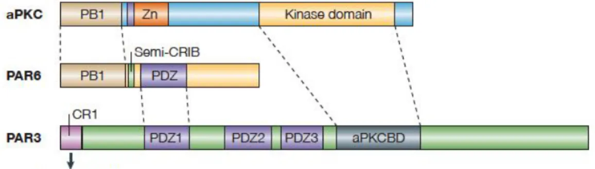
Figure I.5. Schematic representation showing the structures of polarity proteins aPKC, PAR3, and PAR6. ... 14
Figure I.6. Schematic representation of endothelial cell junctions. ... 18
Figure I.7. Schematic structure and cellular localization of VE-cadherin. ... 20
Figure I.8. Schematic representation of the primary and secondary structures of β-catenin. ... 23
Figure I.9. Transport of molecules in endothelial cells. ... 29
Figure I.10. The role of VE-cadherin in VEGF-induced permeability. ... 31
Figure I.11. Splice variants of the human VEGF-A mRNA. ... 33
Figure I.12. The binding properties of VEGFRs (VEGFR1, 2 and 3) with different VEGF ligands. ... 36
Figure I.13. Regulation of cell viability, cell migration, cell proliferation and vascular permeability via VEGFR2 signaling pathways. ... 39
Chapter II
Figure II.1. eNOS-derived NO inhibits transcriptional activity of β-catenin. ... 98 Figure II.2. NO inhibits β-catenin transcriptional activity and EC proliferation induced by Wnt3a. ... 100 Figure II.3. VEGF inhibits Wnt/β-catenin signaling in an eNOS-dependent manner. ... 102 Figure II.4. SNO of Cys466 is responsible for the inhibitory effect of NO on β-catenin transcriptional activity. ... 103 Figure II.5. Cys466 of β-catenin is necessary for NO-mediated disruption of the β-catenin/TCF4 complex. ... 105 Figure II.6. SNO of Cys466 is critical for the inhibitory effects of NO on EC proliferation. 107
Figure II.S1. S1179D-eNOS expression produces significant amounts of NO compared to S1179A-eNOS when co-transfected with β-catenin. ... 108 Figure II.S2. Transcriptional activity and cell proliferation induced by VP16-LEF1 are insensitive to the effects of NO. ... 109
Chapter III
Figure III.1. RNA-seq experimental design and workflow. ... 136 Figure III.2. Validation of sample preparation before sequencing. ... 137 Figure III.3. Distribution patterns of total identified genes and regulated genes ... 138 Figure III.4. GO enrichment analysis of genes regulated by eNOS knockdown and a heat map showing fold changes of selected genes. ... 139 Figure III.5. GO enrichment analysis of genes differentially expressed in VEGF-treated cells and VEGF-treated eNOS-depleted cells. ... 140 Figure III.6. Hierarchical clustering of genes in VEGF-treated cells and VEGF-treated
eNOS-depleted cells. ... 141 Figure III.7. Expression of selected eNOS-knockdown-regulated genes was validated by
qRT-LIST OF TABLES
Chapter I
Table I.1. Analysis of nitrosylated peptides of β-catenin stimulated with GSNO. ... 46
Chapter II
Table II.S1.Mutagenesis sense primers (bovine) ... 110 Table II.S2. qRT-PCR primers (bovine) ... 110
Chapter III
Table III.1. Number of reads obtained and identified genes in RNA-seq samples. ... 144 Table III.2. A list of genes whose expression was profoundly regulated by eNOS knockdown in endothelial cells. ... 145 Table III.3. KEGG pathway enrichment analysis of genes regulated by eNOS knockdown. 146 Table III.4. A list of genes whose expression was differentially regulated in eNOS-depleted and VEGF-regulated eNOS-depleted cells. ... 147 Table III.5. KEGG signaling pathways analysis of genes differentially expressed in
ABBREVIATIONS
Akt/PKB : Protein kinase B Ang-1/-2 : Angiopoietin-1/-2 ARM(s) : Armadillo(s)
APC : Adenomatous polyposis coli aPKC : Atypical protein kinase C BAEC : Bovine aortic endothelial cells BBB : Blood brain barrier
Bcl-9 : B-cell CLL/lymphoma 9 protein bFGF : Basic fibroblast growth factor BH4 : Tetrahydrobiopterin
β-TrCP1: Beta-transducin repeat containing protein 1 CaM: Calmodulin
cGMP : Cyclic guanosine 3’, 5’-monophosphate CKIα : Casein kinase Iα
CRD : Cysteine rich domain
CT-CM : Control-conditioned medium (control for Wnt-conditioned medium) Cx : Connexin
DAG : Diacylglycerol
eNOS : Endothelial nitric oxide synthase ECM : Extracellular matrix
EGF : Epidermal growth factor EPC : Endothelial progenitor cell
ERK : Extracellular signal-regulated kinase FAD : Flavin adenine dinucleotide
FGF : Fibroblast growth factor FMN : Flavin mononucleotide Fz : Frizzled
GM-CSF : Granulocyte macrophage colony stimulating factor GSK3β : Glycogen synthase kinase 3 β
GSNO : S-nitrosoglutathione
GSNOR : S-Nitrosoglutathione reductase HIF-1 : Hypoxia-inducible factor-1 HMG : High mobility group HSP90 : Heat shock protein 90
HUVECs : Human umbilical vein endothelial cells Ig : Immunoglobulin
iNOS : Inducible nitric oxide synthase IL-6, -8 : Interleukin -6, -8
MMPs : Matrix metalloproteinases nNOS : Neuronal nitric oxide synthase
IQGAP1 : Ras GTPase-activating-like protein JAMs : Junctional adhesion molecules
JNK : c-Jun N-terminal kinases KO : Knockout
Krm : Kremen
LDL : Low-density lipoprotein LEF : Lymphoid enhancer factor
LRP5/6 : LDL receptor-related proteins 5 and 6 MAPK : Mitogen-activated protein kinase MMTS : Methyl methanethiosulfonate
NADPH : Nicotinamide adenine dinucleotide phosphate NES : Nuclear export signal
NLS : Nuclear localization signal NO : Nitric oxide
NPC : Nuclear pore complex Nrps : Neuropilins
ODQ : 1H-[1,2,4]oxadiazolo[4,3-a]quinoxalin-1-one PAF : Platelet-activating factor
PAK : p21 activated kinases PAR : Partitioning-defective
PLC : Phospholipase C
PP2A : Protein phosphatase 2A
Rac1 : Ras-related C3 botulinum toxin substrate 1 ROS : Reactive oxygen species
RTK : Receptor tyrosine kinase
RT-qPCR : Reverse transcription quantitative polymerase chain reaction SFK : Src family kinase
sGC : Soluble guanylate cyclase SH2 : Src Homology 2
SNO : S-nitrosylation
siRNA : Small interfering RNA TCF4 : T cell factor 4
TGF-β : Transforming growth factor-β
VCAM-1 : Vascular cell adhesion molecule -1 VEGF : Vascular endothelial growth factor
VEGFR : Vascular endothelial growth factor receptor VE-cadherin : Vascular endothelial cadherin
VSMC : Vascular smooth muscle cells WIF : Wnt inhibitory factor
Wnt3a-CM : Wnt3a conditioned medium WT : Wild type
ACKNOWLEDGEMENTS
Foremost, I would like to express my sincere gratitude to my research supervisor Dr. Jean-Philippe Gratton for his scientific guidance and encouragement throughout my study and during the preparation of this thesis. I am extremely thankful to him for the encouragement and support during the years of my Ph.D study. Besides my advisor, I would like to thank the rest of my thesis committee members for their insightful comments.
My sincere thanks also go to all the fellow labmates for their kind cooperation and support. Chantal, Cécile, Rony, Vanda, Fanny, Malika and Rosemberg have all been great colleages and supportive friends. Working with them is an invaluable experience.
Thanks also extended to all my friends who share the joy for every achievement with me and offer great help to go through a lot of difficulties. They have always been absolutely supportive and great companies.
Finally, I would like to thank all of my family members for their understanding, patience, and their great support, which were critically important for me to complete this study.
I.1. Vasculature, endothelium and angiogenesis
I.1.1 Vascular structure
The vascular system is a complex network hierarchically composed of arteries, veins and capillaries. Blood vessels form a closed tubular circulation system that transports blood and oxygen throughout the body; they are therefore crucial for the development of organs and tissue repair in human (1, 2).
Large vessels, including arteries and veins, are made up of a trilaminate tissue architecture composed of the tunica intima, tunica media and tunica externa (Figure I.1). The innermost layer, the tunica intima, is lined with endothelial cells, and the subendothelial layer underlying them, called the extracellular matrix (ECM), is a highly organized network of proteins and other macromolecules important for cell adhesion, migration and differentiation (3). The second layer, the tunica media, is separated from the tunica intima by an elastic lamina, a connective tissue sheet formed by smooth muscle cells (4). The outermost and densest layer is the tunica externa, which is made up of connective tissues and also contains nerves, fibroblasts, the ECM and capillaries (5). The capillaries are composed of a layer of endothelial cells and connective tissues, connecting arterioles and venules (6).
Figure I.1. Structure of blood vessels.
The circulatory system is a complex network of veins, arteries and capillaries. Arteries and veins contain three layers: tunica intima, tunica media and tunica adventitia. Capillaries consist of a layer of endothelial cells and connective tissue. Adapted from reference (7).
I.1.2 Endothelium and endothelial cells
The endothelium is a type of epithelium that lines the interior of blood and lymphatic vessels (Figure I.1). It forms a semi-permeable barrier between blood or lymph in the lumen and the surrounding tissues to control the passage of immune cells and metabolites. The importance of the endothelium was first recognized in relation to effect on vascular tone through the secretion of molecules that relax or contract vessels to regulate the vascular tone (7).
Endothelial cells that make up the endothelium originate from angioblasts during embryonic development. Angioblasts are derived from the mesoderm, a layer of cells that also give rise to hematopoietic cells, connective tissues, and muscles. In addition, endothelial progenitor cells (EPCs) can home into neovessels sites and differentiate into endothelial cells in adults.
In the human body, approximately 1x10¹³ endothelial cells form the endothelium that lines
the entire vascular system (8). In most adult tissues, the turnover rate of endothelial cells is slow, ranging from a couple of months to years, depending on their localization in different tissues (8). Endothelial cells regulate a variety of functions, including vascular smooth muscle tone, host defence reactions, angiogenesis and vascular homeostasis. Endothelial cells interact with smooth muscle cells in the vascular wall; alternatively, they can also interact with cells and molecules in circulating blood. One of the most well-known functions of endothelial cells is the regulation of vascular tone such as making certain adjustments to adapt with the local environment in a finely controlled mechanism. For example, endothelial cells are able to sense the shear stress generated by blood flow and pass this signal on to the surrounding tissues and smooth muscle cells,
important for the balance of tissue oxygen and metabolic supply, as well as for the remodeling of the vascular structure (9). Another important function of endothelial cells is to provide a barrier between blood and vascular tissues by acting as a semi-permeable layer.
I.1.3 Vasculogenesis and angiogenesis
Vasculogenesis and angiogenesis are two distinct mechanisms for the formation of the vascular network in the embryo. Vasculogenesis gives rise to the heart and the primitive vascular plexus inside the embryo, while angiogenesis is responsible for the remodeling and expansion of this network.
I.1.3.1 Vasculogenesis
Vasculogenesis is defined as the de novo formation of blood vessels. It is a process by which a primary vascular plexus is formed through differentiation of endothelial progenitor cells (48).
Vasculogenesis includes multiple steps: mesoderm formation, blood island differentiation, endothelial cell differentiation and primary capillary plexus formation (Figure I.2) (10). The embryonic mesoderm is the main source of vascular endothelial and hematopoietic progenitor cells; it also acts as the site of vasculogenesis. Induction of mesoderm during gastrulation results in the formation of blood islands on the yolk sac in mice on embryonic day (E) 6.5-7. Hematopoietic precursor cells are located internally in the blood islands and differentiate into
form a vascular lumen, deposit a basal lamina structure and prepare for the formation of a primitive vascular plexus near the site of their origin (10).
Figure I.2. Embryonic vasculogenesis and endothelial cell differentiation.
(A-B) During vasculogenesis, mesodermal cells differentiate into hemangioblasts, whose migration to the yolk sac leads to the formation of primitive blood islands. (C) In the blood islands, the peripheral hemangioblasts differentiate into endothelial cell precursor angioblasts, whereas the internal hemangioblasts differentiate into hematopoietic cells. (D) When activated, endothelial cells migrate and fuse together to become blood islands. These islands then remodel into tubular
I.1.3.2 Angiogenesis
Angiogenesis, a process first described in tumor growth by Folkman et al., defines the formation of new blood vessels from pre-existing ones (12, 13). Angiogenesis takes place throughout development and adulthood, whereas vasculogenesis is generally thought to occur during a limited period early in embryonic development.
I.1.3.2.1 Types of angiogenesis
There are two main types of angiogenesis: sprouting and non-sprouting angiogenesis (also called intussusceptive angiogenesis). Both types contribute to the vascularization of organs and tissues during development (14, 15). Sprouting angiogenesis is defined as a general mechanism of vessel growth that coordinates migrating endothelial tip cells and proliferative endothelial stalk cells (Figure I.3A). Intussusceptive angiogenesis is defined as the process in which vessels are split from preexisting ones via the formation of transvascular tissue pillars (Figure I.3B) (16-18).
Figure I.3. Sprouting angiogenesis and intrussusceptive angiogenesis.
(A) During sprouting angiogenesis, tip cells produce filopodia in response to angiogenic growth factors, whereas stalk cells produce less filopodia but proliferate to form vascular lumen in response to stimuli. (B) During intussusceptive angiogenesis, pre-existing vessels split into two daughter vessels via the insertion of a tissue pillar. Adapted from reference (19).
tip cells
stalk cells
A
B
Sprouting angiogenesis
Intussusceptive angiogenesis
I.1.3.2.1.1 Sprouting angiogenesis
Sprouting angiogenesis is a fundamental mechanism of vessel growth that takes place in several steps. In the presence of angiogenic growth factors, endothelial cells are activated and secrete proteases such as extracellular matrix metalloproteinases (MMPs); this leads to ECM degradation which allows endothelial cells to escape from the vessel wall and move towards the connective tissues. The endothelial cells that migrate at the vascular front are called tip cells. Tip cells contain long filopodia, membrane projections that assembled from parallel actin filaments in tight bundles that probe the environment for directional cues such as growth factors. Stalk cells are the endothelial cells that follow tip cells; they produce less filopodia but proliferate more in response to growth factors (20, 21). Activated endothelial cells can migrate into the surrounding matrix to form sprouts to connect with neighbouring vessels. The sprouts are able to establish lumens in which endothelial cells are differentiated to accommodate the local environment. Finally they mature by remodeling into a complex vascular network to co-ordinate with the existing surrounding vascular network that surrounds them. In summary, sprouting angiogenesis includes endothelial cell migration, proliferation, proteolytic degradation of ECM and the subsequent formation of capillary tubes (22).
One of the main characteristics of sprouting angiogenesis is the specification of endothelial cells into tip cells and stalk cells (23). Tip cells are migratory and polarized, whereas stalk cells are more involved in the formation of tubes, branches and vascular lumens, as well as the establishment of junctions with neighbor cells and the synthesis of basement membrane components (24, 25). Specialization of these two types of endothelial cells is transient and
which are luminized and non-proliferating. This last process further promotes vessel integrity and vasculature stabilization through an increase in cell adhesion and the dampening response produced by stimulatory factors (26, 27).
I.1.3.2.2.2 Non-sprouting (Intussusceptive) angiogenesis
Intussusceptive angiogenesis was first described in a study that observed the appearance in the capillary network of tiny pillars on rat lung microvasculature in casts by scanning electron microscopy (18). It may take place as the result of proliferation of endothelial cells inside the vessel lumen and the subsequent fusion and splitting of the capillaries. In fact, both sprouting and intussusceptive angiogenesis can concur in the vascularization of organs or tissues—including lung, heart and yolk sac–during development, depending on the number of vessels already present when the organ starts to grow quickly. For example, intussusceptive angiogenesis predominates in the lung which contains endothelial precursors and is already vascularized by vasculogenesis, whereas sprouting angiogenesis happens in the brain anlage where there are no angioblasts (15).
During intussusceptive angiogenesis, four phases are necessary for the capillary wall to extend into the lumen and split a single vessel in two. First, a contact between the two facing capillary walls is established. Second, the endothelial cell junctions are permeabilized to allow growth factors and inflammatory cells to migrate into the lumen. Third, a core, filled with cells such as pericytes and myofibroblasts, is formed between the two new vessels at the contact region. During this phase, collagen fibers in the core provide ECM for the growth of the vessel lumen. Finally, the core is removed and a new vessel is formed. Intussusceptive angiogenesis is important due to its role in reorganizing pre-existing cells, which allows an increase in the number of
of importance during development, especially when there is lack of resources to create a rich microvasculature with new cells which will eventually develop into a new vessel (17).
I.1.3.2.2 Proliferation and migration of endothelial cells during angiogenesis
During angiogenesis, growth factors and chemokines can stimulate endothelial cells to break out of their stable position in the vessel wall and coordinate sprouting, branching and new lumenized network formation (28-30). These processes require the coordination of endothelial cell proliferation, migration and polarization in response to a set of molecular cues that are integrated in both space and time.
Endothelial cell proliferation is important during angiogenesis to enable sprouts to expand in length and diameter, and form new vessels. A useful tool to assess endothelial cell proliferation is BrdU labeling, a technique commonly used in the detection of proliferating cells in living tissues. BrdU, a marker of S phase entry, can be incorporated into newly synthesized DNA during cell replication and be detected by specific antibody (31).
Endothelial cell migration can be driven by growth factors such as VEGF and basic fibroblast growth factor (bFGF), a process called chemotaxis or directional migration. Organogenesis, wound healing and immune responses are orchestrated of direction cell migration toward specific locations. During directional migration, endothelial cells adopt a front-rear polarized morphology where their leading edge form membrane protrusions and attach to the underlying substrate though new contacts. It involves the following main events (Figure I.4). First,
lamellipodia are cytoplasmic protrusions formed at the leading edge of migrating cells (32). Second, the protrusion is attached to the focal adhesion, a multimeric protein complex that provides an adhesive link between the intracellular actin and the ECM. Third, the cell body is contracted by stress fibers to allow its forward progress. Stress fibers are actin microfilaments located at the rear of the cell, which contract and pull the trailing edge forward to keep up with the rest of the cell. Finally, adhesions are dissembled and the rear of the cell is released (29, 33).
Figure I.4. Directed migration process of a cell.
For a cell to migrate, it first responds to stimuli by developing filopodia and lamellipodia structures at the leading edge. Cell morphology is modified under the control of a protein family of small GTPases (Rac1, RhoA and Cdc42) and this process is referred to as cell polarization. The lamellipodia formed at the leading edge of cells are stabilized and anchored to the underlying substrates through the de novo formation of focal adhesion attachments, which provides the necessary contraction required for the cell body to move forward. Forward cell movement is achieved through disassembly of focal adhesion and detachment at the rear of the cell, which allows retraction of the tail during active migration. Calpains are a family of calcium-activated proteases, which are able to promote disassembly of focal adhesion to facilitate cell migration. Adapted from reference (34).
As mentioned, when cells are migratory, they develop a front-rear axis of polarity. The front-rear polarity is essential for directional cell migration and is mediated by the localized activation of Rho GTPases and partition defective proteins (PARs). The Rho GTPases are composed of Cdc42, Rac and RhoA (35, 36). Both Cdc42 and Rac are at the front edge of the cell and play a role in the regulation of microtubule rearrangements. Cdc42 is involved in the formation of dynamic filopodia that sense the guidance cues at the beginning of migration while Rac affects actin polymerization associated with the formation of lamellipodia (37). RhoA, on the other hand, is active at the rear of the cells and controls the rear-end retraction, which allows forward mobilization by inducing actomyosin contractility (38). The activation of these proteins must be tightly controlled for the precision of cell orientation. PAR proteins including PAR3 and PAR6 are also widely recoganized as being involved in cell polarity (Figure I.5). The crosstalk between PAR proteins and the Rho family GTPases is critical for cell migration. Atypical protein kinase C (aPKC), another important regulator of polarity, can form a polarity complex with PAR6 at the leading edge of the cell, downstream of Cdc42. Cdc42 promotes aPKC activation when bound to PAR6. The PAR6/aPKC complex in turn phosphorylates PAR3. The abovementioned polarity complexes have been demonstrated to be involved in the initial polarization steps of endothelial cells to modulate migration (39, 40). In addition, PKCζ and β-catenin are known to function in a complex at adherens junctions and to localize at the leading edge together with PAR3 and PAR6 to promote directional and collective endothelial cell migration induced by the growth factor Ang-1 (40).
Figure I.5. Schematic representation showing the structures of polarity proteins aPKC, PAR3, and PAR6.
αPKC comprises a Ser/Thr kinase domain, a PB1 domain and a zinc-finger (Zn) motif. PAR6 is composed of several domains including a PB1 domain, semi-CRIB site and a PDZ domain. PAR3 contains three PDZ domains, a CR1 domain and a αPKC binding domain (aPKCBD). The PB1 (Phox and Bem1) domain, composed of approximately 80 amino acid residues, is conserved among animals, fungi, amoebas, and plants. The PDZ domain is a modular protein-interaction domain specialized for binding to short peptide motifs at the extreme carboxy termini of other proteins. The CRIB domain defines a domain containing the Cdc42/Rac interactive binding, and Semi-CRIB in PAR6 refers to a domain that is about half the usual length of a usual domain. The dotted lines show regions of the proteins that interact with each other. Adapted from reference (39).
I.1.3.2.3 Regulators of angiogenesis
The development of a proper vascular network is critical not only for development and differentiation during embryogenesis, but also for repair of wounded tissues and functional regeneration in the adult organism. Deregulation of angiogenesis is observed in numerous malignant, inflammatory, ischemic, infectious and immune disorders. It is implicated in several
Angiogenesis is important in tumorigenesis because the formation of new blood vessel is required for oxygen and nutrient exchange between the tumor periphery and the hypoxic core (43). Therefore, it is important to maintain a balanced angiogenesis. A considerable number of studies have revealed a variety of angiogenic factors in vertebrates including VEGF, Notch, Ang, Wnt, fibroblast growth factor (FGF) and Transforming growth factor-β (TGF-β).
VEGF is the central regulator of angiogenesis and participates in inducing proliferation, migration, permeability and survival of endothelial cells (44, 45). In physiological angiogenesis, VEGF induces angiogenic sprouting during early postnatal retinal development by controlling tip cell migration and stalk cell proliferation, depending on its spatial distribution (46). Also, in situ hybridization of healing wounds has shown increased expression of VEGF by epidermal keratinocytes and expression of VEGFR1 was elevated in vessels in the wound (47). In tumor angiogenesis, expression of VEGF is upregulated and the growth of tumors in mice can be inhibited by antibodies against VEGF (48).
The Notch pathway is an evolutionarily conserved signaling pathway that is required for normal embryonic development, regulation of tissue homeostasis and maintenance of stem cells in adults (49). Notch signaling controls multiple aspects of angiogenesis including cell specification (tip versus stalk cell), proliferation, motility and filopodia protrusion. Migratory tip cells express relatively high levels of the Notch ligand DLL4, whereas proliferative stalk cells express Jagged-1, a ligand which is able to activate Notch receptors (50, 51). In proliferating endothelial cells, Notch signaling is a negative regulator since evidence has shown that endothelial cell proliferation is increased when Notch signaling is suppressed (23, 52-54). Overactivation of
of the Notch intracellular domain in zebrafish inhibited the migration and generation of the filopodia of endothelial cells (53).
Angiopoietins belong to a family of vascular growth factors that are important in embryonic and postnatal angiogenesis. The most well studied angiopoietins are Ang-1 and Ang-2. Ang-1 is important for endothelial cell migration, adhesion and survival, whereas Ang-2 can disrupt the connections between the endothelium and perivascular cells, promote cell death and induce vascular regression (55). Tie-1 and Tie-2 are receptors for angiopoietins in angiogenesis and both are essential for vascular maturation (55). In Tie-1 null embryos, angiogenesis is not perturbed, however, vessels lose their integrity, leading to hemorrhage and death between E13.5 and birth (56). Tie-2 null embryos die between E9.5 and E12.5, and the vasculature remains poorly organized with only few numbers of endothelial cells and vessel branches (57). Upon Ang-1 binding, Tie-2 receptor molecules cluster to bring their kinase domains in close proximity and to allow trans-phosporylation and the initiation of downstream signal transduction (55).
Additionally, Wnts, a family of secreted growth factors, have been shown to function in angiogenesis and Wnt signaling can promote angiogenesis (58). Inactivation of Norrin, a Wnt ligand, results in Norrie disease which is characterized by ocular vascular defects (59). Abnormality in Frizzled 4, a Wnt receptor, leads to defects in retinal vascular development (60).
I.1.3.3 Neovascularization by endothelial progenitor cells
The dogma that endothelial cells are exclusively differentiated from angioblast was generally accepted. However, the identification of circulating endothelial progenitor cells (EPCs)
express various markers including endothelial markers such as VE-Cadherin, CD31/PECAM-1, KDR, TIE-2, and Flk-1, as well as stem cell markers CD133 and CD45.
Homing and differentiation of EPCs require coordinated multistep signaling events including chemoattraction, adhesion, transmigration and ultimately differentiation into endothelial cells (62). Chemokine Stromal-cell-derived factor-1 and VEGF are known factors that initiate the mobilization of EPCs. EPCs reside within a stem cell niche in the bone marrow. MMP-9, a matrix metalloproteinase involved in the degradation of the ECM, causes the release of soluble Kit ligand from EPCs in the bone marrow, allowing EPCs to mobilize to the peripheral circulation (63). Once at the site, adhesion to activated endothelial cells leads to the transmigration of EPCs through the endothelial cell monolayer, which is an important step for homing of progenitor cells (64, 65). EPCs can then invade and migrate to the same site, where they mature into functional endothelial cells (66).
EPCs are capable of mobilizing into active sites of angiogenesis and function in neovascularization in animal models under ischemia. In mice, EPCs were demonstrated to promote corneal neovascularization by mobilizing in bone marrow in response to ischemia and granulocyte macrophage-colony stimulating factor (GM-CSF) (67). In pathological conditions, such as acute myocardial infarction, the mobilization of EPCs from bone marrow increases in order to induce re-endothelialization and neovascularization. It is believed this could potentially become a promising clinical therapy for vascular repair in coronary artery diseases (61).
I.1.4 Endothelial cell-cell junctions and cell permeability
Cell junctions are composed of protein complexes that provide contact and communication between neighbouring cells. For example, endothelial cells are linked by adhesive proteins that are organized in a complex network of junction structures, mostly composed of adherens, tight and gap junctional complexes (68-70) (Figure I.6). All three types of endothelial cell junctions are required for the maintenance of vascular integrity, which is important for tissue homeostasis.
Figure I.6. Schematic representation of endothelial cell junctions.
Tight junctions are composed of claudins, occludins, JAMs, and ZO proteins. In adherens junctions, VE-cadherin mediates the adhesion between adjacent endothelial cells. The cytoplasmic tail of VE-cadherin binds to β-catenin and p120; this complex in turn binds α-catenin and then mediates the formation of actin bundles. Connexins in gap junctions mediate the transportation of
nucleus actin claudins/ occludins JAMs Tight junctions VE-cad α-catenin VE-cad β-catenin Adherens junctions Gap junctions Connexins p120 β-catenin ZO-proteins
I.1.4.1 Adherens junctions
In endothelial cells, adherens junctions consist of the transmembrane protein cadherins including VE-, N- and M-cadherin and intracellular components such as p120-catenin, β-catenin and α-catenin that link this protein to the actin cytoskeleton (71) (Figure I.6). Adherens junctions perform multiple functions in the establishment of cell–cell adhesion and signal transduction, and are important for cell adhesion, cell survival, cell growth, and paracellular permeability to circulating leukocytes and solutes (68, 72-74).
I.1.4.1.1 VE-cadherin
The cadherin family includes the calcium-dependent adhesion molecules expressed in different tissues. E-cadherin is mostly present in epithelial cells, whereas N-cadherin can be found in the nervous system, smooth muscle cells, fibroblasts and endothelial cells. In endothelial cells, N-cadherin has been found on the cell membrane and clustered at cell-cell junctions (75, 76). VE-cadherin is expressed only in the endothelial cells as an exclusive signature and is one of the first markers expressed in endothelial cell progenitors when they start to be committed to the endothelial lineage (77).
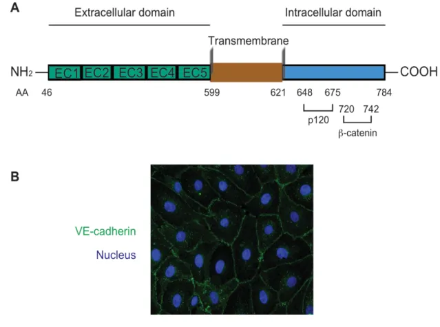
Figure I.7. Schematic structure and cellular localization of VE-cadherin.
(A) Primary structure of mouse VE-cadherin. β-catenin binds to the amino acid sites 720-742 whereas p120-catenin interacts with the juxta-membrane domain amino acid sites 648-675. AA: amimo acide. EC (1, 2, 3…5): extracellular domain (1, 2, 3…5). (B) Immunofluorescence microscopy image of cadherin localization in confluent bovine artery endothelial cells. VE-cadherin is recognized by Goat anti-VE-VE-cadherin (C19) and shown in green and DAPI labeled nucleus in blue. DAPI: 4',6-diamidino-2-phenylindole.
VE-cadherin contains five extracellular repeats (EC1-5), a transmembrane domain and a well-conserved cytoplasmic tail (Figure I.7A). VE-cadherin is localized at cell-cell contacts in confluent endothelial cells (Figure I.7B), where it forms calcium-dependent hemophilic adhesions with adjacent cells in the extracellular domain. The cytoplasmic tail of VE-cadherin can be divided into two regions: a juxtamembrane domain that binds to p120-catenin in order to regulate adherens junction stability and a catenin-binding domain that binds to β-catenin to link cadherins to the actin filament (78, 79). Phosphorylation of Tyr658 in the juxtamembrane domain was sufficient to prevent the binding of p120-catenin, and phosphorylation of Tyr731 at the catenin-binding domain caused dissociation of β-catenin (80).
During embryogenesis, VE-cadherin contributes to the remodeling and maturation of the vasculature (81-84). Deletion of the gene Cdh5, which encodes for VE-cadherin, results in early embryonic lethality due to defects in vascular remodeling and integrity (85). In vivo, the administration of blocking antibodies against VE-cadherin results in a dramatic increase in vascular permeability and hemorrhages during angiogenesis (86, 87). In vitro, VE-cadherin mediates cell contact inhibition of cell growth in endothelial cells and the presence of the last 82 amino acids at the C-terminus of the protein is required for this inhibitory effect (88).
I.1.4.1.2 β-catenin
β-catenin protein was initially identified as a ~90 kDa protein that associates with the cytoplasmic tail of E-cadherin (89, 90). β-catenin is expressed in almost all human tissues (91). In the adherens junctions of endothelial cells, β-catenin acts as a linker between the transmembrane protein VE-cadherin and the cytoplasmic protein α-catenin (72, 92-105).
The primary structure of the human β-catenin protein is composed of an NH2-terminal portion of approximately 150 amino acid, a COOH-terminal region of 115 amino acids and a central structural core of 12 repeats of 42 amino acids called armadillo repeats (ARMs) (106) (Figure I.8A). Each ARM consists of 3 helices (H1, H2 and H3), all together composed of 12 repeats stacked to form a superhelix. The superhelix contains a positively charged groove that is composed of the H3 helices and the side chains with the flanking loop regions (107) (Figure I.8B). The structure is very straight for the first 8 repeats and then curves between repeats 8 and 9 to form a pronounced cleft.
Figure I.8. Schematic representation of the primary and secondary structures of β-catenin.
(A) β-catenin is composed of an N-terminal, C-terminal and a central region that contains 12 armadillo repeats. Tyr654, Lys312, Lys435 and Cys619 of β-catenin are important amino acid sites for VE-cadherin/β-catenin association. β-catenin interacts with α-catenin via its N-terminal region, where the phosphorylation site Tyr142 is located. (B) The β-catenin crystal structure (residues 138–781) shows that each armadillo repeat is composed of three helices that are shown as blue (H1), green (H2) and yellow (H3) cylinders, and the C-terminal domain is shown in red. The C-terminal domain α-helix is called helix C. (B) is adapted from reference (108).
1 2 3 4 5 6 7 8 9 10 11 12
NH
2 Armadillo 781 1 149 666COOH
VE-cadherin A B α-cateninThe β-catenin–cadherin interaction surface spans the entire length of the 12 ARM repeat domain of β-catenin (109). The crystal structure of E-cadherin/β-catenin has been well characterized and it shows that the β-catenin Tyr654 site forms a hydrogen bond with the E-cadherin Asp665 site. Phosphorylation of Tyr654 on β-catenin causes E-E-cadherin to dissociate from β-catenin probably because the phosphate group is too large to be accommodated in the interface and the negatively charged residue electrostatically repels E-cadherin Asp665 (109). Likewise, in endothelial cells, phosphorylation of β-catenin dramatically decreases its binding to VE-cadherin and disassembles the junctions, indicating that tyrosine phosphorylation of β-catenin can modify junctional stability (110). In addition, the Asp674 and Glu682 sites of E-cadherin form salt bridges with the β-catenin Lys435 and Lys312 residues, respectively, contributing to the formation of a complex (109, 111).
β-catenin binds α-catenin with its distal parts of the N-terminal and the adjacent first ARM repeat (112). Tyrosine kinases Fyn, Fer, or c-Met can phosphorylate Tyr142 site of β-catenin that is located in the binding domain and significantly decreasing α-catenin binding to β-catenin (113-115).
β-catenin has been implicated in the development of the vascular system. β-catenin null-mutant embryos show a loss of formation of the mesoderm, suggesting that β-catenin plays a vital role in mouse development during the gastrulation stage (116). Endothelial-specific deletion of the β-catenin gene in mice is embryonic lethal due to defects in vascularization and the development of the heart (117, 118). Moreover, β-catenin is a dual functional protein: in addition to its role in the regulation of the coordination of the cell-cell junction, β-catenin is an important component in
Wnt/β-catenin signaling pathway where it plays a role in the regulation of gene transcription. The role of β-catenin in the Wnt signaling pathway will be discussed in detail in section I.2.3.3.2.
I.1.4.1.3 α-catenin
α-catenin is an actin-binding and -bundling protein that contains a β-catenin binding domain in the N-terminus and actin-binding sites in the C-terminus (112, 119-121). It links the cadherin-β-catenin complex to the actin-based cytoskeleton by binding directly to filamentous actin (F-actin) or by interacting with the actin-binding proteins (93, 95, 96). Actin is a major cytoskeletal component of endothelial cells, which exists as globular actin (G-actin) and as a filamentous polymer called F-actin, a linear chain of G-actin subunits (122, 123).
α-catenin plays an important role in intercellular adhesion during development and differentiation. In Drosophila, cell adhesion is disrupted when α-catenin contains a mutation in the binding site for Armadillo, the homologue of β-catenin in Drosophila (124).
I.1.4.2 Tight junctions
Tight junctions are formed after adherens junctions during intracellular contact formation (125). Tight junctions create a series of contacts between the adjacent cells to close the extracellular space and form a tight intercellular barrier. For example, tight junctions play a central role in establishing the blood-brain barrier (BBB), a highly selective permeable barrier formed by
arteries generally possess more developed tight junctions compared to those in capillaries and venules, due to exposure to high rates of blood flow (127). In addition, the barrier function of tight junctions is to transport between cells and help to maintain cell polarity between the luminal and the abluminal side of the endothelial cells by separating their apical and basolateral compartments (128).
Tight junctions are composed of claudins, occludins, junctional adhesion molecules (JAMs) and zonula occludens (ZOs) (Figure I.6). Claudin family members are the major transmembrane components in the endothelial cell tight junctions that are located at the most apical part of the lateral surface (129). Claudins exhibit hemophilic or heterophilic adhesive properties through their extracellular domains and directly regulate the barrier function of tight junctions (130). Deletion of the claudin-5 gene in mice results in post-natal death due to a defective BBB (131). Occludin is another transmembrane protein in tight junctions containing two extracellular loops (132). Occludin is associated with increased tight junction function and is highly expressed in the BBB. Downregulation of occludin is associated with several diseases including stroke, diabetes and hypoxia (70). Molecules in the JAMs family are type I transmembrane glycoproteins composed of a single transmembrane domain. The extracellular domain of JAMs folds into two immunoglobulin-like domains. JAMs play important roles in the assembly of cell-cell junctions, establishment of cell polarity and regulation of leukocyte migration through endothelial cells (133-135). ZO proteins (ZO-1, ZO-2, and ZO-3) are cytoplasmic scaffold proteins that link transmembrane proteins to the cortical actin cytoskeleton. The localization of ZO-1 in the cell periphery is positively correlated with the confluence of endothelial cells, where tight junctions are formed (136). In endothelial cells, ZO-1 has been implicated in the regulation of adherens
I.1.4.3 Gap junctions
Gap junctions are intercellular channels that directly connect the cytoplasm of adjacent cells in order to transport water, ions and other small molecules between cells. Thus, gap junctions play an important role in cell communication (72). Connexins (Cx), such as Cx43, Cx40 and Cx37 are the main components of gap junctions. Six Connexins combined together to form one connexon; connexons act as channels between adjacent endothelial cells (Figure I.6). Studies show that vascular endothelial cell-specific ablation of Cx43 causes hypotension associated with significant induction of plasma nitric oxide (NO), indicating a role for Cx43 in vascular homeostasis (138). Another report showed that simultaneous deletion of Cx37 and Cx40 in mice resulted in severe vascular abnormalities and eventually lethality on postnatal day 1, underscoring the importance of endothelial cell gap junctions in the development of the vasculature (139).
I.1.4.4 Endothelial cell permeability
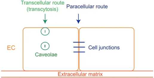
Endothelial cells separate blood from the different tissue compartments and modulate the passage of cells and solutes between blood and tissue. Vascular permeability increases when quiescent endothelium is exposed to stimuli such as growth factors and cytokines (140, 141). The passage of macromolecules, fluids and cells through the endothelial barrier can occur either through (transcellular) or between (paracellular) cells (Figure I.9) (142).
Small molecules are generally transported by transcytosis, which requires a trafficking vesicle system that involves caveolae and their coat proteins, caveolins (143). Caveolae are invaginations of the plasma membrane, which are also a special type of lipid raft. One of the first functions attributed to caveolae in endothelial cells is the ability to transfer molecules from the lumen of blood vessels to the sub-endothelial space by transcytosis. Caveolae can be released from the plasma membrane into cytoplasm during transcytosis, with different sizes between 50 and 100 nm. Caveolae-mediated transcytosis is a critical mechanism used for the transport of albumin, insulin, low-density lipoproteins and hormones across the endothelial barrier (144, 145).
Figure I.9. Transport of molecules in endothelial cells.
The passage of macromolecules, fluids and cells through endothelial cells can take place through transcellular (through caveolae) or paracellular (through cell junctions). Adapted and modified from reference (142).
Transcellular route
(transcytosis) Paracellular route
Caveolae
Extracellular matrix
The passage of cells and large macromolecules is mostly done via the paracellular pathway and is finely controlled by the opening and closing of endothelial cell-cell junctions (142). Several factors, including VEGF, can increase endothelial cell permeability and leukocyte diapedesis through modulation of the VE-cadherin-catenin complex in the plasma membrane (146-149). The initial step of induction of the endothelial cell permeability involves the destabilization of adherens junctions (71, 150). VEGF stimulation can activate the Src family kinase (SFK) to induce tyrosine phosphorylation of VE-cadherin and β-catenin respectively, resulting in the subsequent disassembly of the adherens junctions (Figure I.10A) (151, 152). Another important mechanism in VEGF-mediated endothelial cell permeability is through VE-cadherin internalization, which also requires Src activation. In this scenario, Src kinase-induced phosphorylation of VE-cadherin provokes the internalization of VE-cadherin through clathrin-mediated endocytosis, a process in which cells absorb proteins through the inward budding of the plasma membrane vesicles containing the proteins being absorbed (80, 153-155) (Figure I.10B).
Figure I.10. The role of VE-cadherin in VEGF-induced permeability.
Homophilic interactions of VE-cadherin are expressed in adjacent endothelial cells to maintain the integrity of adherens junctions via the interaction of VE-cadherin with actin cytoskeleton using catenins. VEGF binds to its receptor and induces Src activation, resulting in the phosphorylation of VE-cadherin. (A) Src can induce not only VE-cadherin phosphorylation but also β-catenin phosphorylation, dissociating the cadherin-catenin complex in parallel. (B) Phosphorylation of VE-cadherin leads to its internalization into clathrin-coated pits. The process can cause dissociation of VE-cadherin from catenins and disassembly of cell adherens junctions, ultimately leading to increased permeability. Endosomal VE-cadherin may be recycled to the cell surface to participate in reorganization of adherens junctions in vascular remodeling (153, 156).
actin VE-cad α-catenin VE-cad α-catenin actin β-catenin VE-cad VE-cad P P actin VE-cad α-catenin VE-cad α-catenin β-catenin A B VE-cadher in Endocytosis Recycling VEGF VEGF receptor Src activation β-catenin β-catenin Clathrin VE-cad P β-arrestin P120 P120 P120 β-catenin β-arrestin
Endothelial cell permeability is important since the disruption of intercellular junctions between endothelial cells would increase the passage of macromolecules through the endothelium, thereby providing a pro-angiogenic microenvironment (157, 158). In summary, it has been found that endothelial cell junctions are crucial for cell permeability and angiogenesis in the literature review in this section. In the next section, I am going to discuss the importance of several signaling pathways in endothelial cells that are responsible for their functions, especially those involved in angiogenesis.
I.2. Signaling pathways in endothelial cells
I.2.1 VEGF/eNOS signaling pathway
VEGF binds to its receptors on the surface of endothelial cells and activates intracellular tyrosine kinases, triggering multiple downstream events, such as Phosphatidylinositol 3-kinases (PI3K)/Akt, phospholipase Cγ (PLCγ) and endothelial nitric oxide synthase (eNOS) signaling pathways, that regulate vascular development and function in both healthy organisms and those with diseases such as cancer.
I.2.1.1 VEGF ligands
VEGF was first identified by Senger et al. in 1983 as a functional factor in vascular permeability and vascular development (159). It is well regarded as an endothelial cell-specific mitogen and chemotactic agent that is part of the cysteine-knot growth-factor superfamily.
VEGF is expressed in various types of cells including endothelial cells, vascular smooth muscle cells, macrophages and tumor cells (160). VEGF can be found in mammals as part of a family of structurally homologous secreted glycoproteins: A, B, C, VEGF-D and placental growth factor (P1GF) (161). All the five VEGFs can be alternatively spliced to form multiple isoforms. VEGF-A is generated by different types of cells including endothelial cells under stress conditions, and is highly expressed in tissues undergoing growth (162). The VEGF-A gene is differentially spliced into various isoforms: VEGF121, VEGF145, VEGF165,
VEGF189 and VEGF206 (Figure I.11). VEGF165 is the most predominant and biologically active
isoform and thus will be the form referred to for the remainder of the thesis.
Figure I.11. Splice variants of the human VEGF-A mRNA.
There are eight exons in the VEGF-A gene and alternative splicing results in several isoforms of different sizes: VEGF121, VEGF145, VEGF165, VEGF189 and VEGF206. Adapted from reference
In humans, VEGF is usually produced as a dimeric glycoprotein: a 46 kDa homodimer composed of two 23 kDa subunits (163). Stress conditions such as hypoxia induce VEGF mRNA expression, either through the direct binding of the transcription factor hypoxia inducible factor (HIF) to the regulatory cis-acting enhancer elements located in the VEGF gene, or through the increased stabilization of its mRNA (164, 165). Additionally, a variety of cytokines and growth factors, including epidermal growth factor (EGF), TGF-β and interleukin 6 (IL-6), have been demonstrated to promote VEGF expression in vitro (166, 167). More recent studies have shown that the activation of the Wnt signaling pathway in colonic neoplasia strongly upregulates VEGF expression by modulating the T cell factor 4 binding element found in the VEGF promoter (168).
VEGF plays an important angiogenic role in inducing a directional, chemoattractive cue and is critical for endothelial cell permeability, proliferation and migration (169, 170). Deletion of the VEGF gene on one of the alleles would ultimately cause embryonic lethality due to a variety of defects in angiogenesis, suggesting that expression of VEGF on both alleles is crucial to the formation of a closed circulation system (171, 172). Overexpression of VEGF also leads to early embryonic lethality due to vascular defects, implying that embryonic survival requires the precise control of VEGF-A expression (171, 173).
Besides the activities in the vascular system, VEGF also plays a role in physiological functions such as wound healing, bone formation, hematopoiesis and development (174, 175).
I.2.1.2 VEGF receptors
VEGF factors regulate vascular development, angiogenesis and lymphangiogenesis by binding to a number of homologous receptor tyrosine kinases (RTKs) that are VEGF receptors (VEGFR), including VEGFR1/Flt-1, VEGFR2/Flk-1/KDR, and VEGFR3/Flt4, which are expressed mainly in endothelial cells in blood vessels and in the lymphatic system (176-181). VEGFRs are composed of an extracellular region of seven Ig-like domains, a short transmembrane domain, a juxtamembrane domain, an intracellular region containing a tyrosine kinase domain and a C-terminal tail (Figure I.12). VEGFRs are all essential for VEGF-induced angiogenesis since knockout mice for either isoform can lead to embryonic lethality due to vascular defects (182-185).
VEGFR1 is expressed in hematopoietic stem cells, monocytes, macrophages and vascular endothelial cells. Ablation of the VEGFR1 gene Flt-1 in mice causes impairment of the vasculature and embryonic lethality on E9.0 (182). In the Flt-1-null mutant, abnormal blood island and vascular assembly was observed, suggesting that Flt-1 is important for organization of embryonic vasculature.
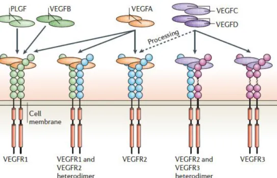
Figure I.12. The binding properties of VEGFRs (VEGFR1, 2 and 3) with different VEGF ligands.
Various VEGF ligands bind to the three VEGFRs, resulting in the formation of different VEGFR homo- or heterodimers. VEGF binds the extracellular part of the receptors that contains seven immunoglobulin-like subunits. VEGFA binds VEGFR1, VEGFR2, or the dimers of the two. The dimers formed by both VEGFB and PLGF bind VEGFR1. Proteolysis processing of VEGF-C and -D enables binding to VEGFR2. Adapted from reference (181).
VEGFR2, which is expressed in vascular and lymphatic endothelial cells, is responsible for most of the mitogenic and chemotactic effects of VEGF. VEGFR2-null mice die in utero between E8.5 and E9.5 as a result of impaired development of hematopoietic and endothelial cells, suggesting its indispensable role in the establishment of the vascular system (185). The phenotypes of VEGFR2-null mice resemble to a great extent those in VEGF-null mice. In addition, VEGFR2 expression is found in the embryonic precursors of endothelial cells, with its highest levels observed during embryonic vasculogenesis and angiogenesis, and during pathological processes such as neovascularization during tumor angiogenesis, confirming the essential role of VEGFR2 in vascular assembly (186, 187).
VEGFR3 is only found in lymphatic endothelial cells and binds both C and VEGF-D (181). Vegfr3-/- mice die due to the defective vascular remodeling before the emergence of lymphatics, indicating that VEGFR3 is essential for blood vascular development (181).
In addition, neuropilins (Nrps) are a family of cell surface receptors involved in VEGF intracellular signaling transduction as co-receptors for VEGFRs (188). There are two NRP homologues including NRP1 and NRP2, which are transmembrane proteins (189, 190). NRP1 may either homomultimerize or form heteromultimers with NRP2 (188). NRP1 is able to bind and form complex with VEGFR2, whereas NRP2 is associated with VEGFR3 in a VEGFC- or VEGFD-dependent manner (191, 192).
I.2.1.3 VEGF signaling pathways
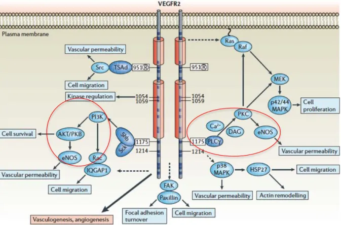
Even though the affinity of VEGFR1 for VEGF is much higher than that of VEGFR2, the effect of VEGF is mainly transduced by VEGFR2 on endothelial cells. VEGF has been shown to activate a number of different intracellular signaling pathways through VEGFR2, including protein kinase C (PKC), PI3K/Akt/PLCγ, mitogen-activated protein kinase (MAPK)/extracellular signal-regulated kinase (ERK), and p38 MAPK (Figure I.13). These signaling pathways play important roles in mediating endothelial cell survival, proliferation, migration, and permeability. Here, we focus on PI3K/Akt and PLCγ signaling because these pathways are involved in the activation of eNOS that results in the generation of NO from endothelial cells (193).
There are several phosphorylation sites which are important for the transduction of signals of VEGF (Figure I.13). For example, Tyr951 of VEGFR2, a binding site for the T-cell-specific adaptor, participates in the regulation of endothelial cell migration (194). Phosphorylation of Tyr1175 on VEGFR2 is essential for VEGF-induced PI3K and PLCγ activation and the downstream events (181). Additionally, Tyr801 is also an autophosphorylation site of VEGFR2 and is essential for activation of PI3K/eNOS signaling and generation of NO (193).
Figure I.13. Regulation of cell viability, cell migration, cell proliferation and vascular permeability via VEGFR2 signaling pathways.
Upon binding of VEGF, VEGFR2 is phosphorylated on several sites including Tyr951, Tyr1054, Tyr1059, Tyr1175 and Tyr1214. Phosphorylation of these sites on VEGFR2 is required to trigger signaling pathways such as PI3K, PLCγ, Src, Ras and p38; these pathways are important in cell survival, cell migration, cell proliferation, vascular permeability, focal adhesion turnover and actin remodeling. Adapted from reference (181).