2005/234
Cytosolic persistence of mouse brain CYP1A1 in chronic
heme deficiency*
Ralf P. Meyer
1,**, Raija L.P. Lindberg
2, Francine
Hoffmann
2and Urs A. Meyer
31
Department of Neuropathology, Neurozentrum,
University of Freiburg, Breisacherstraße 64, D-79106
Freiburg, Germany
2
Pharmazentrum, Department of Research and
Neurology, Klingelbergstraße 50-70, CH-4056 Basel,
Switzerland
3
Biozentrum, Department of Pharmacology,
Klingelbergstraße 50-70, CH-4056 Basel, Switzerland
**Corresponding authore-mail: [email protected]
Abstract
Previous work has demonstrated that the function of
extrahepatic cytochrome P450 CYP1A1 is dependent on
the availability of heme. CYP1A1 is involved in the
acti-vation of polyaromatic hydrocarbons. In the present
study we used a transgenic mouse model with chronic
impairment of heme synthesis – female porphobilinogen
deaminase-deficient (PBGD
-/-) mice – to investigate the
effects of limited heme in untreated and
b-naphthofla-vone (b-NF)-treated animals on the function of CYP1A1
in brain. The heme content of PBGD
-/-mice was
dimin-ished in the liver and brain compared to wild types. In
the liver, partial heme deficiency led to less potent
induc-tion of CYP1A1 mRNA after b-NF treatment. In the brain,
CYP1A1 protein was detected not only at the
endoplas-mic reticulum (ER), but also in the cytosol of PBGD
-/-mice. Furthermore, 7-deethylation of ethoxyresorufin, an
indicator of CYP1A1 metabolic activity, could be restored
by heme in cytosol of PBGD
-/-mouse brain. Independent
of the genotype, we found only one cyp1a1 gene
prod-uct, indicating that the cytosolic appearance of CYP1A1
most likely did not originate from mutant alleles. We
con-clude that heme deficiency in the brain leads to
incom-plete heme saturation of CYP1A1, which causes its
improper incorporation into the ER membrane and
per-sistence in the cytosol. It is suggested that diseases
caused by relative heme deficiency, such as hepatic
por-phyrias, may lead to impaired hemoprotein function in
brain.
Keywords: apoprotein; cytochrome P450; membrane
insertion; porphyria; transgenic mouse.
*Parts of this report were presented at the 15th International Symposium on Microsomes and Drug Oxidations (MDO), Mainz, Germany, July 2004. The study was awarded with the poster prize.
Introduction
Heme is one of the body’s pivotal molecules. It fulfils a
vital role in cellular oxygen sensing and utilization in all
living organisms from bacteria to humans (Padmanaban
et al., 1989). Heme is the prosthetic group in numerous
hemoproteins, including P450 cytochromes (CYPs)
(Ran-garajan and Padmanaban, 1989; Zhang et al., 1998;
Abraham et al., 2000). CYPs play an important role in the
oxidative metabolism of numerous endogenous and
for-eign compounds (Nelson, 2004). CYPs are active in drug
metabolism and clearance, in cholesterol homeostasis,
and in steroid hormone regulation, not only in the liver,
but also in the brain (Thuerl et al., 1997; Meyer et al.,
2001a,b; Ourlin et al., 2002; Handschin and Meyer, 2003).
CYPs perform their manifold functions as functional
holo-enzymes saturated with heme (Correia and Meyer, 1975;
Jover et al., 1996). Thus, the regulation of heme
availa-bility is of major importance for CYP function. Cellular
levels of heme are regulated by its rate of synthesis and
degradation by the key enzymes d-aminolevulinate
syn-thase 1 (ALAS-1) and the heme oxygenase (HO) system
(Jover et al., 1996; Maines, 2000). However, regulation of
heme availability for CYPs is tissue-dependent. Previous
studies in vitro and in mice, both with pharmacologically
induced acute heme deficiency, have revealed
differen-ces in heme saturation of CYPs and pointed to a role of
heme in the incorporation of apocytochromes into the
endoplasmic reticulum (ER) membrane (Meyer et al.,
2002). A regulatory heme pool tightly regulates heme
synthesis in the liver, but apparently less so in
extrahe-patic tissue (Bissell and Hammaker, 1976b; Giger and
Meyer, 1983).
Porphyrias are inherited or acquired disorders that are
attributable to partial defects in the enzymes of heme
synthesis (porphyrin metabolism), usually in association
with endogenous or exogenous stressors (Daniell et al.,
1997). In certain so-called hepatic porphyrias, patients
have neuropsychiatric symptoms, including autonomic
neuropathy with abdominal pain, vomiting, and
hyperten-sion, and central neuropathies, including motor
weak-ness, paralysis and mental symptoms (Sassa, 2002).
These symptoms may possibly originate from impaired
heme protein function in the brain. Candidate
hemopro-teins in the brain are the nitric oxide synthase, guanylate
cyclase system, tryptophan dioxygenase, mitochondrial
cytochromes, and several isoforms of the CYP
superfam-ily (Jover et al., 2000).
In the present study, we investigated if expression and
function of CYP1A1 is affected in vivo by chronic heme
deficiency in the brain. CYP1A1 is involved in the
meta-bolic activation of polyaromatic hydrocarbons
(Hankin-son et al., 1991). We used a female transgenic mouse
model mimicking one form of hepatic porphyria, acute
intermittent porphyria (AIP). These mice carry a targeted
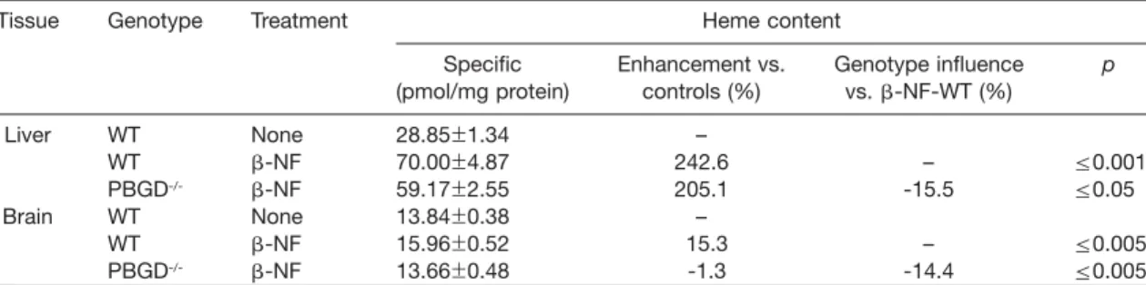
Table 1 Heme content of mouse liver and brain after treatment with b-naphthoflavone.
Tissue Genotype Treatment Heme content
Specific Enhancement vs. Genotype influence p
(pmol/mg protein) controls (%) vs. b-NF-WT (%)
Liver WT None 28.85"1.34 – WT b-NF 70.00"4.87 242.6 – F0.001 PBGD-/- b-NF 59.17"2.55 205.1 -15.5 F0.05 Brain WT None 13.84"0.38 – WT b-NF 15.96"0.52 15.3 – F0.005 PBGD-/- b-NF 13.66"0.48 -1.3 -14.4 F0.005
WT, wild type. Specific values represent pooled organs from two mice (mean"SEM; ns3). Treatment with b-NF was performed as described in the materials and methods section.
disruption of porphobilinogen deaminase (PBGD), the
third enzyme of the heme pathway (Lindberg et al., 1996,
1999). PBGD
-/-mice have a defined chronic disturbance
of heme biosynthesis, which can be challenged by
CYP-inducing factors. Therefore, we treated the mice with
b-naphthoflavone (b-NF), an inducer of CYP1A1. Here
we demonstrate that gene expression, heme saturation,
and enzyme activity of CYP1A1 are impaired in PBGD
-/-mice after treatment with b-NF. Our results indicate that
diseases causing heme deficiency, such as AIP, could
lead to impaired hemoprotein function in brain.
Results
Measurement of heme content
Initial experiments were designed to determine whether
heme content in the liver and brain of mice is increased
by b-NF-treatment and affected by the PBGD
-/-geno-type. In wild-type mice, b-NF treatment increased heme
content in both liver and brain, with much stronger
enhancement in liver than in brain (Table 1). In PBGD
-/-mice, the b-NF-dependent enhancement of heme
con-tent was lower in liver and brain compared to wild-type
data (Table 1). It is noteworthy that the heme content in
brains of b-NF-treated PBGD
-/-mice was similar to that
found in untreated wild-type mice (Table 1).
Regulation of cyp1a1 mRNA expression in liver
In a subsequent experiment, we analyzed if the
signi-ficant reduction in heme content of b-NF-treated
PBGD
-/-mice compared to b-NF-treated wild-types
corresponds to diminished cyp1a1 gene transcription.
Therefore, the expression of mRNA in liver of untreated
wild-types, b-NF-treated wild-types and b-NF-treated
PBGD
-/-animals was investigated by Northern blot
(Fig-ure 1). As expected, b-NF treatment led to a significant
increase in cyp1a1 mRNA transcription in both
geno-types (pF0.005). Nevertheless, expression of cyp1a1
mRNA originating from b-NF-treated heme-deficient
PBGD
-/-mice was clearly lower compared to
b-NF-treat-ed wild-type mice (Figure 1, lanes 1 and 2), indicating a
negative effect of PBGD deletion and reduced heme
availability for cyp1a1 gene transcription (pF0.05). No
mRNA was detectable in untreated liver (Figure 1, lane
3). This finding is due to the fact that gene transcription
of cyp1a1 is usually very low in untreated animals
(Gon-zalez, 1990). GAPDH control data showed no difference
(Figure 1).
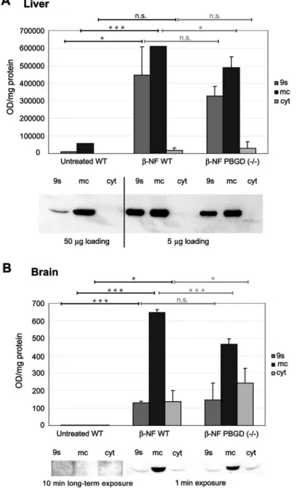
Expression of CYP1A1 protein in cellular
subfractions of liver and brain
We next investigated by immunoblot how b-NF treatment
and heme deficiency affect expression and subcellular
localization of CYP1A1 protein in liver and brain (Figure
2). In the liver, the genotype-dependent inducibility of
CYP1A1 protein was in accordance with our findings
investigating mRNA expression (see Figure 1). b-NF
treatment led to the expected induction of CYP1A1 in
both wild-types and PBGD
-/-mice (pF0.001), but with
less potency in subfractions of PBGD
-/-mice, for which
a lower CYP1A1 protein signal was observed compared
to wild-type mice (Figure 2A) (pF0.05). CYP1A1 was
located almost exclusively in microsomes, independent
of treatment and genotype.
In brain, we observed a striking difference in CYP1A1
expression
between
b-NF-treated
wild-types
and
PBGD
-/-mice (Figure 2B). b-NF treatment led to a
1.4-fold increase in CYP1A1 in microsomes of wild-types
compared to PBGD
-/-mice (pF0.001). In addition,
cyto-solic CYP1A1 was detected, which was two-fold higher
in the PBDG
-/-mice compared to the wild-type animals
(pF0.05). In untreated mice, CYP1A1 was not detectable
in the brain (Figure 2B).
Ethoxyresorufin-O-deethylase (EROD) activity of
CYP1A1 in mouse brain is dependent on genotype
and b-NF induction
The investigation of CYP1A-specific EROD activity in
brain subfractions matched our findings from
immuno-blotting experiments. In untreated mice, no or only
neg-ligible EROD activity could be found in any subfraction,
whereas treatment of the mice with b-NF led to
remark-able increase in EROD activity in all subfractions tested
(Figure 3). EROD activity was found predominantly in the
microsomes, with no significant differences between
wild-types and PBGD
-/-mice (Figure 3A). The EROD
activity detectable in the cytosol revealed clear
depend-ence on genotype after treatment of mice with b-NF.
Cytosolic EROD activity of PBGD
-/-mice appeared to be
two-fold higher than in wild-types, which reflects a
remarkable fraction of non-membrane bound CYP1A1 in
the brain (Figure 3A) (pF0.005). This
genotype-depend-ent difference in cytosolic EROD activity remained,
Figure 1 Expression of cyp1a1 mRNA in liver from wild type and PBGD-/-mice (Northern blot analysis).
(1) b-NF-induced wild types; (2) b-NF-induced PBGD-/- mice;
and (3) untreated wild types, with 5
mg of total RNA loaded on
each lane. In the case of cyp1a1 mRNA, a 294-bp probe located on exon 7 was used for hybridization (see materials and meth-ods). GAPDH was used as a reference. Statistical significance is demonstrated by asterisks (**pF0.005; *pF0.05).although less striking, after the addition of 8
m
Mhematin
(Figure 3B). Addition of hematin is reported to saturate
and reconstitute the CYP apoprotein fraction (Omiecinski
et al., 1980). We observed a three-fold enhancement of
EROD activity in cytosol in PBGD
-/-mice, and five-fold
enhancement in wild-types, after the addition of hematin
(Figure 3B). In microsomes, however, hematin caused a
reduction in EROD activity (pF0.05).
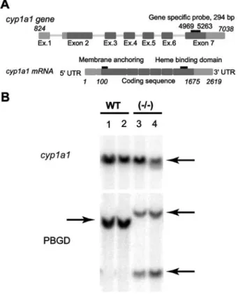
Gene products of cyp1a1
To examine if cytosolic CYP1A1 originates from an allelic
or splice variant of cyp1a1 leading to either cytosolic or
microsomal protein, we generated a gene-specific probe
designed to recognize sequences of exon 7. This probe
should allow the detection of CYP1A1 gene products
with differences in the membrane-anchoring sequence
(Gonzalez et al., 1984) (Figure 4A).
However,
hybridization
of
mouse
genomic
DNA
obtained from wild-type and PBGD
-/-mice with the
294-bp probe revealed that, most likely, only one cyp1a1 gene
product exists in the mouse, independent of the
geno-type (Figure 4B). Therefore, the solubility of CYP1A1 in
brain may not originate from an allelic or splice variant of
the cyp1a1 gene.
Discussion
CYP1A1 is expressed predominantly in extrahepatic
tis-sue and abundantly in brain (McFayden et al., 1998; Iba
et al., 2003). Our previous in vitro studies have suggested
that heme deficiency leads to increased accumulation of
CYP1A1 in the cytosol of COS-1 cells and untreated
male mice (Meyer et al., 2002). This is supported by
the present in vivo data in female PBGD
-/-mice. The
PBGD
-/-mouse provides a unique model of chronic
impairment of heme synthesis (Lindberg et al., 1996,
1999). After b-NF-treatment, the chronic partial heme
deficiency in these mice led to impaired CYP1A1
expres-sion and enzyme activity in mouse liver and brain. More
surprisingly, CYP1A1 protein and activity were detected
in increased amounts in the cytosol of mouse brain after
b-NF treatment of PBGD
-/-mice compared to wild-type
mice. The use of female mice in the present in vivo study
has the advantage that, in contrast to males (Meyer
et al., 2002), in untreated mice, CYP1A1 mRNA, protein
and EROD activity were not detectable in both liver and
brain. CYP1A1 protein is known to be highly inducible in
several brain regions, corresponding to elevated levels of
microsomal EROD activity (Morse et al., 1998). Therefore,
the genotype-dependent effects of the CYP1A1 inducer
b-NF on heme content, CYP1A1 expression and enzyme
activity under conditions of chronic heme deficiency, as
investigated in the present study, substantially extend
data from our previous in vitro study mimicking the
untreated situation (Meyer et al., 2002).
We have speculated that the availability of heme
effi-ciently regulates the incorporation of apocytochrome
CYP1A1 into the ER membrane, at least in extrahepatic
tissue. Interestingly, this effect of heme deficiency,
lead-ing to cytosolic accumulation of CYP1A1, is not
observed in the liver (Omiecinski and Juchau, 1980;
Mey-er et al., 2002). This can be explained by the efficient
adaptation of ALAS-1 to increased heme demands, such
as drug treatment, in liver and intestine, which is
medi-ated by nuclear receptor signaling by CAR and PXR
(Fra-ser et al., 2003). In extrahepatic tissue, ALAS-1 obviously
adapts differently to heme demands or drug treatment,
as stimulation of ALAS-1 by drugs is less marked than
in the liver (Reed et al., 1989). Although the brain
pro-duces heme required for synthesis and turnover of its
own hemoproteins (De Matteis et al., 1981), most of the
constitutively synthesized heme is used for cell
homeo-stasis and the generation of carbon monoxide and nitric
oxide, and is not drug inducible (Ingi et al., 1996; Jover
et al., 2000; da Silva et al., 2001). Heme oxidation in brain
is effectively controlled and balanced by the activity of
the heme oxygenase isoforms HO-1 and HO-2 (Maines,
2000).
Moreover, administration of exogenous heme to
por-phyria patients and to healthy volunteers increased
P450-mediated drug metabolism (Bonkovsky et al.,
1991). Our present study suggests that this increase is
due to heme effects in extrahepatic tissues,
reconstitut-ing undersaturated apocytochromes and/or leadreconstitut-ing to
greater incorporation of cytoplasmic apocytochromes
into the ER membrane, elevating their enzyme activity.
This concept is corroborated by significantly enhanced
levels of cytosolic EROD activity in b-NF-treated mice
after the exogenous addition of hematin. Extrahepatic
CYP apoproteins are reported to have low affinity for their
heme prosthetic group, but a comparatively high
turn-over number when in the holoenzyme state (Omiecinski
et al., 1980). The effect of hematin in lowering
microso-mal EROD activity may be explained by the fact that,
after induction, the membrane-bound fraction of CYP1A1
is in the holoenzyme state and excessive heme may be
inhibitory (Namkung et al., 1983). Conversely, this
con-cept of high turnover numbers of extrahepatic CYPs in
the holoenzyme state can most likely explain our finding
that, if no exogenous heme is added, there is largely no
difference in microsomal EROD activity after b-NF
induc-tion between wild-type and PBGD
-/-mice, whereas
CYP1A1 protein is clearly reduced in PBGD
-/-mice.
The mechanistic aspects of heme incorporation into
CYP apoproteins and the role of this process in
membrane incorporation is still poorly understood. Our
observation that heme deficiency leads to cytosolic
Figure 2 Expression of CYP1A1 protein in subfractions of liver and brain of wild-type and PBGD-/-mice.
Scanning densitometry evaluation of immunoblots with monoclonal antibodies mab 1-7-1 raised against CYP1A1 (Park et al., 1982). (A) CYP1A1 immunoblot of liver subfractions from female mice. Values represent a pool of two untreated animals and a pool of two b-NF-treated animals of each genotype (mean"SEM; ns3) The mapped blot is a representative of these experiments. (B) CYP1A1 immunoblot of brain subfractions from female mice. Values represent a pool of four untreated animals and a pool of two b-NF-treated animals of each genotype (mean"SEM; untreated, ns3; b-NF-treated, ns4). The mapped blot is a representative of these experi-ments. Protein from 9000 g supernatant (9s), microsomes (mc) and cytosol (cyt) were separated by SDS-PAGE. Immunosignals were visualized using enhanced chemiluminescence (ECL). Statistical significance is demonstrated by asterisks (***pF0.001; *pF0.05; n.s., not significant).
accumulation of CYP1A1 may be important for further
clarification of this process. Several previous studies
pro-vided evidence for the concept that heme incorporation
and exchange as well as membrane insertion of CYP
iso-forms is a dynamic process between the cytoplasm and
ER membranes. The biosynthesis of apocytochrome was
shown to precede that of heme, so that incorporation of
heme into the nascent apoprotein must take place prior
to or in close relation to membrane insertion (Meier et al.,
1984; De Matteis and Marks, 1996). Furthermore, CYP
heme can dissociate reversibly from its apoprotein to
allow heme exchange (Bissell and Hammaker, 1976a). It
was suggested from studies in testis that a less stable
form of CYP may be present in extrahepatic tissues that
shows impaired association with heme (Maines and
Jollie, 1984). These data corroborate our findings that a
small but significantly regulated fraction of enzymatically
active CYP1A1 holoenzyme is present in the cytosol of
wild-type and PBGD
-/-mice (Figure 3A).
We presume that the availability of heme differentially
regulates the amount of specific CYP isoforms. We have
demonstrated that partial chronic heme deficiency led to
Figure 3 Formation of resorufin by 7-deethylation of 7-ethoxyresorufin in subfractions of mouse total brain from untreated and b-NF-treated wild-type and PBGD-/-mice.
Incubations were performed with 6
m
M7-ethoxyresorufin (A) and in the presence of 8m
Mhematin (B). Values represent a pool of four untreated animals, and a pool of two b-NF-treated animals of each genotype (mean"SEM; ns3). Statistical significance is demonstrated by asterisks (***pF0.001; **pF0.005; *pF0.05; n.s., not significant).Figure 4 Detection of gene products of CYP1A1 and PBGD with gene-specific probes by Southern blot analysis.
(A) Schematic representation of cyp1a1 gene and mRNA struc-ture. Localization of the cyp1a1 gene-specific probe, the heme-binding domain and the membrane-anchoring domain are indicated. (B) Phosphoimage of the hybridization of genomic DNA from wild-type and PBGD-/-mice with wa-32Px-ATP-labeled
CYP1a1 and PBGD gene-specific probes. The DNA was digest-ed with EcoRI.
less potent induction of cyp1a1 mRNA in liver after
treat-ment of PBGD
-/-mice with b-NF. This is consistent
with significant inhibition of CYP2A5 in liver of these
PBGD
-/-mice after phenobarbital treatment (Jover et al.,
2000). The mechanism by which impaired heme
synthe-sis decreases CYP expression is not completely
under-stood and is still the subject of some controversy. Several
reports demonstrate that heme stabilizes the mRNA of
target genes and is therefore required for effective gene
transcription, e.g., for CYP1A1, CYP2B1/2 and HO-1
(Kloepper-Sams and Stegeman, 1994; Sultana et al.,
1997; Alam et al., 2003). Consistent with these studies,
we speculate that heme deficiency may lead to impaired
CYP1A1 mRNA stability, which results in reduced
gene transcription. However, other studies showed that
some CYP isoforms, including cyp2b10 and CYP2B1/2,
revealed less or no modulation of CYP mRNA in liver by
partial heme deficiency (Hamilton et al., 1988; Jover et
al., 2000). Furthermore, other hemoproteins, namely
neu-ronal nitric oxide synthase and soluble guanylate cyclase,
are reported to function normally in mice with limited
heme (Jover et al., 2000). Our findings are in agreement
with previous clinical studies in porphyria patients and
healthy volunteers showing that the effect of heme in
increasing drug metabolism is related to specific CYP
isoforms (Mustajoki et al., 1994). Currently, we are
inves-tigating the isoform specificity of this effect using
GFP-fused CYP1A, CYP2C, and CYP3A isoforms. This may
provide information about subcellular localization and the
regulation of these CYPs after drug treatment or
phar-macologically induced heme deficiency.
In conclusion, the results of the present study suggest
that partial chronic heme deficiency affects cyp1a1
expression, as well as CYP1A1 holoenzyme function. In
partial heme deficiency, CYP1A1 in brain is incompletely
saturated with heme. Incorporation of the isoform into ER
membranes is impaired and it remains in the cytosol.
Consequently, the enzyme activity of CYP1A1 is altered.
This concept may apply to other extrahepatic P450
cyto-chromes and other hemoproteins. Diseases causing
heme deficiency, such as AIP, could apparently lead to
impaired hemoprotein function in brain. It has been
strongly suggested that symptoms such as autonomic or
peripheral neuropathies originate from such impaired
brain hemoprotein function rather than from the putative
neurotoxic heme precursor d-aminolevulinic acid (ALA),
as the neuropathy in PBGD
-/-mice developed in the
pres-ence of almost normal ALA levels (Lindberg et al., 1999;
Schuurmans et al., 2001; Sassa, 2002). Therefore, the
present study on CYP1A1 regulation in heme-deficient
mice may contribute to the understanding of CYP
func-tion and regulafunc-tion in brain and the proceeding
mecha-nisms in chronic heme deficiency.
Materials and methods
Animals
Three female C57Bl/6J mice (age 1 year) of each genotype (wild type or PBGD-/-) were injected twice intraperitoneally with 80 mg
b-NF/kg in corn oil. Four female wild-type mice were used as controls. The animals were maintained in a 12-h light/dark cycle in a controlled-environment animal facility and had free access to standard rodent chow and tap water. Mice were anesthetized with sodium pentobarbital, the left ventricle of the heart was punctured and the animal was perfused with cold isotonic saline to remove blood from the organs. Livers and brains were excised, frozen in liquid nitrogen and stored at -808C until use.
Chemicals
7-Ethoxyresorufin, resorufin, and b-NF were purchased from Aldrich (Buchs, Switzerland). Bovine serum albumin and NADPH were obtained from Sigma (Buchs, Switzerland). Glucose-6-phosphate, glucose-6-phosphate dehydrogenase, PCR free nucleotide premix and the DNA ladder were purchased from Roche Diagnostics (Mannheim, Germany). Protoporphyrin IX flu-orescence standard and hemin were purchased from Porphyrin Products (Logan, UT, USA). Trizol-RNA preparation kit and M-MLV reverse transcriptase were obtained from Life Technol-ogies (Basel, Switzerland). Gel purification and DNA preparation kits were purchased from Qiagen (Basel, Switzerland). For clon-ing, the pGEM-T easy vector (Promega-Catalys AG, Wallisellen, Switzerland) was used. Taq-DNA polymerase was obtained from Perkin-Elmer (Rotkreuz, Switzerland). PCR primers were synthe-sized by Intron (Kaltbrunn, Switzerland). All other reagents were from commercial sources at the highest purity available.
RNA purification, cDNA synthesis and probe
generation
Total RNA of frozen tissue was isolated using the Trizol-RNA purification protocol (Life Technologies). RNA was reverse-tran-scribed in 50 mMTris-HCl, pH 8.3, 75 mMKCl, 10 mM dithio-threitol (DTT), 3 mM MgCl2, 200
m
M of each deoxynucleotidetriphosphate, 200 U/mg RNA M-MLV reverse transcriptase, 28 U of ribonuclease inhibitor (RNAsin, Promega, Madison, WI, USA) and 2
m
MoligowdT14(A/C/G)x primer. The mixture was incubatedat 408C for 1 h.
Specific primers for the gene-specific cyp1a1 probe were designed as follows: the cyp1a1 forward primer (sense strand) corresponded to nucleotides 1375–1393 (59-GGG TGA CCC AAA CGA GTT-39), and the cyp1a1 reverse primer to nucleotides 1669–1687 (59-TGA AGA TGC TGA GGA CCA-39) (GenBank accession number K02588). As a loading control, a gene-spe-cific probe for detection of the standard sn-glyceraldehyde-phosphate dehydrogenase (GAPDH) was used (Lindberg et al., 1996).
Analysis of mRNA using Northern blotting
For analysis of mRNA by the Northern blot technique we gen-erally followed the protocol described by Sambrook et al. (1989). A 5-mg sample of liver total RNA was subjected to denaturing electrophoresis on 1% agarose formaldehyde gels using 3-(N-morpholino)propanesulfonic acid (MOPS) as buffering substance
as described by Sambrook et al. (1989). The gel was run for 4 h at 80 V (constant voltage). RNA was then transferred to a nylon membrane (GeneScreen, NEN Research Products, Boston, MA, USA) by blotting overnight in 10= SSC buffer (150 mM NaCl, 15 mMsodium citrate). The RNA was attached to the membrane by UV cross-linking (Stratagene, Basel, Switzerland). Hybridi-zation was carried out in buffer containing 50% deionised for-mamide, 5= SSC, 5= Denhardt’s solution, 1% SDS and 10% (w/v) dextran sulfate. Before being added to the hybridization solution, the PCR-labeled probe was boiled for 5 min in 300
ml
of 10 mg/ml salmon sperm DNA and quickly chilled on ice. Hybridization was carried out for 20 h at 428C. Washes were performed in 2= SSC/0.1% SDS at RT for 30 min and in 2= SSC/1% SDS at 658C for 10–20 min. Membranes were exposed to X-ray film using intensifying screens.Analysis of genomic DNA using Southern blotting
Genomic DNA from wild-type and PBGD-/-mice was used for
Southern blotting. The DNA was digested with EcoRI and trans-ferred to a GeneScreen nylon membrane as described by Lind-berg et al. (1996). Hybridization was carried out in buffer containing 1M NaCl, 1% SDS and 10% (w/v) dextran sulfate. Before being added to the hybridization solution, the w32
Px-PCR-labeled probe was boiled for 5 min in 400
ml of 10 mg/ml salmon
sperm DNA and quickly chilled on ice. Hybridization was car-ried out for 15 h at 658C. Washes were performed twice in 2= SSC/0.1% SDS at RT for 5 min and in 2= SSC/1% SDS at 658C for 10–20 min. Membranes were exposed to X-ray film using intensifying screens.Homogenization and subfractionation of mouse
tissue
Homogenization and subfractionation of mouse tissue was per-formed as described previously (Meyer et al., 2002). Homoge-nate was centrifuged at 9000 g (Omiecinski et al., 1980). The 9000 g supernatant was then ultracentrifuged at 170 000 g (rmax)
for 30 min at 48C using a Beckman TLA120.2 rotor in a Beckman bench-top ultracentrifuge. The 170 000 g pellet was designated as ‘microsomes’, and the 170 000 g supernatant as ‘cytosol’. The protein content of the subfractions was measured according to Lowry et al. (1951). NADPH-cytochrome P450 oxidoreductase was used as a marker to check membranous contamination of the cytosol (Na¨slund et al., 1988). All methods used were proved to generate high-integrity subcellular organelles and cytosol with negligible membranous contamination (Meyer et al., 2002).
Ethoxyresorufin-O-deethylase (EROD) assay
EROD was measured according to the method of Burke et al. (1985), using an end-point fluorimetric determination. Samples equivalent to a protein amount of 40
mg for liver or 200
mg for
brain were preincubated in 100 mMsodium phosphate buffer, pH 7.4, containing 20% glycerol, 0.2 mMEDTA, 0.5 mMDTT, 5 mMMgC12, 1 mg/ml bovine serum albumin, 6m
Msubstrate,and 3.4
mg of purified rat cytochrome c reductase for 2 min at
378C. NADPH (0.5 mM) was added and the reaction was allowed to proceed for 15 min or 2 h at 378C in darkness and under 100% oxygen (Omiecinski et al., 1980). Freshly prepared hema-tin (Correia and Meyer, 1975) at a concentration of 8m
Mwas added to the reaction mixture 5 min after the start. Incubation was stopped by the addition of methanol and the mixture was centrifuged at 14 000 g for 10 min. Fluorescence of the product and identically handled resorufin standards were measured using a Perkin-Elmer LS50B luminescence photometer (Perkin Elmer). An identical set of samples boiled for 15 min at 958C was used as blanks.Immunoblot using monoclonal antibody mab-1-7-1
against CYP1A1
Protein from subfractions of liver (50 and 5
mg/lane for untreated
and b-NF-treated mice, respectively) and brain (500mg/lane)
was precipitated with methanol and resolved on 9% SDS poly-acrylamide gels. Proteins were transferred to polyvinylidene difluoride (PVDF) membranes (Immobilon P, Millipore AG, Vol-ketswil, Switzerland) in buffer (125 mMTris, 960 mM glycine). Incubations were performed with CYP1A1 monoclonal anti-body mab-1-7-1 (diluted 1:500) (Park et al., 1982), followed by exposure to horseradish peroxidase-conjugated IgG (goat anti-mouse) at a dilution of 1:3000. The immunopositive bands were visualized with enhanced chemiluminescence (ECL; Amersham-Pharmacia GmbH, Du¨bendorf, Switzerland). The intensity of the immunostained bands was evaluated by scanning densitometry using Tina 2.10 software (Raytest GmbH, Straubenhardt, Ger-many), and the results were analyzed statistically.Heme determination
Heme content was determined by measurement of protopor-phyrin IX fluorescence as previously described (Carvalho et al., 1997; Meyer et al., 2002). Brain or liver tissue was treated with 2Moxalic acid (100
ml) at 958C for 30 min. Samples were
sub-sequently resuspended in 900ml of PBS and centrifuged at
15 000 g for 15 min. Fluorescence emission in the supernatant was determined spectrofluorimetrically (Perkin Elmer LS50B). Excitation and emission wavelengths were set to 405 and 600 nm, respectively. The background was evaluated by meas-uring fluorescence in non-boiled samples. A standard curve of protoporphyrin IX was run in parallel.Statistical analysis
Data were analyzed statistically using an unpaired two-group t-test (two-tailed t-test) to demonstrate the influence of both b-NF treatment and genotype. Data processing was performed using Microsoft Excel 2003 and SPSS 11.0.1 software. Results were considered significant with pF0.05 for alterations due to b-NF treatment or genotype. In cases where b-NF or different genotype did not cause an effect (p)0.05) the results were con-sidered not significant (n.s.).
Acknowledgments
This study was supported by grants from the Deutsche Fors-chungsgemeinschaft (DFG Me 1544/1-1 and Me 1544/4-1) (to R.P.M.) and the Swiss National Science Foundation (to U.A.M.).
References
Abraham, N.G., Jiang, S., Yang, L., Zand, B.A., Laniado-Schwartzman, M., Marji, J., Drummond, G.S., and Kappas, A. (2000). Adenoviral vector-mediated transfer of human heme oxygenase in rats decreases renal heme-dependent arachidonic acid epoxygenase activity. J. Pharmacol. Exp. Ther. 293, 494–500.
Alam, J., Killeen, E., Gong, P., Naquin, R., Hu, B., Stewart, D., Ingelfinger, J.R., and Nath, K.A. (2003). Heme activates the heme oxygenase-1 gene in renal epithelial cells by stabilizing Nrf2. Am. J. Physiol. Renal Physiol. 284, F743–F752. Bissell, D.M. and Hammaker, L.E. (1976a). Cytochrome P-450
heme and the regulation of delta-aminolevulinic acid synthe-tase in the liver. Arch. Biochem. Biophys. 176, 103–112.
Bissell, D.M. and Hammaker, L.E. (1976b). Cytochrome P-450 heme and the regulation of hepatic heme oxygenase activity. Arch. Biochem. Biophys. 176, 91–102.
Bonkovsky, H.L., Healey, J.F., Lourie, A.N., and Gerron, G.G. (1991). Intravenous heme-albumin in acute intermittent por-phyria: evidence for repletion of hepatic hemoproteins and regulatory heme pools. Am. J. Gastroenterol. 86, 1050–1056. Burke, M.D., Thompson, S., Elcombe, C.R., Halpert, J., Haa-paranta, T., and Mayer, R.T. (1985). Ethoxy-, pentoxy- and benzyloxyphenoxazones and homologues: a series of sub-strates to distinguish between different induced cytochromes P-450. Biochem. Pharmacol. 34, 3337–3345.
Carvalho, H., Bechara, E.J., Meneghini, R., and Demasi, M. (1997). Haem precursor d-aminolaevulinic acid induces acti-vation of the cytosolic iron regulatory protein 1. Biochem. J.
328, 827–832.
Correia, M.A. and Meyer, U.A. (1975). Apocytochrome P-450: reconstitution of functional cytochrome with hemin in vitro. Proc. Natl. Acad. Sci. USA 72, 400–404.
da Silva, J.L., Zand, B.A., Yang, L.M., Sabaawy, H.E., Lianos, E., and Abraham, N.G. (2001). Heme oxygenase isoform-specif-ic expression and distribution in the rat kidney. Kidney Int.
59, 1448–1457.
Daniell, W.E., Stockbridge, H.L., Labbe, R.F., Woods, J.S., Anderson, K.E., Bissell, D.M., Bloomer, J.R., Ellefson, R.D., Moore, M.R., Pierach, C.A., et al. (1997). Environmental chemical exposures and disturbances of heme synthesis. Environ. Health Perspect. 105 (Suppl. 1), 37–53.
De Matteis, F. and Marks, G.S. (1996). Cytochrome P450 and its interactions with the heme biosynthetic pathway. Can. J. Physiol. Pharmacol. 74, 1–8.
De Matteis, F., Zetterlund, P., and Wetterberg, L. (1981). Brain 5-aminolaevulinate synthase. Developmental aspects and evi-dence for regulatory role. Biochem. J. 196, 811–817. Fraser, D.J., Zumsteg, A., and Meyer, U.A. (2003). Nuclear
tors constitutive androstane receptor and pregnane X recep-tor activate a drug-responsive enhancer of the murine 5-aminolevulinic acid synthase gene. J. Biol. Chem. 278, 39392–39401.
Giger, U. and Meyer, U.A. (1983). Effect of succinylacetone on heme and cytochrome P450 synthesis in hepatocyte culture. FEBS Lett. 153, 335–338.
Gonzalez, F.J. (1990). Molecular genetics of the P-450 superfam-ily. Pharmacol. Ther. 45, 1–38.
Gonzalez, F.J., Mackenzie, P.I., Kimura, S., and Nebert, D.W. (1984). Isolation and characterization of full-length mouse cDNA and genomic clones of 3-methylcholanthrene-induci-ble cytochrome P1-450 and P3-450. Gene 29, 281–292. Hamilton, J.W., Bement, W.J., Sinclair, P.R., Sinclair, J.F., and
Wetterhahn, K.E. (1988). Expression of 5-aminolaevulinate synthase and cytochrome P-450 mRNAs in chicken embryo hepatocytes in vivo and in culture. Effect of porphyrinogenic drugs and haem. Biochem. J. 255, 267–275.
Handschin, C. and Meyer, U.A. (2003). Induction of drug metab-olism: the role of nuclear receptors. Pharmacol. Rev. 55, 649–673.
Hankinson, O., Brooks, B.A., Weir-Brown, K.I., Hoffman, E.C., Johnson, B.S., Nanthur, J., Reyes, H., and Watson, A.J. (1991). Genetic and molecular analysis of the Ah receptor and of Cyp1a1 gene expression. Biochimie 73, 61–66. Iba, M.M., Storch, A., Ghosal, A., Bennett, S., Reuhl, K.R., and
Lowndes, H.E. (2003). Constitutive and inducible levels of CYP1A1 and CYP1A2 in rat cerebral cortex and cerebellum. Arch. Toxicol. 77, 547–554.
Ingi, T., Chiang, G., and Ronnett, G.V. (1996). The regulation of heme turnover and carbon monoxide biosynthesis in cultured primary rat olfactory receptor neurons. J. Neurosci. 16, 5621–5628.
Jover, R., Hoffmann, K., and Meyer, U.A. (1996). Induction of 5-aminolevulinate synthase by drugs is independent of increased apocytochrome P450 synthesis. Biochem. Bio-phys. Res. Commun. 226, 152–157.
Jover, R., Hoffmann, F., Scheffler-Koch, V., and Lindberg, R.L. (2000). Limited heme synthesis in porphobilinogen deami-nase-deficient mice impairs transcriptional activation of spe-cific cytochrome P450 genes by phenobarbital. Eur. J. Biochem. 267, 7128–7137.
Kloepper-Sams, P.J. and Stegeman, J.J. (1994). Turnover of hepatic microsomal cytochrome P4501A protein and heme in beta-naphthoflavone-induced Fundulus heteroclitus. Mol. Mar. Biol. Biotechnol. 3, 171–183.
Lindberg, R.L.P., Porcher, C., Grandchamp, B., Ledermann, B., Bu¨rki, K., Brandner, S., Aguzzi, A., and Meyer, U.A. (1996). Porphobilinogen deaminase deficiency in mice causes a neu-ropathy resembling that of human hepatic porphyria. Nat. Genet. 12, 195–199.
Lindberg, R.L., Martini, R., Baumgartner, M., Erne, B., Borg, J., Zielasek, J., Ricker, K., Steck, A., Toyka, K.V., and Meyer, U.A. (1999). Motor neuropathy in porphobilinogen deami-nase-deficient mice imitates the peripheral neuropathy of human acute porphyria. J. Clin. Invest. 103, 1127–1134. Lowry, O.H., Rosebrough, N.J., Farr, A.L., and Randall, R.J.
(1951). Protein measurement with the Folin phenol reagent. J. Biol. Chem. 193, 265–275.
Maines, M.D. (2000). The heme oxygenase system and its func-tions in the brain. Cell Mol. Biol. 46, 573–585.
Maines, M.D. and Jollie, D.R. (1984). Dissociation of heme met-abolic activities from the microsomal cytochrome P-450 turn-over in testis of hypophysectomized rats. J. Biol. Chem. 259, 9557–9562.
McFayden, M.C., Melvin, W.T., and Murray, G.I. (1998). Regional distribution of individual forms of cytochrome P450 mRNA in normal adult human brain. Biochem. Pharmacol. 55, 825–830.
Meier, P.J., Gasser, R., Hauri, H.P., Stieger, B., and Meyer, U.A. (1984). Biosynthesis of rat liver cytochrome P-450 in mito-chondria-associated rough endoplasmic reticulum and in rough microsomes in vivo. J. Biol. Chem. 259, 10194–10200. Meyer, R.P., Hagemeyer, C.E., Knoth, R., Kurz, G., and Volk, B. (2001a). Oxidative hydrolysis of scoparone by cytochrome P450 Cyp2c29 reveals a novel metabolite. Biochem. Bio-phys. Res. Commun. 285, 32–39.
Meyer, R.P., Knoth, R., Schiltz, E., and Volk, B. (2001b). Possible Function of astrocyte cytochrome P450 in control of xeno-biotic phenytoin in the brain: in vitro studies on murine astro-cyte primary cultures. Exp. Neurol. 167, 376–384.
Meyer, R.P., Podvinec, M., and Meyer, U.A. (2002). Cytochrome P450 CYP1A1 accumulates in the cytosol of kidney and brain and is activated by heme. Mol. Pharmacol. 62, 1061–1067. Morse, D.C., Stein, A.P., Thomas, P.E., and Lowndes, H.E.
(1998). Distribution and induction of cytochrome P450 1A1 and 1A2 in rat brain. Toxicol. Appl. Pharmacol. 152, 232–239. Mustajoki, P., Mustajoki, S., Rautio, A., Arvela, P., and Pelkonen, O. (1994). Effects of heme arginate on cytochrome P450-mediated metabolism of drugs in patients with variegate por-phyria and in healthy men. Clin. Pharmacol. Ther. 56, 9–13. Namkung, M.J., Faustman-Watts, E., and Juchau, M.R. (1983).
Hematin-mediated increases of benzo(a)pyrene mono-oxy-genation in maternal, fetal and placental tissues of inducible
and non-inducible mouse strains. Dev. Pharmacol. Ther. 6, 199–206.
Na¨slund, B.M.A., Glauman, H., Warner, M., Gustafsson, J.A., and Hansson, T. (1988). Cytochrome P450 b and c in the rat brain and the pituitary gland. Mol. Pharmacol. 33, 31–37. Nelson, D.R. (2004). P450 nomenclature and overview. URL:
http://drnelson.utmem.edu/cytochromep450.html.
Omiecinski, C.J. and Juchau, M.R. (1980). Activators of cyto-chrome P-450 dependent monooxygenation reactions. Proc. West. Pharmacol. Soc. 23, 9–10.
Omiecinski, C.J., Namkung, M.J., and Juchau, M.R. (1980). Mechanistic aspects of the hematin-mediated increases in brain monooxygenase activities. Mol. Pharmacol. 17,
255–232.
Ourlin, J.C., Handschin, C., Kaufmann, M., and Meyer, U.A. (2002). A link between cholesterol levels and phenobarbital induction of cytochromes P450. Biochem. Biophys. Res. Commun. 291, 378–384.
Padmanaban, G., Venkateswar, V., and Rangarajan, P.N. (1989). Haem as a multifunctional regulator. Trends Biochem. Sci.
14, 492–496.
Park, S.S., Fujino, T., Guengerich, F.P., and Gelboin, H.V. (1982). Monoclonal antibodies that inhibit enzyme activity of 3-methylcholanthrene-induced cytochrome P-450. Cancer Res. 42, 1798–1808.
Rangarajan, P.N. and Padmanaban, G. (1989). Regulation of cytochrome P-450b/e gene expression by a heme- and phe-nobarbitone-modulated transcription factor. Proc. Natl. Acad. Sci. USA 86, 3963–3967.
Reed, C.J., van den Broeke, L.T., and De Matteis, F. (1989). Drug-induced protoporphyria in the olfactory mucosa of the hamster. J. Biochem. Toxicol. 4, 161–164.
Sambrook, J., Fritsch, E.F., and Maniatis, T. (1989). Molecular Cloning. (Cold Spring Harbor, NY, USA: Cold Spring Harbor Laboratory Press).
Sassa, S. (2002). The porphyrias. Photodermatol. Photoimmu-nol. Photomed. 18, 56–67.
Schuurmans, M.M., Hoffmann, F., Lindberg, R.L., and Meyer, U.A. (2001). Zinc mesoporphyrin represses induced hepatic 5-aminolevulinic acid synthase and reduces heme oxygen-ase activity in a mouse model of acute hepatic porphyria. Hepatology 33, 1217–1222.
Sultana, S., Nirodi, C.S., Ram, N., Prabhu, L., and Padmanaban, G. (1997). A 65-kDa protein mediates the positive role of heme in regulating the transcription of CYP2B1/B2 gene in rat liver. J. Biol. Chem. 272, 8895–8900.
Thuerl, C., Otten, U., Knoth, R., Meyer, R.P., and Volk, B. (1997). Possible role of cytochrome P450 in inactivation of testos-terone in immortalized hippocampal neurons. Brain Res. 762, 47–55.
Zhang, L., Hach, A., and Wang, C. (1998). Molecular mechanism governing heme signaling in yeast: a higher-order complex mediates heme regulation of the transcriptional activator HAP1. Mol. Cell Biol. 18, 3819–3828.