Abstract Introduction: During the treatment of a malu-nited transverse acetabular fracture, a hitherto unde-scribed extended avulsion of the labrum from the stable acetabular fragment was found. Based on the labral patho-morphology present in this case, the hypothesis was put forward that traumatic acetabular labral avulsions are a constant phenomenon in transverse acetabular fractures. Patients and methods: Fourteen patients underwent cap-sulotomy and/or surgical dislocation of the involved hip to facilitate open reduction and internal fixation of trans-verse acetabular fractures. Results: In all cases, the labrum was partially or completely detached from the superior ac-etabular rim. In eight cases with bucket-handle tears of the labrum from the stable superior fragment, the injured portion was resected back to normal margins. In one case the labrum was avulsed with an attached piece of bone and was repaired by screw fixation. Small separations of the labrum from the underlying acetabular rim occurred at the level of the fractures in five cases with minor displace-ment and received no treatdisplace-ment. Conclusion: With dis-placed transverse acetabular fractures, consideration should be given to opening the joint at the time of open reduction and internal fixation to look for associated intracapsular injuries. An avulsed portion of the labrum should be left if stable and undamaged. If unstable and damaged, it is probably better resected and if unstable but intact and/or attached to a bony fragment, it should be repaired. Keywords Hip joint · Acetabulum · Trauma · Fracture · Surgery
Introduction
This study is based on the treatment experience in a pa-tient who developed a malunion 6 months after a transverse acetabular fracture. To surgically address the malunion, it was necessary to perform an intraarticular osteotomy. The intra-operative assessment of the joint revealed that the labrum was avulsed from the bony acetabular rim along the entire length of the superior fragment. Moreover, the labrum was interposed between the superior contour of the head closed to the fracture area of the acetabular roof. The labrum remained attached to the medially translated inferior fragment. There was a full thickness chondral de-fect on the superior aspect of the femoral head with well-demarcated borders that appeared to have been caused by the superior fragment during the medial displacement of the femoral head. A large band-like area of partial thick-ness cartilage loss with indistinct borders, which matched the region where the avulsed labrum had abraded the ar-ticular surfaces of both the femoral head and the acetabu-lum, was present.
The labral pathomorphology present in this case and observed in fourteen consecutive cases thereafter suggests that traumatic acetabular labral avulsions may interfere with an anatomic reduction and/or contribute to the unsat-isfactory late results in acetabular fractures[10, 11, 13]. It is the scope of this paper to report on pathomorphologies identified during open surgery of transverse acetabular frac-tures, which provides evidence that labral injuries are part of the intracapsular pathology and may contribute to pain and progressive arthritis.
Patients and methods
Six female and eight male patients underwent open reduction and internal fixation for a transverse acetabular fracture revealing a significant displacement (>5 mm) [11] in nine patients (7 transtec-tal and 2 infratectranstec-tal fractures) and a minor displacement (<5 mm) [11] in five patients (4 transtectal and 1 infratectal fractures). Sur-gery was performed between day 1 and day 9 after the fracture (Ta-ble 1); time for the surgery ranged between 2 and 8.5 h (Ta(Ta-ble 2). Michael Leunig · John B. Sledge · Thomas J. Gill ·
Reinhold Ganz
Traumatic labral avulsion from the stable rim:
a constant pathology in displaced transverse acetabular fractures
Arch Orthop Trauma Surg (2003) 123 : 392–395 DOI 10.1007/s00402-003-0575-z
Received: 3 June 2002 / Published online: 23 August 2003 O R I G I N A L A RT I C L E
M. Leunig (✉) · J. B. Sledge · T. J. Gill · R. Ganz Department of Orthopedic Surgery, University of Berne, Inselspital, CH-3010 Berne, Switzerland
Tel.: +41-31-6328920, Fax: +41-31-6323600, e-mail: [email protected]
393
The hip joint was explored by extending the traumatic capsular tear (six patients) or by extending the traumatic capsular tear and performing a surgical dislocation (eight patients) (Table 2). When surgical dislocation was performed using an anterior or posterior approach, the femoral head was dislocated anteriorly by flexion, adduction and external rotation of the lower extremity [5]. The round ligament, if not traumatically sheared off, had to be tran-sected to optimize visualization during dislocation.
Labral lesions were characterized according to location, size, stability and evidence of intra-substance damage. Depending on the type of labral damage, lesions were resected, reattached or left untreated (Table 2). Anatomic reconstruction of the articular sur-face was attempted before or after treatment of the labral pathol-ogy, depending on whether the labrum had to be repaired or re-sected (displacement <2 mm) [7]. The post-operative treatment in-cluded partial weight bearing for at least 8 weeks and was depen-dent on the degree of the radiographic consolidation of the frac-ture. Follow-up (ranging from 4.6 to 6.9 years) was performed clinically, reflected by the Merle d’Aubigne score [1], and radio-graphically, reflected by the Tönnis score [14].
Results
At the time of surgery the labrum was in one piece in all 14 cases, but was at least partially avulsed from the
ac-etabular rim. Nine lesions (all in displaced fractures) were defined as unstable, e.g., able to interpose the avulsed por-tion of the labral between the articular surfaces, and five were defined as stable. The type and location of the un-stable avulsions were consistent in each patient (Table 2). Eight of the nine unstable labral avulsions were at the junction of the labrum with the bony acetabular rim, while one was avulsed with an attached fragment of bone. All nine disrupted labra remained attached to the medially dis-placed acetabular fragment, but were detached from the stable superior fragment along its entire length (Fig. 1). The damaged intrasubstance portion of the bucket-handle tears and the five stable tears all occurred at the fracture site through the acetabular rim. All femoral heads had some degree of traumatic chondral injury in the superior region, caused by impaction of the head against the jagged inferior border of the superior fragment at the time of the medial dislocation.
The eight unstable avulsions that separated at the junc-tion of the labrum to the acetabular rim had all more or less severe damage to the labral substance and were there-fore resected. The avulsed portions were resected back to
Table 1 Patient and fracture
characteristics Case Patient Fracture Delay of
surgery
Gender Age Classification Location Dislocation
1 male 18 yrs B1.1 transtectal >5 mm 9d
2 male 19 yrs B2.3 transtectal >5 mm 3d
3 male 21 yrs B2.2 transtectal <5 mm 1d
4 female 31 yrs B1.1 transtectal >5 mm 1d
5 female 59 yrs B2.1 infratectal >5 mm 4d
6 male 47 yrs B3.3 transtectal <5 mm 8d
7 female 47 yrs B1.3 transtectal >5 mm 2d
8 female 52 yrs B2.1 transtectal <5 mm 8d
9 male 62 yrs B1.3 infratectal >5 mm 8d
10 female 23 yrs B1.1 transtectal >5 mm 1d
11 female 41 yrs B2.3 transtectal >5 mm 2d
12 male 19 yrs B1.2 infratectal <5 mm 2d
13 male 29 yrs B1.2 transtectal >5 mm 4d
14 male 25 yrs B2.3 transtectal <5 mm 2d
Table 2 Surgical management
SP Smith-Peterson, KL Kocher-Langenbeck, ExIF ex-tended Iliofemoral
Case Time for Approach Joint inspection Labrum tear surgery
Location Stable Treatment
1 3.5 h SP capsulotomy superior no Resection
2 3.5 h KL dislocation superior no Repair
3 4.0 h SP capsulotomy anterior yes No
4 6.0 h ExIF capsulotomy superior no Resection
5 4.5 h SP capsulotomy superior no Resection
6 2.5 h KL dislocation posterior yes No
7 3.5 h KL dislocation superior no Resection
8 4.0 h SP capsulotomy posterior yes No
9 6.0 h ExIF dislocation superior no Resection
10 4.0 h KL dislocation superior no Resection
11 8.5 h KL capsulotomy superior no Resection
12 3.0 h KL capsulotomy posterior yes No
13 4.0 h SP dislocation superior no Resection
their stable attachment on the inferior fragment with ta-pering of the corners to produce a stable transition. One labrum with an attached fragment of bone was reduced and secured with screw-osteosynthesis. The five stable tears were left untreated. After internal fixation, but prior to capsular closure, the patients had a 2-mm drill hole made in the femoral head to assess vascularity [6]. In all cases bleeding was identified, demonstrating continued perfu-sion of the femoral head.
After a mean follow-up of 5.6 years (range 4.6–6.9 years), four patients received a THA (highest age group), two fur-ther patients revealed a low Merle d’Aubinge and radio-graphic score despite a good postoperatice reduction (ex-ception: case 9) (Table 3). In three of the four patients re-quiring a THA (two osteoporotic postmenopausal women, one psychotic woman), there was a loss of reduction early during the postoperative period. One patient had signifi-cant traumatic femoral head damage (Fig. 1). Of the re-maining ten patients, seven patients had a good to excel-lent Merle d’Aubinge score. Two of those patients who had a fair Merle d’Aubigne score, had low radiographic
scores. One patient was lost to follow-up. Until the last follow-up, there was no apparent avascular necrosis.
Discussion
Traumatic labral damage was found in all patients of this study, suggesting that it may be important to consider the possible contribution of intracapsular pathology when treat-ing acetabular fractures. When the acetabular fracture is significantly displaced, the attached labrum must either be torn at the fracture site or avulsed from the stable fracture fragment. Based on our experience, the attachment of the labrum to the bony acetabular rim appears to be the pri-mary site of failure since all patients revealed labral avul-sions, which has not ever been reported in large series [10]. This is different from two column fractures, where tensile properties of the labrum, which is still attached to the bone, may contribute to the so-called apparent congruency. With both column fractures, the acetabulum no longer has bony continuity to the remainder of the pelvis, so the labrum is most likely the structure that minimizes fragment displace-ment, maintaining the correct relationship irrespective of the displacement from the remainder of the pelvis [10]. Considering the physiologic function of the labrum in the maintenance of the joint sealing function, which is re-quired for sufficient cartilage lubrication and distribution of joint pressures, labral pathologies should be addressed [2, 3]. Moreover, the labrum is known to contain nocicep-tive fibers [9] and thus might cause symptoms if left un-treated. Although the labrum reveals a rich peripheral vas-cularity possessing the potential to heal [9], the literature reports mixed results for the non-operative treatment of labral pathologies, most of them being nontraumatic. 394
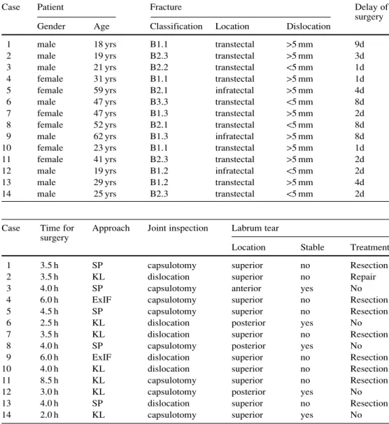
Fig. 1 a Anteroposterior pelvic radiograph of a male patient (case
11) with medially displaced femoral head and inferior fragment in a transverse acetabular fracture of the right hip. b Intraoperative photograph of the same patient showing a hemicircular labral avul-sion (probed by a forceps) from the superior acetabular rim (white dots). Note the cartilage lesion (case 11) seen on the femoral head (black star)
Table 3 Follow-up
Case Postoperative Follow-up Md’A OA
reduction yrs pts Grade
1 <2 mm 6.9 yrs 18 0 2 <2 mm 6.0 yrs 18 0 3 <2 mm 6.7 yrs 16 0 4 <2 mm 6.6 yrs 18 0 5 <2 mma 6.4 yrs 16 (THA) 3b 6 <2 mm 6.0 yrs 16 1–2c 7 >2 mm d d d 8 <2 mma 5.6 yrs 16 (THA) 3 9 >2 mm 5.5 yrs 14 (THA) 3 10 <2 mm 4.9 yrs 18 0 11 <2 mme 4.8 yrs 15 (THA) 2 12 <2 mm 4.7 yrs 11 1–2 13 <2 mm 4.6 yrs 17 0 14 <2 mm 4.6 yrs 11 1–2
THA total hip arthroplasty, OA osteoarthritis aearly (<2 months) postoperative loss of reduction bpreexisting bilateral OA (grade 2)
cpreexisting bilateral pistol grip deformity dlost to follow up
395
Based on our findings the question arises, which strat-egy should be followed for treating labral injuries associ-ated with transverse acetabular fractures. Ikeda et al. did have seven of eight patients respond to non-operative care with protected weight bearing [8], while Fitzgerald only had seven of 56 patients respond to non-operative treat-ment [4]. Since only those patients whose non-operative treatment failed had their labral lesions examined, it is not known how the lesions in those who responded to non-op-erative treatment differed from those who did not. Opera-tive resection of the damaged portion of the labrum, how-ever, has been reported to provide excellent pain relief and good range of motion, but its effect on the long-term function of the hip has yet to be defined [4, 12].
In this study, eight unstable avulsions were resected back to stable margins (Fig. 2a, b), one labrum and its attached piece of bone was reduced and stabilized (Fig. 2c, d), and five lesions were stable and were thought to require no treat-ment. The risk to the vascularity of the acetabular fragments was minimized by utilizing the traumatic capsular rents to perform the capsulotomy. The follow-up of 5.6 years should be sufficient to propose that capsulotomy and/or anterior surgical dislocation at the time of surgery do not increase the risk of avascular necrosis of the femoral head or of the acetabular fracture fragments. The overall clinical result of the current study showed good to excellent Merle d’Aubinge scores in 11 of 13 cases (one case was lost to
follow-up). However, four of the 11 cases were converted into THA, revealing good to excellent results after osteo-synthesis in seven of 13 cases only. If fractures converted into THA are considered as surgery-, patient- (three cases of early loss of fracture reduction) or trauma-related (one case of femoral head damage) failures, seven of the nine patients available for follow-up (78%) showed good to ex-cellent results. Despite the fact that the data match those reported from the classical studies [10, 11], we believe our series is too small to perform conclusive comparisons with other studies.
In summary, significantly displaced transverse acetab-ular fractures constantly revealed avulsions of the labrum from the stable rim. Although not all labral injuries cause reduction problems or later symptoms, as shown by the excellent classical studies obtained with this group of ac-etabular fractures [10, 11], we recommend evaluating and treating intra-capsular injuries at the time of open reduc-tion and internal fixareduc-tion of severely displaced transverse acetabular fractures. The high incidence of extended labral injuries found and the possibility of interposition between the head and the repaired acetabulum suggest that these injuries may negatively impact the outcome if neglected.
References
1. d’Aubigne R, Postel M (1954) Functional results of hip arthro-plasty with acrylic prosthesis. J Bone Joint Surg 36-A:451–475 2. Ferguson S, Bryant J, Ganz R, Ito K (2000) The influence of the acetabular labrum on hip joint cartilage consolidation: a poroelastic finite element model. J Biomech 33:953–960 3. Ferguson SJ, Bryant JT, Ganz R, Ito K (2000) The acetabular
labrum seal: a poroelastic finite element model. Clin Biomech 15:463–468
4. Fitzgerald RH Jr (1995) Acetabular labrum tears. Diagnosis and treatment. Clin Orthop 311:60–68
5. Ganz R, Gill TJ, Gautier E, Ganz K, Krügel N, Berlemann U (2001) Surgical dislocation of the adult hip. A technique with full access to femoral head and acetabulum without the risk of avascular necrosis. J Bone Joint Surg 83-B:1119–1124 6. Gill T, Sledge J, Ekkernkamp A, Ganz R (1998) Intraoperative
assessment of femoral head vascularity after femoral neck frac-ture. J Orthop Trauma 12:474–478
7. Heeg M, Klasen H, Visser J (1990) Operative treatment for ac-etabular fractures. J Bone Joint Surg 72-B:383–386
8. Ikeda T, Awaya G, Suzuki S, Okada Y, Tada H (1988) Torn acetabular labrum in young patients. Arthroscopic diagnosis and management. J Bone Joint Surg 70-B:13–16
9. Kim YT, Azuma H (1995) The nerve endings of the acetabular labrum. Clin Orthop 320:176–181
10. Letournel E, Judet R (1993) Fractures of the acetabulum. Springer, Berlin Heidelberg New York
11. Matta JM (1996) Fractures of the acetabulum: accuracy of re-duction and clinical results in patients managed operatively within three weeks after injury. J Bone Joint Surg 78-A:1632– 1645
12. Nelson MC, Lauerman WC, Brower AC, Wells JR (1990) Avulsion of the acetabular labrum with intraarticular displace-ment. Orthopedics 13:889–891
13. Tile M (1995) Fractures of the pelvis and acetabulum. Williams and Wilkins, Baltimore
14. Tonnis D (1976) Normal values of the hip joint for the evalua-tion of X-rays in children and adults. Clin Orthop 119:39–47
Fig. 2a–d Schematic drawings of unstable (a, b) and stable (c, d)
avulsion. In unstable labral avulsion (a) the labrum is not attached to the acetabular rim and prone to dislocate into the joint. In such cases the labrum had to be resected (b). In stable labral avulsions (c), partial attachment of the labrum to the acetabular rim was pre-sent. In cases of osseus avulsions, refixation was attempted (d)