Translational medicine
Stem and progenitor cell-based therapy in
ischaemic heart disease: promise, uncertainties,
and challenges
Jo
¨ rn Tongers
1*
, Douglas W. Losordo
2, and Ulf Landmesser
3*
1
Department of Cardiology and Angiology, Hannover Medical School, Carl-Neuberg Strasse 1, 30625 Hannover, Germany;2
Feinberg Cardiovascular Research Institute and Program in Cardiovascular Regenerative Medicine, Northwestern University Feinberg School of Medicine, Chicago, IL 60611, USA; and3
Department of Cardiology, Cardiovascular Center, University Hospital Zurich, Ra¨mistrasse 100, CH-8091 Zurich, Switzerland
Received 3 September 2010; revised 13 December 2010; accepted 21 January 2011; online publish-ahead-of-print 28 February 2011
In the absence of effective endogenous repair mechanisms after cardiac injury, cell-based therapies have rapidly emerged as a potential novel therapeutic approach in ischaemic heart disease. After the initial characterization of putative endothelial progenitor cells and their potential to promote cardiac neovascularization and to attenuate ischaemic injury, a decade of intense research has examined several novel approaches to promote cardiac repair in adult life. A variety of adult stem and progenitor cells from different sources have been examined for their potential to promote cardiac repair and regeneration. Although early, small-scale clinical studies underscored the potential effects of cell-based therapy largely by using bone marrow (BM)-derived cells, subsequent randomized-controlled trials have revealed mixed results that might relate, at least in part, to differences in study design and techniques, e.g. differences in patient population, cell sources and prep-aration, and endpoint selection. Recent meta-analyses have supported the notion that administration of BM-derived cells may improve cardiac function on top of standard therapy. At this stage, further optimization of cell-based therapy is urgently needed, and finally, large-scale clinical trials are required to eventually proof its clinical efficacy with respect to outcomes, i.e. morbidity and mortality. Despite all promises, pending uncertainties and practical limitations attenuate the therapeutic use of stem/progenitor cells for ischaemic heart disease. To advance the field forward, several important aspects need to be addressed in carefully designed studies: comparative studies may allow to discriminate superior cell populations, timing, dosing, priming of cells, and delivery mode for different applications. In order to predict benefit, influencing factors need to be identified with the aim to focus resources and efforts. Local retention and fate of cells in the therapeutic target zone must be improved. Further understanding of regenerative mechanisms will enable optimization at all levels. In this context, cell priming, bionanotechnology, and tissue engineering are emerging tools and may merge into a combined bio-logical approach of ischaemic tissue repair.
-Keywords Stem and progenitor cells † Bionanotechnology † Cell-based therapy † Ischaemic cardiomyopathy † Ischaemic heart disease † Myocardial infarction
Background
Treatment of acute myocardial infarction (AMI) and ischaemic car-diomyopathy includes rapid revascularization to limit ischaemic damage and consecutive left ventricular (LV) dysfunction and remodelling, and optimized secondary prevention strategies aiming to attenuate progression of cardiac dysfunction and vascular disease. As a result, the prevalence of heart failure from
post-ischaemic cardiac dysfunction rather increases1causing a substan-tial morbidity.2 Furthermore, despite modern medical therapy, there is a substantial number of patients with ischaemic heart disease refractory to current therapeutic approaches lacking further treatment options, i.e. patients with angina pectoris and no option of interventional or surgical revascularization.
After the initial description of putative endothelial progenitor cells (EPCs) more than a decade ago,3extensive research in the field of
*Corresponding author. Tel:+49 511 532 5402/6627, Fax: +49 511 532 3357, Email:[email protected](J.T.); Tel:+41 44 255 9595, Fax: +41 44 255 4401, Email:[email protected](U.L.)
regenerative medicine has undermined the long-standing dogma that some differentiated organs such as the heart cannot be repaired in post natal life. We are standing on the merge to the era of biological repair in ischaemic cardiovascular disease after the potential of cardiac repair by a variety of stem and progenitor cell populations has been revealed in pre-clinical and early clinical studies.
In the first part of this review, we recapitulate the status quo of adult cell-based therapy in ischaemic heart disease with a clear focus on randomized-controlled clinical trials where available. In addition, we chose to include smaller-size, uncontrolled clinical studies where randomized-controlled data are not available and interesting insights are suggested. Due to space limitations, we were unfortunately not able to include all clinical studies. In the second part, we critically reflect limitations, uncertainties, and chal-lenges of current approaches before finally discussing potential roadmaps of future developments in the field of cell-based cardiac repair. For a comprehensive review of stem and progenitor cell biology, the reader is referred to other in-depth reviews.4–8
Clinical experience from
cell-based therapy
By definition, stem cells are capable to self-renew and to generate progenitor cells that continue to differentiate into committed mature cells. Progenitor cells, hence, are more
lineage-determined, and therefore carry a more limited differentiation potential and may proliferate for a finite number of divisions and lack a self-renewal capacity. In this nomenclature, CD133 is a marker of premature, rather undifferentiated, barely lineage-committed stem and progenitor cells that is lost early during differ-entiation, whereas expression of CD34 is maintained to later stages.
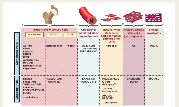
The therapeutic use of unselected bone marrow cells that contain stem and progenitor cells initially gained most momen-tum and has been evaluated farthest in the clinic setting. More recently, other adult stem and progenitor cells, such as circulating stem and progenitor cells, resident cardiac stem cells, and mesenchymal stem cells (MSCs) are being used in translational studies for clinical applications (Figure 1). Skeletal myoblasts (SMs) constitute another cell category that was considered suit-able for cardiac repair. Each cell population seems to carry its own profile of advantages, disadvantages, practical limitations, and translational practicability.9
In the field of ischaemic heart disease, stem and progenitor cell-based therapies have been applied after AMI, after remote (chronic) myocardial infarction (CMI) with cardiac dysfunction and/or in ischaemic cardiomyopathy, and for patients with intract-able (chronic) angina pectoris that is not amenintract-able to revasculari-zation and is refractory to medical therapy. Dependent on the targeted entity, different delivery routes have been used for cell transfer.
Figure 1 Clinically examined as well as emerging stem and progenitor containing cell populations, and their delivery routes for the treatment of ischaemic heart disease.
Delivery routes for stem and progenitor
cell-based therapies
Conceptually, the goal is to safely deliver an optimal number of effective cells as selective as possible to the therapeutic target zone via a minimally invasive route. So far, cells have been trans-planted into the heart overly via intracoronary infusion or intra-myocardial injection (Figure1).
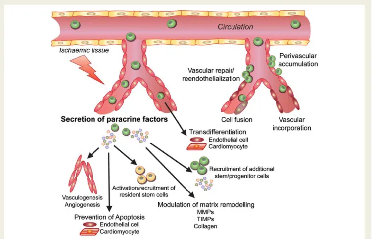
Adapting the elegant, minimal-invasive technique lent from inter-ventional cardiology for transvascular approaches, stem cells can be homogenously distributed via intracoronary infusion. To reduce spill-back and, in turn, optimize the contact time of cells and coronary vessel wall, cells are infused in block-balloon tech-nique.10 This approach is only feasible in patients with a patent target vessel and, therefore, no option for non-revascularized areas. In addition, its efficiency is impaired by a complex multistep process of vessel adhesion and transmigration to allow infused stem cells to invade the tissue. Then, homing of invaded cells is also dependent on chemoattraction towards factors secreted from the ischaemic tissue.11 In line with this concept, cardiac homing of early EPCs after intracoronary infusion was increased in patients with an acute compared with chronic MI.12 As a side note, although intravenous infusion of stem/progenitor cells initially appeared effective,13low target zone cell concentration due to off-target homing14and biological redistribution15strongly challenged this transvascular strategy which cannot be favoured anymore.
The more invasive intramyocardial cell injection, on the other hand, overcomes some of these hurdles and appears particularly
suitable in the case of non-revascularized infarct-related vessels and/or ischaemic cardiomyopathy. Cells can be injected into the myocardium from the epimyocardial side, commonly during open-heart surgery, or endomyocardial side by means of needle-tipped delivery catheters. Detouring the bloodstream has also been par-ticularly attractive for large cell types such as MSCs and SMs. On the downside, injection-related puncture of the viable or necrotic myocardium comes with some risk, i.e. of ventricular perforation. Further, injections lead to inhomogeneous distribution of cell clus-ters in malperfused tissue. In this scenario, an elegant and now commonly used catheter-based option is to guide the endomyo-cardial injection by electromechanical mapping (EMM, e.g. NOGA mapping). This technique enables to focus the cells to ischaemic but viable (hibernating) myocardium.
For the future advance, however, it has to be kept in mind from the clinician’s perspective that the more invasive the technique and the higher percentage of co-morbidities introduced, the higher the procedural risk in this group of commonly elderly, multimorbid patients, who will be eligible for cell-based therapies.
Unselected bone marrow-derived cells
The use of unselected BM-derived mononuclear cells (BMCs) is clearly the most examined cardiac cell-based therapy in clinical studies (Figure2) with a clinical follow-up experience up to 5 years.16To some extent, this development is the result of the pragmatic attractive-ness of BMCs: (i) BMCs are rather easy to harvest, (ii) yielded cell numbers do not limit clinical applications, (iii) while it remains
unknown which cell types are more potent or have particular potent repair properties, BMCs contain an ‘un-narrowed’ composition of cells including fractions of stem and progenitor cells, and (iv) their prep-aration does not need prolonged ex vivo manipulation.
Acute myocardial infarction
After early-phase clinical studies had suggested the safety and feasi-bility of intracoronary BMC infusion after AMI,10,17–19several mid-sized, randomized, partly placebo-controlled trials have generated mixed results. The randomized-controlled REPAIR-AMI and BOOST trials showed an improvement of global LV ejection frac-tion (LV-EF) without significant changes of LV end-diastolic volumes 4 – 6 months after cell transfer.20,21A REPAIR-AMI sub-study revealed that the increase in LV-EF did not occur at the expense of increases in end-systolic or end-diastolic volumes.22 Two other landmark studies, on the other hand, did not observe a significant improvement in LV function or dimensions at 4- to 6-month follow-up,23,24 although Janssens et al.23 observed a reduction in infarct size 4 months after intracoronary cell transfer in a study with very early BMC administration (i.e. within 24 h). Although definite reasons for these mixed results remain elusive, differences in study protocol and design, including time from reper-fusion to cell injection, type, number, and isolation technique of cells, or follow-up design have been discussed. Whereas BMCs were infused within the first 7 post-infarct days in most trials, in Janssen’s trial, cells were injected within the 24 h after AMI.23In the ASTAMI trial, magnetic resonance imaging (MRI) was not per-formed until 2 – 3 weeks after cell transfer, whereas only echocar-diography was done at baseline. Further, cells by contrast were prepared differently following the Lymphoprep technique.24 Sub-sequently, follow-up data on the REPAIR-AMI and BOOST collec-tives have become available. In REPAIR-AMI, the improvement of LV function was sustained after 12 months20and was associated with a significant reduction in major adverse cardiovascular events after AMI over this period;25 an observation that carried on until 2 years of follow-up.26Hence, in BOOST, the difference in LV-EF between the groups was no longer significant after 18 months,27although an echocardiographic substudy revealed a per-sistent enhanced diastolic function from BMC application.28 Recently, 5-year follow-up data from the BOOST collective did not show a sustained benefit on systolic and diastolic LV function after a single BMC infusion.16 Interestingly, subgroup analyses suggested that patients with a more severely impaired LV function may have a benefit from BMC administration, whereas patients with a rather preserved LV function post-MI may not benefit.
In the controlled but non-randomized BALANCE study,29 hae-modynamics, measures of LV function and geometry, contractility, infarct size, exercise capacity, and, of note, mortality remained improved up to 5 years after BMC infusion when compared with a matched, non-randomized control group. However, these results should be interpreted with caution, given the non-randomized design of the study. Another non-randomized study from China with a long-term follow-up has recently suggested that BMC administration may lead to an improved LVEF after 4 years.30 Notably, the results of the recently published REGENT trial are discussed below, because one study arm received selected CD34+/KDR+BMCs.
Chronic myocardial infarction
In patients with CMI, less experience from clinical studies is avail-able. The early-phase non-randomized IACT study by Strauer et al.31 suggested that intracoronary BMC transfer more than 5 months after MI may result in smaller infarct size, better LV-EF, and wall movement velocity associated with signs of higher cardiac metabolism in the infarcted myocardium. This application has been revisited by a carefully designed cross-over study where intracoronary BMC transfer more than 3 months after MI led to a significant improvement of LV-EF related to enhanced regional contractility in the area of cell application.32 Of note, this effect was also visible even in patients who crossed-over from control or treatment with circulating progenitor cells.32
Strauer et al.33have recently reported the long-term follow-up data on the intracoronary application of BMCs in patients with chronic heart failure (CHF) due to ischaemic cardiomyopathy from the non-randomized STAR study. Over a 5-year follow-up, intracoronary BMC administration was not associated with adverse events. The authors reported an improved LV perform-ance, quality of life, and survival in patients with CHF who agreed to BMC application when compared with the control group with a similar LV-EF, who did not agree to BMC treatment.33 However, due to the non-randomized design of the study, these results need to be interpreted with caution.
Refractory myocardial ischaemia
Another set of early-phase studies has addressed the effect of BMCs in patients with refractory myocardial ischaemia lacking options for revascularization. These studies jointly suggest that BM-derived cell injection via transepicardial (e.g. during bypass surgery) or transen-docardial (e.g. guided via EEM) may improve subjective endpoints such as angina frequency or heart failure symptoms, and/or measures of global/regional wall motion and perfusion.34–38In the randomized-controlled, double-blind trial conducted by van Ram-shorst et al.,39 intramyocardial application of BMCs resulted in a modest but significant improvement of myocardial perfusion as assessed by SPECT, angina severity, and quality of life during a 3-month follow-up in patients with severe angina (classes III – IV), despite optimal medical therapy, ineligible for myocardial revascu-larization, and evidence of myocardial ischaemia at baseline. Simi-larly, 6 months after direct injection of autologous BMCs in patients with severe refractory angina, a significant improvement of exercise time, LV function, and NYHA functional class was observed in the PROTECT-CAD trial.40 The data of the ACT34-CMI trial in a similar patient population are discussed below. Cumulatively, the studies discussed above suggest feasibility and some studies suggest efficacy of BMC-based therapy in acute/ chronic MI as well as chronic refractory angina. Recent meta-analyses that summarized more than 1000 patients showed a significant improvement in LV-EF after BMC therapy on top off standard treatment.41–43It needs to be acknowledged, however, that LV-EF might not have been the ideal endpoint to clearly detect efficacy (also see Endpoint selection). Overseeing safety data from up to 5 years, no sustained safety issues associated with the use of unselected BMCs have been observed after cardiac transplantation in clinical studies.
Circulating/mobilized CD133
1and
CD34
1stem and progenitor cells
CD133 and CD34 are the most commonly used single markers for the enrichment of haematopoietic stem cells (HSCs). The observation that selection of certain cell populations, i.e. CD34+ cells from total mononuclear circulating cells, may augment their potency for cardiac repair has raised the interest in using selected cell populations as opposed to unselected BMCs.44 Baseline levels of circulating stem/progenitor cells are known to be low, which restricts their therapeutic use. To lever-age their therapeutic potential, these cell types can be pharma-ceutically mobilized from the BM into the circulation in order to be isolated and enriched (i.e. leukapheresis). Driven by the history of the field, EPCs, a potential progeny of HSCs, have been in the focus (for review see45,46).
CD1331cells
CD133+ cells are more immature and less lineage-determined than CD34+ cells. Intracoronary infusion of selected CD133+ cells after recent AMI has been evaluated in a small-scale, non-randomized clinical study that revealed improved LV-EF paralleled by a reduction in myocardial perfusion defect after 4 months.47 However, more coronary events, such as stent occlusion and in-stent restenosis, were observed after CD133+ cell transfer. The authors further reported a time-dependent adverse remodel-ling of the infarct-related artery with accelerated luminal loss and a reduced conductance after CD133+ cell transfer.48 However, when CD133+ cells were injected locally during bypass surgery in patients with recent AMI, no safety concerns became evident in a small, non-randomized study, whereas regional wall motion was improved associated with better myocardial perfusion and via-bility.49Transepicardial injection of CD133+cells into the border zone of MI during operative revascularization in patients who had previous MI was reported by Stamm et al.50,51in a small, uncon-trolled study. Since no adverse events were reported, treatment was considered safe and feasible. The conclusion that this treat-ment resulted in improved LV function, however, is limited by the lack of a control group.50,51
CD341cells
CD34+ cells contain more endothelial lineage-determined cells than CD133+cells and are therefore considered as a cell popu-lation enriched for ‘early‘ EPCs. In the randomized-controlled REGENT trial, unselected and CD34+/CXCR4+-selected BMCs were examined for their effects in LV function after MI in patients with reduced LV function (EF 40%).52 After 6 months, LV-EF increased by 3% in patients treated with unselected BMCs, 3% in patients receiving CD34+/CXCR4+-selected BMCs, and remained unchanged in the control group. There were, however, no signifi-cant differences in absolute changes of LV-EF between the groups.52There was a trend in favour of BMC efficacy in individuals with most severely impaired LV dysfunction, as has been observed in several other studies using BMCs after acute MI.53A potential limitation of this trial was that MRI measurements of LV function were only completed in a subgroup of patients.
We and others have evaluated intramyocardial injection of CD34+ progenitors in coronary artery disease (CAD) patients with chronic angina. Our Phase I/IIa pilot study suggested safety and feasibility as well as a positive trend of bioactivity as evidenced by SPECT perfusion imaging 6 months after CD34+ progenitor cells were injected into the hibernating myocardium in patients with refractory chronic angina; transendocardial delivery was guided by EMM (NOGA mapping) to identify ischaemic but still viable myocardium.54These trends were further substantiated by our subsequent Phase IIb randomized-controlled, multicentre ACT34-CMI trial.55In ongoing studies, this concept is under evalu-ation for patients critical limb ischaemia (clinicaltrials.gov: NCT00616980). So far, no safety issues are evident with the use of CD34+cells. Whether further selection using the combination of different markers augments cellular repair capacity has to be determined in the future.
Mesenchymal stem cells
Mesenchymal stem cells or stromal cells constitute another poten-tial option for stem/progenitor cell-based therapy, and their use is also evaluated as a potential allogeneic approach based on their immunomodulatory properties. Mesenchymal stem cells are stromal cells present in various tissues, such as BM and adipose tissue. Reflecting their paracrine activity, they exert anti-inflammatory and anti-apoptotic effects.56Local immunosuppres-sive action has fuelled hope of an allogeneic MSC transplantation as an ‘of-the-shelf’ cell-based therapy to surround practical limit-ations of cell resources and ex vivo culture often required in the autologous setting.
The regenerative capacity of MSCs in general and the contro-versially discussed aspect of immune privilege57,58 of allogeneic MSCs needs to be evaluated in men. Autologous and allogeneic MSC transfer is currently under investigation; however, clinical data are scarce. In a randomized-controlled, double-blinded Phase I study, intravenous application of allogeneic MSCs after acute MI did not raise safety concerns and as assessed by a global symptom score might be efficacious over a period of 6 months.59 In early studies, MSCs were injected intravenously; however, the pulmonary passage of these large cells may be problematic.60 Since these positive efficacy data stand in con-trast to negative experience from intravenous BMC applications, this set of data should still be considered with caution. Sub-sequently, intracoronary application of MSCs after AMI has been evaluated in two non-randomized early-phase studies by Chen et al. A high-dose of BM-derived MSCs (6× 10¹⁰) resulted in a significant improvement of LV-EF in several modal-ities,61 whereas a lower dose (5× 10⁶) did not improve LV function in chronic ischaemic cardiomyopathy in a subsequent study by the same authors.62 Still, MSC treatment resulted in an improved exercise capability and heart failure symptomatol-ogy after 3 months.
Adipose tissue can also serve as a source of MSCs, i.e. adipose tissue-derived stem cells (ADSCs). Two clinical trials—APOLLO and PRECISE—are exploring the safety, feasibility, and efficacy of freshly isolated ADSCs with the CellutionTMsystem (Cytori
Cardiac-derived cardiovascular stem
and progenitor cells
Until recently, our dogma was that the fully differentiated heart has no capability for cell turnover and self-repair. In their elegant observation, Bergmann et al.63showed evidence for in-men cardi-omyocyte renewal at a rate of 1% per year in younger adults and 0.5% in the elderly. In post-natal hearts, various subtypes of tissue-resident cardiac stem and progenitor cells (CSCs) classified by surface antigens and transcription markers have been reported, although it is undetermined whether these subtypes have clearly distinct phenotypes. Cardiac stem and progenitor cells, which have been suggested to be capable of creating cardiomyocytes and all surrounding cell types, are a promising candidate-at least in theory-to provide contractility and vascularization.64
In the light of the fact that their genuine number is low, CSCs isolated from endomyocardial biopsies have successfully been expanded ex vivo to leverage this therapeutic concept.65Dr Marban’s group has proposed a population of potential clinical relevance that has been identified by expanding CSCs from self-adherent clusters (cardiospheres) under certain conditions, i.e. cardiosphere-derived stem cells (CDCs).66,67There is still some controversy on the car-diomyogenic potential of cardiospheres.68,69Dr Field’s group has recently suggested by using genetic cell tracking that there are tem-poral limitations for the ability of cardiac-resident c-kit+ cells to acquire a cardiomyogenic phenotype, i.e. that the cardiomyogenic population is present in neonatal hearts but largely lost in adult mouse hearts and suggested that elucidation of the underlying mol-ecular mechanisms may permit a more robust cardiomyogenic induction in adult-derived cardiac c-kit+cells.70
Early-stage clinical studies currently address safety and feasibility of cardiosphere-derived stem and progenitor cell transplantation. In the SCIPIO trial (clinicaltrials.gov: NCT00474461), patients with ischaemic cardiomyopathy from CMI who undergo bypass surgery received an intracoronary infusion of autologous CSCs iso-lated from their right atrial appendages. Further, patients with ischaemic LV dysfunction from recent MI receive an intracoronary injection of CDCs in the ongoing Phase I randomized dose-escalation CADUCEUS trial (clinicaltrials.gov: NCT0089336).
Skeletal myoblasts
The concept of resident stem cells (i.e. satellite cells), which are quiescent under normal conditions, contribute to new myocytes in response to injury is rather well established for skeletal muscu-lature. Skeletal myoblasts can be conveniently isolated from muscle biopsies and ex vivo expanded for therapeutic use. After pre-clinical evidence showed their repair effects in ischaemic myocardium,71,72 SMs have been thought to transdifferentiate into cardiomyocytes. In the meanwhile, pre-clinical data indicate that SMs maintain a skeletal muscle fate,73 infrequently fuse with cardiomyocytes,74 and hardly couple electromechanically with the host myocardium.75
The clinical value of SMs is uncertain. Initially, several uncon-trolled early-phase studies generated promising results for the therapeutic utility of SMs in CHF, suggesting an improved global and regional contractility and/or viability in the infarct zone. In most studies, SMs were overly injected via a
transepicardial route during graft surgery,76–78 whereas only one study reported the transendocardial injection of myoblasts as safe and feasible.79 The most comprehensive clinical evalu-ation of SMs, so far, has been the multicentre, randomized-controlled, double-blind Phase II MAGIC trial.80 Herein, autolo-gous, for 3 weeks ex vivo expanded SMs were directly adminis-tered around the scar tissue in patients with ischaemic cardiomyopathy who underwent bypass surgery. After 6 months, primary endpoints, namely global and regional LV func-tion assessed by echocardiography, were not significantly changed. Notably, the authors reported a significant reduction in LV volumes in the high-dose group. The longest clinical experience with SM transfer, however, has been published by Dib et al. In their non-randomized, uncontrolled Phase I study, patients undergoing surgery for revascularization or implantable cardioverter-defibrillator implantation showed an increase in LV-EF and viability, whereas no adverse events were observed up to 4 years transepicardial injection of autologous SMs.81
Fuelled by an earlier report from Menasche et al.76 where the use of myoblasts was associated with arrhythmic events in 40% of the patients, SMs have been considered to be pro-arrhythmogenic. In the subsequent MAGIC trial, no significant increase in arrhythmic events was reported, although it must be acknowledged that there was a trend towards more arrhythmias in SM-treated patients despite treatment with amiodarone a priori.80 Hence, none of the other early phase above nor the 4-year surveillance by Dib et al.81 has added evidence for SM-induced sudden death. Chachques et al. hypothesized that the arrhythmicity of SMs may be conditioned by the bovine serum used during ex vivo expansion for 3 weeks (cellular cardio-myoplasty) that may constitute an antigenic and thereby inflamma-tory substrate. A strictly autologous preparation using the corresponding donor serum in this rather small-sampled study did not lead to any arrhythmias over 1 – 2 years of follow-up.82 As the majority of the above-mentioned, early-phase studies did not report arrhythmic events, it remains vague whether this par-ticular procedure stabilizes SMs electrically. There is still debate about the potential pro-arrhythmicity of different adult stem/pro-genitor cells, in particular the SMs.83,84 Further, in contrast to earlier hesitations to infuse SMs intracoronary in the light of a potential risk for microembolization due to their cell size, there were no adverse events following transcoronary-venous SMs deliv-ery observed in the POZNAN trial.85The potential risk of arrhyth-mias and sudden death needs, however, ultimate clarification for this cell type.
Endogenous mobilization of stem/
progenitor cells
In contrast to exogenous cell transfer requiring invasive delivery, adult stem/progenitor cells can be mobilized from the BM by sys-temic administration of certain cytokines [e.g. granulocyte colony-stimulating factor (G-CSF)] to augment circulating levels of these cells and, thereby, enhance ischaemic tissue repair via cir-culating stem/progenitor cells. This concept has been transferred from the haematologists who isolate HSCs from the bloodstream before ablation of the BM in stem cell transplantation.
Earlier, the authors of the much debated MAGIC trial reported that G-CSF mobilization after MI was associated with an unex-pected higher rate of in-stent restenosis at the culprit lesion, whereas intracoronary infusion of peripheral blood stem cells appeared safe and potentially efficacious; because of this safety issue, the study was discontinued.86Subsequently, in a controlled, non-randomized study by Ince et al.,87 G-CSF mobilization of CD34+BMCs shortly after angioplasty in AMI improved LV func-tion and metabolic activity and attenuated LV remodelling up to 12 months without accentuation of the restenosis rate or late adverse events. In the same line, Valgimigli et al.88showed an unremarkable safety profile for G-CSF in patients with AMI, although LV per-fusion or function was unchanged 6 months after treatment. Fur-thermore, G-CSF did not improve LV wall motion or perfusion over the period of 1 month in patients with stable CAD.89 Of note, there was a trend towards more ischaemia, and in 2 out of 16 patients reinfarction occurred.89Early clinical experience with stem cell mobilization by G-CSF from 10 trials, including 445 patients with AMI, was recently summarized in a meta-analysis by Zohlnho¨fer et al. Herein, the authors concluded that cumulat-ively the data do not support efficacy of endogenous stem cell mobilization by G-CSF.90 Without evidence for efficacy on the one side, and a potentially questionable safety profile on the other (i.e. with respect to restenosis), large-scale trials, which would be needed to clarify the clinical potential, could not be advocated. Interestingly, very early G-CSF administration (,12 h) has recently been reported to attenuate ventricular remodelling after large, anterior MI, whereas systolic function and myocardial perfusion were unchanged in the STEM-AMI collective.91
In recent years, pharmaceutical compounds emerged to modu-late well-defined cascades of stem/progenitor cell biology and homing. Continuing the concept of endogenous stem cell mobiliz-ation, Zaruba et al.92 proposed a combined strategy of G-CSF with dipeptidylpeptidase IV (DPP-IV) inhibition to improve cardiac homing of mobilized stem/progenitor cells, which is currently under clinical investigation in the SITAGRAMI trial in patients after AMI. This concept proposes that inhibition of DPP-IV that is cleaving one of the main stem cell attracting chemokines, SDF-1a, will improve homing of mobilized stem/progenitor cells. More recent results from our group and others described the CXCL12/ CXCR4 axis, the SDF-1a pathway, as a relevant regulator of endogenous cell mobilization.93 In pre-clinical studies, the CXCR4-antagonists Plerixafor (known as Mobozil or AMD3100) mobilizes stem/progenitor cells from the BM, increases BM-derived cell incorporation in the ischaemic border zone, and improves cardiac function post-MI.93Because this agonist has clinically been used as HIV therapeutic without relevant side effects for years, its safety profile appears favourable. To our knowledge, no clinical data are available on any of these pharmaceutical strategies to thera-peutically modulate endogenous stem/progenitor cell trafficking.
Emerging stem cell types with a potential
for cardiomyogenic differentiation
Embryonic stem cells
Embryonic stem cells (ESCs) posses several features that are con-ceptually attractive for cardiac repair. Embryonic stem cells are
pluripotent, which means that they have the ability to differentiate into all cell lineages.94,95On the other hand, there are substantial ethical and regulatory concerns with their retrieval. The limited availability further hinders its therapeutic utility. In addition, there is a risk for malignancies (i.e. intra- and extracardiac teratoma or other tumour formation) associated with the use of ESCs,96,97as it has recently become evident in a patient who developed a likely donor-derived multifocal brain tumour after treatment with foetal neural stem cells.97 At the same time, reliable modalities to regulate and control differentiation in a targeted and controlla-ble fashion are challenging, although several approaches to limit tumour formation have been explored, including strategies to enhance cardiopoietic programming of ESCs.98
Furthermore, following allogeneic application, ESCs may trigger an immune response in the recipient. Finally, it remains elusive whether human ESCs efficiently structurally engraft and electrome-chanically integrate into ischaemic myocardium. The above-described limitations are some of the hurdles that currently hinder the transition of ESC-based therapy into clinical translation.
Inducible pluripotent cells
In 2006, Takahashi and Yamanaka99 reported that differentiated murine fibroblasts could be reprogrammed into stem cells with the capacity to form all three germ layers and termed these cells induced pluripotent stem cells (iPS). Nuclear reprogramming with ectopic stemness factors has opened the opportunity to gen-erate autologous patient-derived iPS from adult somatic cells.99,100 The ability of both mouse and human iPS to differentiate in func-tional cardiomyocytes has recently been demonstrated.101,102
The study of iPS from selected cohorts of patients is an innova-tive way to uncover molecular mechanisms of disease, such as nicely illustrated in the study of Moretti et al.,103who generated pluripotent stem cells from dermal fibroblasts of patients with long-QT syndrome type-1 and subsequently induced their differen-tiation into functional cardiac myocytes that recapitulated the elec-trophysiological features of the disorder.104
Functionally, intramyocardially injected undifferentiated iPS, but not parental fibroblasts, engrafted and improved contractile func-tion of infarcted myocardium while attenuating adverse remodel-ling in immunocompetent mice.105 Only in immunocompetent mice, the environment after cardiac transplantation was permissive for differentiation, whereas in immunodeficient mice, tumour development was observed, which highlights the importance of immune surveillance to prevent tumour growth.105New technol-ogies such as small molecule screen and epigenetic modifications most probably will establish further potentially more safe options for the generation of iPS. It will, however, be crucial to validate the epigenetic and phenotypic stability in the reprogrammed state. Also, reprogramming pluripotency in somatic cells comes with the risk of tumorigenicity as described above. Furthermore, iPS become immunologically relevant while they differentiate with up-regulation of histocompatibility antigens. This aspect would narrow the range of applications to the autologous setting.106As described above, several important hurdles have to be resolved before a clinical translation of iPS-based therapies can be considered, including safety issues to prevent tumour growth that may require determination of the appropriate
differentiation of iPS-derived cells and removal of residual undiffer-entiated cells before transplantation.107In addition, upscaling for an effective iPS-based cell generation will be required. Several impor-tant considerations in the proposed mechanisms of cell-based therapy for cardiac repair and its further development are described in the Supplementary material online and depicted in Figures3–5.
Future directions of cell-based
therapy: a roadmap
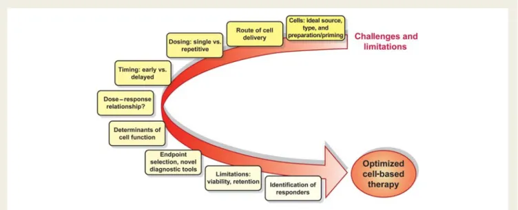
Various strategies have been proposed to support stem cells in the hostile environment of ischaemic tissue characterized by ischaemia, acidosis, inflammation, and oxidative stress. Beyond the modu-lation of cell homing, viability, engraftment, and retention, it becomes more and more clear that true repair of damaged myo-cardium will need more than a single-dose administration of cells.
Priming of stem and progenitor cells to
enhance their therapeutic efficacy
The concept to pre-treat or modify stem/progenitor cells before application ( priming) and thereby enhance their therapeutic potency has evolved from earlier pre-clinical observations.108–114 These strategies basically target any function step that influences cell fate from the application on: adhesion/transmigration,
homing, migration, engraftment, survival, cell – cell interaction, repair capacity, differentiation, and retention. Potential tools for modification include drugs, small molecules, naked and vector-facilitated plasmids, and epigenetic reprogramming (for more details see115,116). Priming of dysfunctional autologous cells from cardiovascular patients via any of these tools may allow for a ‘reset-ting of impaired biopotency’.
Among multiple targets stemming from pre-clinical evaluation, the following examples are under clinical investigation: we and others have identified a reduced endothelial NO synthase-dependent NO production as an important mechanism limiting the functional repair capacity of endogenous progenitor cells in patients with diabetes or hypertension.108,117In the randomized, placebo-controlled ENACT-AMI trial, the therapeutic use of eNOS-overexpressing EPCs is presently studied in patients with large MI (clinicaltrials.gov: NCT00936819).118 As another example of modified stem/progenitor cells, in the MESAMI II trial (clinicaltrials.gov: registration in process) designed by Roncalli and colleagues, patients with no-option chronic ischaemic cardio-myopathy will receive an intramyocardial injection of MSCs pre-treated with melatonin. This concept is based on pre-clinical data showing that pre-treatment with the pineal hormone increases survival, paracrine activity, and therapeutic efficiency of MSCs in a rodent ischaemia model.119 Another way to boost paracrine and differential cell functions is to pre-stimulate cells before appli-cation.120,121This concept is currently translated into patients with
heart failure from ischaemic cardiomyopathy by pre-incubation of BM-derived MSCs with growth factors to guide their transition into cardiopoietic cells (C-Cure trial, clinicaltrials.gov: NCT00810238). As no clinical study of primed stem cells has been completed yet, it is going to be interesting whether these strategies will hold promise in clinical applications. Many more targeted cell modifications can be expected to be in transition from bench to bedside at present.
Bionanotechnology to support cell-based
therapies
In addition to the harmful biochemical milieu, loss of tissue archi-tecture and loss of matrix support within the infarcted myocar-dium detrimentally affect cell – cell and cell – matrix interaction and, in turn, regulation of dependent pathways that are functionally inevitable for the homeostasis of transplanted cells. The rapidly evolving field of bionanotechnology allows to specifically design biomaterials to support transplanted cells within this ischaemic environment.122Herein, the structure, dimensions, and shape of constructs are pivotal to better mimic the native architecture of extracellular matrix. An optimal biomaterial to support cell therapy should provide a three-dimensional environment to enhance biomechanical properties of extracellular matrix; a purpose for which controlled organization at nano-scale is needed (for more details see123). In some biomatrices, bioactive signals can be incorporated to specifically modulate stem cell biology while supporting them structurally.124
The strategy to support cell transfer has rapidly gained attention triggered by exciting pre-clinical data. In murine models, nanofibres that self-assemble into a matrix recruited endogenous progenitors to the myocardium and supported transplantation of cardiomyo-cytes providing a microenvironment.125 In principle, biomaterials can be custom-designed to optimally fit the organ-specific micro-environment.126 Furthermore, bioactive signals can be incorpor-ated in some biomaterials to additionally enhance cell survival, retention, proliferation, and differentiation. In this context,
Padin-Iruegas et al.127 reported that an insulin-like growth factor carrying nanofibre enhances CSC-dependent repair of cardiac injury.128In our hands, the combination of a linage-specific opti-mized, self-assembling nanofibre enhances the potency of cell-therapy in ischaemic tissue repair.129Also, bioactive sequences of biologically attractive paracrine factors, e.g. SDF-1, can more effectively be presented via biomaterials with the aim to recruit endogenous into or support exogenously applied cells in ischaemic myocardium.130
Although emerging results for the role of bionanomaterials in cell-based ischaemic tissue repair are promising, there has not been precedence in humans. Before any clinical translation, several aspects such as dosing, dynamics and kinetics of bioactivity, biodegradability, occurrence and excretion of breakdown pro-ducts, and immune compatibility have to be carefully addressed.
Tissue engineering
Tissue engineering has now been advanced for several years. In particular aiming at replacement of the myocardium, the break-through in this field has been hindered by several challenges. While cell seeding of construct backbones has worked well for proliferative and hypoxia-tolerant cells, it has been difficult to achieve tissue-like densities with cells with low proliferation rate such as cardiomyocytes,122 although successful generation and transplantation of engineered heart tissue grafts has been reported in small animal models.131Achieving sufficient perfusion of larger constructs is challenging. Passive diffusion is generally tolerated up to a distance of ,0.1 – 0.2 mm. In the case of longer distances, dysfunctionality and necrosis occur as a failure of sufficient diffu-sion.132At present, bigger, more complex constructs such as myo-cardium (1.0 – 1.5 cm) cannot be maintained by simple diffusion and need anastomosis to a vascular network. Since generation of a stable and functional construct microenvironment remains criti-cal, stem and progenitor cells display a strong potential to acceler-ate tissue engineering for the purpose of seeding but also for the creation of a vascular networks.133
Combined strategies of biological repair
As stated above, single-dose transfer of a single cell type into a deranged environment will likely not be sufficient to ‘regenerate’ a complex tissue such as the heart. More recent insights suggest that combined strategies of biological repair more closely resemble native biological mechanisms and, thus, may be more potent than current one-stop, single-cell strategies (Figure5). Conceptually, the combination of different types of stem/progenitor cells with stromal cells is functionally very attractive aiming at synergisms. Different cell (sub-) populations may exert different regenerative effects. Unselected and selected cells, for example, showed differ-ent spatial patterns of homing.15Melero-Martin et al.133recently described striking evidence for the synergistic interaction of EPCs and MSCs, which contain relevant pericytes, to create a sub-stantial and functional vascular network in a Matrigel assay that was stable for up to 4 weeks.
To add yet another level of complexity, supplemental tools that are aimed to improve efficacy of cell transfer have become available. Engraftment of cardiac-derived stem cells (CSCs) after intramyocar-dial application, for example, is improved after sealing the injection site with fibrin glue to attenuate the known leakage of cell suspen-sion.134Low-energy shock wave treatment of the target tissue has been shown to enhance recruitment of circulating EPCs by enhan-cing local expression of chemoattracting growth factors in mice.135 This technology is thought to prepare the ischaemic target zone for cell transfer and thereby indirectly augment cell homing and retention. In the ongoing Cellwave study (clinicaltrials.-gov: NCT00326989), extracorporal shock wave pre-treatment before intracoronary application of BM-derived progenitor cells is already under clinical evaluation in patients with CMI. In addition, homing of cells via the intracoronary route can be improved by dis-rupting cell-carrying microbubbles via ultrasound. Without evoking undesired damage, this technology appears to facilitate vascular transmigration by creating capillary pores.136,137
With all limitations and shortcomings in mind, the idea to realize effective cardiac tissue repair by using only one cell type is likely challenging. Depending on further mechanistic and clinical insights, we envision combined, precisely timed, multi-step approaches that incorporate various stem and stromal cells, paracrine factors, and specifically bioengineered tools for the future cardiac regenerative medicine in order to advance this exciting field to the next level, the level of true ischaemic tissue repair.
Summary
We are currently in the phase of translation of cardiac cell-based therapy, and good translation without any surprise takes time and efforts. After the initial hype revolving cell-based cardiac tissue repair led to a rather rapid translation into early-phase clini-cal trials, we have now reached the stage where conclusive answers and thoughtful fine-tuning, which includes a step back to the bench to find answers, are needed. There is some consensus that cell-based therapy has potential beneficial effects on top of standard medial therapy as it has been expressed by the task force of European Society of Cardiology.138Apart from uncertain-ties and limitations discussed above, several pressing tasks need to be addressed: sustained efficacy needs to be proven for each cell type depending on the treated entity. Comparative studies may allow discriminating superior cell populations, sources, timing, dosing, and delivery mode considering with regard to certain appli-cations. Safety profiles dependent on cell type, delivery route, and underlying disease need to be characterized by long-term follow-up of larger collectives. To substantiate these aspects, large-scale, multicentre trials are clearly needed. In order to predict benefit, influencing factors need to be identified with the aim to focus resources and efforts. Local retention and fate of cells in the therapeutic target zone must be improved. Further understanding of regenerative mechanisms will enable optimization at all levels. Cell priming, bionanotechnology, and tissue engineer-ing are emergengineer-ing tools and may merge into a combined biological approach of ischaemic tissue repair.
After a hectic decade, it is time to catch breath and focus efforts on concise pre-clinical work, and thoughtfully designed, sufficiently powered clinical trials to find answers to important remaining questions, to clarify on uncertainties, and to overcome limitations with the aim to optimize cell-based therapy. Despite all promise, eventually, we are challenged to show robust effects on disease progression, morbidity, and mortality associated with an accepta-ble safety profile to advance the promising concept of cell-based therapy to clinical routine.
Supplementary material
Supplementary material is available at European Heart Journal online.
Funding
J.T. was supported by the American Heart Association (Midwest Affili-ate), the German Heart Foundation, and Solvay Pharmaceuticals. D.W.L. was supported by NIH grants R01 HL53354, R01 HL77428, R01 HL80137, and R01 HL95874. U.L. was supported by Swiss National Research Foundation grants 310030-122339 and 33CM30-124112/1 and the Zurich Center of Integrated Human Physiology.
Conflict of interest: none declared.
References
1. Fang J, Mensah GA, Croft JB, Keenan NL. Heart failure-related hospitalization in the U.S., 1979 to 2004. J Am Coll Cardiol 2008;52:428 – 434.
2. Gheorghiade M, Bonow RO. Chronic heart failure in the United States: a mani-festation of coronary artery disease. Circulation 1998;97:282 – 289.
3. Asahara T, Murohara T, Sullivan A, Silver M, van der Zee R, Li T, Witzenbichler B, Schatteman G, Isner JM. Isolation of putative progenitor endo-thelial cells for angiogenesis. Science 1997;275:964 – 967.
4. Hirschi KK, Ingram DA, Yoder MC. Assessing identity, phenotype, and fate of endothelial progenitor cells. Arterioscler Thromb Vasc Biol 2008;28:1584 – 1595. 5. Aicher A, Zeiher AM, Dimmeler S. Mobilizing endothelial progenitor cells.
Hypertension 2005;45:321 – 325.
6. Hristov M, Erl W, Weber PC. Endothelial progenitor cells: mobilization, differ-entiation, and homing. Arterioscler Thromb Vasc Biol 2003;23:1185 – 1189. 7. Dimmeler S. Regulation of bone marrow-derived vascular progenitor cell
mobil-ization and maintenance. Arterioscler Thromb Vasc Biol 2010;30:1088 – 1093. 8. Yi BA, Wernet O, Chien KR. Pregenerative medicine: developmental paradigms
in the biology of cardiovascular regeneration. J Clin Invest 2010;120:20 – 28. 9. Segers VF, Lee RT. Stem-cell therapy for cardiac disease. Nature 2008;451:
937 – 942.
10. Strauer BE, Brehm M, Zeus T, Kostering M, Hernandez A, Sorg RV, Kogler G, Wernet P. Repair of infarcted myocardium by autologous intracoronary mono-nuclear bone marrow cell transplantation in humans. Circulation 2002;106: 1913 – 1918.
11. Frangogiannis NG, Smith CW, Entman ML. The inflammatory response in myo-cardial infarction. Cardiovasc Res 2002;53:31 – 47.
12. Schachinger V, Aicher A, Dobert N, Rover R, Diener J, Fichtlscherer S, Assmus B, Seeger FH, Menzel C, Brenner W, Dimmeler S, Zeiher AM. Pilot trial on determinants of progenitor cell recruitment to the infarcted human myocardium. Circulation 2008;118:1425 – 1432.
13. Barbash IM, Chouraqui P, Baron J, Feinberg MS, Etzion S, Tessone A, Miller L, Guetta E, Zipori D, Kedes LH, Kloner RA, Leor J. Systemic delivery of bone marrow-derived mesenchymal stem cells to the infarcted myocardium: feasi-bility, cell migration, and body distribution. Circulation 2003;108:863 – 868. 14. Aicher A, Brenner W, Zuhayra M, Badorff C, Massoudi S, Assmus B, Eckey T,
Henze E, Zeiher AM, Dimmeler S. Assessment of the tissue distribution of trans-planted human endothelial progenitor cells by radioactive labeling. Circulation 2003;107:2134 – 2139.
15. Hofmann M, Wollert KC, Meyer GP, Menke A, Arseniev L, Hertenstein B, Ganser A, Knapp WH, Drexler H. Monitoring of bone marrow cell homing into the infarcted human myocardium. Circulation 2005;111:2198 – 2202. 16. Meyer GP, Wollert KC, Lotz J, Pirr J, Rager U, Lippolt P, Hahn A, Fichtner S,
Schaefer A, Arseniev L, Ganser A, Drexler H. Intracoronary bone marrow cell transfer after myocardial infarction: 5-year follow-up from the randomized-controlled BOOST trial. Eur Heart J 2009;30:2978 – 2984.
17. Fernandez-Aviles F, San Roman JA, Garcia-Frade J, Fernandez ME, Penarrubia MJ, de la Fuente L, Gomez-Bueno M, Cantalapiedra A, Fernandez J, Gutierrez O, Sanchez PL, Hernandez C, Sanz R, Garcia-Sancho J, Sanchez A. Experimental and clinical regenerative capability of human bone marrow cells after myocardial infarction. Circ Res 2004;95:742 – 748.
18. Assmus B, Schachinger V, Teupe C, Britten M, Lehmann R, Dobert N, Grunwald F, Aicher A, Urbich C, Martin H, Hoelzer D, Dimmeler S, Zeiher AM. Transplantation of Progenitor Cells and Regeneration Enhancement in Acute Myocardial Infarction (TOPCARE-AMI). Circulation 2002;106: 3009 – 3017.
19. Schachinger V, Assmus B, Britten MB, Honold J, Lehmann R, Teupe C, Abolmaali ND, Vogl TJ, Hofmann WK, Martin H, Dimmeler S, Zeiher AM. Transplantation of progenitor cells and regeneration enhancement in acute myo-cardial infarction: final one-year results of the TOPCARE-AMI Trial. J Am Coll Cardiol 2004;44:1690 – 1699.
20. Schachinger V, Erbs S, Elsasser A, Haberbosch W, Hambrecht R, Holschermann H, Yu J, Corti R, Mathey DG, Hamm CW, Suselbeck T, Assmus B, Tonn T, Dimmeler S, Zeiher AM. Intracoronary bone marrow-derived progenitor cells in acute myocardial infarction. N Engl J Med 2006; 355:1210 – 1221.
21. Wollert KC, Meyer GP, Lotz J, Ringes-Lichtenberg S, Lippolt P, Breidenbach C, Fichtner S, Korte T, Hornig B, Messinger D, Arseniev L, Hertenstein B, Ganser A, Drexler H. Intracoronary autologous bone-marrow cell transfer after myocar-dial infarction: the BOOST randomised controlled clinical trial. Lancet 2004; 364:141 – 148.
22. Schachinger V, Assmus B, Erbs S, Elsasser A, Haberbosch W, Hambrecht R, Yu J, Corti R, Mathey DG, Hamm CW, Tonn T, Dimmeler S, Zeiher AM. Intracoron-ary infusion of bone marrow-derived mononuclear cells abrogates adverse left ventricular remodelling post-acute myocardial infarction: insights from the rein-fusion of enriched progenitor cells and infarct remodelling in acute myocardial infarction (REPAIR-AMI) trial. Eur J Heart Fail 2009;11:973 – 979.
23. Janssens S, Dubois C, Bogaert J, Theunissen K, Deroose C, Desmet W, Kalantzi M, Herbots L, Sinnaeve P, Dens J, Maertens J, Rademakers F, Dymarkowski S, Gheysens O, Van Cleemput J, Bormans G, Nuyts J, Belmans A, Mortelmans L, Boogaerts M, Van de Werf F. Autologous bone
marrow-derived stem-cell transfer in patients with ST-segment elevation myo-cardial infarction: double-blind, randomised controlled trial. Lancet 2006;367: 113 – 121.
24. Lunde K, Solheim S, Aakhus S, Arnesen H, Abdelnoor M, Egeland T, Endresen K, Ilebekk A, Mangschau A, Fjeld JG, Smith HJ, Taraldsrud E, Grogaard HK, Bjornerheim R, Brekke M, Muller C, Hopp E, Ragnarsson A, Brinchmann JE, Forfang K. Intracoronary injection of mononuclear bone marrow cells in acute myocardial infarction. N Engl J Med 2006;355:1199 – 1209.
25. Schachinger V, Erbs S, Elsasser A, Haberbosch W, Hambrecht R, Holschermann H, Yu J, Corti R, Mathey DG, Hamm CW, Suselbeck T, Werner N, Haase J, Neuzner J, Germing A, Mark B, Assmus B, Tonn T, Dimmeler S, Zeiher AM. Improved clinical outcome after intracoronary admin-istration of bone-marrow-derived progenitor cells in acute myocardial infarction: final 1-year results of the REPAIR-AMI trial. Eur Heart J 2006;27:2775 – 2783. 26. Assmus B, Rolf A, Erbs S, Elsasser A, Haberbosch W, Hambrecht R, Tillmanns H,
Yu J, Corti R, Mathey DG, Hamm CW, Suselbeck T, Tonn T, Dimmeler S, Dill T, Zeiher AM, Schachinger V. Clinical outcome 2 years after intracoronary admin-istration of bone marrow-derived progenitor cells in acute myocardial infarction. Circ Heart Fail 2010;3:89 – 96.
27. Meyer GP, Wollert KC, Lotz J, Steffens J, Lippolt P, Fichtner S, Hecker H, Schaefer A, Arseniev L, Hertenstein B, Ganser A, Drexler H. Intracoronary bone marrow cell transfer after myocardial infarction: eighteen months’ follow-up data from the randomized, controlled BOOST (BOne marrOw trans-fer to enhance ST-elevation infarct regeneration) trial. Circulation 2006;113: 1287 – 1294.
28. Schaefer A, Meyer GP, Fuchs M, Klein G, Kaplan M, Wollert KC, Drexler H. Impact of intracoronary bone marrow cell transfer on diastolic function in patients after acute myocardial infarction: results from the BOOST trial. Eur Heart J 2006;27:929 – 935.
29. Yousef M, Schannwell CM, Kostering M, Zeus T, Brehm M, Strauer BE. The BALANCE Study: clinical benefit and long-term outcome after intracoronary autologous bone marrow cell transplantation in patients with acute myocardial infarction. J Am Coll Cardiol 2009;53:2262 – 2269.
30. Cao F, Sun D, Li C, Narsinh K, Zhao L, Li X, Feng X, Zhang J, Duan Y, Wang J, Liu D, Wang H. Long-term myocardial functional improvement after autologous bone marrow mononuclear cells transplantation in patients with ST-segment elevation myocardial infarction: 4 years follow-up. Eur Heart J 2009;30: 1986 – 1994.
31. Strauer BE, Brehm M, Zeus T, Bartsch T, Schannwell C, Antke C, Sorg RV, Kogler G, Wernet P, Muller HW, Kostering M. Regeneration of human infarcted heart muscle by intracoronary autologous bone marrow cell transplantation in chronic coronary artery disease: the IACT Study. J Am Coll Cardiol 2005;46: 1651 – 1658.
32. Assmus B, Honold J, Schachinger V, Britten MB, Fischer-Rasokat U, Lehmann R, Teupe C, Pistorius K, Martin H, Abolmaali ND, Tonn T, Dimmeler S, Zeiher AM. Transcoronary transplantation of progenitor cells after myocardial infarction. N Engl J Med 2006;355:1222 – 1232.
33. Strauer BE, Yousef M, Schannwell CM. The acute and long-term effects of intra-coronary Stem cell Transplantation in 191 patients with chronic heart failure: the STAR-heart study. Eur J Heart Fail 2010;12:721 – 729.
34. Hamano K, Nishida M, Hirata K, Mikamo A, Li TS, Harada M, Miura T, Matsuzaki M, Esato K. Local implantation of autologous bone marrow cells for therapeutic angiogenesis in patients with ischemic heart disease: clinical trial and preliminary results. Jpn Circ J 2001;65:845 – 847.
35. Tse HF, Kwong YL, Chan JK, Lo G, Ho CL, Lau CP. Angiogenesis in ischaemic myocardium by intramyocardial autologous bone marrow mononuclear cell implantation. Lancet 2003;361:47 – 49.
36. Fuchs S, Satler LF, Kornowski R, Okubagzi P, Weisz G, Baffour R, Waksman R, Weissman NJ, Cerqueira M, Leon MB, Epstein SE. Catheter-based autologous bone marrow myocardial injection in no-option patients with advanced coron-ary artery disease: a feasibility study. J Am Coll Cardiol 2003;41:1721 – 1724. 37. Perin EC, Dohmann HF, Borojevic R, Silva SA, Sousa AL, Mesquita CT, Rossi MI,
Carvalho AC, Dutra HS, Dohmann HJ, Silva GV, Belem L, Vivacqua R, Rangel FO, Esporcatte R, Geng YJ, Vaughn WK, Assad JA, Mesquita ET, Willerson JT. Trans-endocardial, autologous bone marrow cell transplantation for severe, chronic ischemic heart failure. Circulation 2003;107:2294 – 2302.
38. Perin EC, Dohmann HF, Borojevic R, Silva SA, Sousa AL, Silva GV, Mesquita CT, Belem L, Vaughn WK, Rangel FO, Assad JA, Carvalho AC, Branco RV, Rossi MI, Dohmann HJ, Willerson JT. Improved exercise capacity and ischemia 6 and 12 months after transendocardial injection of autologous bone marrow mono-nuclear cells for ischemic cardiomyopathy. Circulation 2004;110:II213 – II218. 39. van Ramshorst J, Bax JJ, Beeres SL, Dibbets-Schneider P, Roes SD, Stokkel MP,
de Roos A, Fibbe WE, Zwaginga JJ, Boersma E, Schalij MJ, Atsma DE. Intramyo-cardial bone marrow cell injection for chronic myoIntramyo-cardial ischemia: a random-ized controlled trial. JAMA 2009;301:1997 – 2004.
40. Tse HF, Thambar S, Kwong YL, Rowlings P, Bellamy G, McCrohon J, Thomas P, Bastian B, Chan JK, Lo G, Ho CL, Chan WS, Kwong RY, Parker A, Hauser TH, Chan J, Fong DY, Lau CP. Prospective randomized trial of direct endomyocardial implantation of bone marrow cells for treatment of severe coronary artery dis-eases (PROTECT-CAD trial). Eur Heart J 2007;28:2998 – 3005.
41. Abdel-Latif A, Bolli R, Tleyjeh IM, Montori VM, Perin EC, Hornung CA, Zuba-Surma EK, Al-Mallah M, Dawn B. Adult bone marrow-derived cells for cardiac repair: a systematic review and meta-analysis. Arch Intern Med 2007; 167:989 – 997.
42. Lipinski MJ, Biondi-Zoccai GG, Abbate A, Khianey R, Sheiban I, Bartunek J, Vanderheyden M, Kim HS, Kang HJ, Strauer BE, Vetrovec GW. Impact of intra-coronary cell therapy on left ventricular function in the setting of acute myocar-dial infarction: a collaborative systematic review and meta-analysis of controlled clinical trials. J Am Coll Cardiol 2007;50:1761 – 1767.
43. Martin-Rendon E, Brunskill SJ, Hyde CJ, Stanworth SJ, Mathur A, Watt SM. Auto-logous bone marrow stem cells to treat acute myocardial infarction: a systematic review. Eur Heart J 2008;29:1807 – 1818.
44. Kawamoto A, Iwasaki H, Kusano K, Murayama T, Oyamada A, Silver M, Hulbert C, Gavin M, Hanley A, Ma H, Kearney M, Zak V, Asahara T, Losordo DW. CD34-positive cells exhibit increased potency and safety for therapeutic neovascularization after myocardial infarction compared with total mononuclear cells. Circulation 2006;114:2163 – 2169.
45. Besler C, Doerries C, Giannotti G, Lu¨scher TF, Landmesser U. Pharmacological approaches to improve endothelial repair mechanisms. Expert Rev Cardiovasc Ther 2008;6:1071 – 1082.
46. Urbich C, Dimmeler S. Endothelial progenitor cells: characterization and role in vascular biology. Circ Res 2004;95:343 – 353.
47. Bartunek J, Vanderheyden M, Vandekerckhove B, Mansour S, De Bruyne B, De Bondt P, Van Haute I, Lootens N, Heyndrickx G, Wijns W. Intracoronary injec-tion of CD133-positive enriched bone marrow progenitor cells promotes cardiac recovery after recent myocardial infarction: feasibility and safety. Circula-tion 2005;112:I178 – I183.
48. Vanderheyden M, Vercauteren S, Mansour S, Delrue L, Vandekerckhove B, Heyndrickx GR, Van Haute I, De Bruyne B, Timmermans F, Wijns W, Bartunek J. Time-dependent effects on coronary remodeling and epicardial con-ductance after intracoronary injection of enriched hematopoietic bone marrow stem cells in patients with previous myocardial infarction. Cell Transplant 2007; 16:919 – 925.
49. Ahmadi H, Baharvand H, Ashtiani SK, Soleimani M, Sadeghian H, Ardekani JM, Mehrjerdi NZ, Kouhkan A, Namiri M, Madani-Civi M, Fattahi F, Shahverdi A, Dizaji AV. Safety analysis and improved cardiac function following local autolo-gous transplantation of CD133(+) enriched bone marrow cells after myocardial infarction. Curr Neurovasc Res 2007;4:153 – 160.
50. Stamm C, Westphal B, Kleine HD, Petzsch M, Kittner C, Klinge H, Schumichen C, Nienaber CA, Freund M, Steinhoff G. Autologous bone-marrow stem-cell transplantation for myocardial regeneration. Lancet 2003;361:45 – 46. 51. Stamm C, Kleine HD, Westphal B, Petzsch M, Kittner C, Nienaber CA,
Freund M, Steinhoff G. CABG and bone marrow stem cell transplantation after myocardial infarction. Thorac Cardiovasc Surg 2004;52:152 – 158. 52. Tendera M, Wojakowski W, Ruzyllo W, Chojnowska L, Kepka C, Tracz W,
Musialek P, Piwowarska W, Nessler J, Buszman P, Grajek S, Breborowicz P, Majka M, Ratajczak MZ. Intracoronary infusion of bone marrow-derived selected CD34+CXCR4+ cells and non-selected mononuclear cells in patients with acute STEMI and reduced left ventricular ejection fraction: results of random-ized, multicentre Myocardial Regeneration by Intracoronary Infusion of Selected Population of Stem Cells in Acute Myocardial Infarction (REGENT) Trial. Eur Heart J 2009;30:1313 – 1321.
53. Landmesser U. Bone marrow cell therapy after myocardial infarction. What should we select? Eur Heart J 2009;30:1310 – 1312.
54. Losordo DW, Schatz RA, White CJ, Udelson JE, Veereshwarayya V, Durgin M, Poh KK, Weinstein R, Kearney M, Chaudhry M, Burg A, Eaton L, Heyd L, Thorne T, Shturman L, Hoffmeister P, Story K, Zak V, Dowling D, Traverse JH, Olson RE, Flanagan J, Sodano D, Murayama T, Kawamoto A, Kusano KF, Wollins J, Welt F, Shah P, Soukas P, Asahara T, Henry TD. Intramyo-cardial transplantation of autologous CD34+ stem cells for intractable angina: a phase I/IIa double-blind, randomized controlled trial. Circulation 2007;115: 3165 – 3172.
55. Losordo DW, Henry T, Schatz RA, Lee JS, Costa M, Bass T, Schaer G, Niederman A, Mendelsohn F, Davidson C, Waksman R, Soukas PA, Simon D, Chronos N, Fortuin FD, Huang PP, Weintraub N, Yeung A, Rosenfield K, Wong SC, Taussig A, Rava AN, Sherman W, Kereiakes D, Strumpf RK, Port S, Pieper K, Adams PX, Harrington R. Abstract 5638: Autologous CD34+ cell therapy for refractory angina: 12 month results of the phase II ACT34-CMI study. Circulation 2009;120:S1132-a-.
56. Pittenger MF, Martin BJ. Mesenchymal stem cells and their potential as cardiac therapeutics. Circ Res 2004;95:9 – 20.
57. Amado LC, Saliaris AP, Schuleri KH, St John M, Xie JS, Cattaneo S, Durand DJ, Fitton T, Kuang JQ, Stewart G, Lehrke S, Baumgartner WW, Martin BJ, Heldman AW, Hare JM. Cardiac repair with intramyocardial injection of allo-geneic mesenchymal stem cells after myocardial infarction. Proc Natl Acad Sci USA 2005;102:11474 – 11479.
58. Poncelet AJ, Vercruysse J, Saliez A, Gianello P. Although pig allogeneic mesench-ymal stem cells are not immunogenic in vitro, intracardiac injection elicits an immune response in vivo. Transplantation 2007;83:783 – 790.
59. Hare JM, Traverse JH, Henry TD, Dib N, Strumpf RK, Schulman SP, Gerstenblith G, DeMaria AN, Denktas AE, Gammon RS, Hermiller JB Jr., Reisman MA, Schaer GL, Sherman W. A randomized, double-blind, placebo-controlled, dose-escalation study of intravenous adult human mesenchymal stem cells (prochymal) after acute myocardial infarction. J Am Coll Cardiol 2009;54:2277 – 2286.
60. Fischer UM, Harting MT, Jimenez F, Monzon-Posadas WO, Xue H, Savitz SI, Laine GA, Cox CS Jr. Pulmonary passage is a major obstacle for intravenous stem cell delivery: the pulmonary first-pass effect. Stem Cells Dev 2009;18: 683 – 692.
61. Chen SL, Fang WW, Ye F, Liu YH, Qian J, Shan SJ, Zhang JJ, Chunhua RZ, Liao LM, Lin S, Sun JP. Effect on left ventricular function of intracoronary trans-plantation of autologous bone marrow mesenchymal stem cell in patients with acute myocardial infarction. Am J Cardiol 2004;94:92 – 95.
62. Chen S, Liu Z, Tian N, Zhang J, Yei F, Duan B, Zhu Z, Lin S, Kwan TW. Intracor-onary transplantation of autologous bone marrow mesenchymal stem cells for ischemic cardiomyopathy due to isolated chronic occluded left anterior des-cending artery. J Invasive Cardiol 2006;18:552 – 556.
63. Bergmann O, Bhardwaj RD, Bernard S, Zdunek S, Barnabe-Heider F, Walsh S, Zupicich J, Alkass K, Buchholz BA, Druid H, Jovinge S, Frisen J. Evidence for car-diomyocyte renewal in humans. Science 2009;324:98 – 102.
64. Beltrami AP, Barlucchi L, Torella D, Baker M, Limana F, Chimenti S, Kasahara H, Rota M, Musso E, Urbanek K, Leri A, Kajstura J, Nadal-Ginard B, Anversa P. Adult cardiac stem cells are multipotent and support myocardial regeneration. Cell 2003;114:763 – 776.
65. Bearzi C, Rota M, Hosoda T, Tillmanns J, Nascimbene A, De Angelis A, Yasuzawa-Amano S, Trofimova I, Siggins RW, Lecapitaine N, Cascapera S, Beltrami AP, D’Alessandro DA, Zias E, Quaini F, Urbanek K, Michler RE, Bolli R, Kajstura J, Leri A, Anversa P. Human cardiac stem cells. Proc Natl Acad Sci USA 2007;104:14068 – 14073.
66. Smith RR, Barile L, Cho HC, Leppo MK, Hare JM, Messina E, Giacomello A, Abraham MR, Marban E. Regenerative potential of cardiosphere-derived cells expanded from percutaneous endomyocardial biopsy specimens. Circulation 2007;115:896 – 908.
67. Johnston PV, Sasano T, Mills K, Evers R, Lee ST, Smith RR, Lardo AC, Lai S, Steenbergen C, Gerstenblith G, Lange R, Marban E. Engraftment, differentiation, and functional benefits of autologous cardiosphere-derived cells in porcine ischemic cardiomyopathy. Circulation 2009;120:1075 – 1083.
68. Andersen DC, Andersen P, Schneider M, Jensen HB, Sheikh SP. Murine ‘cardio-spheres’ are not a source of stem cells with cardiomyogenic potential. Stem Cells 2009;27:1571 – 1581.
69. Davis DR, Ruckdeschel Smith R, Marban E. Human cardiospheres are a source of stem cells with cardiomyogenic potential. Stem Cells 2010;28:903 – 904. 70. Zaruba MM, Soonpaa M, Reuter S, Field LJ. Cardiomyogenic potential of
C-kit(+)-expressing cells derived from neonatal and adult mouse hearts. Circula-tion 2010;121:1992 – 2000.
71. Murry CE, Wiseman RW, Schwartz SM, Hauschka SD. Skeletal myoblast trans-plantation for repair of myocardial necrosis. J Clin Invest 1996;98:2512 – 2523. 72. Taylor DA, Atkins BZ, Hungspreugs P, Jones TR, Reedy MC, Hutcheson KA,
Glower DD, Kraus WE. Regenerating functional myocardium: improved per-formance after skeletal myoblast transplantation. Nat Med 1998;4:929 – 933. 73. Reinecke H, Poppa V, Murry CE. Skeletal muscle stem cells do not
transdiffer-entiate into cardiomyocytes after cardiac grafting. J Mol Cell Cardiol 2002;34: 241 – 249.
74. Reinecke H, Minami E, Poppa V, Murry CE. Evidence for fusion between cardiac and skeletal muscle cells. Circ Res 2004;94:e56 – e60.
75. Reinecke H, MacDonald GH, Hauschka SD, Murry CE. Electromechanical coup-ling between skeletal and cardiac muscle. Implications for infarct repair. J Cell Biol 2000;149:731 – 740.
76. Menasche P, Hagege AA, Vilquin JT, Desnos M, Abergel E, Pouzet B, Bel A, Sarateanu S, Scorsin M, Schwartz K, Bruneval P, Benbunan M, Marolleau JP, Duboc D. Autologous skeletal myoblast transplantation for severe postinfarc-tion left ventricular dysfuncpostinfarc-tion. J Am Coll Cardiol 2003;41:1078 – 1083. 77. Herreros J, Prosper F, Perez A, Gavira JJ, Garcia-Velloso MJ, Barba J, Sanchez PL,