BACKGROUND
With advancing age arteries stiffen, reducing arterial compliance and leading to the development of systolic hypertension and to a substantial increase in pulse pressure. An augmented pulse pressure can be a predictor of the development of hypertension, which has been linked to several cardiovascular diseases including atherosclerosis, and to pathologies such as diabetes and renal dysfunction. In this study, we tested the hypothesis that reduced wall compliance induces pulse-pressure–mediated changes in arterial wall metabolism and remodeling.
METHODS
Porcine carotid arteries were perfused for 24 h using an ex vivo arterial support system. Control arteries were exposed to a pulse shear stress (6 ± 3 dynes/cm2) combined with a pulse pressure of 80 ± 10 mm Hg, yielding a physiological cyclic stretch of 4–5%. A reduced compliance group was also studied, in which arteries were wrapped with an external band, thereby decreasing cyclic stretch to levels <1%.
RESULTS
The experimentally reduced compliance caused a decreased contraction capacity induced by norepinephrine(NE), and this was associated with lower levels of α-smooth muscle cell-actin (α-SMC-actin) and desmin protein expressions. Arteries that were exposed to a reduced cyclic stretch exhibited a higher level of matrix metalloproteinase-2 (MMP-2) expression activity as well as an increase in Ki67 expression, thereby suggesting that matrix degradation and cellular proliferation had been initiated. Furthermore, the expression of plasminogen activator inhibitor-1 (PAI-1) in stiffened arteries was lower than in the control arteries. CONCLUSIONS
These findings underline the importance of cyclic stretch in the maintenance of a differentiated and fully functional phenotype of vascular SMCs, as well as in the regulation of migratory properties, proliferation, and matrix turnover.
Am J Hypertens 2008; 21:425-431 © 2008 American Journal of Hypertension, Ltd.
Effects of Reduced Cyclic Stretch on Vascular Smooth
Muscle Cell Function of Pig Carotids Perfused Ex Vivo
Veronica Gambillara
1, Tyler Thacher
1, Paolo Silacci
1and Nikos Stergiopulos
1The first two authors contributed equally to this work.
1Laboratory of Hemodynamics and Cardiovascular Technology, Swiss Federal
Institute of Technology, Lausanne, Switzerland. Correspondence: Tyler Thacher ([email protected])
Received 10 September 2007; first decision 10 October 2007; accepted 4 December 2007; advance online publication 24 January 2008. doi:10.1038/ajh.2007.72
© 2008 American Journal of Hypertension, Ltd.
Arterial stiffening is considered to be an accurate marker of the aging process, and is associated with several cardiovascular dis-ease conditions such as hypertension and atherosclerosis, and with pathologies including diabetes and renal dysfunction.1,2
A consequence of arterial stiffening is an overall decrease in sys-temic compliance, augmenting vascular impedance and wave reflection, and leading to an increase in systolic and pulse pres-sures. Augmented pulse pressure is now recognized as a strong predictor of coronary heart disease.3 Another consequence of
arterial stiffening is a decrease in arterial diameter pulsation over the heart cycle, or cyclic circumferential stretch.
Cyclic circumferential stretch is considered a potent bio-mechanical stimulus, a reduction in which can alter the smooth muscle cell (SMC) phenotype. Two proteins that are upregulated during SMC differentiation are α-SMC-actin,4
and desmin.5 α-SMC-actin is one of the most abundant
pro-teins found in SMCs and is crucial to contractile function.6
Desmin is a crucial component in muscle-cell architecture
and is highly expressed in fully differentiated SMCs. Therefore, α-SMC-actin and desmin can be used as markers of SMC dif-ferentiation, providing insight into smooth muscle contractile characteristics with regard to phenotype.
A reduction in arterial compliance is also suspected to ini-tiate certain cellular responses that are known to trigger the remodeling process, including cellular proliferation and migration rates,4,7,8 processes involved in atherosclerosis, and
intimal wall thickening.9,10 Arterial remodeling is initiated
by the breakdown of extracellular matrix, thereby increasing the migratory capabilities of cells moving from the media to the intima. Matrix metalloproteinases (MMPs) are a family of proteases capable of degrading extracellular matrix upon acti-vation. MMP-2 has been implicated as a key player in arterial remodeling.11 MMP activation, allowing for neo-intima
deve-lopment, may be regulated by the plasmin system,12 namely
plasminogen activator inhibitor-1 (PAI-1).13 Studying the
reg-ulation of MMP-2 and PAI-1 in response to altered wall com-pliance may provide an insight into certain aspects of arterial remodeling.
The effects of cyclic stretch on the regulation of endothelial cell signaling or SMC tone have been studied mostly in cells cultured either on stretch chambers or in elastic tubes, wherein cell–cell and matrix–cell interactions are missing. A few in vivo studies have been reported, wherein cyclic stretch has been
suppressed by wrapping a stiff band around a blood vessel. However, total suppression of cyclic stretch by a non-extensi-ble cuff is not physiological and does not permit the study of reduced cyclic stretch because, with the exception of calcified arteries, some residual diameter pulsation always persists. We have therefore designed experiments in which arterial segments are perfused ex vivo under physiologically relevant pressure and flow conditions. Control arteries are subjected to either a physi-ological cyclic stretch (5%) or to a reduced cyclic stretch (1%). We addressed the short-term effects of reduced cyclic stretch on SMCs in their native tissue environment in which cell–cell and cell–matrix interactions are preserved. We focused our study on the effects of reduced cyclic stretch on SMC pheno-type, contractile function, migration, proliferation, and induc-tion of vascular remodeling.
METHODS
Arterial groups. Porcine left internal carotid arteries were
obtained from the Bell slaughterhouse (Cheseaux, Switzerland) hortly after sacrifice of the animals, which were 6-month-old pigs weighing 120–150 kg. Adventitial tissue was removed and a 3.5 cm segment, 1 cm distal to the bifurcation was excised. After cleaning, the arterial segments were mounted onto the
ex vivo arterial support system (see description later in the
text). The segments were stretched longitudinally to 1.3 times the unstretched and unpressurized length. In oder to simulate decreased compliance, a silicon cuff of 6.0 or 8.0 mm (depend-ing upon the outer diameter), was placed around the arte-rial segment. The reduction in circumferential cyclic stretch obtained with the cuff was ~80%, as compared to the uncuffed arterial segment.
Arterial perfusion system. The ex vivo arterial perfusion
sys-tem used in this study enables the perfusion of isolated arte-rial segments under precise control of perfusion pressure and flow. Details on ex vivo arterial support system have been given earlier.14 The arterial segments were perfused for 24 h with
M199-EBS (Amersham) containing 5% fetal calf serum. The medium was constantly gas-infused with 5% CO2 and 95% air. The perfusion flow to the arterial segments was adapted to create a pulsatile unidirectional shear stress with a mean value of 6 dyne/cm2, amplitude of 3 dyne/cm2 and frequency of 1 s.
The mean perfusion pressure was set at 80 mm Hg, and pulse pressure amplitude was ±10 mm Hg. The resulting strain values were 4–5% for the uncuffed segment, which is in the physi-ological range of pulsatile stretch for the porcine carotid, and <1% for the cuffed segment, simulating a less compliant arte-rial segment.
It is worth noting that if shear stress is calculated on the basis of the diameter of a pressurized vessel under static con-ditions prior to the introduction of the pulsatile flow, slightly higher shear stresses will actually result in the cuffed vessel as compared to the non cuffed segment. In order to minimize this effect, the diameter should be measured first under pressurized, static flow conditions. Then, on the basis of this initial value, the flow rates should be set, and the shear stress calculated. Once
an acceptable flow and the desired cyclic stretch are achieved, the diameter is remeasured, taking its average value throughout the stretching cycle. This new value is then used for fine-tuning flow rates and shear stress. The flow is also rechecked through-out the 24-h perfusion, making fine adjustments, if necessary, in order to maintain the proper shear stress and cyclic stretching.
SMC analysis. The arterial rings were tested before and after
the perfusion experiment to determine vasocontractility. They were then mounted in an organ chamber (EMKA Technology), equilibrated in Krebs solution at 37 °C, gassed with 95% O2 and 5% CO2. The resting tension was adjusted to 2 g. The arterial rings were pre-contracted with 60 mmol/l KCl until a constant maximum contraction was reached. Then norepinephrine (NE) and prostaglandin F2-α (PG-F2α) dose–response curves were obtained, with the NE and PG-F2α concentrations vary-ing between 10−8 mol/l and 10−5 mol/l. Endothelial
functional-ity was a criterion for inclusion in our study, and was assessed using Bradykinin (Bk) (data not shown).
Protein extraction and analysis. Protein expression and
gelati-nolytic activity were assessed using immunoblot and zymogra-phy techniques, respectively, as described earlier.14 Protein was
extracted from the samples with a Brij-35 lyses buffer (50 mmol/l Tris pH 7.5, 1 mol/l NaCl, 2 mol/l Urea, 0.1% Brij-35, and 1 pro-tease inhibitor cocktail (Roche)). 20 µg of protein was subjected to electrophoresis, and then transferred to a nitrocellulose fil-ter (Amersham). The filfil-ters were incubated with mouse anti-α-SMC-actin antibody15 (1:500, kind gift from the Gabbiani
group in Geneva, Switzerland), mouse antidesmin antibody (1:500, Dako), rabbit anti-PAI-1 antibody (1:200, Santa Cruz Biotechnology), mouse antiMMP-2 antibody (1:500 Chemicon International), mouse anti- Glyseraldehyde-3-phosphate dehy-drogenase antibody (1:500; VWR), followed by enhanced chemiluminescence–peroxidase-labeled mouse or anti-rabbit antibodies (1:1000, Amersham). Protein expressions were normalized to the Glyseraldehyde-3-phosphate dehydrogenase protein expression. MMP-2 activity was determined using gel zymography and expressed as the ratio of the active form over the total MMP-2 (latent and active forms) for each sample.
Immunofluorescence and in situ zymography. At the end of
the perfusion period, part of the arterial segment was rinsed with 0.9% NaCl, snap-frozen in OCT compound (Tissue-Tek) and stored at −80 °C for later analysis. Serial sections of 5 µm were cut, air-dried, and fixed in 100% acetone for 5 min at −20 °C for carrying out α-SMC-actin, desmin, Ki67, and PAI-1 staining. The sections were incubated with 0.1% Triton X-100 in phosphate-buffered saline for 10 min, then incu-bated for 60 min with α-SMC-actin (1:20, kind gift from the Gabbiani group in Geneva, Switzerland), desmin (1:50, Dako), Ki67 (1:40, Chemicom), or with rabbit anti-PAI-1 antibody (1:20, Santa Cruz Biotechnology) in 10% normal goat serum in phosphate-buffered saline. The sections were then incu-bated with a rabbit anti-mouse immunoglobulin G rabbit fluorescein- conjugate (1:100, Amersham) or a goat anti-mouse
immunoglobulin G rhodamine-conjugate (1:250, Amersham) as secondary antibody for 45 min. All steps were performed at room temperature. The sections were examined using a Zeiss Axiovert 135 microscope.
For detection of apoptosis, an in situ cell death detection kit was used (Roche). Cryopreserved tissue sections were fixed and permeated. 50 µl of a TdT-mediated dUTP nick end labeling reaction mixture was added on the sample and stored for 60 min at 37 °C in the dark. After being washed with phosphate-buffered saline, the samples were analyzed under a fluorescence microscope. Negative and positive sam-ples have been used in order to validate the experiment. For the purpose of quantifying the apoptotic cells, we normalized the positive TdT-mediated dUTP nick end labeling stained cells to the total number of cells counted through nucleus detection of 4,6-diamidino-2-phenylindole in four different arteries in each group.
For in situ zymography, vessel sections were incubated at 37 °C for 5 h with a fluorogenic gelatin substrate (DQ gelatin; Molecular Probes) at a concentration of 25 µg/ml in a zymo-graphy buffer. For evaluating the hypothesis that increases in fluorescence were caused by enhanced MMP activity, for some of the arterial sections an inhibitor of such enzymes, 1-10-phenanthroline, was also added to the reaction buffer at a final concentration of 50 µmol/l. Proteolytic activity was detected through green fluorescence (530 nm).
Statistics. The data are reported as mean values ± s.d. Student’s t-tests were performed to assess significant differences. A value
of P < 0.05 was considered significant.
RESULTS
SMC contraction capacity in response to cyclic stretch
The results were arrived at by first normalizing the percent-age change in contraction after addition of of NE and PG-F2α (in concentrations ranging between 10−8 and 10−4 mol/l) to
the contraction obtained with 60 mmol/l of KCl. This pro-tocol was performed for the freshly harvested arteries, the normal group, and the reduced-stretch group. The contrac-tile data from each normal and reduced-stretch experiment was then subtracted from the contractile data obtained from its corresponding freshly harvested segment. Therefore the dose–response curves for NE and PG-F2α represent the aver-age absolute increase in contractile response caused by the perfusion conditions.
NE-derived contraction capacity rose by 60% in arteries sub-mitted to a normal cyclic stretch when compared with arteries exposed to a reduced cyclic stretch (Figure 1a, P < 0.01), but this rise in contraction capacity manifested itself only at higher concentrations of NE.
PG-F2α, which followed the same trends as NE at lower con-centrations but with a much lower overall contractile response, exhibited insignificant results (Figure1b, P < 0.1). The lar-gest difference in PG-F2α-mediated contraction between our reduced and normal stretch cases, occured at a dosage of 1.E-7 mol/l (not significant). The largest absolute values of
con-traction occured at 1.E-6 (not significant). At the final point, 1.E-5, both values approach zero, signifying that the average differences in contractile response in our experiments do not differ from those of the control segment.
Influence of cyclic stretch on SMC phenotype
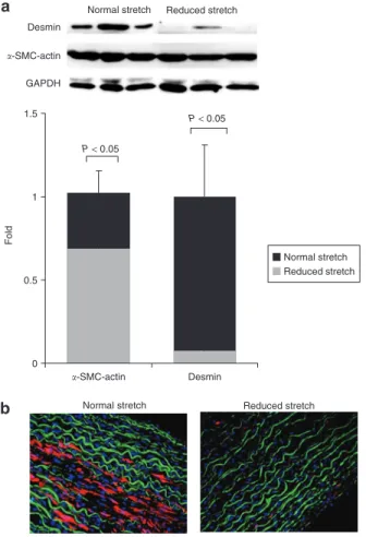
In order to investigate the effect of cyclic stretch on SMC phe-notype, we assessed the protein expression of α-SMC-actin and desmin, both markers of SMC differentiation. A significant dif-ference in SMC phenotype was observed between the arteries exposed to physiological stretch and those exposed to reduced cyclic stretch. Reducing cyclic stretch causes a decrease of 30% in the protein expression α-SMC-actin (Figure 2a, P < 0.05) and 85% in that of desmin (Figure 2a, P < 0.02). These results were further supported by immunofluorescence of arterial sections. In arteries submitted to a normal stretch (Figure 2b, left), desmin expression could be easily detected in the media, whereas in sections with reduced compliance very little desmin expression was found (Figure 2b, right).
Effect of reduced cyclic stretch on MMPs expression and activation
In arteries exposed for 24 h to reduced cyclic stretch, MMP-2 expression was 50% higher than in those exposed to normal cyclic stretch (Figure 3a, P < 0.01). Zymography assays also revealed a higher activation of MMP-2 in arteries exposed to a reduced stretch (Figure 3b, P < 0.05).
0 20 40 60 80 100 120 a b Reduced stretch P < 0.01 Dose (M)
1.E−08 1.E−07 1.E−06 1.E−05 1.E−04
Dose (M)
1.E−08 1.E−07 1.E−06 1.E−05
% Increase in contractio n Normal stretch Reduced stretch Normal stretch 0 2 4 6 8 10 12 14 % Increase in contractio n
Figure 1 | Norepinephrine and prostaglandin F2-∝ dose –response curves. (a) Norepinephrine dose–dependent constriction capacity in porcine carotid artery segments after 1 day of ex vivo perfusion with a normal cyclic stretch (4–5%) and a reduced stretch (<1%). The data are represented as mean values ± s.d., n = 6. (b) Prostaglandin F2-α dose-dependent constriction capacity in porcine carotid artery segments immediately after harvesting and after 1 day of ex vivo perfusion with a normal cyclic stretch (4–5%) and a reduced stretch (<1%). The data are expressed as mean values ± s.d., n = 3.
In order to analyze the distribution of gelatinolytic activ-ity throughout the arterial wall, we performed an in situ
zymo graphy. The increase in MMP expression that was
observed in arteries exposed to reduced cyclic stretch corre-lated with the increase in gelatinolytic activity in the arterial wall (Figure 3c, A). This gelatinolytic activity was distributed
mainly in the tunica media, and was present in both groups. Localization of gelatinolytic activity was reduced in the pres-ence of the inhibitor 1-10-phenanthroline, thereby establish-ing the specificity of the assay (Figure 3c, B).
Effect of cyclic stretch on PAI-1 protein expression and localization
The expression level of PAI-1 was higher in arteries exposed to normal cyclic stretching. Cuffed arteries presented a 20% decrease in PAI-1 expression after 1 day of perfusion
(Figure 4a, P < 0.05).
Immunostaining analysis allowed localization of PAI-1 expression, mainly in the media, in arteries exposed to normal
cyclic stretch (Figure 4b, left). By contrast, for arteries exposed to a reduced cyclic stretch, PAI-1 protein was downregulated and expressed in the media and endothelium (Figure 4b, right).
Normal stretch a
b
Reduced stretch
Normal stretch Reduced stretch
Desmin �-SMC-actin GAPDH 0 0.5 1 1.5 Normal stretch Reduced stretch P < 0.05 P < 0.05 Fold �-SMC-actin Desmin
Figure 2 | Desmin protein expression. (a) Representative immunoblot showing α-smooth muscle cell-actin (α-SMC-actin) and desmin protein bands in arteries exposed to normal and reduced cyclic stretch. The data are expressed as mean values ± s.d., n = 6. (b) Desmin was localized by immunostaining on cross-sections of arteries exposed to normal and reduced cyclic stretch. Desmin is seen in red, elastin in green, and nuclei in blue. All images were taken at identical contrast settings and luminescence levels at ×20 original magnification. n = 4. GAPDH, glyseraldehyde-3-phosphate dehydrogenase. 0 0.5 1 1.5 2 MMP-2 Normal stretch Reduced stretch MMP-2 GAPDH P < 0.01 Reduced stretch Normal stretch Fold Normal stretch Normal stretch Reduced stretch
Reduced stretch 0 1 P < 0.05 1.4 Latent MMP-2 Active MMP-2 1.2 0.8 0.6 0.4 0.2 Fold a b c A B
Reduced stretch Normal stretch
MMP-2 activation
Figure 3 | Matrix metalloproteinase (MMP) expression. (a) The effect of cyclic stretch on MMP-2 protein expression, visualized by immunoblotting. MMP-2 protein expression was normalized to total glyseraldehyde-3-phosphate dehydrogenase (GAPDH) expression. The data are expressed as mean values ± s.d., n = 6. (b) The effect of MMP-2 activity evaluated by zymography. The data are expressed as mean values ± s.d., n = 4. (c) A, The effect of cyclic stretch on total MMP activation, as assessed by gelatinolytic activity; B: proof of specificity of the assay using the inhibitor 1-10-phenanthroline. All images were taken at identical contrast settings and luminescence levels at ×20 original magnification. n = 4.
Effect of cyclic stretch on proliferation
In order to investigate the effect of reduced cyclic stretch on cel-lular proliferation, we stained arteries exposed to normal and reduced cyclic stretch with Ki67. We noticed that proliferating cells were localized predominantly in the media (Figure 5a).
We observed a significant 2.2-fold increase in cellular prolif-eration in the reduced stretch group when compared with the normal stretch group (Figure 5b).
Effect of cyclic stretch on cell apoptosis
There was no statistical difference in apoptosis between the two groups of arterial segments (Figure 6b). Arteries exposed to normal cyclic stretch showed 9 ± 3% cell death, whereas arteries with artificially reduced cyclic stretch showed 7 ± 3% cell death, thereby suggesting that reducing the compliance does not induce higher apoptosis rates. We noticed that pos-itive-stained cells were present mainly in the regions closer to the intima in both the groups (Figure 6a).
DISCUSSION
Effects of cyclic stretch on SMC phenotype and contractile function: Arterial stiffening decreases wall compliance and
this, in turn, reduces cyclic stretch. Cyclic stretch is known to be a potent mechanical stimulus to endothelial cells and vascu-lar SMCs. In order to study the effects of reduced cyclic stretch, we cuffed carotid arteries perfused under pulsatile pressure
ex vivo. The experimentally reduced wall compliance induces
changes in cellular responses and matrix turnover. Perfusion for 1 day with a reduced cyclic stretch leads to drastic decrease in contractile capacity when mediated by NE, of the order of 60% of arteries perfused with a normal cyclic stretch. PG-F2α, which also induces a contractile response, although to a much smaller extent than NE, followed a similar trend to NE, yet yielded insignificant results. Contractile phenotype markers such as α-SMC-actin and desmin have been earlier shown to decrease in arteries submitted to a reduced cyclic stretch. In cuffed vessels, α-SMC-actin, the most abundant protein in the cytoskeleton, diminished by 30%, and the desmin expression
Normal stretch Reduced stretch a
b Normal stretch Reduced stretch
Normal stretch Reduced stretch
PAI-1 GAPDH 0 P < 0.05 Fold 0.8 0.4 1.2 PAI-1 expression
Figure 4 | Plasminogen activator inhibitor-1 (PAI-1) expression. (a) Representative immunoblot showing PAI-1 protein bands in arteries exposed to normal and reduced cyclic stretch. The data are expressed as mean values ± s.d., n = 6. (b) PAI-1 was visualized by immunostaining on cross-sections of arteries exposed to normal and reduced cyclic stretch. PAI-1 is visualized in green, elastin in red, and nuclei in blue. All images were taken at identical contrast settings and luminescence levels at ×20 original magnification. n = 4. GAPDH, glyseraldehyde-3-phosphate dehydrogenase.
a b Reduced stretch Normal stretch 0 0.5 1 1.5 2 2.5 3 3.5 4 Reduced stretch Normal stretch Ki67 expression F ol d P < 0.01
Figure 5 | Proliferation localization and expression. (a) Proliferation was visualized by staining arterial sections exposed to normal and reduced cyclic stretch with Ki67. Cells expressing Ki67 are seen in green and elastin in red. Images were taken at identical contrast settings and luminescence levels at ×20 original magnification. n = 4. (b) The percentage of Ki67-positive cells per arterial section was calculated by dividing the number of positively stained cells (colocalized with a nucleus) by the total number of cells (n = 4).
b Reduced stretch Normal stretch 0 2 4 6 8 10 12 14 % % Apoptotic cells
a Normal stretch Reduced stretch
Figure 6 | Apoptosis localization and expression. (a) Apoptosis was visualized using a TdT-mediated dUTP nick end labeling reaction kit on cross-sections of arteries exposed for 1 day to normal and reduced cyclic stretch. Apoptotic cells are visualized in red and nuclei in blue. All images were taken at identical contrast settings and luminescence levels at ×20 original magnification. n = 6.
(b) The percentage of apoptotic cells in arterial sections was calculated by dividing the number of positively stained cells by the total number of cells (n = 6).
was almost completely removed. This suggests that reduced cyclic stretch leads to changes in SMC phenotype, apparently from contractile to synthetic. These results confirm earlier findings that cyclic stretch regulates the expression of some markers of SMC differentiation.4,8 In our experiments, arteries
perfused under reduced cyclic stretch have shown a decreased contraction in response to NE when normalized to the maxi-mal contraction induced by 60 mmol/l of KCl. Given the fact that the maximum contraction induced by KCL did not differ between our two groups, we hypothesize that the decrease in contractile response to NE in cuffed arteries is linked to a change in phenotype.16,17 Our hypothesis seems to be in
accordance with the findings of Li et al., who demonstrated that human spindle-shaped clones exhibit a higher expression of contractile phenotype markers and a stronger response to NE stimulation than do the epithelioid SMCs.16 The
conclu-sions from the PG-F2α data are more ambiguous, but they do suggest that reducing the cyclic stretch may also affect certain receptors responsible for SMC contraction.
Cyclic stretch in relation to vascular wall remodeling, migra-tion, proliferation and apoptosis: Previously reported in vitro
effects of mechanical strain on SMCs include increased cell proliferation and deposition of a matrix necessary for the organization and development of arteries, and later for vas-cular remodeling during atherosclerosis and hypertension.8,18
There is increasing evidence that changes in cyclic stretch lead to vascular wall remodeling, a process that involves deg-radation of the extracellular matrix through stimulation of MMPs.19–21 We observed an increase in MMP-2 expression
in arteries submitted to a reduced cyclic stretch when com-pared with arteries submitted to a normal cyclic stretch. The activation of MMP-2 was also higher in the cuffed arteries. These results suggest that, in arteries with artificially reduced cyclic stretch, a vascular remodeling process is triggered, and this may lead to structural changes similar to those observed in in vivo studies.22–24 In situ zymography was used for
assess-ing global gelatinase activity and for evaluatassess-ing overall matrix turnover. When compared against data from the normal stretch group, these results confirm that under reduced cyclic stretch the matrix turnover cycle is being augmented.
Plasmin is a potent activator of most MMPs and is regulated by PAI-1. Downregulation of PAI-1 suggests an increased activation of MMPs.25 Several groups of researchers have
reported the involvement of MMPs and the fibrinolytic (plas-min/plasminogen) system as key factors in cell migration and tissue remodeling.13,26 We have shown a decrease in PAI-1 in
arteries perfused with reduced cyclic stretch. Fluorescence data revealed that PAI-1 was predominantly localized at the endothelium, whereas, in arteries perfused with normal cyclic stretch, PAI-1 was primarily expressed by SMCs. Therefore a reduced circumferential cyclic stretch induces a simultane-ous decrease in PAI-1 expression and increase in MMP-2 activity. These effects, together with cell migration and pro-liferation, are suspected to contribute to the increased inti-mal thickening.18,27 In addition, as discussed earlier, the cells
in arteries with reduced cyclic stretch dedifferentiated into a
more synthetic phenotype known to have higher proliferation rates and mobile capacities.28,29
In our study, we found no difference in apoptosis rates between the normal and reduced-stretch groups. This seems to partly contrast with the findings of Courtman et al., who observed no changes in proliferation but marked an increase in apoptosis of SMCs in the media of infrarenal rabbit aortas which had been cuffed to reduce cyclic stretch. Courtman et al. also observed significant atrophy and necrosis. The differences may be attributable to differences in design and duration of the two studies. Courtmann et al. performed long-term
in vivo experiments and analyzed the consequences of the
stiff external banding on vessel wall integrity after 6 weeks. Furthermore, their band was quite tight, reducing the diam-eter by 25% and totally unloading arterial media from circum-ferential stresses.30 Our study diminished but did not totally
abolish cyclic stretch, leaving a residual pulsation of the order of 1%, which is closer to the physiological pulsation in aged or stiffened conduit arteries. Also, our study describes changes in SMC phenotype and migration potential after only 1 day of
ex vivo perfusion.
In summary, the current study demonstrates that a decrease in cyclic stretch leads to changes in SMC phenotypes, and to a concurrent decrease in NE-induced contraction capacity. Furthermore, the increase in MMP-2 is associated with the decrease in PAI-1 as well as with the increase in Ki67, thereby suggesting that the decrease in cyclic stretch may promote wall remodeling, SMC proliferation, and migration. Because reduction in cyclic stretch is often the result of a stiffened arte-rial tree, this work gives insights into possible consequences of arterial stiffening on large-vessel wall physio-pathology.
Acknowledgments: We would like to offer special thanks to Dominique Seppey and Chantal van den Broek for their help with this work. Disclosure: The authors declared no conflict of interest.
1. Zieman SJ, Melenovsky V, Kass DA. Mechanisms, pathophysiology, and therapy of arterial stiffness. Arterioscler Thromb Vasc Biol 2005; 25:932–943. 2. Lehoux S, Tedgui A. Signal transduction of mechanical stresses in the vascular
wall. Hypertension 1998; 32:338–345.
3. Benetos A, Safar M, Rudnichi A, Smulyan H, Richard JL, Ducimetieère P, Guize L. Pulse pressure: a predictor of long-term cardiovascular mortality in a French male population. Hypertension 1997; 30:1410–1415.
4. Reusch P, Wagdy H, Reusch R, Wilson E, Ives HE. Mechanical strain increases smooth muscle and decreases nonmuscle myosin expression in rat vascular smooth muscle cells. Circ Res 1996; 79:1046–1053.
5. Watson PA, Hannan R, Carl LL, Giger KE. Desmin gene expression in cardiac myocytes is responsive to contractile activity and stretch. Am J Physiol 1996; 270:C1228–C1235.
6. Saga H, Kimura K, Hayashi K, Gotow T, Uchiyama Y, Momiyama T, Tadokoro S, Kawashima N, Jimbou A, Sobue K. Phenotype-dependent expression of alpha-smooth muscle actin in visceral smooth muscle cells. Exp Cell Res 1999; 247:279–292.
7. Langille BL. Arterial remodeling: relation to hemodynamics. Can J Physiol Pharmacol 1996; 74:834–841.
8. Birukov KG, Shirinsky VP, Stepanova OV, Tkachuk VA, Hahn AW, Resink TJ, Smirnov VN. Stretch affects phenotype and proliferation of vascular smooth muscle cells. Mol Cell Biochem 1995; 144:131–139.
9. Thyberg J, Hedin U, Sjolund M, Palmberg L, Bottger BA. Regulation of differentiated properties and proliferation of arterial smooth muscle cells. Arteriosclerosis 1990; 10:966–990.
10. Liu SQ, Goldman J. Role of blood shear stress in the regulation of vascular smooth muscle cell migration. IEEE Trans Biomed Eng 2001; 48:474–483.
11. Abbruzzese TA, Guzman RJ, Martin RL, Yee C, Zarins CK, Dalman RL. Matrix metalloproteinase inhibition limits arterial enlargements in a rodent arteriovenous fistula model. Surgery 1998; 124:328–334; discussion 334–325. 12. Dewyer NA, Sood V, Lynch EM, Luke CE, Upchurch GR Jr, Wakefield TW, Kunkel S,
Henke PK. Plasmin inhibition increases MMP-9 activity and decreases vein wall stiffness during venous thrombosis resolution. J Surg Res 2007; 142:357–363. 13. Peng L, Bhatia N, Parker AC, Zhu Y, Fay WP. Endogenous vitronectin and
plasminogen activator inhibitor-1 promote neointima formation in murine carotid arteries. Arterioscler Thromb Vasc Biol 2002; 22:934–939.
14. Gambillara V, Montorzi G, Haziza-Pigeon C, Stergiopulos N, Silacci P. Arterial wall response to ex vivo exposure to oscillatory shear stress. J Vasc Res 2005; 42:535–544.
15. Skalli O, Ropraz P, Trzeciak A, Benzonana G, Gillessen D, Gabbiani G. A monoclonal antibody against alpha-smooth muscle actin: a new probe for smooth muscle differentiation. J Cell Biol 1986; 103:2787–2796.
16. Chien S, Li S, Shyy YJ. Effects of mechanical forces on signal transduction and gene expression in endothelial cells. Hypertension 1998; 31:162–169.
17. Sartore S, Franch R, Roelofs M, Chiavegato A. Molecular and cellular phenotypes and their regulation in smooth muscle. Rev Physiol Biochem Pharmacol 1999; 134:235–320.
18. Bendeck MP, Zempo N, Clowes AW, Galardy RE, Reidy MA. Smooth muscle cell migration and matrix metalloproteinase expression after arterial injury in the rat. Circ Res 1994; 75:539–545.
19. Galis ZS, Khatri JJ. Matrix metalloproteinases in vascular remodeling and atherogenesis: the good, the bad, and the ugly. Circ Res 2002; 90:251–262.
20. Chesler NC, Ku DN, Galis ZS. Transmural pressure induces matrix-degrading activity in porcine arteries ex vivo. Am J Physiol 1999; 277:H2002–H2009.
21. Lehoux S, Lemarie CA, Esposito B, Lijnen HR, Tedgui A. Pressure-induced matrix metalloproteinase-9 contributes to early hypertensive remodeling. Circulation 2004; 109:1041–1047.
22. Benetos A, Laurent S, Hoeks AP, Boutouyrie PH, Safar ME. Arterial alterations with aging and high blood pressure. A noninvasive study of carotid and femoral arteries. Arterioscler Thromb 1993; 13:90–97.
23. Fath SW, Burkhart HM, Miller SC, Dalsing MC, Unthank JL. Wall remodeling after wall shear rate normalization in rat mesenteric arterial collaterals. J Vasc Res 1998; 35:257–264.
24. Galis ZS, Johnson C, Godin D, Magid R, Shipley JM, Senior RM, Ivan E. Targeted disruption of the matrix metalloproteinase-9 gene impairs smooth muscle cell migration and geometrical arterial remodeling. Circ Res 2002; 91:852–859. 25. Lijnen HR. Plasmin and matrix metalloproteinases in vascular remodeling.
Thromb Haemost 2001; 86:324–333.
26. Redmond EM, Cullen JP, Cahill PA, Sitzmann JV, Stefansson S, Lawrence DA, Okada SS. Endothelial cells inhibit flow-induced smooth muscle cell migration: role of plasminogen activator inhibitor-1. Circulation 2001; 103:597–603. 27. Glagov S. Intimal hyperplasia, vascular modeling, and the restenosis problem.
Circulation 1994; 89:2888–2891.
28. Bochaton-Piallat ML, Gabbiani G, Pepper MS. Plasminogen activator expression in rat arterial smooth muscle cells depends on their phenotype and is modulated by cytokines. Circ Res 1998; 82:1086–1093.
29. Hao H, Gabbiani G, Bochaton-Piallat ML. Arterial smooth muscle cell heterogeneity: implications for atherosclerosis and restenosis development. Arterioscler Thromb Vasc Biol 2003; 23:1510–1520.
30. Courtman DW, Cho A, Langille L, Wilson GJ. Eliminating arterial pulsatile strain by external banding induces medial but not neointimal atrophy and apoptosis in the rabbit. Am J Pathol 1998; 153:1723–1729.