Florian M. Buck Juerg Hodler Marco Zanetti Claudio Dora Christian W. A. Pfirrmann Received: 22 March 2010 Revised: 8 June 2010 Accepted: 20 June 2010 Published online: 26 July 2010 # European Society of Radiology 2010
Ultrasound for the evaluation
of femoroacetabular impingement
of the cam type. Diagnostic
performance of qualitative criteria
and alpha angle measurements
Abstract Objective To develop and assess a technique to evaluate cam type femoroacetabular impingement (FAI) using ultrasound (US).Methods
Fifty patients (24 women, 26 men) were included (mean age: 39.1years; age range: 16–59). US images of the anterior and anterosuperior contour of the femoral neck were obtained and analysed in 50 patients. Non-spherical shape of the head-neck junction (cam deformity), bony protuberances at the femoral neck, shape of the femoral neck (waist deficiency) and alpha angle were assessed. Magnetic reso-nance (MR) arthrography served as the standard of reference. Diagnostic performance and receiver operating characteristics (ROC) curves were calculated.ResultsBased on MR arthrography 28 patients had cam-type FAI. On US, an anterosuperior cam deformity was seen in 40/44 patients (Reader 1/Reader 2;
sensitiv-ity 93%/89%, specificity 36%/14%). A bony protuberance anterosuperiorly in 23/13 patients (sensitivity 71%/ 32%, specificity 86%/82%) and an anterosuperior waist deficiency in 19/ 35 patients (sensitivity 25%/54%, specificity 100%/54%). Sensitivity and specificity of the other criteria were lower than 70% (average of Reader 1 & 2).ConclusionA techni-que to evaluate cam type FAI using US is presented. The detection of an anterosuperior cam deformity is sen-sitive, and presence of an anterosu-perior bony protuberance is specific for cam FAI. Alpha angle measure-ments are not helpful in establishing the diagnosis.
Keywords Femoroacetabular impingement . Cam deformity . Ultrasound . Alpha angle . Magnetic resonance imaging
Introduction
Femoroacetabular impingement is a known aetiology of premature osteoarthritis of the non-dysplastic hip [1–5]. Cam and pincer types of FAI have been proposed [1,6]. In young athletic individuals FAI of the cam type is predominant. A prevalence of 17% in men and 4% in women has been reported [7]. The cam type is charac-terised by a non-spherical shape of the femoral head at the junction to the femoral neck (cam deformity), reduced waist of the femoral neck, and bony protuberances generally at the anterior and anterosuperior aspect of the femoral neck [4,8–11]. This deformity leads to jamming of the femoral head into the anterior and anterosuperior
acetabulum resulting in early chondral and labral damage due to recurrent microtrauma [12–14].
Cam impingement may be treated by open or arthro-scopic restoration of a physiological waist and removal of bony protuberances [5, 15, 16] at a low rate of complications [2, 16, 17]. These procedures aim to prevent or delay additional degeneration of the hip joint. Patients with no or mild osteoarthritis have a substantially better outcome than patients with advanced cartilage damage [15].
Groin pain is a very early complaint of patients suffering from FAI but its differential diagnosis is very wide: stress fractures of the femoral neck, iliopsoas tendonitis, tears of the adductor tendons, nerve entrapment
F. M. Buck
:
J. Hodler:
M. Zanetti:
C. W. A. PfirrmannDepartment of Radiology,
Orthopedic University Hospital Balgrist, Zurich, Switzerland
C. Dora
Department of Orthopedic Surgery, Orthopedic University Hospital Balgrist, Zurich, Switzerland
F. M. Buck ())
University Clinic Balgrist,
Forchstrasse 340, 8008, Zurich, CH, Switzerland
e-mail:florian.buck@gmail.com Tel.: +41-77-4080952
syndromes and inguinal hernia are only a few of the possible underlying conditions leading to groin pain, especially in athletes [18]. Therefore, a history of groin pain is an early, but non-specific finding of FAI. Diagnosis is additionally suspected based on clinical examination (impaired internal rotation andflexion of the hip joint) [1,
19,20]. Plain radiography and magnetic resonance (MR) imaging are used to confirm the diagnosis, to exclude some of the differential diagnostic possibilities, and to assess the degree of joint damage for treatment planning. Early recognition of FAI is important because patients with osteoarthritic changes do substantially worse post-operatively [5, 15, 16, 21, 22]. Delay in diagnosing the underlying condition or misdiagnosis may be associated with prolonged training interruptions, unnecessary medi-cal and surgimedi-cal treatments, and last but not least potentially more pronounced cartilage damage [1, 5,23]. Therefore, a cost-effective, fast and widely available technique for early detection of patients with FAI is of interest. An examination technique based on ultrasound (US) would meet these criteria. Thus, the purpose of our study was to develop and assess a technique to evaluate femoroacetabular cam deformity using US.
Materials and methods
The institutional review board approved the study. Written informed consent was obtained from all patients.
Patients
Patients with suspected FAI of the cam-type based on clinical examination were included in the study. Clinical examination was performed by the referring orthopaedists and included tests for the evaluation of internal rotation and flexion of the hip joint [14, 24–26]. Patients with inflammatory diseases, tumours, previous hip surgery, developmental hip dysplasia, and vascular necrosis of the femoral head were excluded from the study. In total, 50 patients (mean age: 39.1 years; age range: 16–59) out of 68 consecutive patients referred for MR arthrography from the orthopaedic outpatient clinic were included in the study. There were 24 women (mean age: 40.1 years; age range: 18–58) and 26 men (mean age: 32.6; age range: 16–59).
Eighteen patients (mean age: 35.2 years; age range: 16– 59) had to be excluded because of previous surgery to the hip joint (n=8), developmental dysplasia of the hip (n=6), and avascular necrosis of the femoral head (n=2).
Clinical information about the patients was gathered from the referring orthopaedists. In particular, the presence or absence of groin pain, increase of groin pain upon physical exercise, pain at night, and impaired internal rotation and flexion of the hip was documented in each patient.
Ultrasound
Ultrasound was performed before MR arthrography using a curved array transducer with 2–5 MHz frequency range (iU22 Ultrasonography System, C5-2 curved array trans-ducer, Philips Medical Systems, DA Best, The Nether-lands). US was performed by a radiologist with 4 years of experience in US who was blinded as to the MRfindings (BFM). The examination was performed with the patient in a supine position and neutral position of the hip joint and leg.
Longitudinal images of the anterior and anterosuperior head-neck contour were obtained in a transverse oblique plane parallel to the axis of the femoral neck (Fig.1) and labelled“anterior contour” or “anterosuperior contour” for the reviewers. The anterior contour was evaluated with the transducer perpendicular to the skin surface. The ante-rosuperior contour was evaluated in almost the same position, but with the transducer slightly more cranial and angled 45° caudally. All US images were saved in the picture archiving and communication system (PACS).
Analysis of ultrasound
Two musculoskeletal radiologists with 14 years (ZM) and 18 years (HJ) of experience in musculoskeletal radiology analysed all US images independently and were blinded to the results of MR imaging. Three qualitative criteria were evaluated at the anterior and anterosuperior head-neck contour: Presence or absence of a non-spherical head-neck junction (cam deformity) (Fig. 2), presence or absence of focal bony protuberances at the femoral neck (Fig.3), and shape of the osseous contour of the femoral waist (concave,flat, convex) (Fig. 4). In the qualitative evalua-tion, a cam deformity was defined as the presence of a non-spherical head-neck junction (Fig. 2) evaluated by visual judgement before measurement of the alpha angle. The labrum was not assessed.
Quantification of the cam deformity (alpha angle measurement) was performed in a five-step procedure (Fig. 1): First, a tangent line from the distal insertion of the joint capsule (point A in Fig.1e) to the femoral head contour was drawn (Fig. 1e). This line served as an approximation for the femoral neck axis. Then, a circle was defined by three points (Points B, C, D in Fig.1f) on the contour of the femoral head. To prevent measurement errors due to a femoroacetabular cam deformity, all three points were placed on the spherical portion of the proximal femoral head contour. The first point (Point B in Fig.1f) was placed where the tangent line drawn in step 1 touched the femoral head contour. The second point (Point D in Fig. 1f) was placed on the most proximal femoral head contour that was visible. The third point (Point C in Fig.1f) was placed in the middle between the first two points.
In a third step, the centre axis of the femoral neck was drawn as a parallel line to the first tangent line and
the centre of the femoral head (Fig. 1f). Then, the point where the femoral head contour crossed the circle defined in step 2 was identified (white arrow, Fig. 1f). Finally, the alpha angle was measured (Fig. 1g) in accordance with the method described by Nötzli and co-workers [25].
MR arthrography
Intra-articular contrast media were injected in a standardised fashion by a musculoskeletal radiologist. Afterfluoroscopic confirmation of the intra-articular position of the tip of the needle with 1 mL of an iodinated contrast agent (iopamidol
Fig. 1 Photograph illustrating the US probe position to evaluate the anterior (a, b) and anterosuperior (c) femoral neck contour in a hip phantom consisting of a human hip skeleton embedded in anatomically shaped acrylic glass (a) and a patient (b, c) and the corresponding US image of the anterior osseous contour as evaluated in b (d). Measurement of the alpha angle was performed in a five-step procedure: First, a tangent line from the distal insertion (a) of the joint capsule (arrowheads) to the femoral head contour was drawn (e). This line served as an approximation for the femoral neck axis. Then, a circle was defined by three points (b–d) on the contour of the femoral head (f). The first point (Point B in f) was placed where the tangent line drawn in step 1 touched the
femoral head contour. The second point (Point D in f) was placed on the most proximal femoral head contour that was visible. The third point (Point C in f) was placed in the middle between thefirst two points. To prevent measurement errors due to a femoroacetabular cam deformity, all three points were placed on the concentric portion of the proximal femoral head contour. In a third step, the centre axis of the femoral neck was drawn as a parallel line to thefirst tangent line and the centre of the femoral head (f). Then, the point (arrow) where the femoral head contour crossed the circle defined in step 2 was identified (f). Finally, the alpha angle was measured (g) in accordance with the method described by Nötzli and co-workers [25]
200 mg/mL, Iopamiro 200, Bracco, Milan, Italy), 8 mL of a diluted MR contrast agent (gadopentetate dimeglumine, Magnevist, Bayer Pharma, Berlin, Germany) at a concen-tration of 2 mmol/L were injected.
MR imaging was performed with one of two 1.5-T systems (Avanto or Espree; Siemens Medical Solutions, Erlangen, Germany). The examination was performed in the supine position with the hip joint in a neutral position. The following sequences were acquired: coronal T1-weighted spin-echo sequence (section thickness, 3 mm; repetition time, 604 ms; echo time, 13 ms; field of view, 16 cm; matrix, 512×512), coronal intermediate-weighted fast spin-echo sequence with fat saturation (section thick-ness, 3 mm; repetition time, 3520 ms; echo time, 39 ms; field of view, 16 cm; matrix, 512×512; turbo factor, 7), sagittal water excitation three-dimensional double-echo steady-state sequence (section thickness, 1.7 mm; repeti-tion time, 25 ms; echo time, 9 ms;field of view, 15 cm; matrix, 512 × 512), sagittal T1-weighted spin-echo sequence (section thickness, 4 mm; repetition time, 550 ms; echo time, 13 ms; field of view, 16 cm; matrix, 512×512), transverse oblique (parallel to the long axis of the femoral neck) water-excitation true fast imaging with steady-state precession sequence (section thickness, 1.25 mm; repetition time, 8.9 msec; echo time, 3.3 msec; flip angle, 28°; intersection gap, none; field of view, 17 cm; matrix, 512×512). The transverse oblique
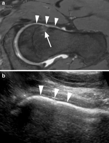
three-Fig. 2 A 34-year-old man with a laterally increasing radius of the femoral head at the anterior contour consistent with cam impingement (arrows, normal radius; dashed arrows, increased radius). Comparison of the imaging techniques. a MRI. b US
Fig. 3 A 36-year-old woman with a bony protuberance at the anterosuperior head-neck junction (arrow) consistent with cam impingement. Comparison of the imaging techniques. a MRI. b US
Fig. 4 A 40-year-old man with considerable waist deficiency consistent with cam impingement (arrowheads). Comparison of the anterior contour of the femoral head-neck junction using MRI (a) and US (b). Bone marrow edema pattern is seen at the typical location of the anterior aspect of the femoral neck (arrow in a)
dimensional data set was used for radial reformations by using the long axis of the femoral neck as a rotation axis [8].
Standard of reference
Two musculoskeletal radiologists with 2 years (BFM) and 10 years (PCWA) of experience in musculoskeletal radiology analysed all MR arthrographic images in consensus. The radiologists were not involved in the US evaluation and were blinded to its results. In the presence of a cam deformity and/or femoral waist deficiency the diagnosis of FAI of the cam type was established, disregarding the alpha angle measurement. The result of the MR evaluation served as a standard of reference.
Statistical analysis
Diagnostic performance of the qualitative criteria (sensi-tivity, specificity, positive and negative predictive value, and accuracy) was calculated. Receiver operating charac-teristics (ROC) curve analysis for alpha angles measured on ultrasound was performed. Interobserver agreement was evaluated using kappa statistics for qualitative criteria and intraclass correlation coefficient (ICC) for quantitative criteria. The results of the evaluation on the US and MR images were compared using descriptive statistics (qual-itative criteria) and Pearson correlation coefficient (PCC; alpha angle measurements). SPSS (version, 16.0 mac; SPSS, Chicago, IL, USA) software was used for statistical analysis.
Results
Forty-seven patients suffered from groin pain, whereas the pain increased in 36 patients upon physical exercise (Table1). Three patients had pain at night and 41 patients had impaired internal rotation and flexion of the hip.
The results of the analysis of the ultrasound examina-tions are presented in Table 2. Interobserver agreement was slight to moderate according to Landis and Koch [27] (Table 3). The calculated diagnostic performances of all the items evaluated are shown in Table4.
Mean anterior and anterosuperior alpha angle measured by reader 1 was 64.8° (range, 31°–89°; standard deviation, 12.5°) and, 69.5° (range, 43°–94°; standard deviation, 11.7°), respectively. Reader 2 measured a mean anterior and anterosuperior alpha angle of 57.1° (range, 34°–83°; standard deviation, 15.0°) and 72.7° (range, 38°–90°; standard deviation, 11.2°), respectively. The ICC for the quantitative interreader agreement was moderate for both anterior and anterosuperior alpha angle measurements (Table 3).
The results of the evaluations on the US and MR images matched as follows: The presence of a cam deformity at the anterior contour matched in 48% (n= 24) for Reader 1 and in 66% (n=33) for Reader 2; at the anterosuperior contour in 64% (n=32) for Reader 1 and 56% (n=28) for Reader 2. Bony protuberances at the anterior contour matched in 48% (n=24) for Reader 1 and in 60% (n=30) for Reader 2. Corresponding values for bony protuberances anterosuperiorly were 78% (n=39) for Reader 1 and 50% (n=25) for Reader 2. Concerning the presence of an anterior waist deficiency, Reader 1 agreed with the evaluation on MR images in 66% (n=33) and Reader 2 in 78% (n=39); anterosuperiorly 50% (n=25) and 32% (n=16), respectively.
The alpha angle measurements of reader 1 showed a strong, significant relationship with the measurements on MR images (anterior measurements: PCC 0.891, p<0.001;
Table 1 Symptoms of included patients
Symptom Number of
patients
FAI No FAI
Groin pain 47 28 19
Increase of groin pain upon physical exercise
36 22 14
Pain at night 3 1 2
Impaired internal rotation andflexion of the hip
41 25 16
Abbreviations: FAI Patients with FAI; No FAI Patients without FAI.
Table 2 Sonographicfindings
Finding Reader 1 Reader 2
Non-Spherical Head-Neck Junction (Cam Deformity)
Anterior 29 18
Anterosuperior 40 44
Focal Bony Protuberances at the Femoral Neck (Osseous Bump)
Anterior 17 8
Anterosuperior 23 13
Flat or Convex Osseous Contour of the Femoral Neck (Waist Deficiency)
Anterior 19 7
Anterosuperior 35 24
Table 3 Interobserver agreement Ultrasound qualitative evaluation
Kappa P
Presence of a cam deformity
Anterior 0.196 0.126
Anterosuperior 0.265 0.050
Presence of a bony protuberance at the femoral neck
Anterior 0.335 0.008
Anterosuperior 0.168 0.191
Waist deficiency
Anterior 0.420 0.000
Anterosuperior 0.252 0.048
Alpha angle measurements
ICC P
Alpha angle Anterior 0.515 0.006
Anterosuperior 0.509 0.007 Abbreviations: ICC Intraclass correlation coefficient.
anterosuperior: PCC 0.889, p<0.001). The measurements of reader 2, however, showed only a poor to moderate relationship (anterior measurements: PCC 0.425, p=0.002; anterosuperior: PCC 0.199, p=0.165).
ROC curves (Fig. 5) were plotted for the alpha angle measurements. The results are provided in Table5. ROC analysis for anterior alpha angle measurements demon-strated areas under the curve of 0.581 (p=0.328) for
Table 4 Diagnostic performance of qualitative read-out criteria
TP TN FP FN Sens Spec PPV NPV Acc
Presence of a cam deformity Anterior R1 19 12 10 9 68% 55% 66% 57% 62% R2 14 18 4 14 50% 82% 78% 56% 64% Anterosuperior R1 26 8 14 2 93% 36% 65% 80% 68% R2 25 3 19 3 89% 14% 57% 50% 56%
Presence of a bony protuberance Anterior R1 11 16 6 17 39% 73% 65% 48% 54% R2 6 20 2 22 21% 91% 75% 48% 52% Anterosuperior R1 20 19 3 8 71% 86% 87% 70% 78% R2 9 18 4 19 32% 82% 69% 49% 54% Waist deficiency Anterior R1 14 17 5 14 50% 77% 74% 55% 62% R2 25 12 10 3 89% 55% 71% 80% 74% Anterosuperior R1 7 22 0 21 25% 100% 100% 51% 58% R2 15 13 9 13 54% 59% 63% 50% 56%
Abbreviations: TP true-positive. TN true-negative. FP false-positive. FN false-negative. Sens sensitivity. Spec specificity. PPV positive predictive value. NPV negative predictive value. Acc accuracy. R1 reader 1. R2 reader 2.
Fig. 5 Receiver operating characteristics (ROC) curves for ultrasound alpha angle measurements. Solid lines show the results of the anterior measurements (Reader 1, thick line; Reader 2, thin line). Dashed lines
show the results of the anterosuperior measurements (Reader 1, thick line; Reader 2, thin line)
Reader 1 and 0.665 (p=0.047) for Reader 2. Anterosuper-iorly, the areas under the curve were 0.688 (p=0.023) for Reader 1, and 0.588 (p=0.291) for Reader 2.
Discussion
It is possible to visualise the anterior and anterosuperior osseous contour of the femoral neck and, in the opinion of the authors, look for the typical osseous con figu-ration causing FAI of the cam type using US. However, no criterion with equally high specificity and sensitivity was found. Based on the osseous contour of the femoral neck, we defined and evaluated three qualitative criteria (cam deformity, waist deficiency and bony protuberan-ces) and proposed a technique to measure the alpha angle. To the best of our knowledge, this is the first time evaluation of FAI of the cam type was attempted using US.
Ultrasound is widely available, inexpensive, and does not involve radiation exposure to the patient examined. These attributes are important for the examination of young patients. In particular, young men dedicated to high
performance sports could benefit when cam FAI is
diagnosed before damage of the joint occurs. In the country where the authors’ institution is located screening for FAI of young men during army recruitment is currently evaluated. Standardised clinical measurement of the internal rotation of the hip joint is performed. Individuals with FAI of the cam type could be advised to change their type of sport (for example no martial arts, no ice hockey) or surgical treatment at an early stage may be initiated [28–30].
Detection of an anterosuperior cam deformity was a sensitive finding for FAI. The presence of an anterosupe-rior bony protuberance and a femoral waist deficiency were specific findings. However, looking at Table 4 the negative predictive value (NPV) for the presence of a cam deformity at the anterosuperior osseous contour was between 50% and 80%.
Likewise, the positive predictive values (PPV) for the presence of an anterosuperior bony protuberance and a femoral waist deficiency and interreader agreement were not high enough for the recommendation of US as a screening tool for FAI.
In the clinical situation, this makes additional imaging such as plain radiographs and MRI necessary in these patients. From a patients perspective one-stop-shop
imag-ing is to be preferred and this would certainly hamper the implementation of US.
The anterosuperior osseous contour seems to be more useful for the assessment of cam FAI than the anterior contour. Thisfinding is in line with an article by Pfirrmann and co-workers [8] evaluating the specific location of cam deformities at the femoral head-neck junction. The authors demonstrated a predominance of the cam deformity at the anterosuperior aspect compared with the anterior aspect of the femoral neck.
In our study, the measurement of alpha angles did not prove to be helpful. There may be two possible reasons for this. First, US and the measurement technique we have developed may have some limitations: We made the assumption that the femoral neck axis is parallel to, or at least in a constant relationship with, a line drawn from the insertion of the joint capsule at the femoral neck to the femoral head. Additionally, any malalignment of the US transducer to the femoral neck could have led to distortion of the osseous contour of the femoral neck and consec-utively to measurement errors.
Second, the alpha angle itself could be an unreliable criterion for diagnosing FAI. Several recent articles support the thesis that alpha angle measurements are not very helpful in the evaluation of FAI. Lohan et al. [31] found a considerable variability of alpha angle measure-ments performed on MR images. Statistically they found no value of alpha angle measurements in suggesting the presence or absence of cam FAI. Nouh and co-workers [32] assessed the value of a subjective assessment of the alpha angle on MR images. Measurement of the alpha angle served as the standard of reference. Because of the quite low areas under the curve (≤0.606) of the ROC analysis they concluded that subjective assessment of alpha angles is not optimal unless one is quite confident about a bony abnormality.
Because many intra-articular abnormalities, like labral tears and chondral lesions are not accessible to US, US has limitations in the evaluation of patients with groin pain. Because up to 76% of cases of FAI are of a mixed type (cam FAI and pincer FAI combination) and because it is not possible to address the pincer component with US, additional imaging may be needed for a comprehensive evaluation of FAI [33, 34]. In our study, there were no patients with a pincer FAI only. However, in the hands of an experienced ultrasonographer the presented qualitative criteria can be useful in suggesting the diagnosis of a FAI as the cause of the patients’ complaints and a cam FAI component can be assessed.
Table 5 Receiver operating characteristics (ROC) curve analysis: area under the curve of the alpha angle measurements using US
Measurement Area SD P 95% confidence interval
Lower bound Upper bound
Anterior R1 0.581 0.082 0.328 0.412 0.741
R2 0.665 0.077 0.047 0.514 0.815
Anterosuperior R1 0.688 0.078 0.023 0.536 0.841
R2 0.588 0.082 0.291 0.426 0.749
Contrary to the US evaluation of hips in newborns, the mature skeleton is fully mineralised and therefore not all parts of the hip joint may be visualised. This renders US evaluation of the hip in adults considerably more difficult. In our study, US evaluation of alpha angles is charac-terised by a moderate interreader agreement (ICC 0.509– 0.515) only. Falliner et al. [35] and Simon et al. [36] reported a superior interreader agreement in ultrasound angle measurements at DDH (developmental dysplasia of the hip) screening in newborns (ICC 0.72–0.74) compared with the ultrasound angle measurements in our study. This superior interreader agreement can be partly explained by the new measurement techniques in our study and the mineralised skeleton in adults.
An alternative to MR imaging and US for visualising cam deformities is CT with 3D reconstruction [24]. 3D CT-based hip models may be used for kinematic hip analysis. However for CT of the pelvis radiation exposure is necessary which may be a problem because typically young patients suffer from cam impingement.
Limitations of our study include the selected group of patients from an orthopaedic clinic. The lack of a true gold standard, such as surgery, to prove cam-type impingement was another limitation. Because the use of US in the evaluation of FAI has not been presented before, data on
the interobserver variation with respect to repeated measurements would be beneficial. Concerning the alpha angle measurements, angulation of the US probe with respect to the femoral neck could potentially lead to different measurements because the measurement techni-que strongly relies on the tangent line from the distal insertion of the joint capsule to the femoral head contour. Finally, during the US examination, the observer may have lost his“blindness” to the presence of FAI based on symptoms that the patient had.
Ultrasound examination should preferably be per-formed in combination with standardised physical exami-nations and in collaboration with experienced orthopaedic surgeons. Based on the described qualitative criteria, it is possible to evaluate cam FAI using US and decide whether an additional plain radiograph or MR examina-tion is required to substantiate the diagnosis of FAI and to demonstrate secondary damage of the cartilage and the labrum.
In conclusion, a technique to evaluate cam type FAI using US is presented. The detection of an anterosuperior cam deformity is a sensitive, and the presence of an anterosuperior bony protuberance is a specific finding for a cam FAI. Alpha angle measurements are not helpful in establishing the diagnosis.
References
1. Lavigne M, Parvizi J, Beck M, Siebenrock KA, Ganz R, Leunig M (2004) Anterior femoroacetabular impingement. Part I. Techniques of joint preserving surgery. Clin Orthop 418:61–66
2. Murphy S, Tannast M, Kim YJ, Buly R, Millis MB (2004) Debridement of the adult hip for femoroacetabular impingement. Clin Orthop Relat Res 429:178–181
3. Tanzer M, Noiseux N (2004) Osseous abnormalities and early osteoarthritis. Clin Orthop Relat Res 429:170–177 4. Ito K, Minka-Il MA, Leunig M, Werlen
S, Ganz R (2001) Femoroacetabular impingement and the cam-effect. J Bone Joint Surg Br 83-B:171–176
5. Ganz R, Parvizi J, Beck M, Leunig M, Notzli H, Siebenrock KA (2003) Femoroacetabular impingement. A cause for osteoarthritis of the hip. Clin Orthop Relat Res 417:112–120 6. Beck M, Kalhor M, Leunig M, Ganz R
(2005) Hip morphology influences the pattern of damage to the acetabular cartilage: femoroacetabular impingement as a cause of early osteoarthritis of the hip. J Bone Joint Surg Br 87:1012–1018
7. Gosvig KK, Jacobsen S, Sonne-Holm S, Gebuhr P (2008) The prevalence of cam-type deformity of the hip joint: a survey of 4151 subjects of the copenhagen osteoarthritis study. Acta Radiol 49:436–441
8. Pfirrmann CW, Mengiardi B, Dora C, Kalberer F, Zanetti M, Hodler J (2006) Cam and pincer femoroacetabular impingement: characteristic MR arthrographicfindings in 50 patients. Radiology 240:778–785
9. Panzer S, Augat P, Esch U (2008) CT assessment of herniation pits:
prevalence, characteristics, and potential association with morphological predictors of femoroacetabular impingement. Eur Radiol. doi:10.1007/ s00330-008-0952-7
10. Beaule PE, Zaragoza E, Motamedi K, Copelan N, Dorey FJ (2005) Three-dimensional computed tomography of the hip in the assessment of
femoroacetabular impingement. J Orthop Res 23:1286–1292 11. Kassarjian A, Yoon LS, Belzile E,
Connolly SA, Millis MB, Palmer WE (2005) Triad of MR arthrographic findings in patients with cam-type femoroacetabular impingement. Radiology 236:588–592
12. Tannast M, Goricki D, Beck M, Murphy SB, Siebenrock KA (2008) Hip damage occurs at the zone of femoroacetabular impingement. Clin Orthop Relat Res 466:273–280
13. Ito K, Minka MA 2nd, Leunig M, Werlen S, Ganz R (2001)
Femoroacetabular impingement and the cam-effect. A MRI-based quantitative anatomical study of the femoral head-neck offset. J Bone Joint Surg Br 83:171–176
14. Ganz R, Leunig M, Leunig-Ganz K, Harris WH (2008) The etiology of osteoarthritis of the hip: an integrated mechanical concept. Clin Orthop Relat Res 466:264–272
15. Beck M, Leunig M, Parvizi J, Boutier V, Wyss D, Ganz R (2004) Anterior femoroacetabular impingement. Part II. Midterm results of surgical treatment. Clin Orthop 418:67–73
16. Stahelin L, Stahelin T, Jolles BM, Herzog RF (2008) Arthroscopic offset restoration in femoroacetabular cam impingement: accuracy and early clinical outcome. Arthroscopy 24(51– 57):e51
17. Peters CL, Erickson JA (2006) Treatment of femoro-acetabular impingement with surgical dislocation and debridement in young adults. J Bone Joint Surg Am 88:1735–1741 18. Omar IM, Zoga AC, Kavanagh EC et al
(2008) Athletic pubalgia and“sports hernia”: optimal MR imaging technique andfindings. Radiographics 28:1415– 1438
19. Jaeger M, Wild A, Westhoff B, Krauspe R (2004) Femoroacetabular
impingement caused by a femoral osseous head-neck bump deformity: clinical, radiological, and experimental results. J Orthop Sci 9:256–263 20. Wyss TF, Clark JM, Weishaupt D, Notzli
HP (2007) Correlation between internal rotation and bony anatomy in the hip. Clin Orthop Relat Res 460:152–158 21. Schmid MR, Notzli HP, Zanetti M, Wyss TF, Hodler J (2003) Cartilage lesions in the hip: diagnostic effectiveness of MR arthrography. Radiology 226:382–386
22. Philippon MJ, Briggs KK, Yen YM, Kuppersmith DA (2009) Outcomes following hip arthroscopy for femoroacetabular impingement with associated chondrolabral dysfunction: minimum two-year follow-up. J Bone Joint Surg Br 91:16–23
23. Ilizaliturri VM Jr (2009) Complications of arthroscopic femoroacetabular impingement treatment: a review. Clin Orthop Relat Res 467:760–768 24. Kubiak-Langer M, Tannast M, Murphy
SB, Siebenrock KA, Langlotz F (2007) Range of motion in anterior
femoroacetabular impingement. Clin Orthop Relat Res 458:117–124
25. Notzli HP, Wyss TF, Stoecklin CH, Schmid MR, Treiber K, Hodler J (2002) The contour of the femoral head-neck junction as a predictor for the risk of anterior impingement. J Bone Joint Surg Br 84:556–560
26. Tannast M, Kubiak-Langer M, Langlotz F, Puls M, Murphy SB, Siebenrock KA (2007) Noninvasive three-dimensional assessment of femoroacetabular impingement. J Orthop Res 25:122–131 27. Landis JR, Koch GG (1977) The
measurement of observer agreement for categorical data. Biometrics 33:159–174 28. Bizzini M, Notzli HP, Maffiuletti NA
(2007) Femoroacetabular impingement in professional ice hockey players: a case series of 5 athletes after open surgical decompression of the hip. Am J Sports Med 35:1955–1959
29. Koh J, Dietz J (2005) Osteoarthritis in other joints (hip, elbow, foot, ankle, toes, wrist) after sports injuries. Clin Sports Med 24:57–70
30. Shindle MK, Voos JE, Heyworth BE et al (2007) Hip arthroscopy in the athletic patient: current techniques and spectrum of disease. J Bone Joint Surg Am 89 (Suppl 3):29–43
31. Lohan DG, Seeger LL, Motamedi K, Hame S, Sayre J (2009) Cam-type femoral-acetabular impingement: is the alpha angle the best MR arthrography has to offer? Skeletal Radiol 38:855– 862
32. Nouh MR, Schweitzer ME, Rybak L, Cohen J (2008) Femoroacetabular impingement: can the alpha angle be estimated? AJR Am J Roentgenol 190:1260–1262
33. Tannast M, Siebenrock KA, Anderson SE (2007) Femoroacetabular
impingement: radiographic diagnosis– what the radiologist should know. AJR Am J Roentgenol 188:1540– 1552
34. Philippon MJ, Kuppersmith DA, Wolff AB, Briggs KK (2009) Arthroscopic findings following traumatic hip dislocation in 14 professional athletes. Arthroscopy 25:169–174
35. Falliner A, Schwinzer D, Hahne HJ, Hedderich J, Hassenpflug J (2006) Comparing ultrasound measurements of neonatal hips using the methods of Graf and Terjesen. J Bone Joint Surg Br 88:104–106
36. Simon EA, Saur F, Buerge M, Glaab R, Roos M, Kohler G (2004) Inter-observer agreement of
ultrasonographic measurement of alpha and beta angles and the final type classification based on the Graf method. Swiss Med Wkly 134:671– 677