HAL Id: tel-02130621
https://tel.archives-ouvertes.fr/tel-02130621
Submitted on 16 May 2019HAL is a multi-disciplinary open access archive for the deposit and dissemination of sci-entific research documents, whether they are pub-lished or not. The documents may come from teaching and research institutions in France or abroad, or from public or private research centers.
L’archive ouverte pluridisciplinaire HAL, est destinée au dépôt et à la diffusion de documents scientifiques de niveau recherche, publiés ou non, émanant des établissements d’enseignement et de recherche français ou étrangers, des laboratoires publics ou privés.
Structure-function relationships of the lysine
decarboxylase from Pseudomonas aeruginosa
Diego Carriel
To cite this version:
Diego Carriel. Structure-function relationships of the lysine decarboxylase from Pseudomonas aerug-inosa. Biomolecules [q-bio.BM]. Université Grenoble Alpes, 2017. English. �NNT : 2017GREAV011�. �tel-02130621�
THÈSE
Pour obtenir le grade de
DOCTEUR DE LA COMMUNAUTÉ UNIVERSITÉ
GRENOBLE ALPES
Spécialité : Biologie Structurale et Nanobiologie
Arrêté ministériel : 25 mai 2016
Présentée par
Diego CARRIEL
Thèse dirigée par Irina GUTSCHE et Co-dirigée par Sylvie ELSEN
préparée au sein de l’Institut de Biologie Structurale dans l'École Doctorale de Chimie et Sciences du Vivant
Structure-function relationships of
the lysine decarboxylase LdcA
from Pseudomonas aeruginosa
Thèse soutenue le 15 Mai 2017, devant le jury composé de : M. Axel HARTKE
Professeur des Universités, UCBN, Caen (Rapporteur) Mme. Anne-Marie DI GUILMI
Ingénieur-chercheur, CEA Paris-Saclay (Rapporteur) M. Bertrand TOUSSAINT
Professeur des Universités, UGA, Grenoble (Président) Mme. Patricia RENESTO
Directeur de Recherche, CNRS, Grenoble (Examinateur) Mme. Irina GUTSCHE
Directeur de Recherche, CNRS, Grenoble (Directeur de thèse) Mme. Sylvie ELSEN
1 Abstract
The lysine decarboxylase (LDC) belongs to a family of decameric PLP-dependent enzymes that catalyse the reaction transforming L-Lysine into cadaverine while consuming a proton. They have been extensively characterised in enterobacteria, where they have been shown to play a crucial role during acid, oxidative stress and antibiotic resistance.
Since mechanisms allowing bacteria to counter stress challenges are important for displaying full virulence, we wondered if the opportunistic bacterium Pseudomonas aeruginosa could be using LdcA to counter stress conditions that have already been described for enterobacteria. During my PhD, we addressed this question by using different but complementary.
First of all, we used promoter-gene fusions and western-blot analysis to determine the conditions in which ldcA was expressed and its product synthesized.
In parallel, we constructed an ldcA mutant and its complemented strain to understand whether LdcA was involved in acid and oxidative stress response. We used manual screenings and high-throughput technologies (Biolog) and we discovered that the cadaverine produced by LdcA is needed for full growth fitness when growing in minimal medium using L-glutamate as carbon source. Since slow growing phenotypes are linked to heightened bacterial persistence and because cadaverine has been shown to reduce the persisters population, we also examined if the presence of LdcA is modifying the amount of persisters during carbenicillin treatment.
Finally, by combining phylogenetic and structural analysis, we discovered that LdcA belongs to a different subgroup of bacterial LDCs. Sequence alignments show that key residues needed for binding ppGpp are not present in the predicted binding site which also suggests that the enzymatic activity is not inhibited by this molecule. These observations are coherent with the fact that LdcA seems to be close to Arginine Decarboxylase (ADC) from E. coli which does not bind ppGpp.
Our work shows that, in spite of the fact that LdcA catalyses the same enzymatic reaction and shares the same structural fold than enterobacterial lysine decarboxylases, it is not implicated in acid stress or oxidative stress responses. Its role is linked to physiological effects of cadaverine and to the relationship between L-lysine and L-Arginine catabolism.
2 Remerciements
Toutàd a o dàjeàtie sà à e e ie à àIrina Gutsche uià aàa ueillià o eàso àdeu i eàdo to a t.à Cette expérience extraordinaire a été possible grâce à toi et à ton support inestimable, tu as toujours ussià à eà e o te àleà o aleàpe da tàlesà o e tsàdiffi ilesàetàtuà asàt a s isàtaàpassio àpou àlaà science. De la même manière je remercie à Sylvie Elsen uià aà sui ià p ati ue e tà pe da tà laà lo gueu à deà aà th seà età uià aà guid à au cours de cette thèse. La qualité de ton encadrement s ie tifi ue,à ai sià ueà tesà ualit sà hu ai esà o tà pe isà d aff a hi à etteà tapeà a e à u à g a dà bagage des connaissances et de savoir-faire.
Je tiens à remercier à Madame le Docteur Ina Attrée ainsi que le personnel de son laboratoire qui aàa ueilliàetàapp isàta tàdesà hoses.
Je souhaite remercier très vivement Madame le Docteur Anne-Marie Di Guilmi du CEA Paris Saclay et Monsieur le Professeur Axel Hartke deà l U i e sit à deà Cae à Basse-Normandie. Ils ont accepté d t eàlesà appo teu sàdeà eà a us it,àjeàleu àsuisà e o aissa tàdeàleu àpatie eàetàdeàleu àte psà investi pour juger ce travail.
Je tiens également à remercier à Madame le Docteur Patricia Renesto deàl U i e sit àdeàG e o leàetà Monsieur le Professeur Bertrand Toussaint uiào tàa ept àd t eàe a i ateu sàdeà eàt a ail,à eà uià
aàpe isàdeàp ofite àdeàleu àe pe tiseàpe da tàlaàsoute a eàdeàth se.
Je souhaite remercier Madame le docteur Celine Brochier-Armanet deàl U i e sit àdeàL o àetàdeàson doctorant Pierre Garcia uià o tàpe isàdeàd eloppe à esà o p te esàda sàleàdo ai eàdeàlaà phylogénie.
Je remercie aussi à Madame le docteur Joanna Timmins deàl IB“à uià aàsui iàdepuisà o à aste àetà qui a participé aux CST de cette thèse. Egalement j ai e aisà e e ie à à Mada eà leà do teu à Sandrine Ollagnier uiàaàpa ti ip àau àC“Tàetà uià aàpa tag àso àe pe tise.
Pou à leu à o seil,à leu sà soutie sà età o e tsà u o à aà pa tag sà j ad esseà u à g a dà e ià à David, Guillaume, Caroline, Yann, Jan, Clarissa, Matt, Leandro, Isai, Julien, Angélique et tous ces gens qui
o tàaid à à alise à eàt a ail.
Jeà ad esseàsu toutà à esàparents qui ont toujours été un support et source de conseil inestimable au cours de cette thèse. Malgré la distance ils ont été toujours là pour moi de manière i o ditio elle.àIlsà o tàd o t à ueàlesà esàso tà à ot eàpo t e.
Finalement, je dédie cette thèse à Megghane, mon amour et meilleure amie, tu as toujours montré ueàtuà o aisàe à oiàetàtuàl asà o t àtoutàauàlo gàdeà esàdu esàa esàdeàth se,àsa sàtoiàjeà au aià pasàpuàa i e àjus u auàl .àLes mots ne seront jamais suffisants pour te montrer ma gratitude pour ce support inestimable.
3
Table of contents
Thesis presentation and objectives 17
Part I: Introduction 19
Chapter I: Pseudomonas aeruginosa 20
1. The genome of Pseudomonas aeruginosa 21
1.1. The genome of PAO1 strain 21
1.2. The pangenome of P. aeruginosa 23
1.2.1. The core genome 23
1.2.2. The accessory genome 23
2. Clinical importance of P. aeruginosa 25
2.1. P. aeruginosa an opportunistic pathogen 25
2.2. P. aeruginosa creates persistent infections in CF patients 26
3. Antibiotic Resistance 28
3.1. Intrinsic mechanisms of antibiotic resistance 28 3.2. Acquired mechanisms of antibiotic resistance 30 3.3. Antibiotic persistence and the stringent response in P. aeruginosa 32
4. The Lifestyle of P. aeruginosa 34
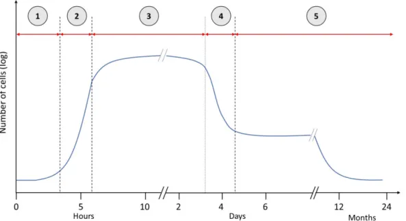
4.1. The planktonic lifestyle 35
4.1.1. The planktonic growth under laboratory conditions 36
4.1.1.1. The lag phase 36
4.1.1.2. The exponential phase 36
4.1.1.3. The stationary phase 37
4.1.1.4. The death phase 37
4.1.1.5. The late stationary phase 38
4.2. Biofilm 40
4.2.1. The molecular components of the biofilm matrix 40
4.2.1.1. Polysaccharides 41
4.2.1.2. Extracellular DNA 41
4.2.1.3. Matrix Proteins 41
4.2.1.4. Surfactants 41
4
4.2.1.6. Polyamines 42
4.3. The regulatory « switch » between planktonic and biofilm state 43 4.3.1. The Gac/Rsm signaling system 43
4.3.2. The c-di-GMP signaling pathway 45
4.3.3. The cAMP signaling pathway 45
4.3.4. Quorum sensing signaling 46
4.3.4.1. N-acyl homoserine lactones (AHL) 46
4.3.4.2. 2-alkyl-4-quinolones (AHQs) 47
4.3.4.3. The IQS system 47
5. The metabolism of P. aeruginosa 48
5.1. The aerobic metabolism of P. aeruginosa 49
5.1.1. The electron transport chain in P. aeruginosa 49
5.1.2. Regulation and functioning of the respiratory chain under oxygen conditions 51 5.1.3. Oxidative stress response in P. aeruginosa. 52
5.2. The anaerobic metabolism 56
5.2.1. Denitrification in P. aeruginosa 56
5.2.2. Anaerobic fermentation processes in P. aeruginosa 58
5.2.3. Pyruvate fermentation in P. aeruginosa 60
Chapter II. The lysine decarboxylase 61
1. The roles of the lysine decarboxylase in bacterial physiology 61
1.1. The inducible Lysine decarboxylase LdcI 62
1.1.1. LdcI is part of the acid and oxidative stress response 62 1.1.2. LdcI affects virulence in pathogenic E. coli and Shigella sp. 64
1.1.3. Regulation of the inducible Lysine decarboxylase LdcI 65
1.1.4. Other factors regulating the LDAR system 66
1.2. LdcC from E. coli 66
1.3. LDC as a virulence factor 66
1.4. The role of cadaverine in bacteria 67
2. The structure characteristics of LDCs from AAT-fold family 68
5
2.2. The core domain 69
2.3. C-terminal domain 69
2.4. The decameric structure of the Lysine decarboxylase 72
3. The enzymatic activity of LDCs 73
4. The molecular partner of the Lysine decarboxylase 74 4.1. LdcI interacts with the MoxR AAA+ ATPase RavA 74
Part II: Materials and Methods 76
1. Strains, plasmids and oligonucleotides 77
1.1. P. aeruginosa & E. coli strains 77
1.2. Oligonucleotides 78
1.3. Plasmids 79
2. Microbiology and cell culture 80
2.1. Growth media 80 2.2. Antibiotics 82 2.3. Culture of P. aeruginosa 82 2.3.1. PAO1 82 2.3.2. CHA 83 2.4. E. coli culture 83
2.5. Follow up of cell growth and density 83
2.6. Strain conservation 84
3. Molecular Biology Techniques 84
3.1. Polymerase chain reaction 84
3.2. SOE PCR (Splicing by Overlap Extension- Polymerase Chain Reaction) technique 85
4. Mutagenesis and complementation 86
4.1. Triparental mating 86
4.2. Allelic exchange by using pEXG2 suicide plasmid 86
4.3. Complementation by using miniCTX PldcA(CDS) 88
4.4. Chemically competent P. aeruginosa 89
4.5. Transformation of P. aeruginosa 89
5. Biochemistry 89
6
5.2. Measurement of β-galactosidase activity 89
5.3. Measurement ldcA expression by luminescence and fluorescence 90
5.4. Separation of proteins by SDS-PAGE and Western Blotting 90
5.4.1. Preparation of cytosolic extracts of P. aeruginosa 90
5.4.2. Denaturing Polyacrylamide gel electrophoresis (SDS-PAGE) 91
6. Western Blotting 92
6.1. Purification of LdcA for antibody production 92
6.2. Immunisation protocol for obtaining LdcA antibodies from rabbits 93
6.3. Immunodetection of LdcA in purified samples or in cytosolic extracts 93
7. Microbial physiology 94
7.1. Biolog 94
7.1.1. Evaluation of pH susceptibility of P. aeruginosa by using BIOLOGTM 94
7.1.2. Evaluation of antibiotic resistance of P. aeruginosa by using BIOLOGTM 95 7.2. Testing of antibiotics susceptibility by the CLSI guidelides. 97
7.3. Assay of bacterial persistence to antibiotics. 98
7.4. T3SS-dependent cytotoxicity against macrophages 98
8. Bioinformatics 98
8.1. Promoter analysis. 98
8.2. Genetic environment of LDCs in Enterobacteria and P. aeruginosa 99
8.3. LdcA Philogeny 102
8.3.1. Dataset assembly 102
8.3.2. Phylogenetic inference 102
Part III: Results 104
Chapter I: The identification of the lysine decarboxylase from the AAT-fold family in P. aeruginosa 105
1. Bioinformatics analyses of LdcI and LdcC homologues in P. aeruginosa 105 2. The genetic environment of PA1818 108
3. Discussion 110
Chapter II: The expression of the Lysine decarboxylase in P. aeruginosa 111
7
2. Constructions of different ldcA promoter fusions 116
3. Characterization of anti-LdcA antibodies for Western Blot analysis. 119
4. Expression of ldcA promoter in minimal medium 120
5. Expression of ldcA promoter in rich LB medium 123
6. Seek for regulators of ldcA expression 125
7. Characterization of ldcA expression in biofilm-like and anaerobic conditions. 128 8. Effect of acid and oxidative stress on the ldcA expression 130
9. Discussion 131
Chapitre III: The Role of the lysine decarboxylase in P. aeruginosa 135
1. Assessment of the role of LdcA in bacterial stress response by HT method 136 1.1 Effect of pH on the growth fitness of P. aeruginosa 137
1.2 Effect of decarboxylases in promoting fitness at pH 4.5 138 1.3 Effect of deiminases in promoting fitness at pH 9.5 141 1.4 Effect of LdcA in antibiotic resistance, oxidative stress response, and detoxification
of other harmful molecules 143
2. The role Lysine decarboxylase and polyamines during growth 149 2.1 Importance of Lysine decarboxylase in carbon metabolism 149 2.2 The importance of polyamines when growing on L-glutamate 156 3. The role of LdcA in the persistence to carbenicillin. 160 4. Assessment of the importance of LdcA in virulence of P. aeruginosa 164
5. Discussion 167
5.1 P. aeruginosa LdcA does not participate in active stress response mechanisms 167 5.2 The cadaverine is needed for optimal growth in L-glutamate and L-aspartate 168 5.3 The role of cadaverine in bacterial persistence 170 5.4 The role of cadaverine produced by LdcA in virulence 171 Chapter IV: Phylogenetic analysis of the Lysine decarboxylase from P. aeruginosa 172
1. Phylogeny of Lysine decarboxylases LdcA 172
2. Discussion 173
8 1. The genetic environment of the ldcA shares characteristics with enterobacterial Lysine
decarboxylases 175
2. The expression of the LdcA is associated to the stationary phase and biofilm lifestyle 175
3. The role of LdcA in persistence 176
4. The role of LdcA in growth fitness 176
5. LdcA and virulence 177
6. LdcA and biofilms 177
7. Bioinformatic analysis of the ppGpp binding site 178
8. Enzymatic activity of LdcA and the role ppGpp 179
9. Structural determination of LdcA 180
10. LdcA forms a new group of LDC in bacteria 181
Appendices 183
Appendix I 184
Appendix II 185
9 List of tables
Table 1. Summary of the characteristics of the genome of 4 different reference strains of P.
aeruginosa 21
Table 2. Efflux pumps that have been characterized in P. aeruginosa with their corresponding
expelled antibiotic and bibliographic reference 29
Table 3. Enzymatic activities of Lysine decarboxylases (AAT-fold) found in literature 74 Table 4. List of strains used during the study 77-78 Table 5. List of oligonucleotides used during the study 78 Table 6. List of plasmids used and constructed for the thesis 79
Table 7. The antibiotics used for the thesis project 82
Table 8. Composition of resolving gel of SDS-PAGE 91
Table 9. Composition of the stacking gel of SDS-PAGE (5% polyacrylamide) 92
Table 10. Testing conditions in plate PM10 95
Table 11. Testing conditions in plate PM11C 96
Table 12. Testing conditions in PM15B 97
Table 13. Sequences of transcription factors that were searched in ldcA promoter region by RSAT 99 Table 14. List of Enterobacteria presenting problems in annotations with ldcI and ldcC genes 101 Table 15. Percent identity matrix obtained after multiple sequence alignments using the amino acid sequences of PA1818, PA4115, PA1346, LdcI, LdcC, AdiA with the program MUSCLE 106 Table 16. Predicted Regulatory Elements found by bioinformatics analysis 115 Table 17. Growth fitness of the strains PAO1, PAO1 ΔldcA, PAO1 Δldc ldcA in different media 154 Table 18. Growth of the strains PAO1, PAO1 ΔldcA, PAO1 Δldc ldcA in different growth media159 Table 19 Survivors from antibiotic persistence experiments 164 Table 20. List of amino acid transporters linked to L-glutamate metabolism 169 Table 21. Enzymatic parameters of LdcI and LdcA in presence or absence of ppGpp 179 Table 22. Resume of the different characteristics of LdcI and LdcC from E. coli and LdcA from
10 List of figures
Figure 1. Pseudomonas aeruginosa 20
Figure 2. The pangenome of P. aeruginosa 24
Figure 3. Colonization process of P. aeruginosa in CF lung 27 Figure 4. Drug targets identified in P. aeruginosa and the resistance mechanisms against antibiotics
32 Figure 5. P. aeruginosa stringent response and persister formation 33
Figure 6. Virulence factors used by P. aeruginosa to infect epithelial cells during acute infection 35
Figure 7. The five phases of the bacterial growth in planktonic lifestyle 38 Figure 8. P. aeruginosa exhibit parallel lifestyle extremes in the environment and human host
39 Figure 9. Schematics of the process of biofilm formation 42 Figure 10. The Gac/Rsm cascade in P. aeruginosa is genetically linked to c-di-GMP through SadC 44 Figure 11. Diagram of the QS circuitry in P. aeruginosa 48
Figure 12. The electron chain of P. aeruginosa 50
Figure 13. Diagram of the respiratory chain of P. aeruginosa 51 Figure 14. Schematic model of the regulatory network controlling the multiple terminal oxidases
in P. aeruginosa 52
Figure 15. Diagram showing the principal regulatory, enzymatic and molecular scavengers involved
in oxidative stress response in P. aeruginosa 55
Figure 16. Reduction of nitrate to dinitrogen during Pseudomonas aeruginosa denitrification 57 Figure 17. Schematic representation of arginine fermentation in P. aeruginosa 59
Figure 18. Schematic model of pyruvate fermentation in P. aeruginosa 60 Figure 19. Diagram of the different roles of the lysine decarboxylase in bacteria 64 Figure 20. Regulation of cadBA operon by CadC in E. coli 65
Figure 21. Decarboxylation reaction of Lysine 68
Figure 22. Lysine decarboxylases of the two different structural families 70 Figure 23. The C-terminal of LdcI is determinant for RavA binding 71 Figure 24. The decameric structure of the lysine decarboxylase LdcI from E. coli 72
11 Figure 26. Schematic presentation of the mechanism of mutagenesis of ldcA by using suicide
plasmid pEXG2-mut-ldcA 87
Figure 27. Diagram of the complementation of ldcA mutants with integrative plasmid
miniCTX-pldcA-ldcA 88
Figure 28. Conserved domain analysis of different predicted Lysine decarboxylases from
P. aeruginosa and E. coli using the program CDD 107
Figure 29. Multiple sequence alignment of the LDC candidates 108 Figure 30. Genetic environment of ldcI and ldcC from E. coli and ldcA from P. aeruginosa 110 Figure 31. Bioinformatics analysis of the promoter region of ldcA gene 115 Figure 32. Assay of promoter-reporter gene fusions between ldcA and gfp, lacZ and lux operon
118 Figure 33. Design and Purification and western blotting of recombinant LdcA 120 Figure 34. Activity of ldcA promoter in MMP minimal medium 122 Figure 35. Expression profil of ldcA in rich medium LB 124 Figure 36. Expression profile of PldcA-lacZ fusions in CHA strains in different media 127 Figure 37. Expression of ldcA in aerobic biofilm-like and in anaerobic planktonic conditions 129 Figure 38. Effect of acid stress (pH5) and oxidative stress (1mM H2O2) on the expression of ldcA
130 Figure 39. Interaction network of ldcA expression and regulation 133 Figure 40. Growth fitness experiments of P. aeruginosa in minimal medium at different pHs 138 Figure 41. Effect of decarboxylases in promoting fitness at pH 4.5 140 Figure 42. Effect of deiminases in promoting fitness at pH 4.5 142 Figure 43. Effect of Aminoglycosides and Fluoroquinolones in growth fitness of PAO1, PAO1 ΔldcA,
PAO1 ΔldcA ldcA 144
Figure . Effect of Cepholosporines and β-lactams in growth fitness of PAO1, PAO1 ΔldcA, PAO1
ΔldcA ldcA 145
Figure 45. Effect of Tetracyclines and cyclic polypeptides, amphenicols, toxic ions, non-ribosomal peptides in growth fitness of PAO1, PAO1 ΔldcA, PAO1 ΔldcA ldcA 146 Figure 46. Effect of toxic molecules in the growth fitness of PAO1, PAO1 ΔldcA, PAO1 ΔldcA ldcA
147 Figure 47. Growth of PAO1, PAO1 ΔldcA, PAO1 ΔldcA ldcA in minimal medium with Glucose or
Glutamate 149
Figure 48. Catabolicpathways of L-proline, L-histidine and L-glutamine 153
Figure 49. Doubling time of the strains PAO1, PAO1 ΔldcA, PAO1 Δldc ldcA in growth medium with
12 Figure 50. Effect of polyamines when growing minimal medium with glutamate for strains PAO1,
PAO1 ΔldcA, PAO1 ΔldcA ldcA 157
Figure 51. Minimal inhibitory concentrations (MIC) of carbenicillin against P. aeruginosa in Mueller
Hinton Broth 162
Figure 52. Persistence assays in two different growth media (rich medium MHB and minimal
medium MMP) treated with 500µg/ml of carbenicillin 163
Figure 53. LDH release by murine macrophages RAW 264.7 infected with PAO1, PAO1 ΔldcA, PAO1
ΔldcA ldcA 165
Figure 54. Macroscopic motiliy assays of P. aeruginosa 166 Figure 55. Phylogenetic analysis of Lysine, Arginine and Ornithinine Decarboxylases in the bacterial
kingdom 173
Figure 56. 3D structure of the ppGpp binding site in LdcI 178 Figure 57. Enzymatic activity of LdcI and LdcA in presence and absence of ppGpp 179 Figure 58. Structure of LdcA from P. aeruginosa 180 Figure 59. Diagram of central carbon and nitrogen metabolism of P. aeruginosa when growing on
glucose 184
Figure 60. Diagram of central carbon and nitrogen metabolism when P. aeruginosa grows on
13 List of abbreviations
AAA+ - ATPases Associated with a Variety of Cellular Activities AAT – aspartate aminotransferase
AC – adenylate cyclase
ADC – Arginine decarboxylase ADP – Adenosine diphosphate AHL – Acyl homoserine lactone AHQ – 2-alkyl-4-quinolone
ANR – Anaerobic transcriptional regulator AR – Alanine racemase
ASL – Airway surface liquid ATP – Adenosine triphosphate AUC – Area under the curve
BLASTP – Basic local alignment search tool protein CAD - Cadaverine
CAMHB – Cation Adjusted Mueller Hinton Broth cAMP – Cyclic adenosine monophosphate CDD – Conserved domain database c-di-GMP – Cyclic diguanylate CDS – Coding DNA sequence CF – Cystic fibrosis
CFTR – Cystic fibrosis transmembrane conductance regulator CFU – Colony forming units
CLSI – Clinical & laboratory standards Institute
CRISPR – Clustered regularly interspaced short palindromic repeats CTD – C terminal domain
CV – Column volume DGC – Diguanylate cyclase DNA – Deoxyribonucleic acid
14 dNTP – Deoxynucleoside triphosphate
FNR – Fumarate and nitrate reductase GDP – Guanosine diphosphate
GFP – Green fluorescent protein GTP – Guanosine triphosphate H+ - Proton
HHQ – 4-hydroxy-2-heptylquinoline HMM – Hidden markov models
ICE – Integrative and conjugative models ICU – Intensive care units
IQS – Integrated quorum sensing IS – Insertion sequence
kb - Kilobase kDa – Kilodaltons
KEGG – Kyoto encyclopedia of genes and genomes LB – Lysogeny broth
LDAR – Lysine dependent acid stress response LDC – Lysine decarboxylase
LPS - Lipopolysaccharide
MIC – Minimal inhibitory concentration MMP – Minimal medium P
MOI – Multiplicity of infection
MUSCLE - Multiple sequence comparison by log-expectation NADH – Nicotinamide adenine dinucleotide (reduced)
NADPH – Nicotinamide adenine dinucleotide phosphate (reduced) OD600nm – Optical density at 600 nanometers
ODC – Ornithine decarboxylase OMP – Outer membrane proteins OMV – Outer membrane vesicules
15 ONPG - ortho-nitrophenyl- β-D-galactopyranoside
ORF – Open reading frame
PBS/T – Phosphate buffer saline/ tween 20 PCR – Polymerase chain reaction
PDE - Phosphodiesterase
PIA – Pseudomonas isolation agar PLP – p rido al ’ phosphate Pmf – proton motive force
ppGpp - guanosine tetraphosphate PQS – Pseudomonas quinolone system PUT - Putrescine
QS – Quorum sensing
RGP – Regions of genome plasticity RLU – Relative light units
RNA – Ribonucleic acid RNAP – RNA polymerase RPM – revolution per minute ROS – Reactive oxygen species
RSAT – Regulatory sequence analysis tool
SDS PAGE – sodium dodecyl sulfate polyacrylamide gel electrophoresis SOD – Superoxide dismutase
SOE PCR – Splicing by overlap extension polymerase chain reaction SPM - Spermidine
STEC – Shigatoxin producing Escherichia coli T3SS – Type three secretion system
TA – Toxin antitoxin WT – Wild type
17 Thesis presentation and objectives
The lysine decarboxylase (LDC) belongs to a family of decameric PLP-dependent enzymes that catalyze the reaction transforming L-Lysine into cadaverine while consuming a proton and producing a CO2 molecule. They are part of the bacterial arsenal against stress
conditions such as acid stress, oxidative stress and antibiotic treatment. In enterobacteria like Escherichia coli, two paralogs are present, LdcI and LdcC. LdcI takes part in acid stress response by buffering bacterial cytoplasm and the surrounding extracellular environment. LdcC, in its turn, is produced during stationary phase and also when bacteria face fluoroquinolone treatment. Cadaverine produced by LDCs is known to scavenge reactive oxygen species (ROS) and is capable of blocking outer membrane proteins, thus reducing the permeability of molecules responsible for acid and oxidative stresses.
The laboratory of Irina Gutsche (IBS) has become a leading group in analyzing and solving the structures of the lysine decarboxylases from E. coli by cryo-electron microscopy. In collaboration with Walid Houry lab (University of Toronto, Canada), they discovered that the activity of the LDCs from E. coli is regulated during the stringent response (nutrient starvation) to prevent intracellular L-Lysine depletion. Indeed, the stringent response signal molecule ppGpp can bind directly to LDCs and inhibit their enzymatic activity. Another milestone in the field of the Lysine decarboxylase was the discovery of a cage-like complex formed by LdcI and the AAA+ ATPase RavA. This peculiar complex prevents the inhibition by (p)ppGpp thereby allowing bacteria to face the challenge of both acid and nutrient stresses. Since the role of the lysine decarboxylase has only been studied in enterobacteria such as E. coli, we wondered whether the lysine decarboxylase was also protecting the well-known opportunistic pathogen Pseudomonas aeruginosa from the stress conditions experienced in the host. For instance, P. aeruginosa is a serious health problem for patients affected by Cystic Fibrosis. In particular, one can wonder if under conditions encountered in the lungs of cystic fibrosis patients whose lungs secretions were shown to be acidified and to become oxidative, LdcA is playing a role in promoting fitness of the bacterium.
To tackle our project with P. aeruginosa, eàsta lishedàaà olla o atio à ithàI aàátt e sàla (BiG, CEA), where they study the pathogenesis of P. aeruginosa and possess the proper equipment and expertise to manipulate this bacterium. With the expert advise of Dr. Sylvie
18 Elsen, who is also my thesis co-director, we constructed a project that would allow us to examine the role of LdcA of P. aeruginosa by using different approaches:
• First of all, we used promoter-gene fusions and western-blot analysis to determine the conditions in which ldcA was expressed and its product synthesized.
• In parallel, we constructed an ldcA mutant and its complemented strain to investigate whether LdcA was involved in acid and oxidative stress response, but also in growth fitness and persistence via production of cadaverine.
• Finally, by combining phylogenetic and structural analyses, we discovered that LdcA belongs to a different subgroup of bacterial LDCs.
Before showing our results, I will start by introducing the general characteristics of the genome and physiology of P. aeruginosa and then present what we know about the lysine decarboxylases.
19
20
Chapter I: Pseudomonas aeruginosa
Pseudomonas aeruginosa is a bacterium that belongs to Pseudomonadaceae which is a representative family of the Gammaproteobacteria class. Ità hasà ee à alledà Bacillus pyocyaneus àfo àaàlo gti eà àphysicians because it generates blue pus in the wounds of infected patients. It is a gram-negative non-sporulating bacillus, measuring 1- 5 µm long and 0.5 - 1 µm wide. It is mobile in liquid media and on solid surfaces and its motility depends on a polar monotrichous flagellum and type IV pili.1 P. aeruginosa is a mesophilic bacterium that
is capable of growing in temperatures ranging from 4°C to 42 °C, with an optimum growth temperature between 30°C and 37°C.2,3 It is also a neutralophile and is found in aquatic and
terrestrial environments that span a pH range from 4.5 to 9.5. More than 50 different organic and inorganic molecules can be degraded by the metabolism of P. aeruginosa. It uses efficiently O2 as the principal electron acceptor but has also the capability of using
nitrates for respiration. When oxygen and nitrates or nitrites are unavailable, the bacterium is capable of fermenting L-Arginine for growth and pyruvate for survival only.3,4
All these properties give P. aeruginosa the ability to thrive in most natural and man-made environments and explain why the bacterium can ultimately infect multiple hosts such as humans and other mammals, insects, nematodes, amebae and even plants5,6.
Figure 1. Pseudomonas aeruginosa. Up left: Electron microscope image of P. aeruginosa biofilm. Up right: Culture of P. aeruginosa on solid agar, the green fluorescent pigment pyoverdin gives the bacterium its
characteristic color. Left: P. aeruginosa infection in burn wound patient. Images obtained from Pr. Filloux website and from microbeworld.org and medetek.co.uk
21 1. The genome of Pseudomonas aeruginosa
1.1. The genome of PAO1 strain
The genome of the strain PAO1 was the first one to be sequenced in the year 2000. As presented in table 1, PAO1 has a genome size of 6.3 mega base pairs (Mbp) with 5,684 predicted open reading frames (ORF)7 (Pseudomonas.com). Around 40% of the genome
consists of hypothetical and conserved hypothetical ORFs with unknown function7,8. The
expression of the genome is coordinated by a complex regulatory network consisting at least of 690 genes (12% of total genome) encoding for sigma factors and anti-sigma factors, transcriptional regulators and two-component regulatory systems9. The regulatory network
integrates and controls important biological process such as energy metabolism, cell division, biofilm formation, cell-to-cell communication, production of virulence factors and guarantees a rapid adaptation to environmental changes10.
Table 1. Summary of the characteristics of the genome of 4 different reference strains of P.
aeruginosa11.
Strain PAO1 PA7 PA14 LESB58
Genome Size (bp) 6264404 6588339 6537648 6601757
G+C content 66.6 66.5 66.3 66.3
protein coding genes 5684 6286 5892 5925
%coding 89 89 89 88
structural RNAs 77 75 72 81
Pseudogenes 5 8 - 34
Assigned function
Translation, ribosomal structure and biogenesis 205 206 205 199
Transcription 516 530 537 501
Replication, recombination and repair 160 235 185 145
Cell cycle control, cell division, chromosome partitioning 34 37 35 34 Posttranslational modification, protein turnover,
chaperones 200 215 210 201
Cell wall/membrane/envelope biogenesis 265 260 266 261
Cell motility and secretion 150 152 154 149
Inorganic ion transport and metabolism 376 355 377 313
Signal transduction mechanisms 337 346 345 337
Energy production and conversion 329 336 340 330
Carbohydrate transport and metabolism 252 250 249 196
Amino acid transport and metabolism 587 571 590 490
Nucleotide transport and metabolism 108 105 110 104
Coenzyme transport and metabolism 191 192 192 210
Lipid transport and metabolism 244 245 248 234
Secondary metabolites biosynthesis, transport and
catabolism 205 198 212 171
General function prediction only 756 759 771 603
22 A disproportionate large number of outer membrane proteins (OMP) are encoded in P. aeruginosa genome compared to other species. 150 genes are predicted to be outer membrane proteins (OMP) involved in iron uptake, antibiotic export and secretion of virulence factors essential for pathogenicity7. Consistent with its environmental versatility,
300 cytoplasmic membrane transport systems are encoded and at least 200 appear to be involved in the transport of nutrients and other molecules such as dicarboxylates, sugars, fatty acids, alcohols, polyalcohols, glycols, aromatic compounds, amines and amino acids7. In
comparison with E. coli, P. aeruginosa exhibits a limited number of genes encoding sugar transporters and uses the Enter-Doudoroff Pathway glycolytic pathway (Enter-Doudoroff Pathway) and an aerobic, oxidative metabolism12. A la geà u e à ofà ge esà i ol edài à
β-oxidation are present and ensure the versatile metabolic capacity of oxidizing a wide variety of organic compounds in a similar way as in Mycobacterium tuberculosis7.
The P. aeruginosa genome appears to contain several undescribed drug efflux systems, predominantly of the Resistance-Nodule-Division (RND) and Major Facilitator Superfamily (MFS) families. The number of predicted drug efflux systems from the (MFS), small multi-drug resistance (SMR), ATP-binding cassette (ABC) and multidrug and toxic compound extrusion (MATE) families is like that of other organisms such as E. coli, Bacillus subtilis and M. tuberculosis. However, PAO1 P. aeruginosa contains many more predicted AcrB/Mex-type RND multidrug efflux systems (10 genes) than E. coli (4), B. subtilis (1) and M. tuberculosis (0)7.
An important number of virulence factors is also encoded in the genome of P. aeruginosa PAO1 such as toxins (exotoxin A), proteases (elastase, alkaline protease, protease IV), phospholipases, flagellum, pili, fimbriae and secretion systems (T1SS, T2SS, T3SS, T5SS, T6SS). Operons encoding the biosynthetic genes for virulence factors such as rhamnolipids, lipopolysaccharides, phenazines, siderophores and polysaccharides (alginates, pel, psl) are also present13–26.
23 1.2. The pangenome of P. aeruginosa
Today high throughput sequencing technologies allowed us to access to the nucleotide sequences of more than 2200 genomes from different P. aeruginosa strains. Nevertheless, in the databases of the NCBI and PATRIC we found that less than 200 different genomes have been completely sequenced and annotated2,7.The analysis of the sequenced genomes has
revealed that the pan-genome of P. aeruginosa can be divided in two different groups of genes:
1.2.1. The core genome
It encodes a set of metabolic and pathogenic factors shared by all P. aeruginosa strains, irrespective of origin. The core genome constitutes approximately 90% of the total genome and is highly conserved (>98% identity) from strain to strain. Today it is estimated to be composed of more than 4000 ORFS. Its G+C percentage is 67.1%27–32. Despite research
efforts, there is limited knowledge about the function of more than 1000 genes in P. aeruginosa core genome (figure 2).
1.2.2. The accessory genome
It encompasses genes that are found in some P. aeruginosa strains but not others. In figure
2, we found a representation of P. aeruginosa pangenome in which we see both the
accessory genome and the core genome. The accessory genome appears as segments that are not scattered randomly throughout the core genome but are inserted at specific sites, called Regions of Genome Plasticity (RGPs). The accessory genome can be grouped in four categories: (i) integrative and conjugative elements (ICEs), (ii) replacement islands, (iii) prophages and phage-like elements, and (iv) transposons, insertion sequences (ISs), and integrons. Horizontal gene transfer is by far the most important process contributing to its evolution. This is reflected by its G+C percentage of 62.4% (median). Up-to-date more than 10000 genes have been classified as part of the accessory genome, a number that should increase with the addition of newly sequenced P. aeruginosa strains27–32.
24 Figure 2. The pangenome of 5 strains of P. aeruginosa27. The pangenome is a compilation of all the genes from all the strains of a single species. In this figure, the core genes from the strain PA14 were used as matrix for putting the accessory genes from strains PA2192, C3719, PAO1 and PACS2. The golden circle indicates the core genome, while the second circle shows the totality of genes that have been annotated for all the strains back in 2008. The third circle indicates the position of tRNA and is generally used to identify insertion sites. The fourth circle, in black, indicates the RGPs which can be sites of insertions of common or unique genomic islands and bacteriophage genomes, or the result of deletions of segments of DNA in one or more strains. The accessory genomes from each strain are shown in different colours: PACS2 (turquoise), C3719 (violet), PA2192 (green), PA14 (blue) and PAO1 (red). The outer green arrows show the positions of rRNAs.
25 2. Clinical importance of P. aeruginosa
2.1. P. aeruginosa is an opportunistic pathogen
P. aeruginosa can cause both acute and chronic infections in its host. Acute P. aeruginosa infections are invasive and cytotoxic and frequently result in substantial tissue damage, systemic spread, sepsis, and mortality. The pathogenesis of acute infections relies upon the expression of many surface-exposed and secreted virulence factors, including: toxins, proteases (delivered by a type II secretion system; T2SS), type IV pili (Tfp), flagella and a type III secretion system (T3SS) that can inject a set of eukaryote specific effectors across the plasma membrane of target cells33.
Chronic infections are minimally invasive and noncytotoxic and they involve the formation of biofilms that protect bacteria against assault by the host immune system and provide resistance to antibiotics34,35. Thus, chronic infections rarely result in systemic spread, but
instead lead to unrelenting non-productive host infammation that contributes to the resulting morbidity and mortality35.
P. aeruginosa is seldom a member of the normal microbial flora in humans. Clinical studies report that colonization rates for specific sites in humans are 0 to 2% for skin, 0 to 3.3% for the nasal mucosa, 0 to 6.6% for the throat, and 2.6 to 24% for fecal samples36. However,
when patients are hospitalized, colonization rates may exceed 50%, especially among patients in which the cutaneous or mucosal barriers have been damaged by mechanical ventilation, tracheotomy, catheters, surgery, or severe burns36. Immunocompromised
patients have higher risks of colonization by this organism, and patients that have received antimicrobial therapy were also shown to suffer from increased colonization by P. aeruginosa 36.
Healthcare statistics indicate that P. aeruginosa has become the second leading cause of nosocomial pneumonia, the third most common cause of urinary tract infections, the fourth most frequently isolated pathogen in infections in surgical sites, and the seventh pathogen responsible for bloodstream infections and the leading cause of pneumonia among pediatric patients in the intensive care unit (ICU)36.
P. aeruginosa presents a serious therapeutic challenge for treatment of both community-acquired and nosocomial infections, making appropriate antibiotics are essential
26 for the restablishment of infected patients36. Unfortunately, selection of appropriate
antibiotics is complicated by the ability of P. aeruginosa to develop resistance to multiple classes of antibacterial agents, even during the course of treating an infection. Epidemiological outcome studies have shown that infections caused by drug-resistant P. aeruginosa are associated with significant increases in morbidity, mortality, need for surgical intervention, length of hospital stay and chronic care, and overall cost of infection treatment36. Healthcare statistics from ICUs around the world reveals that, in the periods
between 2003-2007, the rates of antibiotic resistance among P. aeruginosa isolates show that : fluoroquinolone resistance is around 30% in US while more than 50% in the rest of the world ; piperacillin and piperacilline-tazobactam resistance concerns almost 20% in US while 50% in the world ; amikacin resistance is found in less than 10% in US but in 30% around the globe ; imipenem and meropenem resistance is seen in 25% in US isolates while almost 40% in world ones and finally cefepime resistance touches 10% of US isolates and shows an alarming 70% in isolates from the rest of the planet36–38 . The virulence factors that enable P.
aeruginosa to infect its host will be presented later in this chapter.
2.2. P. aeruginosa creates persistent infections in CF patients
Another clinical setting where P. aeruginosa has become an important health concern is Cystic Fibrosis (CF) in which the organism infects and colonizes patiens from the early childhood39,40. CF is a recessive inherited disorder caused by the presence of mutations in
the gene encoding cystic fibrosis transmembrane conductance regulator (CFTR) protein, with an incidence of 1 in 2500 live births. CFTR functions as an ion channel that transports chloride ions across epithelial cell surfaces41–43.
In CF airways, CFTR dysfunction modifies the composition and the properties of the airway surface liquid (ASL). ASL is a mucus layer which protects airway surfaces against irritants and infecting micro-organisms. The ASL becomes thin and viscous making the gel-forming mucins dry and compacted on top of the epithelial layers. The dehydration of mucin blocks the clearance of bacteria allowing them to colonize and grow44,45 This default
generates chronic inflammation generating a toxic pro-inflammatory local microenvironment, which damages the lung and the innate immunity, further facilitating infections. It is under this hostile context that P. aeruginosa must overcome challenges such
27 as osmotic stress, an acidified ASL, competition from other colonizers, nutritional inadequacy, antibiotics, acid and oxidative stresses, etc,in order to survive45,46.
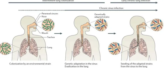
Figure 3, shows the initial process of colonization of the airways in CF patients and
theàesta lish e tàofàea l à h o i àlu gài fe tio .àB iefl ,àpatie t sàai a s,à othàlu gsàa dà sinuses, are colonized by environmental P. aeruginosa. The sinus environment provides a protected niche were the bacteria can survive against waves of antibiotic treatment and immune response compared to the lung airways where they can be completely eradicated. The same survivors in the sinus are capable of intermittent colonization of the lung which can at first be controlled by antibiotic treatments. However, the cycles of intermittent colonization and eradication allows the progressive acquisition of mutations that increases the fitness and adaptability of bacteria. The genetically adapted strains can then migrate to the lower airways and generate chronic infections that are very difficult to eradicate.
The final outcome of the chronic infection is a constant but slow damage of the lung tissue until breathing is no longer possible. Under these dreadful conditions, a lung transplant is often the only long term treatment for these patients explaining why their lifetime is limited to around 40 years47,48.
Figure 3. Colonization process of P. aeruginosa in CF lung. Environmentally acquired P.
aeruginosa start invading airways of CF patients creating acute infections. Sinuses become the niche were the bacteria can survive antibiotic treatment and immune system. Bacteria from the sinuses can migrate and reinfect the lungs. The cycles of colonization-eradication allow the accumulation of mutations that gradually increase the fitness of the bacteria. Genetically adapted bacteria start to become more resilient and establish chronic infection in the patient48.
28 One of the most important factors that enhance the survival of P. aeruginosa against the stress conditions in the CF lung is the sigma factor 22 also known as AlgU47,48. Activation
of AlgU leads to a coordinated downregulation of central metabolism, motility and virulence, and a concurrent upregulation of genes affecting membrane permeability and efflux, and also genes that are involved in heat shock, osmotic and oxidative stress response. AlgU also is responsible for the production of alginate, which is a polysaccharide that is secreted to protect bacteria and that seems to be part of a general envelope stress response47,48.
Overproduction of alginate has been linked to a phenomenon called mucoid conversion, which arises from mutations in the gene encoding the anti-sigma factor MucA that regulates the functioning of AlgU by sequestration. The consequence of this mutation is a constitutive activation of AlgU. However, it has been reported that the mucoid conversion is a reversible process because mucA seems to be a repetitive target for selective mutations during chronic pathogenesis47,48.
3. Antibiotic Resistance
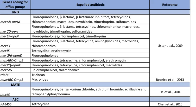
3.1. Intrinsic mechanisms of antibiotic resistance
Once a human host has developed a P. aeruginosa infection, the most important decision to sa eàaàpatie t sàlifeàisàtheàad i ist atio àofàaàp ope àa ti ioti àt eat e t.àO eàofàtheà iggestà problems encountered by infected patients is that P. aeruginosa is a highly resistant organism against antibiotherapy. This is explained by the fact that P. aeruginosa possesses an arsenal of enzymes, efflux pumps and virulence mechanisms that allow the bacteria to resist and persist against treatments, and they are shown in Figure 47,36. The expulsion of
antibiotics from the cytoplasm to the outside of the cell is performed by using sophisticated efflux pumps that belong to 5 different families: Resistance-nodule-division (RND), major superfacilitator superfamily (MFS), small multi-drug resistance (SMR), ATP-binding cassette (ABC) and multidrug and toxic compound extrusion (MATE)7,36. Four families use the energy
from pmf to expel antibiotics while one family, the ABC uses ATP, to pump and keep antibiotics far from their cellular targets. Table 2 presents the best characterized families in P. aeruginosa and shows that the RND family is capable of pumping most of the antibiotics used by healthcare systems.
29 Other resistance mechanisms, depend on the modification of LPS mediated by the products of the arnBCADTEF-ugd and pmrCAB operons. The original lipid A is modified by the addition of 4-amino-4-deoxy-L-arabinose and phosphoethanolamine. This « alternative » LPS is responsible for generating resistance against colistin and polymyxin B49–51.
Table 2. Efflux pumps that have been characterized in P. aeruginosa with their corresponding expelled antibiotic and bibliographic reference36,52–54.
Enzymatic mechanisms are also present, as it is the case for AmpC that possesses an extended spectrum of activities against β-lactams. This enzyme has also been detected inside outer membrane vesicules (OMVs) in biofilms. OMVs are liposome-like containers that act as molecular decoys to trap and neutralize all kinds of environmental aggressions such as antibiotics55.
The strong selective preasure generated by the antibiotherapy of the infected patient favours the accumulation of mutations, ending up in the amplification of resistant phenotypes. Mutations on the DNA gyrase (gyrA and gyrB)56 and the topoisomerase IV (parC
and parE)57 have been described to confer resistance to fluoroquinolones by modifying the
binding site of the antibiotic. Genes coding for
efflux pumps Expelled antibiotic Reference
RND
mexAB-oprM
Fluoroquinolones, β-la ta s,àβ-la ta aseài hi ito s,àtet a li es,à chloramphenicol macrolides, novobiocin, trimethoprim, sulfonamides
mexCD-oprJ
Fluo o ui olo es,àβ-la ta s,àtet a li es,à hlo a phe i olà a olides,à novobiocin, trimethoprim, sulfonamides
mexEF-oprN Fluoroquinolones,chloramphenicol, trimethoprim
mexXY
Fluo o ui olo es,àβ-la ta s,àtet a li e,àa i ogl osides,à a olides,à chloramphenicol
mexJK Tetracycline, erythromycin
mexGHI-opmD Fluoroquinolones
muxABC-OmpB Fluoroquinolones, tetracycline, chloramphenicol, erythromycin
mexPQ-opmE Fluoroquinolones, tetracycline, chloramphenicol, macrolides
mexMN Chloramphenicol, thiamphenicol
triABC Triclosan
muxABC-OmpB Macrolides Beceiro et al., 2013
MATE
pmpM
Fluoroquinolones, benzalkonium chloride, ethidium bromide, acriflavine and
tetraphenylphosphonium He et al., 2004
ABC
PA4456 Tetracycline Chen et al., 2015
30 Another important antibiotic resistance mechanism involves the porin OprD. OprD is a specialized porin which has a specific role in the uptake of positively charged amino acids such as lysine. Decrease in the expression of oprD and/or mutations that disrupt the translational production of a functional porin for the outer membrane are associated to a heightened antibiotic resistance against imipenems but not carbapenems58. Overexpression
of efflux pumps by accumulation of mutations in the regulatory network controlling their expression is also a mechanism that generates multidrug-resistant phenotypes in P. aeruginosa strains36.
3.2. Acquired mechanisms of antibiotic resistance
P. aeruginosa is also capable of acquiring resistance genes carried on mobile genetic elements that are obtained from other microorganisms in the environment. Acquired drug-resistance mechanisms include the drug-resistance towards β-lactam antibiotics by the acquisition of genes coding for β-lactamases of the following types: class A serine β-lactamases; class A extended-spectrum β-lactamases; class A carbapenemases; class B metallo- β-lactamases, class D OXA-type enzymes. The resistance towards amiglycosides arises from the acquisition of enzymes modifying aminoglycosides such as phosphorylases, acetyltransferases and nucleotidyl transferases. The other mechanism relies on enzymes encoded by the genes rmtA, rmtB, rmtC, rmtD and armA, that modify the target 16S rRNA36.
All these mechanisms explain the impressive ability of P. aeruginosa to develop antibiotic resistance. For this reason, treatments combining a variety of antibiotics are the basis for successful treatment against P. aeruginosa. Nevertheless, this is not always enough to avoid the appearance of drug resistant phenotypes. The great quantity of antibiotic resistance mechanisms and the genetic flexibility of P. aeruginosa demonstrate that this bacterium is aà se iousà healthà o e à a dà thatà it sà possible that no effective antibiotic treatment will be available if the discovery of new antibiotic molecules and the understanding of resistance mechanisms do tà keepà upà ithà theà e olutio à ofà theà microorganism.
32 Figure 4. Drug targets identified in P. aeruginosa and the resistance mechanisms against antibiotics. Left: colistin and polymyxin B are responsible for the disorganization of LPS and the perforation of the outer membrane, β-lactams block peptidoglycan biosynthesis, aminoglycosides and tetracyclins and chloramphenicols interfere with the functioning of the ribosome, blocking protein synthesis, fluoroquinolones hinder the functioning of topoisomerases needed for DNA replication. Right: P. aeruginosa modifies the composition of LPS to prevent the binding of colistin and polymyxin B, mutations in topoisomerases impede the binding of fluoroquinolones to their targets, chromosomaly and plasmid encoded enzymes inactivate or modify antibotics, pmf and ATP-using efflux pumps drive out antibiotics from the cytoplasms, inhibition of porins (OprD) prevents the entrance of antibiotics, finally alginate production serves as a physical barrier for antibiotic diffusion.
3.3. Antibiotic persistence and the stringent response in P. aeruginosa
Persisters are one of the main reasons for recurrent and chronic infections of P. aeruginosa. They are known to withstand antibiotic treatments and spawn a new infecting population upon removal of antibiotic treatment. It has been described that persisters are abundant in
P. aeruginosa biofilms, which is the hallmark of longterm infections particularly in CF59,60.
Persisters are defined as subpopulations of cells occurring at very low frequency, which stochastically emerge in the presence of stress61,62. They show very slow growth and they
are capable of surviving in stressful conditions where the viability of the majority of the population is severely impaired. Upon stress removal, persisters turn back to normal growth to propagate while regaining normal sensitivity to stress. Such persistence was suggested to be based on the heterogeneity of population by means of epigenetic mechanisms, not genetic mutations63.
Even though, the molecular mechanisms determining the process of persister formation are not completely understood, various studies have provided evidences showing the link between stringent response and persistence.
Stringent response happens when bacteria like E. coli or P. aerugionsa encounter amino acid deprivation. This condition activates RelA and SpoT, which start synthezing ppGpp molecules. In association with the transcriptional regulator DksA (global regulator of metabolism), ppGpp interacts with RNA polymerase and inhibits the transcription of ribosomal RNA promoters. This inhibitory impact is concomitant with activation and upregulation of pathways for amino acid biosynthesis and the transcription of stress response genes64–67. Amato et al. (2013) found that stringent responses are linked to the
33
emergence of persisters by involving ppGpp based regulatory events67. Figure 5 shows the
current knowledge linking the stringent response and persistence in P. aeruginosa.
Figure 5. P. aeruginosa stringent response and persister formation. Stringent response is triggered by particular stresses such as amino acid and fatty acid starvation, iron/phosphate depletion and oxidative stress. The (p)ppGpp alarmone is a key determinant for stringent response and it is elevated by RelA/SpoT enzymes. Generally, (p)ppGpp elevation, PolyP (inorganic polyphosphate) and Lon protease complex interfere with normal biological processes in favour of bacterial survival via arrest of (?) metabolism, cell growth and cell division (dashed gray pathways are best understood for the E. coli model, but not or partially characterized in P. aeruginosa). In E. coli, (p)ppGpp signaling is linked to toxin (T)-antitoxin (A) system via activation of the Lon protease leading to the formation of persisters displaying dormant and antibiotic resistance phenotypes (dashed orange line). Generally, the TA complex is stable under normal conditions suppressing toxin activity and further expression of cognate genes. Upon antitoxin degradation, toxin becomes active to hinder biological processes. In the case of P. aeruginosa HigB/A and HicA/B, the toxin components, HigB or HicA,perform endoribonuclease (RNase) activity on mRNA molecules. In P. aeruginosa, the (p)ppGpp alarmone is linked to the production of ROS scavengers probably via QS or RpoS regulators and Lon activity is required for biofilm formation, motility, virulence and antibiotic resistance. Furthermore, the TA system(HigB/A) downregulates biofilm formation and virulence factor production while T3SS (type 3 secretion system) can be found upregulated. Although, the (p)ppGpp signaling, Lon protease activity and TA modules (i.e., HigB/A, HicA/B, and likely more complexes) are present in P. aeruginosa, their link to resistance to antibiotics and other stresses is poorly understood. AA, amino acids; QS, quorum sensing; RNAP, RNA polymerase. CM, cytoplasmic membrane; OM, outer membrane68.
34
The persister state is typically based on the activity of genetically encoded toxin-antitoxin (TA) modules particularly in response to antibiotics. However, it is also proposed to be activated by RelA or SpoT and (p)ppGpp as it is the case E. coli69.
Basically, the toxin element is a stable protein while the antitoxin appears as a protein or as a small RNA. Two mechanisms are described for the effect of small RNA antitoxins: i) they can inhibit the toxin translation by pairing the toxin mRNA, ii) they can inactivate the toxin by direct binding. When antitoxins are proteins they inhibit the activity of the toxin by protein-protein interactions.70,71. Under stressful conditions, the antitoxin releases the toxin
which starts interfering with key cellular processes such as DNA replication, tRNA synthesis, membrane components and ATP generation. The inhibition of cellular proliferation leads to the formation of a dormant or persister cell.
Among the TA systems that are known in P. aeruginosa we find HigB/HigA and HicA/HicB72,73. Both systems present toxins having RNAse activity, that have a role in the
regulation of virulence factors, biofilm formation and in the formation of persisters against ciprofloxacin74.
Overall, the mechanisms of persister formation are a new emerging field that will be extremely important to understand and overcome the chronic infections of P. aeruginosa.
4. The Lifestyle of P. aeruginosa
The two different types of infection phenotypes observed in human disease, namely acute and chronic, have been associated to two different lifestyles of the bacterium: the planktonic and the biofilm modes of growth.
These two different lifestyles are coordinated by a complex network of regulatory factors that determine phenotypical traits that are responsible for the adaptation of the bacteria to their environment but also its pathogenicity and aggressiveness.
35 4.1. The planktonic lifestyle
In the planktonic lifestyle, individual or small groups of free living P. aeruginosa have the capacity to evade or defend against protozoan and metazoan predators, including higher metazoans such as the human host.75. To do this, P. aeruginosa relies in the expression of
virulence factors that are shown in figure 6. Among them, the Type 3 Secretion System (T3SS) is a molecular machine capable of injecting toxins (ExoS/T: ADP ribosyltransferase and GTPase, ExoY: promiscuous nucleotidyl cyclase, ExoU: phospholipase) directly into eukaryotic cells; it is one of the most powerful weapons from P. aeruginosa which is crucial for acute infection33,76,77. The T3SS and its toxins allow P. aeruginosa to resist and defend
against phagocytosis and predation in its environment. Another important mechanism to avoid predation and facilitate dispersal is motility. P. aeruginosa possesses a monotrichous flagellum necessary for swimming and a type IV pili that is used to twitch on solid surfaces33.
Moreover, when P. aeruginosa reaches high numbers, other virulence factors such as Type 2 secretion system (T2SS) and its associated proteases are produced, and the bacterium synthesizes and secretes molecules such as surfactants and pigments35. The combination of
these virulence factors associated to the planktonic lifestyle allow P. aeruginosa to generate acute infections that are invasive and cytotoxic and frequently results in substantial tissue damage, sepsis and mortality. However, it is important to highlight that virulence factors associated to the planktonic lifestyle have been detected during the sessile lifestyle, suggesting that both environmental cues and lifestyle are determinant for the expression of virulence factors35.
Figure 6. Virulence factors used by P. aeruginosa to infect epithelial cells during acute infection. Factors such as adhesins and secreted toxins, proteases, effector proteins and pigments that facilitate adhesion, modulate or disrupt host cell pathways and target the extracellular matrix.76.