HAL Id: hal-00019883
https://hal.archives-ouvertes.fr/hal-00019883
Submitted on 18 Mar 2006
HAL is a multi-disciplinary open access
archive for the deposit and dissemination of
sci-entific research documents, whether they are
pub-lished or not. The documents may come from
teaching and research institutions in France or
abroad, or from public or private research centers.
L’archive ouverte pluridisciplinaire HAL, est
destinée au dépôt et à la diffusion de documents
scientifiques de niveau recherche, publiés ou non,
émanant des établissements d’enseignement et de
recherche français ou étrangers, des laboratoires
publics ou privés.
Immunostimulating properties and three-dimensional
structure of two tripeptides from human and cow caseins
Jean Berthou, D. Migliore-Samour, Alain Lifchitz, Jean Delettré, D. Floc’H,
Pierre Jollès
To cite this version:
Jean Berthou, D. Migliore-Samour, Alain Lifchitz, Jean Delettré, D. Floc’H, et al.. Immunostimulating
properties and three-dimensional structure of two tripeptides from human and cow caseins. FEBS
Letters, Wiley, 1987, 218 (1), pp.55-58. �10.1016/0014-5793(87)81017-7�. �hal-00019883�
Volume 218, number 1, 55-58 FEB 04765 June 1987
Immunostimulating properties and three-dimensional
structure of two tripeptides from human and cow caseins
J. B e r t h o u * , D. M i g l i o r e - S a m o u r +, A. L i f c h i t z * , J. D e l e t t r 6 * , F. F l o c ' h x a n d P. Joll6s +
+ Laboratoire des Prot~ines (Unit~ C N R S 1188 allibe ~ I ' I N S E R M ) , Universit~ de Paris 1I, 45 rue des Saints-P~res, F 75270 Paris Cedex 06, *Laboratoire de Minbralogie-Cristallographie associ~ au CNRS, Universitb Pierre et Marie Curie,
4 place Jussieu, Paris and × Rh4ne-Poulenc Sant~, Centre de Recherches de Vitry, Vitry-sur-Seine, France
Received 6 April 1987
Some tripeptides obtained by enzymic digestion of caseins possess immunomodulating properties. In order to correlate activity and structure, X-ray analysis has been applied to two of them Leu-Leu-Tyr and Gly-
Leu-Phe.
Immunostimulation; Casein; Immunomodulating tripeptide; 3-dimensional structure
1. I N T R O D U C T I O N 2. M A T E R I A L S A N D M E T H O D S The major part o f the studies so far devoted to
chemically defined immunomodulating substances has been performed with compounds o f bacterial origin or with their synthetic analogues [1]. How- ever, we previously observed that enzymic frag- ments obtained from h u m a n casein, the main protein fraction of maternal milk which is usually man's first food, exhibit immunostimulating properties [2]. The purification, the sequence, the synthesis and some biological properties o f an im- munostimulating hexapeptide (Val-Glu-Pro-Ile- Pro-Tyr) from h u m a n fl-casein have been de- scribed [3]. The presence o f some further biologi- cally active short peptides has been detected in h u m a n as well as in cow milk caseins. This paper deals with the characterization o f two active tripeptides (Gly-Leu-Phe and Leu-Leu-Tyr) and their molecular c o n f o r m a t i o n in the solid state by single crystal X-ray diffraction, as a first attempt to correlate biological activity and structure. Correspondence address: P. Joll~s, Laboratory of Pro- teins, University of Paris V, 45, rue des Saint-P~res, F 75270 Paris C6dex 06, France
2.1. P u r i f i c a t i o n p r o c e d u r e s
Two peptides Gly-Leu-Phe and Leu-Leu-Tyr were obtained f r o m delipidated casein digested by non-pretreated trypsin (Worthington): the digest was filtered on Sephadex G-50 (Pharmacia) [2]. Biologically active fractions were submitted to suc- cessive chromatographies. Gly-Leu-Phe, f r o m human casein, was obtained after DEAE-Sepha- dex A-25 c h r o m a t o g r a p h y (fraction III), filtration on Sephadex G-15, Dowex 50 x 4 chromatography (Bio-Rad) and H P L C [3]. The last H P L C (Waters chromatograph, model A L C / G P C 204) was achieved on a / z B o n d a p a k C18 column ( 3 0 0 × 9 mm; Waters associates) eluted at a flow rate o f 1 m l / m i n with a buffer containing 5.5°70 acetonitrile (Baker chemicals) in 0.1070 trifluoroacetic acid (TFA; Uvasol, Merck). Leu-Leu-Tyr, f r o m bovine casein, was obtained after CM-Trisacryl M (IBF) chromatography with a 0.01 M Tris-HCl, p H 4.5, buffer and H P L C (Gilson Chromatograph) with the same column as above. The ultimate H P L C was achieved with a buffer containing 12.5070 acetonitrile in 0.1070 TFA. The corresponding syn- thetic peptides were obtained from Bachem.
Volume 218, number 1 FEBS LETTERS June 1987 2.2. Biological assays
2.2.1. In vitro test
Stimulation o f phagocytosis o f sheep red blood cells (SRBC) by murine peritoneal macrophages [3] was expressed as the increase in the number o f macrophages which had at least ingested two SRBC for a total o f 100 cells examined. Stimula- tion o f secretion o f hemolytic antibodies against SRBC by murine spleen cells [3] was expressed as the increase in the percentage o f hemolysis mea- sured by absorbance at 542 nm.
2.2.2. In vivo test
Resistance o f mice against infection with Kleb- siella pneumoniae [3] was tested after administra- tion o f various doses o f peptides 24 h before infectious challenge. Animals were observed 10 days following infection, mortality being recorded daily.
2.3. Crystallization
Leu-Leu-Tyr crystals were obtained from a dimethylformamide/water (2:1, v/v) mixture and Gly-Leu-Phe crystals from a methanol/water (1 : 3, v/v) one, by slow evaporation at r o o m tempera- ture. Both were very thin needle-shaped crystals elongated along the c axis. Leu-Leu-Tyr crystal- lized as a chloride and Gly-Leu-Phe as a hydrate. 2.4. X-ray analysis and structure determination
The crystals were analyzed by X-ray techniques. Parameters and space groups were determined from Weissenberg camera photographs. The data sets were collected on a Philips P W 1100 single crystal automatic diffractometer with CuK~ radia- tion, A = 0.15418 nm. The crystals were mounted along the c axis. The structure determinations were solved by direct methods [5] and refined by the full-matrix least-squares methods using the Affine program [6] with an isotropic temperature factor for all atoms except the hydrogens.
Table 1
Immunostimulant activities of two casein tripeptides
Peptides Stimulation of phagocytosis a Enhancement of resistance against Klebsiella pneumoniae b Concentration % stimulation Way of Doses Mean survival Survival on Index of P ~
~uM) injection (mg/kg) time (days) day + 10 stimu- lation ts (tc) Assay I Assay 2 Gly-Leu-Phe 30 146 (28.6) 151 (28) i.v. 5 4.7 (2.67) 6/15 (4/30) 176 13.55 1 4.6 4/15 172 1.35 3 160 157 s.c. 5 3.4 (2.67) 3/15 (4/30) 127 41.27 0.3 127 121 1 5.5 7/15 2~5 4.19 Leu-Leu-Tyr 30 148 (28.6) 141 (28) 3 145 148 0.3 122 128
Stimulation of antibody secretion against SRBC c Concentration % hemolysis °7o stimulation P ~ ~M)
10 52.6 (45) 116 0.043
1 41.0 90.6 0.161
a Figures indicate % increase in phagocytosis over controls x 100. Between parentheses are the values of % phagocytosis in control cultures. Underlined figures are significantly different from controls (t-test of Student-Fisher)
b An index, ts/tc × 100, was calculated for each experimental group (ts = mean survival time of treated mice, tc = mean survival time of control mice). Statistical analysis was performed by the distribution-free K-sample test of Jonkheere [4] to determine significant results ( P ¢ 5 , underlined figures)
¢ Figures indicate the °7o increase in hemolytic antibody secretion over controls x 100. Between parentheses are the values of % hemolysis in controls. Underlined figures are significantly different from controls (t-test of Student- Fisher)
Volume 218, number 1 FEBS LETTERS June 1987 O O O tD O O ® ® G b
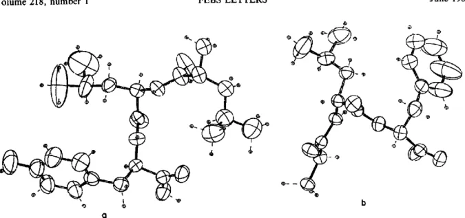
Fig. 1. Conformation of Leu-Leu-Tyr (a) and Gly-Leu-Phe (b) tripeptides. Molecular conformations are seen along the c axis.
3. R E S U L T S A N D D I S C U S S I O N 3.1. Immunostimulating properties
A n enzymic digest o f h u m a n casein was submit- ted to a series o f c h r o m a t o g r a p h i c purification steps and o f i m m u n o m o d u l a t i n g tests, and an ac- tive tripeptide was characterized (table 1). The natural as well as the synthetic peptide Gly-Leu- P h e showed a significant activity on the phago- cytosis o f SRBC by mouse peritoneal macro- phages; f u r t h e r m o r e it protected the mice against an infection with KI. pneumoniae at 1 m g / k g when administered subcutaneously and to a lesser but still significant degree intravenously. The ana- logous peptide Gly-Phe-Leu which also occurs in h u m a n casein displayed also weak but significant activities when the same biological tests were used. The peptide Leu-Leu-Tyr f r o m cow casein in- creased again the phagocytosis o f SRBC by mouse m a c r o p h a g e s but failed to protect the mice against infection with Ki. pneumoniae; however it slightly but significantly stimulated the a n t i b o d y secretion against SRBC by murine spleen cells.
3.2. X-ray analysis and molecular conformation The crystal parameters determined by X - r a y techniques were for peptide Leu-Leu-Tyr, which crystallized in the o r t h o r h o m b i c system, space group P2~2121: a = 26.074/k, b = 17.591 A, c = 5.224 A. F o r peptide Gly-Leu-Phe, which crystal-
lized in the monoclinic system, space group B2, the parameters were: a -- 18.229 ,~,, b = 18.781 A, c -- 5.917 A, 7 = 110°65. The two structures were refined up to a R = 8°7o and 5%, respectively [6]. Fig. 1 shows the structure o f each peptide mole- cule. Their molecular c o n f o r m a t i o n s are defined by the torsion angles [7] f r o m which some r e m a r k s can be drawn: (a) all the peptide bonds are in trans- c o n f o r m a t i o n and all Ccr-CONH-Cxr groups deviate very slightly f r o m planarity (w: 180°); (b) in peptide Leu-Leu-Tyr, the two ~ values are o f the same magnitude ( - 1 0 0 ° and - 8 8 °) indicating that it is already taking a regular helix c o n f o r m a - tion, whereas in peptide Gly-Leu-Phe the molecule has a m o r e stretched c o n f o r m a t i o n as the phenylalanine residue has a ~ value o f - 174°8; (c) the side chains are very agitated as shown by ther- mal ellipsoids (fig. 1).
The crystal packing o f the two tripeptides is shown in fig.2. As expected no intramolecular hydrogen bonds were observed. The molecules are held together through an intermolecular hydrogen b o n d network. In b o t h peptides hydrogen bonds occur between a carbonyl group a n d the amino g r o u p o f a neighbouring molecule. The stacking o f the molecules along the c axis is a result o f the Van der Waals forces. Peptide Gly-Leu-Phe has a zwit- terion ion structure as suggested by its carboxyl group dimensions (C-O: 1.22 ,~,), while the water molecules contribute to the stabilization o f the
Volume 218, number 1 FEBS LETTERS June 1987
.j
9
i!
t
°,)
F ,
,?
--
Fig.2. Crystal structures of Leu-Leu-Tyr (a) and Gly-Leu-Phe (b). The crystal structures are viewed along the c axis.
whole. In peptide Leu-Leu-Tyr, the amino end is charged (NH~-) and stabilized by C1-.
The striking feature o f the crystal structure o f peptide Gly-Leu-Phe is the hydrophobic channel made up o f leucine and phenylalanine residues packed together along the a axis, while all hydro- philic groups are clustered outside the channel. The segregation o f the h y d r o p h o b i c side chains contributes to stabilize the peptide crystal struc- ture. Whether the extended c o n f o r m a t i o n o f this tripeptide is a molecular p r o p e r t y or results f r o m crystal packing remains to be checked.
The aim o f this study is to initiate a correlation between the structures and biological activities o f immunostimulating peptides and later to deduce the topology o f their biological targets. The pre- sent note reports for the first time the three- dimensional structures o f two immunostimulating peptides: other related structures are currently
58
under investigation as obviously further data must be accumulated.
R E F E R E N C E S
[1] Werner, G.H., Floc'h, F., Migliore-Samour, D. and Joll~s, P. (1986) Experientia 42, 521-530. [2] Joll~s, P., Parker, F., Floc'h, D., Migliore, D.,
Alliel, P., Zerial, A. and Werner, G.H. (1982) J. Immunopharmacol. 3, 363-369.
[3] Parker, F., Migliore-Samour, D., F!oc'h, F., Zerial, A., Werner, G.H., Joll~s, J., Casaretto, M., Zahn, H. and Joll~s, P. (1984) Eur. J. Bio- chem. 145, 677-682.
[4] Jonkheere, A.R. (1954) Biometrika 41, 133-145. [5] Germain, G., Main, P. and Woolfson, M.M.
(1971) Acta Cryst. A27, 368-376.
[6] Delettr6, J., Mornon, J. and Lepicard, G. (1980) Acta Cryst. B36, 1430-1435.
[7] IUPAC-IUB, Commission on Biochemical Nomenclature (1970) J. Mol. Biol. 52, 1-17.