HAL Id: hal-02020673
https://hal-univ-rennes1.archives-ouvertes.fr/hal-02020673
Submitted on 16 May 2019HAL is a multi-disciplinary open access
archive for the deposit and dissemination of sci-entific research documents, whether they are pub-lished or not. The documents may come from teaching and research institutions in France or abroad, or from public or private research centers.
L’archive ouverte pluridisciplinaire HAL, est destinée au dépôt et à la diffusion de documents scientifiques de niveau recherche, publiés ou non, émanant des établissements d’enseignement et de recherche français ou étrangers, des laboratoires publics ou privés.
Quantitative live-cell imaging and 3D modeling reveal
critical functional features in the cytosolic complex of
phagocyte NADPH oxidase
Cornelia S Ziegler, Leila Bouchab, Marc Tramier, Dominique Durand, Franck
Fieschi, Sophie Dupré-Crochet, Fabienne Mérola, Oliver Nüße, Marie Erard
To cite this version:
Cornelia S Ziegler, Leila Bouchab, Marc Tramier, Dominique Durand, Franck Fieschi, et al.. Quanti-tative live-cell imaging and 3D modeling reveal critical functional features in the cytosolic complex of phagocyte NADPH oxidase. Journal of Biological Chemistry, American Society for Biochemistry and Molecular Biology, 2019, 294 (11), pp.3824-3836. �10.1074/jbc.RA118.006864�. �hal-02020673�
Accepted
manuscritpt
Quantitative live-cell imaging and 3D modeling reveal critical functional features in thecytosolic complex of phagocyte NADPH oxidase
Cornelia S. Ziegler1, Leila Bouchab1, Marc Tramier2, Dominique Durand3, Franck Fieschi4 Sophie
Dupré-Crochet1, Fabienne Mérola1, Oliver Nüße1,*, Marie Erard1,*
From the 1Laboratoire de Chimie Physique, CNRS, Univ. Paris-Sud, Université Paris-Saclay, 91405, Orsay France, 2Univ Rennes, CNRS, IGDR [(Institut de génétique et développement de Rennes)] – UMR 6290, BIOSIT – UMS 3480, F-35000 RENNES, France, 3Institute for Integrative Biology of the Cell (I2BC), CEA, CNRS UMR 9198, Univ. Paris-Sud, Université Paris-Saclay, Gif-sur-Yvette, France, 4Univ. Grenoble Alpes, CNRS, CEA, Institut de Biologie Structurale, F-38044 Grenoble, France
Running title: a new 3D model of the NADPH oxidase cytosolic complex
*to whom correspondence should be addressed: Marie Erard; marie.erard@u-psud.fr or Oliver Nüsse; oliver.nusse@u-psud.fr
Keywords : intrinsically disordered proteins ; unstructured domain; quaternary structure; NOX family;
NADPH oxidase; protein-protein interaction; FRET-FLIM; FCCS; fluorescent protein
ABSTRACT
Phagocyte NADPH oxidase produces superoxide anions, a precursor of reactive oxygen species (ROS) critical for host responses to microbial infections. However, uncontrolled ROS production contributes to inflammation, making NADPH oxidase a major drug target. It consists of two membranous (Nox2 and p22phox) and three cytosolic subunits (p40phox, p47phox, and p67phox) that undergo structural changes during enzyme activation. Unraveling the interactions between these subunits and the resulting conformation of the complex could shed light on NADPH oxidase regulation and help identify inhibition sites. However, the structures and the interactions of flexible proteins comprising several well-structured domains connected by intrinsically disordered protein segments are difficult to investigate by conventional techniques such as X-ray crystallography, NMR, or cryo-EM. Here, we developed an analytical strategy based on FRET– fluorescence lifetime imaging (FLIM) and fluorescence cross-correlation spectroscopy (FCCS) to structurally and quantitatively characterize NADPH oxidase in live cells. We characterized the inter- and intramolecular interactions of its cytosolic subunits by elucidating their conformation, stoichiometry, interacting fraction, and affinities in live cells. Our results revealed that the three subunits have a 1:1:1 stoichiometry and that nearly 100% of them are present in complexes in living cells. Furthermore, combining FRET data with small-angle X-ray scattering (SAXS) models and published crystal structures of isolated domains and subunits, we built a 3D model of the entire cytosolic complex.
The model disclosed an elongated complex containing a flexible hinge separating two domains ideally positioned at one end of the complex and critical for oxidase activation and interactions with membrane components.
Numerous proteins contain intrinsically disordered regions (1,2). Their growing number challenges the paradigm of a strong relationship between structure and function. Those proteins require new strategies to study their shape and their interactions. Here we focus on proteins composed of structured domains linked by disordered segments. Those segments and thus the whole protein can adopt a broad ensemble of conformations. These ensembles are highly influenced by their environment. In particular, the molecular crowding, the local ion concentrations, pH or viscosity present in live cells may change the set of accessible conformations. Nevertheless, when they are associated with diseases, knowledge about both their structure and their affinity with partners is essential to identify potential drug targets in medicinal chemistry. Their structural analysis calls for new in situ analytical strategies (1,2). Here, we combined live cell FRET-FLIM and FCCS approaches to propose an integrated analytical workflow for structural and quantitative studies of a protein complex composed of such multi-domain proteins in their native environment. We applied this workflow to the intra- and inter-molecular interactions of the cytosolic factors of the phagocyte NADPH oxidase complex.
The phagocyte NADPH oxidase is one of the seven isoforms of the Nox family and a major
Accepted
manuscritpt
enzyme of the immune system due to itspronounced production of microbicidal reactive oxygen species (ROS). A pathological hyperactivity of this oxidase leads to chronic inflammation, which is associated with cardiovascular diseases, stroke, and chronic obstructive pulmonary disease (COPD) (3). The activity is regulated by the spatio-temporal organization of the intra- and intermolecular interactions of its cytosolic subunits, p40phox, p47phox, and p67phox, forming a soluble complex before activation. Upon activation, these subunits translocate together with the small GTPase Rac to the membrane subunits Nox2 and p22phox to form the active oxidase complex. The protein interactions within the cytosolic complex were extensively studied in vitro (Fig. 1) (3). In addition to the interactions between p40phox and p67phox or p47phox and p67phox, an interaction between the SH3 domain of p40phox and the PRR domain of p47phox has been observed but its physiological relevance is not clear (4). The structure of p40phox was entirely solved (5). The presence of intrinsically disordered regions in p47phox and p67phox, predicted from their sequence (6), prevented the crystallization of the whole subunits. Only domains either isolated or in interaction were solved (3). To date, neither the spatial organization nor the stoichiometric composition of the entire complex in live cells was clarified. Using live cell FRET-FLIM and FCCS approaches with fluorescent protein-tagged subunits, we demonstrated a 1:1:1 stoichiometry of the three subunits, estimated their affinity and analyzed their spatial organization. Finally, we used these findings to elaborate a new 3D in silico model of the entire cytosolic complex in the live cell situation. This model shows an elongated complex and a flexible hinge. It is fully compatible with the multiple steps of oxidase activation. In addition, it can guide the identification of potential sites for anti-inflammatory drug targets to regulate the NADPH oxidase activity.
Results
FP tagged subunits are correctly expressed and are functional
The cytosolic subunits p40phox, p47phox and p67phox were tagged with fluorescent proteins (FPs, Table S1), either cyan (CFP: mTurquoise or Aquamarine) (7), yellow (YFP: Citrine) (8), or red (RFP: mCherry) (9). The size of the fusion proteins expressed by COS7 cells was verified by Western Blot (Fig. S1A). To assess whether the FP-labelled subunits are able to reconstitute the active oxidase complex, we used COSNox2/p22 cells stably expressing the membranous subunits Nox2/p22phox and Rac, but no endogenous cytosolic subunits (10). COSNox2/p22 were
transiently transfected with the three cytosolic subunits with or without a FP-tag (Fig. 2A). The production of superoxide anions was monitored by a luminometry assay sensitive to extracellular ROS (Fig. 2B). The ROS production started upon activation with phorbol myristate acetate (PMA) and stopped immediately after addition of diphenyleneiodonium (DPI), a NADPH oxidase inhibitor. All constructs allowed a pronounced ROS production (Fig. 2C). The production with N-terminal tagged p47phox and p67phox was lower in comparison to the C-terminal tagged variants but still much higher than the non-transfected cells. The presence of the FP-tags slowed down the ROS production and consequently delayed the time point at which the maximal signal was reached (Fig. S1B and C). Taken together, these observations show that all our FP-tagged subunits are able to reconstitute an active NADPH oxidase complex and are fully suitable to explore their spatial organization and affinity.
Förster resonance energy transfer (FRET) is observed between fluorescent proteins at both ends of individual subunits and is not strongly modified in presence of their partners.
We then tagged each of the cytosolic subunits p40phox, p47phox and p67phox simultaneously at both termini with a donor (D) and an acceptor (A) FP for Förster resonance energy transfer (FRET), resulting in so-called tandems (schemes Fig. 3, Table S1). We used as a positive control a simple D/A tandem in which the donor and acceptor FPs were linked by a flexible 27 amino acids long peptide (11). Tandems were expressed in COS7 cells and FRET was monitored by fluorescence lifetime imaging (FLIM). The apparent FRET efficiencies, Eapp, were derived
from the average fluorescence lifetime of the donor FP measured in individual cells (Eq. 1 to 3 in experimental procedures). Eapp for the p40phox,
p47phox and p67phox tandems were significant, although lower than in the simple D/A tandem (Eapp = 8.2 ± 1.2%, 12.3 ± 1.2%, 7.7 ± 1.1 % and
32.7 ± 1.8%, respectively, Fig. 3B). The low Eapp
values for the subunit tandems are consistent with the much larger size of the central subunits as compared to a 27 amino acids peptide linker.
Next, we studied the influence of the p67phox partner on the p47phox tandem and of p40phox on the p67phox tandem. Either p47phox or p67phox tandems were co-expressed with a plasmid coding for the partner subunit connected to RFP
via the viral P2A peptide (12,13). The P2A
sequence prevents the peptide bond formation and leads to the separate expression of the subunit and the RFP, the presence of the RFP proving the correct expression of the partner subunit. The
Accepted
manuscritpt
co-expression of tandems with RFP alone wastaken as reference (Fig. 3C). The co-expression of RFP-2A-p67phox did not change the apparent FRET efficiency in the p47phox tandem, and similarly, the presence of p40phox did not modify the FRET level of the p67phox tandem (Fig. 3C and Fig. S2), showing that in both cases, the overall geometry of the tandem is not significantly modified upon interaction with their partners.
FRET signals indicate specific interaction between p67phox and p47phox and provide information on their spatial organisation
The bi-molecular interaction between FP-tagged cytosolic subunits was then investigated using similar FLIM-FRET methodologies (Fig. 4). First, we studied the interaction of p67phox and p47phox (Fig. 4A, B, C and F). Upon co-expression of p47phox-CFP / p67phox-YFP, the lifetimes of the CFP donor were on average significantly shorter than the reference value for p47phox-CFP alone (Fig. 4A & 4B). However, as usual in dual expression systems, FRET efficiencies Eapp varied
strongly from cell to cell, depending on the amount of expressed acceptor. We thus determined Eapp as a function of the acceptor
quantity, as estimated from its average fluorescence intensity (Fig. 4C), or as a function of the ratio [A]/[D] (Fig. 4F), using a custom calibration procedure (see SI), as shown by others (14,15). In all cases, the FRET efficiency increases with the absolute or relative amount of acceptor.
For p47phox and p67phox subunits labeled at their C-termini (CC-labeling), Eapp reaches a
maximum value around 12 % at high acceptor levels (Fig. 4C & 4F). This is significantly higher than the negative control, consisting in the co-expression of p47phox-CFP with YFP alone (Fig. 4C and Fig. S3A). As a second negative control, we used a truncated version of p47phox missing the PRR domain (p47phoxCter[1-342]) and leading in
vitro to a complete abrogation of any interaction
with p67phox (16). The co-expression of p47phoxΔCter-CFP with p67phox-YFP gives clearly different FLIM images as compared to p47phox-CFP, with well separated average lifetime distributions (Fig. 4A and 4B), and measured Eapp
values in the range of the first negative control (Fig. 4C and Fig. S3A). The observation of a plateau value for Eapp, well above the values of the
controls, provides a strong evidence for a specific interaction (17). The absence of significant FRET in the case of p47phoxCtershows in addition that the PRR domain of p47phox is required for this interaction in live cells.
We then modified the labeling sites of our FP tags, either by switching them to the N-termini
or by exchanging FP colors between p47phox and p67phox (Fig. 4C & 4F). For the CN–tags, the maximum Eapp of 8% was lower than for the
CC-labeling (Fig. 4F), yet significantly above the negative controls (Fig. S3A). For the NN–tags,
Eapp, remained very low and close to the range of
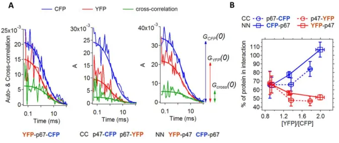
the negative controls (Fig. 4C and Fig. S3A). To evaluate independently the occurrence of an interaction between p47phox and p67phox tagged at their N-termini, we used fluorescence cross-correlation spectroscopy (FCCS). In this technique, the fluctuations of the fluorescence intensities of FP-tagged p47phox and p67phox diffusing in and out a confocal volume in the cytosol of COS7 cells are analyzed by auto- and cross-correlation functions (see Experimental Procedure). A cross-correlation function with a non-null amplitude was observed for p47phox and p67phox tagged either at their CC and NN-termini (Fig. 5A), showing in both cases a co-diffusion of the fluorophores indicative of complex formation. We thus observe a specific interaction between p47phox and p67phox cytosolic subunits for all tested positions and types of FP–tags, with in most cases Eapp well above negative controls.
Apparent FRET efficiencies measured in living cells are very complex average quantities. In addition, the efficiency of energy transfer depends on both the distance and relative orientation between the donor and the acceptor, as predicted by Förster theory (see Experimental Procedure). As the fluorescent proteins are attached to the subunits through variable flexible linkers (Table S1), a large range of relative FP orientations are likely allowed, leading to some averaging of the orientation factor. In the frame of this paper, we will thus assume that major differences in FRET efficiencies are chiefly governed by distance. This is supported by the similar FRET efficiencies observed when different D/A pairs are used, or when the anchoring sites of donor and acceptor are swapped (see for example Fig. 4C & 4F).In this frame, the relative FRET efficiencies observed for donor and acceptor located at different termini provide interesting topological information. The maximum apparent FRET efficiency is higher for the CC–labeling than for the CN–labeling (Fig. 4F). Indeed, the two C termini are known to bind to each other and should be closer than the N-terminus of p67phox and the C-terminus of p47phox, which are separated by p67phox itself (Fig. 1). For the NN–labeling, the distance between FPs is most likely too large to observe FRET, as we have evidence for complex formation through FCCS. The upper distance limit for a FRET-positive situation is about twice the Förster radius of the FRET pair (see experimental procedures), c.a. 100 Å. To fulfill this distance
Accepted
manuscritpt
condition, the N–termini of both subunits shouldpoint to opposite directions in the complex.
FRET signals indicate specific interactions between p40phox and p67phox, and between p40phox and p47phox
We studied similarly the interactions between FP-tagged p40phox and p67phox (Fig. 4D and G, Fig. S3B). For all tag positions (NC, NN, or CC), specific FRET was observed with the same maximum efficiency around 8% at the plateau (Fig. 4D & 4G). The similar maximum FRET efficiencies indicate comparable distances between the different termini of p40phox and p67phox.
The direct p40phox-p47phox interaction was questioned to be of physiological relevance or to be rather an artificial phenomenon resulting from test tube experiments in the absence of p67phox (4). The possible interaction sites were identified in
vitro to be the SH3 domain of p40phox and the C-terminal PRR domain of p47phox, which is also the binding site for p67phox (Fig. 1). We co-expressed p40phox and p47phox or its truncated version p47phoxΔCter with NC or CC labeling (Fig. 4E and 4H). We found specific FRET in both cases with equivalent Eapp, which indicates that the FP-tags at
both termini of p40phox have a similar average geometry relative to the C-terminus of p47phox. When p47phoxΔCter was used instead of full-length p47phox, no significant FRET was observed (Fig. 4E and Fig. S3C). This confirms the requirement of the PRR domain of p47phox for the interaction with p40phox in live cells.
The cytosolic complex has a 1:1:1 composition with a high affinity between the subunits
FRET depends strongly on the number of acceptors in the direct vicinity of the donor and thus allows to explore the stoichiometry of the interaction (18). An uneven stoichiometry of the subunits in the complex (X : Y) will result in different maximum Eapp when the donor and
acceptor are swapped between subunits (Fig. S4). In contrast, even subunit ratios (X : X) will give the same maximum Eapp. On hetero-dimers formed
by p47phox / p67phox as well as by p40phox / p67phox , swapping donor and acceptor did not change the maximum apparent FRET efficiency (Fig. 4F and 4G). We also investigated possible homo-dimerization of the cytosolic subunits by co-expressing each subunit tagged with donor and acceptor FPs in the same cell. In all cases, the Eapp
scatters in the range of the negative controls (Fig. S5), indicating that there are no detectable homo-dimers. Taken together, our findings support a 1:1:1 stoichiometry for the cytosolic complex in the living cell, in agreement with in vitro experiments (19,20).
The amplitudes of the auto- and cross-correlation functions obtained by FCCS provide an estimate of the relative expression levels of the FP tagged subunits, and of their fraction in interaction (Experimental Procedures and SI). The FCCS analysis gives qualitatively similar results with FRET-positive CC-labeling or FRET-negative NN-labeling (Fig. 5B). In both cases, the fraction of interacting protein is correlated to the relative expression level of the proteins. While all p67phox is bound in complex in the presence of a twofold excess of p47phox, the fraction of p47phox interacting with p67phox decreases concomitantly. The apparent fraction of molecules in interaction is somewhat lower for the FRET-positive CC-labeling than for the FRET-negative NN-CC-labeling, which may be ascribed to a FRET-induced decrease of the amplitude of the cross-correlation function (21).
The concentration of bound and free diffusing p47phox and p67phox obtained from the FCCS measurement can also be used to estimate their apparent dissociation constant, 𝐾𝐷𝑎𝑝𝑝(see SI). The median 𝐾𝐷𝑎𝑝𝑝 value is in the range of a few hundred nM, showing a high affinity between p47phox and p67phox in live cells. Such a high affinity, with a 𝐾𝐷 well below 1 µM, is consistent with previous values obtained in vitro ranging from 4 to 32 nM (22).
In conclusion, FRET imaging and FCCS experiments in live cells clearly demonstrate specific interactions between the three cytosolic subunits of the NADPH oxidase and give new insights into their spatial organization in the complexes. In the next paragraphs, we will use this information, together with SAXS, NMR and X-ray crystallography data to build a 3D model of the hetero-trimer. The crystallographic structure of p40phox was solved (5) but p47phox and p67phox are highly flexible and only some domains were crystallized. First, we will describe how several putative conformations of p67phox and p47phox were selected from SAXS experiments. Second, we will assemble the individual subunits and choose along this process the most appropriate conformation of p47phox and p67phox, with the help of the topological information obtained from FRET, to finally propose a 3D model of the hetero-trimer compatible with our live cell experiments, as well as structural and biochemical results available in the literature.
Sets of models for p67phox and p47phox based on SAXS experiments
SAXS analysis consists in producing a set of atomic models compatible with the experimental SAXS curves using existing high-resolution structures of the crystallized domains,
Accepted
manuscritpt
as explained in the section ExperimentalProcedures. For p67phox, we re-examined previous experimental SAXS results (23) while new experiments were performed for the truncated p47phoxCter[1-342] and for p47phox to improve the model previously proposed by Durand et al. (24). For each of the three proteins, we retained a representative selection of possible models, that are discussed in the following steps.
The representative models of p47phoxCter [1-342] were first examined in order to find a model adopting a relative orientation of its PX domain with the SH3 domains compatible with all other previous experimental observations (Fig. S6). First, in the resting state, p47phox adopts an autoinhibited conformation in which the residues of the PX domain that interact with membrane phospholipids during the active phase are poorly accessible (25). Most of the models were rejected using this criterion (Fig. S6A, S6B). Second, the PX domain interacts with the lateral surface of N-terminal SH3 domain including Arg162 and Asp166 (16,25). Therefore, the latter two residues have to be located at the interaction surface between PX and SH3 domains (Fig. S6C). Third, previous kinetic analysis of H/D exchange coupled to mass spectrometry (HDX-MS) identified the residues involved in the intramolecular interaction in the resting state of p47phox, which should also be masked (16).
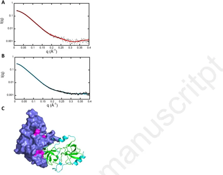
We finally retained one model of p47phox Cter that corresponds to all these criteria (Fig. 6C). The SAXS curve calculated on this model using the program CRYSOL is shown in Fig. 6A together with the experimental data (26). This model of truncated p47phox was then used as starting point for the fit of the experimental SAXS pattern of the full-length p47phox protein (Fig. 6B, Table S4), resulting in a set of full-length models carrying a mainly unstructured C-terminal part. Representative models of the full-length p47phox protein were selected to build the whole 3D model of the hetero-trimer in the next section.
Step by step assembly of a 3D model of the NADPH oxidase cytosolic complex
Assuming a 1:1:1 stoichiometry, the representative SAXS models of full length p47phox and p67phox selected above, and the crystal structure of p40phox were then used to build a model of the hetero-trimer, following a step by step workflow presented in Fig. S7. We know that p40phox and p67phox interact via their PB1 domains (Fig. 1) and the crystal structure of the p40phox -PB1/ p67phox –PB1 interaction has been solved (27). This was used as a template to add first the whole crystal structure of p40phox onto p67phox – PB1 (5) (Step I in Fig. S7). Then we added on the
p67phox-PB1 domain the full length p67phox structural models previously selected. We selected the most appropriate model based on the following criteria, illustrated in Fig. S8. First, we kept the models that had neither clashes nor sterical hindrance with p40phox. Second, considering the maximum dimension of p67phox determined by SAXS (Dmax~160 Å, Table S4) (23,28) and moreover the observation of significant FRET between the two termini of p67phox in the p67phox tandem, the N-terminus of p67phox has to be bent toward its C-terminus. Third, for the p40phox / p67phox interaction, the significant and comparable FRET efficiencies for all tag positions (Fig. 4G) indicates that the termini of p40phox and of p67phox are at similar distances, and significantly below the maximum FRET-compatible distance of 100 Å. The later condition is only fulfilled when p40phox and p67phox adopt a cross-like spatial arrangement (Fig. S8). A representative p67phox SAXS model compatible with all three criteria was selected (Step II in Fig. S7).
The resulting model of the p40phox / p67phox complex became the starting point to add p47phox. The full length p47phox SAXS models were added with the help of the NMR structure of the interaction between the C-termini of p47phox and p67phox, that are bound via their PRR (p47phox) and SH3 (p67phox) domains (29) (Step III in Fig. S7). Most of our full-length p47phox SAXS models lead to steric clashes with p40phox and/or a distance between the N-termini of p47phox and p67phox that is too short to account for the absence of clear FRET observed for NN-labeling of these two subunits (Fig. S9). This leads to the selection of a p47phox SAXS model where p47phox points in the opposite direction of p67phox in a tail-to-tail orientation. This last step results in the complete 3D-model of the p40phox / p47phox / p67phox hetero-trimer (step IV in Fig. S7 and Fig. 7).
Evaluation of our proposed 3D model : significant features and consistency with experimental data
Our assembled model of the NADPH oxidase cytosolic complex reveals an elongated structure where functional domains are connected by flexible peptide linkers (Fig. 7). As already stated, the proposed cross-like arrangement of p40phox and p67phox leads to distances between their termini compatible with significant and equivalent FRET for all combinations (Fig. 7A). The interaction of p47phox with p67phox also brings their C-termini in close vicinity, coherent with the high Eapp measured for CC labeling, while the
intermediate C-N distances are consistent with a lower Eapp for the CN labelling (Fig. 7B).
Accepted
manuscritpt
Interestingly, the FRET efficiency for theCN-tagged subunits is close to the one of the p67phox tandem. Indeed, the C-termini of p47phox and p67phox are very close and separated from the N-terminus of p67phox by a portion of p67phox itself. On the other hand, p47phox is expected to be elongated alone in solution (Dmax by SAXS is in the order of 125 Å, Table S4) but the presence of intramolecular FRET in p47phox tandems in live cells suggests that the subunit can adopt a more compact conformation. Indeed, the flexibility of its unstructured C-terminus clearly allows movements of the N-terminal domain (featured by an arrow in Fig. 7C). The absence of FRET between NN-labelled p47phox and p67phox, suggests that p47phox cannot flip completely towards p67phox but might adopt either a more elongated or more condensed conformations, as well as different angles with respect to the long axis of p67phox. Our model should thus be viewed as a relatively flexible structure with significant variability around its multiple hinges. The Dmax values of
p67phox and the p67phox / p40phox complex are close (Table S2). The Dmax values of p67phox-p47phox and p67phox
-p47phox-p40phox are similar as well (Table S2). This means that the presence of p40phox does not change the overall dimensions of the complex. This is again consistent with the cross-like spatial arrangement of p40phox with p67phox and with the observation of the same Eapp of the p67phox tandem
with or without p40phox. Finally, we find that the calculated SAXS curves using the program CRYSOL on the p67phox -p47phox -p40phox model is in reasonable agreement with the experimental curves published by Yuzawa et al. (Fig. S10) (28).
Discussion
Here we presented an integrated workflow to analyze the intermolecular interactions and the conformation of a complex of proteins formed by structured domains separated by unstructured segments in living cells. This analytical strategy may be adapted to any cytosolic protein complex composed of partially disordered subunits.
We first demonstrate the complementarity of FRET-FLIM and FCCS to characterize live cell interactions, estimate binding affinity and obtain topological information using the same FP-labeling for both techniques. This was made possible owing to the new cyan variants mTurquoise and Aquamarine in the classical CFP/YFP FRET pair (7). Their increased brightness and photostability allowed both improved performances as donors for FLIM and full suitability for two-color FCCS experiments.
The dynamic NADPH oxidase complex depends on protein assembly for activation, and thus the development of inhibitors of this assembly is an attractive concept to regulate its
activity (30,31). For the p40phox-p47phox-p67phox complex, there is a discrepancy between the 1:1:1 stoichiometry found in in vitro experiments (19,20) and the 2 – 3 times higher expression level of p47phox in comparison to p67phox in neutrophils (32). In addition, this higher amount of p47phox compared to p67phox implies that p47phox is present both as a free and complex-bound subunit (32). However, it is not known whether free p67phox exists. Our results revealed two pairwise interactions with a one to one stoichiometry, p47phox / p67phox and p40phox / p67phox that strongly suggest that the ternary complex is assembled in a 1:1:1 stoichiometry inside a live cell. Furthermore, in the presence of an excess of p47phox, all p67phox were bound in complex. In solution, about 10 % to 20 % of recombinant p47phox and p67phox proteins have been detected as dimers (22). We did not observe any dimers by FRET-FLIM. Indeed, in live cells, the unbound cytosolic subunits may encounter a large diversity of potential partners that likely limits dimer formation.
Our live cell FRET results also provide topological information that nicely complements structural data from X-ray crystallography, NMR and SAXS experiments obtained on purified recombinant proteins. The combination of SAXS data and single-molecule FRET in vitro is very useful to investigate protein-folding by integrating complementary information (33-35). The combination of SAXS with FRET measurements of FP-labelled proteins in cellulo provides lower spatial and temporal resolution, however it affords access to the conditions in living cells. To our knowledge, such an approach has not been described before. These pieces of information were integrated to build a new model of the cytosolic NADPH oxidase complex with several noticeable features: (i) p47phox and p67phox interact via their C-termini in a tail-to-tail configuration. Their N-termini are over 100 Å apart, (ii) p67phox and p47phox are not fully elongated and (iii) p40phox and p67phox adopt a cross-like conformation (Fig. 7). The model is consistent with the idea that p47phox is required for the initial assembly of the functional oxidase. Indeed, the PX-domain and the SH3 domains of p47phox are located on one end of the complex, ideally positioned to interact with phospholipids (PX-domain) and with p22phox (SH3 domains) to initiate oxidase assembly. This may then bring p40phox and p67phox closer to the membrane. Indeed, the PX-domain of p40phox also interacts with membrane lipids and the N-terminus of p67phox requires several activation steps mandatory for superoxide production. The flexible hinge between the N-terminal half of p47phox and the rest of the trimeric complex may be required to establish the initial contact with phospholipids
Accepted
manuscritpt
and p22phox and then bend the complex to bringp67phox closer to Nox2. In addition, the proximity of the C-terminus of p47phox with the SH3 domain of p40phox in the model raises the question whether p40phox contributes to stabilize the C-terminus of p47phox at the SH3 domain of p67phox. The position of the p40phox PX domain in the middle of the elongated complex is unfavorable for a role of this domain in the initiation of oxidase assembly since it would require a lateral attachment of the complex on the membrane. However, if p47phox leaves the active complex as recently suggested (36), the p40phox PX domain would be in a favorable position to bind to phosphoinositides in the membrane after its dissociation from PB1 (5) and this would help keeping p67phox in place (37). In p67phox, the TPR and the activation domain are essential for the assembly with Rac and Nox2 and, ultimately, for activation of the oxidase. In the proposed model of the hetero-trimer, presented here in the resting state (Fig. 7), the N-terminal region of p67phox is well exposed and accessible. We assume that the global orientation and the internal flexibility between its domains is fully compatible with the binding to Rac or to Nox2 trough the activation domain (38). At this stage of characterization of the ternary cytosolic complex, it seems that the main limiting step towards activation is p47phox that needs to be activated to promote assembly of the whole cytosolic complex at the membrane. Preventing the conformational changes of the N-terminus of p47phox would probably block the subsequent interactions with the membrane and p22phox and prevent oxidase activation. Therefore, our approach provides critical information for the design of inhibitors that would interfere with key steps in the activation process. Furthermore, the data we obtained and the model are a starting point to investigate the changes that may occur during activation of the oxidase. They may also serve to compare wild-type subunits with mutations found in patients with chronic granulomatous disease in order to understand the phenotype of this oxidase deficiency.
Experimental procedures
Plasmid library and transfection. Plasmids
encoding full length human p47phox (NCF1) and p67phox (NCF2) both embedded in a pEGFP-N1 vector (Clontech) (36), and p40phox (NCF4) embedded in a CMD8 vector (Gift from Marie Claire Dagher) were used as a starting point to build a full library of N- and C-terminal tagged fusion proteins. The cDNA of the subunits were cut out with restriction enzymes or amplified by PCR and inserted in either cyan mTurquoise or Aquamarine-N1 or –C1 vectors (variants of the
pECFP vectors: mTurquoise: T65S-S72A-H148D-S175G-A206K (39), Aquamarine: T65S, H148G (40)), yellow Citrine-N1 or –C1 vectors (the mutation Q69M was introduced into EYFP) (8), or the red mCherry-C1 vector (Clontech, Takara Biotechnology, Co., Ltd). The internal start codon (ATG) was removed by PCR from the FP-N1 vectors or from the subunits embedded in FP-C1 vectors. Table S1 displays an overview of the constructs. In order to build the plasmids coding for RFP-2A-p67phox or p40 phox, we ordered a synthetic gene coding for mCherry-P2A (P2A=ATNFSLLKQAGDVEENPGP) (12) framed by AgeI and HindIII restriction sites (Eurofins). The two enzymes were used to cut out the FP and insert mCherry-2A in pECitrine-p67-C1 or pEmCherry-p40-C1. Primers were purchased from Eurogentec (Kaneka corp., Japan). Plasmids were amplified in DH5α
Escherichia coli, DNA was purified with
E.N.Z.A. ® mini kit 2 (Omega Bio.Tek). For transfection, cells (COS7 or COSNox2/ p22) were seeded either on glass coverslips (Ø 25 mm, thickness 0.13 – 0.16 mm) in 6-well plates for microscopy or in 24-well plates for luminometry one day before the experiment. Cells were transiently transfected with XtremeGene HP (Roche Diagnostics) following the supplier’s instructions and used 24 – 48 h after transfection. Transfection efficiency, monitored by flow cytometry, was constantly between 20 – 30 % for triple transfection. Based on the high reproducibility of transfection efficiencies, we assumed that the subunits without FP-tag were present in the cells at the same average level as the FP-tagged ones.
Cell culture. COS7 cells were purchased from
ATCC and cultured following the supplier’s instructions. COS7 cells stably expressing Nox2 and p22phox (COSNox2/ p22) were kindly provided by M. Dinauer (Washington University School of Medicine, St. Louis, MO) and cultured in medium containing selecting antibiotics (10).
O2•- detection by 012 chemiluminescence. L-012 (100 µM Wako Chemicals) and HRP (20 U/ml) were mixed and added to the well containing transfected COSNox2/ p22 cells a few minutes before the addition of 500 nM of PMA to stimulate the assembly of the NADPH oxidase. 20 µM of DPI was added to stop the O2 •-production. The production was quantified as the relative light units (RLU) area under the curve recorded during 30 mins with a SynergyH1 plate reader (Biotek, USA).
Accepted
manuscritpt
Fluorescence Lifetime Imaging Microscopy (FLIM). Time resolved laser scanning TCSPC
microscopy was performed on a custom made microscope as described previously (40). Briefly, the setup is based on a TE2000 microscope with a 60x, 1.2NA water immersion objective (Nikon). The epifluorescence pathway is equipped with an Hg lamp, a set of filter cubes for the different FPs and a CCD camera (ORCA-AG, Hamamastu Photonics, Table S3). The TCSPC path is equipped with pulsed laser diodes (440 nm for CFPs; 466 nm for YFPs, PicoQuant) driven by a PDL800 driver (20 MHz, PicoQuant). The C1 scanning head (Nikon) probes a 100 x 100 µm maximum field of view. To select the FP fluorescence, dichroic mirrors (DM) and filter sets were used before the detection by a MCP-PMT detector (Hamamatsu Photonics). The signals were amplified by a fast pulse pre-amplifier (Phillips Scientific) before reaching the PicoHarp300 TCSPC module (PicoQuant). Counting rates were routinely between 50 000 and 100 000 cts/s. Transfected cells were kept in PBS at 20°C and studied for 2 h maximum in an Attofluor cell chamber (Thermo Fisher Scientific). The lifetime of a fluorophore is an intensive property, independent of its concentration, which can be precisely monitored even in live cells. A precision of a few percent on lifetime is common (17,41,42,43 ,44). The TCSPC fluorescence decay of all the pixels of the cytosol was computed by the SymPhoTime software (Fig. 3A). The decays were fitted with a mono-exponential fit function (Eq. 1a) for the control cells expressing a donor-fusion protein (without acceptor), the donor being Aquamarine, mTurquoise and Citrine (Fig. 3A, left).
𝐼(𝑡) = 𝐼0 𝑒−𝜏𝑑𝑜𝑛𝑜𝑟𝑡 + 𝐶, [1a]
where C is the constant background.As the donor has a mono-exponential lifetime, its fluorescence decay in the presence of FRET to an acceptor can be fitted with a bi-exponential function (Eq. 1b, Fig. 3A, right).
𝐼(𝑡) = 𝛼𝑙𝑜𝑛𝑔 𝐼0 𝑒 − 𝑡
𝜏𝑙𝑜𝑛𝑔+ 𝛼𝑠ℎ𝑜𝑟𝑡 𝐼0 𝑒−𝜏𝑠ℎ𝑜𝑟𝑡𝑡 + 𝐶,
[1b]
where C is the constant background. 𝛼𝑙𝑜𝑛𝑔 and
𝛼𝑠ℎ𝑜𝑟𝑡 are the proportions of a long (𝜏𝑙𝑜𝑛𝑔) and a
short (𝜏𝑠ℎ𝑜𝑟𝑡) lifetime component respectively.
For fitting, we used a custom made procedure in IGOR Pro (WaveMetrics). The quality of the fits is evaluated by the weighted residual distribution and Pearson’s χ² test (Fig. 3A).
< 𝜏𝐷𝐴> is the average lifetime of the donor-fusion protein, D, in presence of an acceptor-fusion protein, A, (Eq. 2):
< 𝜏𝐷𝐴>= 𝛼𝑙𝑜𝑛𝑔𝜏𝑙𝑜𝑛𝑔+ 𝛼𝑠ℎ𝑜𝑟𝑡𝜏𝑠ℎ𝑜𝑟𝑡 [2]
The apparent FRET efficiency 𝐸𝑎𝑝𝑝 is calculated
using < 𝜏𝐷𝐴> and 𝜏𝐷𝑜𝑛𝑜𝑟 (Eq. 3): 𝐸𝑎𝑝𝑝 = 1 −<𝜏𝐷𝐴>
𝜏𝐷𝑜𝑛𝑜𝑟 × 100 [3]
It was calculated on a cell by cell basis from the fluorescence decays obtained for each cell using for 𝜏𝐷𝑜𝑛𝑜𝑟 the lifetime of the same donor/subunit fusion protein alone.
The apparent FRET efficiencies measured in each cell are complex averages over highly heterogeneous populations of donor molecules engaged in different FRET interactions. Fundamentally, these FRET efficiencies depend on both the distance and relative orientation between donor and acceptor. The determination of distances between FRET partners thus requires some assumption on the orientation factor κ² (45). Unfortunately, the dynamic isotropic regime leading to the popular value κ²=2/3 cannot be applied to fluorescent proteins, since their rotational correlation time in water is around 14 ns
i.e. three to four times longer than the lifetime of
the donor excited state (46 ,47). In the viscous environment of living cells, FPs must be considered as static on the time scale of the FRET interaction. On the other hand, since FPs are fused to the subunits through flexible linkers, they are expected to adopt a whole set of conformations, leading to broadly distributed, but probably non-isotropic and non-homogeneous relative orientations and distances. Therefore, the precise calculation of distances from our FRET data, as proposed for example by Vogel et al (48), is mostly out of reach. Instead, we used the apparent FRET efficiencies to establish distance limits, to guide topological reasoning or to provide comparative information. Indeed, whatever the effective value of κ², the observation of a significant FRET indicates some proximity within the range of the critical Förster distance R°. Typical Förster distances of most fluorescent protein FRET pairs, calculated using κ²=2/3, lie in the range of 50-60 Å (48). R° evolves as (κ²)1/6 and thus varies only slowly with the orientation factor. Using κ²=0.476, a value applicable to fully isotropic and homogeneous static averaging (17,45,49), we obtain quite similar values of R°= 57 Å for Aquamarine / Citrine and 55 Å for Citrine / mCherry. For the maximum, but highly unlikely value κ²=4, these distances would at most extend to 81 Å and 78 Å respectively. In cases of unfavorable relative orientations, R° could on the contrary decrease significantly below 50 Å. Conversely, as the D/A distance approaches twice R°, the FRET efficiency will tend virtually to 0. As a rule of thumb, assuming some degree of orientational and conformational averaging, and considering our practical detection limits, we took
Accepted
manuscritpt
100 Å as the upper distance limit to observe FRETin this study.
The FP-tagged subunits are transiently expressed and the level of expression may strongly vary between cells. We recorded intensity images before any FLIM experiments on the same field of view. The intracellular concentrations are proportional to the average fluorescence intensities. Eapp was commonly plotted as a
function of the fluorescence intensity of the acceptor in the cell, I(A) to observe a FRET-positive situation (17,50-53). As a negative control, the donor/subunit fusion protein was co-expressed with the free acceptor. This control is useful to (i) determine if Eapp is significant and (ii)
to set the threshold for the maximum value of I(A) beyond which unspecific FRET due to molecular crowding starts to contribute to the signals. Indeed, when the cells are crowded with the acceptors, I(A) becomes very high and the donors show unspecific FRET with the acceptors nearby just because of the spatial proximity (17). We discarded such cells.
The plotting of Eapp against [𝐴] [𝐷]⁄ required the
calibration of fluorescence intensities measured in both donor and acceptor channel. The ratio of fluorescence intensities, 𝐼(𝐴) 𝐼(𝐷)⁄ , was transformed in the concentration ratio of fluorescent proteins, [𝐴] [𝐷]⁄ , using a custom calibration procedure of the microscopy setup (see
SI).
Fluorescence Cross Correlation Spectroscopy.
A confocal microscope Leica TCS SP8 SMD (Leica Microsystems) was used. It is equipped with a DMI 6000 CS stand and a 63x/1.2 HC PL APO water immersion objective, a continuous argon laser (514 nm from Leica TCS SP8) and a diode pulsed laser (440 nm, PicoQuant). The signal was selected by 505 nm DM and two bandpass filters (BP 478/22, BP 540/30) and detected by two APD detectors (PicoQuant). For the detection, the SMD module is constituted of a PicoHarp3000 system for TTTR mode of single photon counting (PicoQuant). Cells were kept in an air-conditioned chamber at 30°C. In order to correct for the crosstalk between the CFP and YFP detection channels on the amplitudes of the correlation functions, we used a variant of fluorescence correlation spectroscopy called fluorescence lifetime correlation spectroscopy (FLCS) (54). The fluorescence signal of each channel was corrected from spectral blead-through using FLCS filters as described elsewhere (55,56). In this method combining TCSPC and FCS, the different FCS contributions are separated by using the different fluorescence decay pattern of both fluorophores. The fluctuations of fluorescence intensity were auto- and
cross-correlated and the resulting curves were analyzed using a standard pure diffusion model with SymphoTime (PicoQuant).
The auto-correlation function of fluorescence fluctuations returns the time needed for the fluorophore to cross the confocal observation volume, 𝑉𝐹𝑃, and the number of
molecules, 𝑁𝐹𝑃, present in 𝑉𝐹𝑃. 𝑁𝐹𝑃 is the inverse
of the amplitude to the auto-correlation function, G𝐹𝑃(0) (21). The concentrations of FPs in the observation volume are calculated as following where 𝑁𝐴 is the Avogadro number (Eq. 4):
[𝑌𝐹𝑃] =G 1 𝑌𝐹𝑃(0) 1 𝑁𝐴 1 𝑉𝑌𝐹𝑃,[𝐶𝐹𝑃] = 1 G𝐶𝐹𝑃(0) 1 𝑁𝐴 1 𝑉𝐶𝐹𝑃 [4]
The amplitude of the cross-correlation function, G𝑐𝑟𝑜𝑠𝑠(0) , allows the estimation of the concentration of the stoichiometric p47phox-p67phox complex. p47phox and p67phox are respectively labelled with YFP and CFP (Eq. 5).
[𝐶𝑜𝑚𝑝𝑙𝑒𝑥] = G𝑐𝑟𝑜𝑠𝑠(0) G𝑌𝐹𝑃(0)∙G𝐶𝐹𝑃(0) 1 𝑁𝐴 1 𝑉𝑐𝑟𝑜𝑠𝑠 [5]
In practice, the observation volumes in the two channels, 𝑉𝑌𝐹𝑃 and 𝑉𝐶𝐹𝑃, and 𝑉𝑐𝑟𝑜𝑠𝑠 have always different sizes and are not perfectly coincident. In addition, a fraction of FPs might be non-fluorescent due to non-matured protein, dark state or photobleaching (21). The later was kept here below 10%. In order to minimize the impact of these limits on the detection of interactions, the fractions, 𝑋, of co-diffusing fluorophores that are also the fractions of protein in interaction, were normalized to the average fractions obtained for the p67phox tandem, whose two tags diffuse together (Eq. 6). 𝑋𝑌𝐹𝑃𝑐𝑝𝑙𝑥 =G𝑐𝑟𝑜𝑠𝑠(0) G𝐶𝐹𝑃(0) 〈 G𝐶𝐹𝑃𝑡𝑎𝑛𝑑𝑒𝑚(0) G𝑐𝑟𝑜𝑠𝑠𝑡𝑎𝑛𝑑𝑒𝑚(0)〉 × 100 [6] 𝑋𝐶𝐹𝑃𝑐𝑝𝑙𝑥 =G𝑐𝑟𝑜𝑠𝑠(0) G𝑌𝐹𝑃(0) 〈 G𝑌𝐹𝑃𝑡𝑎𝑛𝑑𝑒𝑚(0) G𝑐𝑟𝑜𝑠𝑠𝑡𝑎𝑛𝑑𝑒𝑚(0)〉 × 100 [6]
In addition, the relative amount of both partners was estimated as (Eq. 7):
[𝑌𝐹𝑃]𝑐𝑝𝑙𝑥 [𝐶𝐹𝑃]𝑐𝑝𝑙𝑥 = G𝐶𝐹𝑃(0) G𝑌𝐹𝑃(0)× 〈 G𝑌𝐹𝑃𝑡𝑎𝑛𝑑𝑒𝑚(0) G𝐶𝐹𝑃𝑡𝑎𝑛𝑑𝑒𝑚(0)〉 [7]
The details for the computation of 𝐾𝐷𝑎𝑝𝑝can be found in SI.
Protein preparation, SAXS data acquisition and processing. p47phox constructs were expressed as a GST fusion protein using a pGex-6P-1 derived vector containing the cDNA corresponding to the full amino acids sequence (from 1 to 397) for the full length p47phox or to a sequence coding for amino acids 1 to 342 for the p47phoxCter construct lacking the C-terminal end of the protein after the AIR motif. For both constructs, the production protocol was the same. The proteins were expressed in E. coli, strain BL21(DE3). Expression was induced with 0.5 mM IPTG, when the cell culture reached an OD of
Accepted
manuscritpt
0.6 at 600 nm. Temperature was then shifted to20°C for an overnight induction. Cells were harvested and resuspended in chilled lysis buffer (50 mM Tris, pH 7.5, 0.3 M NaCl, 1 mM EDTA, 2 mM DTT and Complete EDTA-free protease inhibitor (Roche Diagnostics, Basel, Switzerland)). All following operations were carried out at 4°C. Cells were disrupted by sonication, then centrifuged at 40 000 rpm for 40 min in a Beckman 45 Ti rotor. The supernatant was loaded onto a 4 mL Gluthation-Sepharose 4B column (GE Healthcare) equilibrated in the lysis buffer. Proteins were eluted at 1 mL/min with 50 mL of elution buffer (50 mM Tris, pH 7.5, 50 mM NaCl, and 10 mM Glutathion). Fractions containing GST fusion of the corresponding p47phox construct were pooled and digested overnight at 4°C with PreScission protease (70 units enz. per 40 mg of protein). Due to the genetic construction, digested p47phox possesses 10 and 7 additional residues at its N- and C-terminus, respectively and p47phoxCter only 10 additional residues at its N-terminus. Digestion products were loaded at a flow rate of 1 mL/min onto a MonoS column (GE Healthcare) equilibrated in 50 mM Hepes pH7.5, 50 mM NaCl, 1 mM EDTA, and 2 mM DTT. The proteins were eluted with a 40 mL linear gradient of NaCl (50-500 mM). The resulting protein fractions containing the corresponding p47phox construct were concentrated on a centrifugal concentration device with a 10 kDa cutoff. The protein was then diluted 20 times in Hepes 50 mM pH 7.5, 100 mM NaCl, 1 mM EDTA, 2 mM DTT, and 5% glycerol, and finally re-concentrated at different concentrations where aliquots of p47phox, or p47phoxCter, were taken and stored for SAXS measurements. For p67phox, p47phox and its truncated version, SAXS data processing led to a molecular mass close to that derived from the sequence showing that the protein solutions were devoid of oligomers (Table S4).
SAXS measurements on p47phoxCter were performed on a laboratory instrument Nanostar (Bruker Nanostar, λ = 1.54Å). SAXS data on p47phox full length were collected on the SWING beamline (SOLEIL synchrotron, St Aubin, λ = 1.0Å). Both proteins were studied at different concentrations (see Table S4). SAXS data were normalized to the intensity of the
incident beam, averaged and background subtracted using the program package PRIMUS (57), intensities were put on an absolute scale using water scattering. The final pattern used for fitting was obtained by extrapolating to infinite dilution of the set of curves recorded at the different concentrations.
p47phoxCter was modelled using the program BUNCH (58), which combines rigid-body and ab initio modelling approaches. The program starts from known structures of the two domains PX and SH3 tandem (PDB ID 1KQ6 and 1NG2, respectively) and finds the optimal positions and orientations of domains and probable conformations of the linkers by fitting the SAXS curve calculated on the model to the experimental curve. In a final step, we substituted the dummy residues of the linkers with all atom descriptions using the programs PD2 (59) and SCWRL4 (60). An ultimate adjustment was performed using the program CRYSOL. The modelling was repeated 100 times. Finally, models giving the best agreement with the experimental curve (lowest 2 values) were selected. The same approach was used for p47phox full length using the most probable model describing p47phoxCter and the PRR domain (extracted from the PDB ID 1K4U) as starting point. Models of p67phox were built in the same way starting from the known structures (23). This modelling has shown than p67phox is able to adopt a great variety of very different conformations. Not surprisingly the SAXS data can be fitted using an ensemble of models (EOM approach). (61) All SAXS data are shown in SASBDB (https://www.sasbdb.org) under the SAS codes SASDEJ3, SASDEK3 and SASDEL3.
Molecular structure alignment. PDB structures
and SAXS-based models were imported into the PyMOL Molecular Graphics System, Version 1.8 Schrödinger, LLC.
Statistical Analysis. Data are represented as
means of at least three independent experiments SEM. Significance was tested with one-way ANOVA followed by a Tukey's Multiple Comparison Test using GraphPad Prism version 5.
Accepted
manuscritpt
Acknowledgements: COSNox2/ p22phox cells were a kind gift from Marie Dinauer. This work has benefited from the flow cytometry facility SpICy and the Microscopy Rennes Imaging Centre (MRic) for FCS measurements. Cornelia Ziegler is a recipient of a doctoral grant from IDEX Paris-Saclay. Leïla Bouchab is a recipient of a doctoral grant from Region Île-de -France (DIM Malinf). This work was supported by a FRM grant (Fondation for medical research, DCM20121225747), by the LabEx PALM (ANR-10-LABX-0039-PALM) and by the IDEX Paris Saclay (IRS BioProbe). We thank Julien Marcoux for the preparation of proteins for the SAXS experiments, Sophie Sacquin-Mora and Antoine Taly for fruitful discussion. We thank the staff of the SWING beamline for help during the SAXS measurements.
Conflict of interest: The authors declare that they have no conflicts of interest with the contents of this
Accepted
manuscritpt
References