HAL Id: hal-02448610
https://hal.archives-ouvertes.fr/hal-02448610
Submitted on 22 Jan 2020
HAL is a multi-disciplinary open access
archive for the deposit and dissemination of sci-entific research documents, whether they are pub-lished or not. The documents may come from teaching and research institutions in France or abroad, or from public or private research centers.
L’archive ouverte pluridisciplinaire HAL, est destinée au dépôt et à la diffusion de documents scientifiques de niveau recherche, publiés ou non, émanant des établissements d’enseignement et de recherche français ou étrangers, des laboratoires publics ou privés.
myopathy with neonatal onset
Marc Bitoun, Jorge Bevilacqua, Bernard Prudhon, Svetlana Maugenre, Ana
Lia Taratuto, Soledad Monges, Fabiana Lubieniecki, Claude Cances,
Emmanuelle Uro-Coste, Michèle Mayer, et al.
To cite this version:
Marc Bitoun, Jorge Bevilacqua, Bernard Prudhon, Svetlana Maugenre, Ana Lia Taratuto, et al.. Dy-namin 2 mutations cause sporadic centronuclear myopathy with neonatal onset. Annals of Neurology, Wiley, 2007, 62 (6), pp.666-670. �10.1002/ana.21235�. �hal-02448610�
Dynamin 2 mutations cause sporadic centronuclear myopathy with neonatal onset Marc Bitoun (PhD)P 1,2 P, Jorge A. Bevilacqua (MD, PhD)P 1,3 P, Bernard PrudhonP 1,2 P, Svetlana MaugenreP 1,2
P, Ana Lia Taratuto (MD, PhD)P
4,5 P, Soledad Monges (MD)P 4 P, Fabiana Lubieniecki (MD)P 4 P, Claude Cances (MD)
6, Emmanuelle Uro-Coste (MD)7, Michèle Mayer (MD)8, Michel
Fardeau (MD)P 1,2,9 P, Norma B. Romero (MD, PhD)P 1,2,9, Pascale Guicheney (PhD) P 1,2,9
1. INSERM, U582, Institut de Myologie, Paris, France.
2. Université Pierre et Marie Curie-Paris6, UMR S582, IFR14, Paris, France.
3. Departamento de Neurología y Neurocirugía, HCUCH and Instituto de Ciencias
Biomédicas Universidad de Chile, Santiago, Chile.
4. Hospital Nacional de Pediatría J.P. Garrahan, Buenos Aires, Argentina.
5. Instituto de Investigaciones Neurologicas, FLENI: Departamento de Neuropatologia,
Buenos Aires, Argentina.
6. Hôpital des Enfants, Département de Pédiatrie, Toulouse, France.
7. Service d'Anatomie Pathologique, CHU Rangueil, Toulouse, France.
8. Hôpital Armand Trousseau, Service de Neuropédiatrie, Consultation Maladies
Neuromusculaires, Paris, France.
9. AP-HP, Groupe Hospitalier Pitié-Salpêtrière, Paris, France.
Text: 1477 words
Title: 77 characters
Running head: 37 characters
Abstract: 96 words
Color figures: 2
Number of references: 11
Running title: Dynamin2 mutations cause neonatal CNM
UCorresponding Author:
Dr. Pascale Guicheney, Inserm U582, Institut de Myologie, Groupe Hospitalier
Pitié-Salpêtrière, 75013, Paris, France.
E-mail: [email protected]
Tel : 33 (0) 1.42.16.57.35
Fax: 33 (0) 1.42.16.57.00
Disclosure: The authors have reported no conflicts of interest.
UAcknowledgementsU. We thank the patients and their families for their participation in this study, Dr M Lucero for assistance, Dr T Stojkovic and Dr K Flaningan for helpful advices, the
Institut National de la Santé et de la Recherche Médicale (INSERM), the Association
Française contre les Myopathies (AFM), and the Programme of Collaboration ECOS-SECyT
(N° A02S02) for financial support. Jorge A. Bevilacqua was supported by the Programme
Alban, the European Union Programme of High Level Scholarships for Latin America,
Abstract
We report four heterozygous Dynamin 2 (DNM2) mutations in 5 CNM patients, aged of 1 to
15 years. They all presented with neonatal hypotonia with weak suckling. Thereafter, their
phenotype progressively improved. All patients demonstrated muscle weakness prominent in
the lower limbs and most of them also presented with facial weakness, open mouth, arched
palate, ptosis and ophthalmoparesis. Electrophysiology only showed myopathic changes and
muscle biopsies showed central nuclei and type 1 fibre hypotrophy and predominance. Our
results expand the phenotypic spectrum of DNM2-related CNM from the classical mild form
to the more severe neonatal phenotype.
Keywords: Dynamin 2, Centronuclear Myopathy, Autosomal dominant inheritance. Neonatal
Introduction
The centronuclear myopathies1 are a heterogeneous group of rare neuromuscular
disorders including “Myotubular myopathy” for the severe X-linked recessive form
(XLMTM) due to myotubularin mutations2 and “Centronuclear myopathy” (CNM) for the
autosomal forms. Autosomal CNMs comprise a wide spectrum of phenotypes, ranging from
severe neonatal to mild late-onset familial forms. The most frequent clinical features are
delayed motor milestones, facial and generalised muscle weakness, ptosis and
ophthalmoparesis or ophthalmoplegia3,4. The most prominent histopathological features
consist of a high frequency of centrally located nuclei in the muscle fibres and predominance
and hypotrophy of type 1 fibres3,4,5.
We identified the first mutations responsible for autosomal dominant (AD) CNM in
the DNM2 gene encoding dynamin 2 (DNM2)6, a large GTPase that was mainly implicated in
endocytosis and membrane trafficking7,8. These first 4 heterozygous mutations were all
restricted to the middle domain of the protein and were identified in families or sporadic cases
mostly showing mild, late onset CNM6. Independently, 3 heterozygous mutations in the
Pleckstrin Homology (PH) domain of DNM2 have also been reported in dominant
intermediate Charcot-Marie-Tooth disease type B (DI-CMTB)9.
Here, we report five patients with four novel heterozygous mutations in the PH
domain of the DNM2 leading to sporadic CNM. In contrast to the previously reported DNM2
mutations, these four mutations are associated with a neonatal onset and a more severe
Methods
DNM2 sequencing. The genomic sequence of DNM2 (NT_011295) was used to design
intronic primers to amplify the 22 DNM2 exons, including exons 10bis and 13bis. Polymerase
chain reactions and sequencing were done as previously described6.
Clinical and morphological studies. The clinical data of the five patients were retrospectively
reviewed and were summarized in the Table 1. Motor and sensory nerve conduction studies
were performed using standard procedure. Standard histochemical techniques were performed
on deltoid or quadriceps muscle biopsies from the five patients (Hematein-Eosin, Myosin
Adenosine Triphosphatase preincubated at pH 9.4 or 4.2, and Nicotinamide Adenine
Dinucleotide-Tetrazolium Reductase staining). Digital photographs were obtained with a
Zeiss AxioCam HRc attached to a Zeiss Axioplan Bright Field Microscope. For patients 1, 2
and 3, muscle samples were also fixed in 2.5% glutaraldehyde and embedded in Polybed 812.
Ultrathin sections were stained with uranyl acetate and lead citrate (Reynolds) and examined
Results
DNM2 screening. We identified 4 heterozygous mutations in 5 CNM patients by sequencing
of the coding sequence of DNM2 gene. The mutations were all located in exon 16: a missense
mutation (c.1852G>A) changing alanine 618 to threonine (p.A618T) in patient 1, a missense
mutation (c.1856C>T) changing serine 619 to leucine (p.S619L) in patients 2 and 3, a
missense mutation (c.1856C>G) changing serine 619 to tryptophan (p.S619W) in patient 4,
and a three base pairs deletion (c.1873_1875delGTC) inducing deletion of the valine 625
(p.V625del) in patient 5. Sequences of DNA samples from parents, available for patients 1, 2,
3 and 5, were normal and the paternity was confirmed, indicating de novo mutations.The four
mutations were not found in 100 unrelated healthy control subjects. All mutations were
detected in 3 conserved amino-acids located in the PH domain of DNM2 (Figure 1A).
Clinical data. For the 5 patients, there was no family history of neuromuscular disorder. The
clinical features are summarized in Table 1. The pregnancies were normal except for the
mother of patient 5 affected by pregnancy-induced hypertension. The 5 patients presented
with neonatal hypotonia. None needed respiratory support although three transiently received
oxygen in neonatal period. They all had weak suckling and three required nasogastric tube for
feeding. Three patients presented with marked facial weakness, open mouth and arched palate
in early childhood and four have ptosis and ophthalmoparesis (Figure 1). Motor milestones
were delayed in three patients. Patient 3 was not able to sit, nor control his head or lift his legs
at the age of 1 year and patient 2 walked at the age of 28 months but only with aid. Only
patients 4 and 5 reached independent ambulation at a normal age. All demonstrated
generalized muscle weakness prominent in lower limbs, and distal limb muscles were more
patient 3. Neutrophil counts available for patients 1, 2 and 3 were within the normal range for
age. There were no cardiac abnormalities on electrocardiogram and echocardiography in
patients 2 to 5, while mild apex dilatation was reported in patient 1. Patients 1, 2 and 3 have
no respiratory involvement but patients 4 and 5 developed a restrictive respiratory syndrome
at the age of 10 and 7 years, respectively.
Patient 2 has been studied twice by electromyography (EMG) and nerve conduction velocities
(NCV) at the ages of 4 months and 2 years. These showed myopathic changes, normal
peroneal and median NCV and normal CMAP amplitudes. Patient 3 was similarly evaluated
at 11 days after birth. EMG revealed a myopathic pattern and motor and sensory nerve
conduction studies were normal (Table 1). Patient 5 also showed a myopathic pattern on EMG
at the age of 5 years and had normal motor NCV.
Morphological data. Quadriceps or deltoid muscle biopsies performed from the ages 5
months to 8 years were available for the 5 patients. All biopsies showed predominance and
hypotrophy of type 1 fibres with central nuclei (Figure 2). Percentage of fibres with central
nuclei was 50%, 41%, 44%, and 3%, respectively for patients 1, 2, 3, and 5. NADH-TR
reaction occasionally showed increased oxidative activity around central nuclei. A radial
arrangement of sarcoplasmic strands around the central nuclei was observed in only a few
fibers, mainly in patient 2. In the quadriceps biopsy from patient 3 performed at the age of 5
months, no radial arrangement of sarcoplasmic strands was observed, even by electron
microscopy. Endomysial fibrosis was observed in the biopsies from patients 1 and 2 (Figure
Discussion
In the present study, we identified four novel heterozygous mutations in the Pleckstrin
Homology domain of the Dynamin 2 in young CNM patients who presented with a more
severe phenotype than is typical in autosomal dominant CNM.
The four girls and the boy presently described, shared some XLMTM features10,11
such as severe generalized hypotonia and muscle weakness at birth, ophthalmoparesis and
facial weakness, and normal serum creatine kinase levels, nerve conduction velocities and
cardiac function. However, in these DNM2-related CNM patients, there were no decreased
foetal movements, premature births or long periods of respiratory insufficiency after birth,
while these are often associated with XLMTM. Finally, DNM2-related neonatal CNM is
associated with a relatively good prognosis in comparison with XLMTM in which most of
patients die during the first yearsof life from respiratory failure. In our series, only the boy
was still very weak at the age of one year while the girls progressively became stronger.
However, the two oldest patients developed a restrictive respiratory syndrome at the age of 7
and 10 years and lost deep tendon reflexes.
In the presently described patients, the classical morphological abnormalities
described in late-onset CNM patients, especially concerning the radial arrangement of
sarcoplasmic strands, were less pronounced. One can hypothesise that their scarcity is
probably linked to the young age of the patients. However, in spite of the diagnosis
difficulties, association of central nuclei and type 1 fibre predominance and hypotrophy with
the described clinical findings allowed us to identify a group of 7 CNM-patients and we found
DNM2 mutations in five of them. These results show a genetic heterogeneity in the autosomal
neonatal CNM with a high frequency of mutation of the DNM2 gene and may enable a better
Interestingly, three other DNM2 mutations in the PH domain have been reported in
dominant intermediate Charcot-Marie-Tooth disease type B (DI-CMTB)9, which raises the
possibility that these phenotypes may overlap. Previously, we found mild signs of axonal
sensorimotor neuropathy in addition to predominant myopathic changes in 4 of 10 patients
affected by mild, late-onset CNM harbouring mutations in the middle domain of DNM25. In
the young patients reported here, the available electrophysiological data only show myopathic
changes. It would be certainly of interest to follow the evolution of neurophysiological
parameters in these patients in order to determine a possible progressive peripheral nerve
involvement.
In conclusion, we report the first mutations in the PH domain of DNM2 in 5 patients
with a neonatal onset and relatively severe CNM, with the development of a restrictive
respiratory syndrome at the end of the first decade. These heterozygous de novo mutations
enlarge the spectrum of DNM2-related CNM from the classical mild form to include the
neonatal phenotype, overlapping with the mild end of the spectrum of XLMTM but with a
better prognosis. Our findings show that DNM2 gene should be considered in severe sporadic
References
1. Spiro AJ, Shy GM, Gonatas NK. Myotubular myopathy. Persistence of fetal muscle in an
adolescent boy. Arch Neurol 1966;14:1–14.
2. Laporte J, Hu LJ, Kretz C, et al. A gene mutated in X-linked myotubular myopathy defines
a new putative tyrosine phosphatase family conserved in yeast. Nat Genet 1996;13:175-182.
3. Fardeau M, Tomé F. Congenital myopathies. In: Engel AG, Franzini-Armstrong C, eds.
Myology, 2nd ed. New York: MacGraw Hill, 1994:1500–1504.
4. Jeannet PY, Bassez G, Eymard B, et al. TClinical and histologic findings in autosomal
centronuclear myopathy.T Neurology 2004;62:1484-1490.
5. Fischer D, Herasse M, Bitoun M, et al. Characterization of the muscle involvement in
dynamin 2 related centronuclear myopathy. Brain 2006;129:1463-1469.
6. Bitoun M, Maugenre S, Jeannet P, et al. Mutations in dynamin 2 cause dominant
Centronuclear Myopathy. Nat Genet 2005;37:1207-1209.
7. Jones SM, Howell KE, Henley JR, et al. Role of dynamin in the formation of transport
vesicles from the trans-Golgi network. Science 1998;279:573-577.
8. Praefcke GJ, McMahon HT. The dynamin superfamily: universal membrane tubulation and
9. Züchner S, Noureddine M, Kennerson M, et al. Mutations in the pleckstrin homology
domain of dynamin 2 cause dominant intermediate Charcot-Marie-Tooth disease. Nat Genet
2005;37:289-294.
10. Wallgren-Pettersson C, Clarke A, Samson F, et al. The myotubular myopathies:
differential diagnosis of the X linked recessive, autosomal dominant, and autosomal recessive
forms and present state of DNA studies. Med Genet 1995;32:673-679.
11. Pierson CR, Tomczak K, Agrawal P, et al. X-linked myotubular and centronuclear
Legends
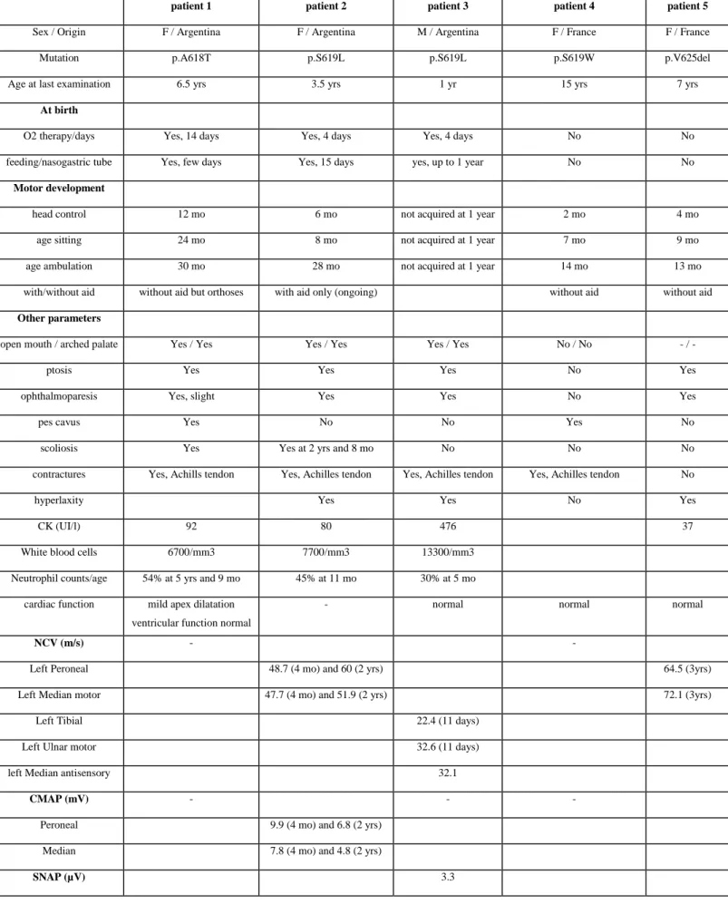
Table 1: Summary of clinical data
F: Female; M: Male, yrs: years, mo: months. -: no information. NVC: nerve conduction
velocities, CMAP: compound muscle action potential, SNAP: sensory nerve action potential.
Figure 1: Clinical features of patients 1, 2 and 3 with early onset CNM with DNM2 mutations in the PH domain.
A. Position of the DNM2 mutations identified in CNM patients. Mutations are specified on
the predicted protein structure including a tripartite GTPase domain (GTPase), a middle
domain (Middle), a Pleckstrin Homology domain (PH), a GTPase effector domain (GED) and
a Proline rich domain (PRD). The new mutations identified in CNM patients are indicated in
red and the already reported mutations in CNM and CMT patients are indicated blue and
green, respectively. All the variants were numbered according to the same isoform, isoform 1
(Accession number: NP_001005360) since this isoform, which includes the four amino-acids
GEIL at positions 516-519 encoded by exon 13bis, is the major one in human brain, muscle,
and primary cultured myoblasts. B: Patient 1 (mutation p.A618T) at the age of 5 years. C:
Patient 2 (mutation p.S619L) at the age of 2 years. D: patient 3 (mutation p.S619L) at the age
of 6 months. All patients had generalized hypotonia, bilateral facial weakness, ptosis and open
mouth. A severe ophthalmoparesis was present in patient 2 (note position of left eye with an
internal deviation on top left panel). Patient 1 had equinovarus and pes cavus.
Figure 2: Histological features in DNM2-related CNM patient
Hematein-Eosin staining (A,B,C,D), ATPase preincubated at pH 9.4 (E,G,H) or pH 4.2 (F)
on muscle biopsies from patients 1 (deltoid biopsy at the age of 5 years), 2 (quadriceps biopsy
at the age of 1 year), 5 (quadriceps biopsy at the age of 2 years) and in a deltoid muscle
biopsy performed at the age of 2 years in one healthy patient (control). Representative fields
of DNM2-related CNM patients showing internal nuclei (A,B,C), type 1 fibre predominance
and hypotrophy (E,F,G) and distorted myofibrillary structure (I,J,K) in comparison with
control (D,H,L). M: electron microscopy for patient 1 showed an halo devoid of organelles
around central nucleus and an appearance of radiating sarcoplasmic strands. N: enlargement
Table 1: Summary of clinical data
patient 1 patient 2 patient 3 patient 4 patient 5
Sex / Origin F / Argentina F / Argentina M / Argentina F / France F / France Mutation p.A618T p.S619L p.S619L p.S619W p.V625del Age at last examination 6.5 yrs 3.5 yrs 1 yr 15 yrs 7 yrs
At birth
O2 therapy/days Yes, 14 days Yes, 4 days Yes, 4 days No No feeding/nasogastric tube Yes, few days Yes, 15 days yes, up to 1 year No No
Motor development
head control 12 mo 6 mo not acquired at 1 year 2 mo 4 mo age sitting 24 mo 8 mo not acquired at 1 year 7 mo 9 mo age ambulation 30 mo 28 mo not acquired at 1 year 14 mo 13 mo with/without aid without aid but orthoses with aid only (ongoing) without aid without aid
Other parameters
open mouth / arched palate Yes / Yes Yes / Yes Yes / Yes No / No - / -
ptosis Yes Yes Yes No Yes
ophthalmoparesis Yes, slight Yes Yes No Yes
pes cavus Yes No No Yes No
scoliosis Yes Yes at 2 yrs and 8 mo No No No contractures Yes, Achills tendon Yes, Achilles tendon Yes, Achilles tendon Yes, Achilles tendon No
hyperlaxity Yes Yes No Yes
CK (UI/l) 92 80 476 37
White blood cells 6700/mm3 7700/mm3 13300/mm3 Neutrophil counts/age 54% at 5 yrs and 9 mo 45% at 11 mo 30% at 5 mo
cardiac function mild apex dilatation ventricular function normal
- normal normal normal
NCV (m/s) - -
Left Peroneal 48.7 (4 mo) and 60 (2 yrs) 64.5 (3yrs) Left Median motor 47.7 (4 mo) and 51.9 (2 yrs) 72.1 (3yrs)
Left Tibial 22.4 (11 days) Left Ulnar motor 32.6 (11 days) left Median antisensory 32.1
CMAP (mV) - - -
Peroneal 9.9 (4 mo) and 6.8 (2 yrs) Median 7.8 (4 mo) and 4.8 (2 yrs)
deep tendon reflexes present present weak at 6 mo present (2 yrs) absent (8 yrs)
present (3 yrs) absent (7 yrs)