The adrenal cortex exhibits sexually dimorphic tissue renewal and disease development
141
0
0
Texte intégral
(2) THÈSE DE DOCTORAT Dimorphisme sexuel du cortex surrénalien dans le renouvellement cellulaire et les pathologies Anaëlle GRABEK Institut de Biologie Valrose Présentée en vue de l’obtention du grade de docteur en Sciences d’Université Côte d’Azur Dirigée par : Andreas Schedl / MarieChristine Chaboissier Soutenue le : 16/04/2019. Devant le jury, composé de : Marie-Christine Chaboissier, Directeur de recherche, Université Côte d’Azur Nick Hastie, Professeur, Université d’Edimbourg Thomas Lamonerie, Professeur, Université Côte d’Azur Serge Nef, Professeur, Université de Genève Andreas Schedl, Directeur de recherche, Université Côte d’Azur Amanda Swain, Directeur de recherche, Université de Londres.
(3)
(4) Dimorphisme sexuel du cortex surrénalien dans le renouvellement cellulaire et les pathologies Jury Président du jury Thomas Lamonerie, Professeur, Université Ĉte d’Azur Rapporteurs Nick Hastie, Professeur, Université d’Edimbourg Amanda Swain, Directeur de recherche, Université de Londres Examinateur Serge Nef, Professeur, Université de Genève Invité Antoine Martinez, Directeur de recherche, Université Clermont Auvergne. 1.
(5) 2.
(6) En français Titre Dimorphisme sexuel du cortex surrénalien dans le renouvellement cellulaire et les pathologies.. Résumé Des différences importantes sont observées dans la prévalence des maladies du cortex surrénalien entre hommes et femmes. En particulier les femmes ont jusqu'à 6 fois plus de probabilités de développer un cancer de la corticosurrénale que les hommes. Malgré cela, les différences et similitudes du renouvellement cellulaire dans le cortex surrénalien ont jusqu’à présent été peu étudiées. La glande surrénale est un organe essentiel au maintien de l’homéostasie corporelle qu’elle régule grâce à la sécrétion d’hormones stéroidiennes. Elle est composée d’une médulla et d’un cortex qui est sous-divisé en plusieurs couches concentriques, de la plus externe à la plus interne : la zone glomérulée, la zone fasciculée et enfin la zone X chez la souris. Le cortex surrénalien est de plus enrobé d’une capsule et d’un mésenchyme. Tout au long du stade adulte, il subit un renouvellement cellulaire important avec des cellules progénitrices localisées au sein de la zone glomérulée qui sont capables de se renouveler et de générer de nouvelles cellules stéroidogéniques qui vont ensuite migrer de façon centripète puis entrer en apoptose lorsqu’elles atteignent la médulla. Au cours de mes travaux de thèse, j’ai pu mettre en évidence la présence d’un renouvellement cellulaire important du cortex surrénalien et sexuellement dimorphe avec un cortex renouvelé en 3 mois chez la femelle et environ 9 mois chez le male. De plus, alors que le renouvellement cellulaire du male repose sur les cellules progénitrices présentes dans la zone glomérulée, chez les femelles, le cortex recrute également des cellules de la capsule. Enfin, des expériences avec des modèles d’inversion de sexe, des gonadectomies et des traitements à la dihydrotestostérone ont pu identifier un effet inhibiteur des androgènes sur le recrutement et la prolifération des cellules progénitrices de la capsule. En parallèle, j’ai étudié un modèle murin exprimant Rspo1 de façon ectopique dans les cellules stéroidogéniques. L’analyse de ce modèle a permis de mettre en évidence le développement, chez ces souris, d’hyperplasie précoce du cortex surrénalien et le développement de tumeurs corticosurrénaliennes avec l’âge, phénotype qui s’est avéré être nettement plus prononcé chez les femelles à partir de la puberté. De plus, les males présentent. 3.
(7) des signes de dégénération cellulaire dès 6 semaines d’âge avec l’apparition de cellules vacuolisées. Ensemble, ces résultats mettent en avant de profondes différences entre mâles et femelles dans l’homéostasie cellulaire du cortex surrénalien et le développement de phénotypes, différences qui pourraient être à l’origine des prévalences plus importantes chez les femmes du développement des maladies de la corticosurrénale.. Mots-clés : Glande surrénale, renouvellement cellulaire, hormones sexuelles, cellules progénitrices. 4.
(8) In English Title The adrenal cortex exhibits sexually dimorphic tissue renewal and disease development.. Abstract Significant differences between men and women are observed in terms of prevalence to adrenal cortical diseases. In particular, women are up to 6 times more likely than men to develop tumors of the adrenal cortex. Despite those striking disparity, differences and similarities in adrenal cortex renewal between sexes have yet to be investigated. The adrenal gland is a vital organ maintaining body homeostasis through the secretion of steroid hormones. It is composed of two endocrine glands, the medulla and the cortex, that is further divided into concentric zones, from the outer to the inner: the zona glomerulosa, the zona fasciculata and a zona reticularis in human or an X-zone in mice. The cortex is surrounded by a capsule that is further enveloped by a layer of mesothelial cells. In the adult stage, the adrenal cortex undergoes constant cellular renewal. Progenitor cells, located in the outer cortex, proliferate and give rise to new steroidogenic cells that centripetally migrate within the cortex, transdifferentiating along the way until they reach the border with the medulla and enter into apoptosis. During my thesis, I have shown that cell renewal in the adrenal cortex is highly sexually dimorphic with the female cortex being fully replaced in less than 3 months, while in males tissue renewal takes about 9 months. In addition, while male cortical renewal relies on progenitors located in the zona glomerulosa, the female cortex also recruits cells from the capsule to replenish the steroidogenic cell population. Finally, using sex reversal mouse models, gonadectomy and dihytrotestosterone treatments, I have identified an inhibitory role of male androgens on the proliferation and recruitment of capsular cells. In parallel, I have also studied a mouse model ectopically expressing Rspo1 in steroidogenic cells. Careful analysis of this model has evidenced the development of cortical hyperplasia and tumors in aging mice, a phenotype that was found to be significantly more pronounced in female than male. In addition, while male adrenals show signs of cellular degeneration in the adrenal cortex as early as 6 weeks of age, this histology is only observed in females of 12 months of age. Together, these results highlight profound differences between. 5.
(9) male and female in tissue homeostasis and phenotype development. These differences could offer an explanation to the unequal prevalence to adrenal diseases in men and women.. Keywords: Adrenal gland, tissue homeostasis, sex hormones, progenitor cells. 6.
(10) Dedicated to my grand-father, Pierre GRABEK.. 7.
(11) 8.
(12) Table of content Dimorphisme sexuel du cortex surrénalien dans le renouvellement cellulaire et les pathologies ........................................................................................... 0 En français .................................................................................................................... 3 Titre .................................................................................................................................... 3 Résumé ............................................................................................................................... 3 In English ...................................................................................................................... 5 Title ..................................................................................................................................... 5 Abstract .............................................................................................................................. 5 Table of content ............................................................................................................ 9 - INTRODUCTION - ....................................................................................................... 11 The adrenal gland ............................................................................................................. 13 The developing adrenal gonadal primordium.................................................................. 15 Development of the adrenal cortex ......................................................................................... 15 Gonadal development and sex determination ........................................................................ 17 Factors involved in adrenal gonadal development .................................................................. 18 WT1 ...................................................................................................................................... 18 GATA 4/6 .............................................................................................................................. 19 PBX1 ..................................................................................................................................... 19 SF1 ........................................................................................................................................ 20 DAX1..................................................................................................................................... 22. The adult adrenal cortex .................................................................................................. 24 Hormonal production ............................................................................................................... 24 The HPA axis......................................................................................................................... 24 The renin-angiotensin-aldosterone system ......................................................................... 25 Steroid synthesis .................................................................................................................. 26 Disease of the adrenal cortex................................................................................................... 27 Congenital adrenal hyperplasia ........................................................................................... 27 Addiso ’s disease ................................................................................................................. 28 Cushi g’s s dro e ............................................................................................................. 28 Adrenocortical tumors ......................................................................................................... 28. 9.
(13) Homeostasis of the adrenal cortex .......................................................................................... 29 Adrenal cortex homeostasis model ..................................................................................... 29 Signaling pathways involved in formation and renewal of the adult cortex ....................... 30 Sonic Hedgehog signaling ................................................................................................ 30 Canonical WNT signaling ................................................................................................. 33 Canonical WNT signaling ............................................................................................. 33 The R-spondin protein family ...................................................................................... 35 WNT/β-catenin signaling in the adrenal cortex .......................................................... 37 cAMP/PKA signaling......................................................................................................... 39 Hippo signaling ................................................................................................................ 40. Scientific context .............................................................................................................. 42 Experimental models........................................................................................................ 44 CRE-LoxP system ...................................................................................................................... 44 Lineage tracing ......................................................................................................................... 45 Sex reversal .............................................................................................................................. 46 RSPO1 Gain of function ............................................................................................................ 48. Aim of the study ............................................................................................................... 51 - RESULTS - .................................................................................................................. 53 Part : Paper. A droge s regulate se -specific ste. cell activit of the adre al corte. .......................................................................................................................................... 55 Part : Paper. Rspo1 over expression induces sex-specific hyperplasia and tissue. degeneratio i the adre al corte ................................................................................ 81 - DISCUSSION - ...........................................................................................................109 Conclusions..................................................................................................................... 111 Perspectives ................................................................................................................... 113 Abbreviations ............................................................................................................117 Nomenclature ............................................................................................................119 Bibliography...............................................................................................................121 Acknowledgements ....................................................................................................137. 10.
(14) - INTRODUCTION -. 11.
(15) 12.
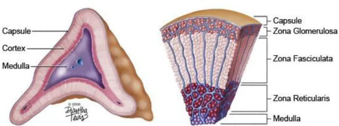
(16) The adrenal gland The adrenal gland is responsible of maintaining body homeostasis through regulation of various processes such as blood pressure, inflammation, stress and metabolism. Despite this important role, the adrenal gland is one of the least studied organs of the human body, it is nevertheless one of the most important one as the absence of adrenals is lethal. The very first description of adrenal tissue was made in the XVI century by Bartholomeus Eustachius (Hiatt & Hiatt, 1997). In 1656, Wharton published "a description of the glands of the entire body," in which he describes a nerve connecting to the adrenal and so suggests a neuroendocrine role for the adrenal gland (Leoutsakos & Leoutsakos, 2008). Almost two hundred years later, a French naturalist and zoologist, Baron George Cuvier, describes two morphologically distinct zones within the gland that will later be known as the cortex and the medulla. However, we had to wait until 1855 to start understanding the vital role of the adrenal glands when Addison first identified patients with severe symptoms resulting from degenerating adrenals. His name was later given to the adrenal insufficiency pathology known as Addison’s disease (Leoutsakos & Leoutsakos, 2008). Another adrenal malfunction, featuring hypercortisolism, was described by Harvey Cushing from whom the disease inherited the name: Cushing’s Syndrome. In 1866, Julius Arnold further described the adrenal cortex and its cellular organization in the different concentric zones (Walczak & Hammer, 2015). Finally, the in vitro synthesis of adrenal hormones was first achieved by Edward Calvin Kendall, Tadeus Reichstein and Philip Showalter Hench, who were awarded with the Nobel Prize in Physiology or Medicine in 1950 (Welbourn, 1991). The adrenal gland maintains its homeostatic role on the entire body by secretion of steroid hormones (and catecholamines). Such a tissue has so far been found exclusively in vertebrates highlighted by the specific expression of adrenal and gonad specific steroid receptors, the most ancient being the estradiol-receptor (ER) (Baker, 2003). We are born with two adrenals, one on each side located above the kidney. They are part of the Hypothalamus-Pituitary-Adrenal (HPA) axis and are highly vascularized to receive stimulating hormones from and release secreted hormones to the whole organism. The adrenal gland is composed of two distinct endocrine glands, the medulla in the center and the cortex surrounding the medulla (Figure 1. Left). Each gland is characterized by its developmental origin as well as the specific set of hormones they are producing. The medulla is derived from the neural crest (ectodermal origin) during development and is composed of chromaffin cells. When exposed. 13.
(17) to stress, these cells secrete catecholamines (such as adrenaline and noradrenaline) to induce the “flight or fight” response. The adrenal cortex is derived from the intermediate mesoderm (mesodermal origin) from which the kidneys and gonads also originate. The adrenal cortex is surrounded by a capsule, itself covered by a mesothelial cell layer, and is subdivided into three concentric layers, from the capsule to the medulla: the zona Glomerulosa (zG), the zona Fasciculata (zF) and either the zona Reticularis (zR) in human or the X-zone in mice (Figure 1. Right).. Figure 1 Schematic drawing of the adrenal gland and the adrenal cortex. Left : Schematic transection of an adrenal gland showing the inner medulla, the surrounding cortex and the envelopping capsule. Right : Detailed schematic of a section of the human adrenal cortex highlighting the different concentric zones.. 14.
(18) The developing adrenal gonadal primordium Development of the adrenal cortex The adrenal cortex and the gonads share a common precursor during embryogenesis. An adrenal-gonadal primordium (AGP) arises from the thickening of the coelomic epithelium of the urogenital ridge at E9.0 in the mouse embryo and is recognized by the expression of the Steroidogenic Factor-1 (Sf1/Ad4bp/Nr5a1), Wilms’ tumor suppressor (Wt1) and Gata4 (Figure 2) (Bandiera et al., 2013; Hatano, Takakusu, Nomura, & Morohashi, 1996; Stallings et al., 2002). Around E10.5, the adrenal-gonadal primordium separates and gives rise to the gonadal primordium and the adrenocortical primordium. By E11, the two organs are fully distinct and the adrenal primordium becomes the fetal adrenal cortex highlighted by the expression of the Fetal Adrenal Enhancer (FAdE) (Figure 2). SF1 has been shown to be strictly required for adrenal cortex and gonad development and Sf1 knockout mice display in agenesis of the two organs (Majdic et al., 2002). Furthermore, the dose of Sf1 has been proven to be important for normal adrenal development. In Sf1+/- animals, the adrenal anlagen were dramatically reduced in size while the gonads were not affected (Bland, Fowkes, & Ingraham, 2004). Consequently, it is thought that the cells expressing higher levels of Sf1 give rise to the adrenal anlagen while the other SF1+ cells, expressing lower Sf1 level, will form the gonads. In addition to SF1, WT1 as well as CITED2 are two transcription factors that have been shown to be crucial for adrenal development and to precede Sf1 expression. Indeed, it is thought that CITED2 acts as a cofactor of WT1 in order to increase Sf1 levels in the AGP and drives the specific differentiation of the adrenal tissue (P. Val, Martinez-Barbera, & Swain, 2007). SF1 is also self-enhancing its expression by binding to the fetal adrenal enhancer located on its intron 4 (FAdE). DAX-1 has been identified as a repressor of FadE, increasing the difference in Sf1 expression between the gonadal and adrenal primordium (Hoivik et al., 2011). As SF1 is maintained throughout the embryonic and adult life, Wt1 expression in the adrenal anlagen is no longer required for its expression and in fact gets quickly abolished after AGP separation (Bandiera et al., 2013). At E12,5, neural crest cells invade the fetal adrenal cortex, migrate to the inner part of the organ and differentiate into chromaffin cells that establish the medulla (Figure 2). Invasion of the chromaffin-to-be cells is followed by encapsulation of the fetal adrenal cortex by WT1+ mesenchymal cells at E14.5 (Bandiera et al., 2013). E14.5 also marks the limit at which fetal cortical cells can become precursors of the adult cortex cell and switch off FAdE expression (Morohashi 2011). By E17.5, only a small proportion of cells are still expressing FAdE and are. 15.
(19) exclusively located at the border with the medulla suggesting that as the adult cortex grows, it takes over the fetal adrenal cortex that becomes restricted to a zone next to the medulla and form the X-zone (Morohashi & Zubair, 2011). However, complete zonation (differentiated zonae glomerulosa and fasciculata) of the adrenal cortex is only fully acquired after birth (Bandiera et al., 2013; Freedman et al., 2013). The final maturation step of the adrenal cortex occurs at puberty. While females maintain an X-zone throughout adulthood until the first pregnancy or aging, the X-zone in males regresses at puberty in response to the rise in androgen secretions (Figure 3) (Hershkovitz, Beuschlein, Klammer, Krup, & Weinstein, 2007; Xing, Lerario, Rainey, & Hammer, 2015).. Figure 2 Schematic representation of adrenal development from the urogenital ridge in the mouse. E=Embryonic day ; P= Postnatal day. Modified from Pihlajoki et al. 2015 (Pihlajoki, Dörner, Cochran, Heikinheimo, & Wilson, 2015).. Figure 3 Schematic representation of the X-zone regression at puberty in the male while it is maintained in female.. 16.
(20) Gonadal development and sex determination As its name suggests, the Adrenal-Gonadal Primordium also gives rise to the gonadal anlagen. From the AGP, the cells expressing lower level of Sf1 separate from the adrenal primordium and give rise to the bipotential gonadal primordium. The primordium is composed of precursor cells that can either become steroid-producing cells or supportive cells (Sertoli cells in male or granulosa cells in female). In addition, primordial germ cells that maintain an undifferentiated state, but do not derive from the intermediate mesoderm, migrate into the developing gonad (Dagmar Wilhelm, Yang, & Thomas, 2013). Two antagonistic pathways in male and female orchestrate sex determination (Figure 4). In females (XX), Wnt4 and Rspo1 expression triggers the level of active β-catenin to increase. βcatenin inhibits Sox9 and male differentiation allowing the development of the ovaries. In males (XY), SRY (sex determining region of the Y chromosome) is transcribed from the Sry gene located on the Y chromosome. Along with SF1, SRY drives the expression of Sox9 inducing PGD and FGF signaling to induce testis differentiation and inhibits β-catenin and ovarian differentiation (Figure 4). The timing at which Sox9 is expressed is crucial for testis development, as a delay causes β-catenin to take over, inhibiting testicular differentiation and driving ovarian differentiation instead (Sekido & Lovell-Badge, 2008). In my research, I have made use of genetic sex-reversal models to investigate the role of the sex chromosomes in the adrenal cortex. The absence of SOX9 in an XY animal (Sf1:Cre(Tg/+); Sox9(flox/flox)) causes gonadal differentiation of the gonads and genitalia towards a female phenotype (Lavery et al., 2011). Reversely, ectopic expression of Sox9 in an XX animal (Wt1:Sox9) triggers male phenotype development (Gregoire et al., 2011; V. P. I. Vidal, Chaboissier, & Schedl, 2002).. 17.
(21) Figure 4 Schematic representation of sex determination and factors involved in the mouse (Kashimada & Koopman, 2010).. Factors involved in adrenal gonadal development WT1 The Wilms’ tumor suppressor 1 (WT1) is a zinc finger transcription factor acting upstream of Sf1 and Dax1 in adrenal-gonadal differentiation. Wt1 knockout mice do not only fail to develop the adrenal gland and the gonad but also lack kidneys demonstrating a role in early differentiation and survival of the uro-genital ridge (Hastie, 2017). Two splicing variants of Wt1 have been shown to be important in the urogenital ridge development: One of the variant contains the lysine-threonine-serine amino acids (+KTS), the other one lacks those (-KTS). In the gonad, the two isoforms have specific roles. For example, the –KTS variant protein seems to be essential to prevent apoptosis in the gonad while the +KTS variant control Sry expression level (Bradford et al., 2009; Hammes et al., 2001). In the adrenal cortex, Wt1 in parallel to Gata4 expression are switched off soon after the separation of the AGP into the adrenal primordium and the gonadal primordium. Forced expression of a Wt1-KTS isoform in the developing adrenal resulted in two distinct cell population in the fetal adrenal cortex at E12,5 including a low Wt1, high Sf1 cell population and a high Wt1, low Sf1, Gata4+ second 18.
(22) population, suggesting that WT1-KTS prevents differentiation of the early adrenal progenitor into steroidogenic lineages (Bandiera et al., 2013). In the adult, a small subset of cells surrounding the capsule displays low levels of Wt1 expression and these cells maintain a progenitor state to steroidogenic lineages. Upon gonadectomy, those cells have been identified to give rise to gonadal-like cells expressing Wt1 and Gata4 as well as low level of Sf1 (Bandiera et al., 2013).. GATA 4/6 GATA binding protein (GATA) 4 and 6 are two transcription factors that have been identified to have important roles in adrenal gonadal development and maintenance. While GATA4 has been shown to be dispensable for adrenal development and adult adrenal maintenance, it is required for initial gonadal differentiation and maturation (Hu, Okumura, & Page, 2013; Kiiveri et al., 2002; Tevosian et al., 2002). Hu et al., highlighted an important role of GATA4 in initiating thickening of the coelomic epithelium at early developmental stages of the genital ridge as well as activation of the early gonadal markers Sf1 and Lhx9 (Baker, 2003; Hu et al., 2013). GATA6 plays a role in the development and maintenance of the adrenal cortex, as deletion of Gata6 resulted in significantly smaller adrenals (Hu et al., 2013; Kiiveri et al., 2002; Tevosian et al., 2002). In addition homozygous double knockout for Gata4 and Gata6 mutant showed complete adrenal agenesis (Tevosian et al., 2002).. PBX1 Pre-B-cells transcription factor 1 (PBX1) is a TALE class homeodomain transcription factor (Lichtenauer et al., 2007; Schnabel, Selleri, & Cleary, 2003). PBX1 can act as a co-activator or co-repressor by binding to other TALE homeodomain proteins including MEIS and PREP to form heterodimers (Lichtenauer et al., 2007). These complexes further interact with HOX proteins, forming heterotrimers and allowing for region-specific DNA binding. Ubiquitous deletion of Pbx1 resulted in embryonic lethality at E15/16 and adrenal agenesis associated with low levels of Sf1 expression demonstrating an important role of PBX1 in the development of the adrenal cortex (Schnabel et al., 2003). In addition, gonads failed to differentiate, yet SF1+ cells were observed in the defective gonads in the Pbx1-/- mutant. According to these results, it was suggested that PBX1 plays an important role in the fate of the adrenal cortex, promoting expansion of the adrenal gonadal primordium through cell proliferation, as well as enhancing Sf1 expression (Schnabel et al., 2003).. 19.
(23) SF1 Steroidogenic factor 1 (SF1/Ad4BP/NR5A1) is the master regulator of steroidogenesis encoded by the nuclear receptor subfamily 5 group A member 1 (NR5A1) gene. SF1 is a transcription factor highly expressed in all steroidogenic tissues including the adrenal cortex, gonads, and the pituitary (Ozisik, Achermann, Meeks, & Jameson, 2003; Parker & Schimmer, 1997). In the embryo, SF1 expression is observed in the uro-genital ridge as early as E9.75 in the mouse and is the earliest marker of the adrenal-gonadal primordium. It is also seen during embryogenesis in the fetal spleen and the hypothalamic primordium (Schimmer & White, 2010). In the adult, Sf1 is expressed in the entire adrenal cortex, the Leydig and Sertoli cells of the testis, the theca and cells in the ovary and the gonadotrope cells (secreting LH and FSH) in the pituitary. Sf1 expression is also observed in the ventromedial part of the hypothalamus, some restricted epithelial structures of the spleen and a few neurons in the hippocampus (Schimmer & White, 2010). In the absence of SF1, mice die perinatally from adrenal insufficiency (Luo, Ikeda, & Parker, 1994). They also lack gonads and develop a male-to-female sex reversal of the external genitalia and of the internal urogenital tracts. This phenotype highlights the essential role of SF1 in adrenal and gonadal development (Buaas, Gardiner, Clayton, Val, & Swain, 2012; Ferraz-deSouza, Lin, & Achermann, 2011; Luo et al., 1994; Schimmer & White, 2010). SF1 is a 461 amino acids proteins of 7 exons that contains a DNA-binding domain (DBD) including two Cys2-Cys2 zinc fingers and a FTZ-F1 box that cooperates to specifically bind with strong specificity, affinity and stability to DNA as a monomer (Figure 5) (Hoivik, Lewis, Aumo, & Bakke, 2010). The FTZ-F1 box is followed by a hinge domain harboring an Activation Function 1 (AF-1), a phosphorylation site and sumoylation sites, essential for the transcriptional activity of SF1. Finally, a Ligand Binding Domain (LBD) harbors an AF-2 domain on its C-terminal that is required for co-factor interactions and transcriptional activity (Figure 5) (Hoivik et al., 2010).. 20.
(24) Figure 5 Schematic drawing of the SF1 protein domains. DBD: DNA Binding Domain; AF: Activation Function; LBD: Ligand Binding Domain; Zn: zinc fingers; NLS: Nuclear Localization Signal; Pro-rich: Proline rich region; FTZ-F1: Fushi-tarazu factor-1 box, P/T/A; DNA binding boxes (Hoivik et al., 2010).. SF1 activates genes required for cholesterol metabolism and steroidogenesis but also has an important role in inducing genes involved in sex determination and differentiation (Hoivik et al., 2010). More specifically, SF1 binds to the promoters of genes encoding cholesterol transport factors such as the steroidogenic acute regulatory protein (StAR) and the sterol carrier protein 2 (SCP2), but also cholesterol metabolizing enzymes for P450 steroidogenic enzymes CYP11A, CYP11B, CYP21 as well as 3β steroid dehydrogenase (3βHSD) and Akr1-b7 (Aigueperse et al., 2001; Caron, Clark, Ikeda, & Parker, 1997; Hoivik et al., 2010; Mclean et al., 2001; Parker & Schimmer, 1997; Sugawara, Holt, Kiriakidou, & Strauss, 1996; Pierre Val, Lefrançois-Martinez, Veyssière, & Martinez, 2003; M. Zubair, Ishihara, Oka, Okumura, & Morohashi, 2006). In line with the importance of SF1 in sex determination and gonad development, Sox9 has been shown to be a direct transcriptional target of SF1 and so have Amh, the FSH receptor and Inha, genes specifically necessary in testis differentiation (Buaas et al., 2012; Schimmer & White, 2010). In addition, the regulation of Dax1 and Sf1 is tightly intertwined. Indeed, Dax1 is activated by SF1 and, in the adrenal, has been shown to have a negative feedback regulation on SF1 transcriptional activity of genes encoding glucocorticoids synthesis enzymes in presence of increased glucocorticoid levels and suggested to have a crucial role in maintaining progenitor capacities through inhibition of steroidogenesis (Hoivik et al., 2010; Kim et al., 2009). Several factors have been identified to bind to the Sf1 promoter and regulate its expression (Figure 6). Sf1 presents an E-box binding domain located in its basal promoter that has been shown to be essential for its regulation. It is the binding site for the nuclear transcription factor Y (CBF), ubiquitous transcription factors, stimulatory protein 1 and 3 (SP1 and SP3) and upstream stimulatory factors (USF) (Figure 6) (Hoivik et al., 2010). POD1 (Capsulin/TCF21). 21.
(25) has also been identified to interact with the basal promoter with a repressive action on the promoter activity (Figure 6) (Hoivik et al., 2010; Pierre Val et al., 2003; M. Zubair et al., 2006). In addition, upstream and downstream regulatory regions have been evidenced to have additional roles in Sf1 regulation. In the fetal adrenal, the Fetal Adrenal Enhancer (FAdE) has been discovered in intron 4 to serve as a positive self-regulator for Sf1 expression and ensures continuous Sf1 expression. However, the FadE activity only starts after Sf1 expression has been initiated and additional factors must initially activate Sf1 expression before it becomes selfregulated (M. Zubair et al., 2006; Mohamad Zubair, Oka, Parker, & Morohashi, 2009). At early developmental stages, HOX proteins are described to interact with PBX1 to bind the FadE binding site for the PBX1-HOX complex located right next to a PREP-PBX1 binding site allowing enhanced transcription. HOX proteins have been shown to specify anterior/posterior identity in the mesoderm and Pbx1 knockout mice failed to develop adrenal glands and differentiated gonads highlighting important roles for those proteins in initiating early differentiation steps of the adrenal-gonadal primordium (M. Zubair et al., 2006). In the AGP, Sf1 expression is also thought to be regulated by WT1 binding to the E-box site and further enhanced by interaction of CITED2 with WT1 with a binding site located upstream of the basal promoter and to initiate adrenal specific differentiation (Figure 6) (P. Val et al., 2007; D. Wilhelm & Englert, 2002). With regards to the gonads, tissue specific activators such as GATA4 and LHX9 (essential factor in germ cell proliferation) have also been described to induce Sf1 expression albeit at a moderate level for GATA4 (Figure 6). Finally, in vitro analysis evidenced SOX9 to have a potential regulatory action on Sf1 expression (Birk et al., 2000; Hoivik et al., 2010; Pierre Val et al., 2003).. Figure 6 Transcription factors binding to the Sf1 basal promoter (Hoivik et al., 2010). DAX1 DAX1, dosage sensitive sex reversal, adrenal hypoplasia congenital, critical region of the X chromosome, gene 1, is another protein encoded by an orphan nuclear receptor family gene:. 22.
(26) NR0B1 (Niakan & McCabe, 2005). NR0B1 is encoded by two exons. The encoded protein of 470 amino acids contains a DNA binding domain and a Ligand Binding Domain (Li et al., 2010; Niakan & McCabe, 2005). Three structures are highly conserved within the nuclear receptor surperfamily and include the region I that encompasses the DNA binding domain (DBD) and the region II and III that are found on the ligand binding domain (LBD) (Niakan & McCabe, 2005). In comparison to other nuclear receptors, DAX1 has the particularity to harbor on its Cterminal three repeats of a 70 amino acid cysteine-rich sequences that include a LXXLL motif instead of the zing-finger DNA-binding domain traditionally found in nuclear receptors. This specificity is thought to confer to DAX1 its transcriptional regulation properties (Lalli, 2014; Niakan & McCabe, 2005). Dax1 has been shown to be expressed as early as E9 in the AGP as well as in the developing gonad, adrenal cortex, anterior pituitary and hypothalamus during embryogenesis. In the adult, its expression is restricted to the adrenal cortex, the gonadotropes (anterior pituitary), the ventro-medial hypothalamus, Leydig and Sertoli cells in the testis and the granulosa and theca cells of the ovary (Li et al., 2010). Interestingly, Dax1 and Sf1 have similar expression patterns. DAX1 has been shown to principally have a negative transcriptional activity and in particular to repress genes that are activated by SF1, including the steroidogenic enzymes and StAR (Lalli, 2014; Niakan & McCabe, 2005). In addition, SF1 induces Dax1 expression that in a feedback loop negatively regulates steroidogenesis. It has been suggested that DAX1 can repress FAdE to transition into the adult adrenal cortex (Lalli, 2014). This was further confirmed in Dax1 knockout mice in which the X-zone fails to regress (Iyer & McCabe, 2004). Even though the specific implications are not known to date, WT1 has been suggested to regulate Dax1 during early developmental stages (Lalli, 2014). In the adult, Dax1 is further enhanced by WNT signaling and glucocorticoids, but repressed by ACTH and its localization in the adrenal cortex is restricted to the subcapsular zone. Finally, DAX1 has been suggested as an essential component to maintain progenitor properties within the zG as it is repressing steroidogenesis and preventing differentiation (Kim et al., 2009). In human, DAX1 mutations have been associated with the cytomegalic form of adrenal hypoplasia congenital (AHG). In the affected patients, adult adrenals lack the mature cortex and the residual adrenal cortex is composed of large vacuolated cells. To this date, it has not been possible to recreate the disease in a mouse model (Iyer & McCabe, 2004; Scheys, Heaton, & Hammer, 2011).. 23.
(27) The adult adrenal cortex Hormonal production The HPA axis The adrenal cortex is communicating with the body via the secretion of steroid hormones. Steroid hormones are only produced by the gonads and the adrenal cortex and encompass the sex hormones (androgens, estrogens, progestins) that regulate the reproductive functions and the adrenal corticosteroids (glucocorticoids and mineralocorticoids) maintaining body homeostasis (Morohashi, 1997). The hormonal production in the adrenal cortex is under the control of two distinct systems: the Hypothalamus-Pituitary-Adrenal (HPA) axis and the ReninAngiotensin axis. Under stress conditions, the hypothalamus produces corticotropin-releasing hormone (CRH) that is sensed by the pituitary and induces adrenocorticotropic hormone (ACTH) release by the corticotropes. ACTH is transported through the bloodstream and induces production of glucocorticoids in the zona fasciculata (Figure 7). Glucocorticoids act on many organs and participate in the regulation of major homeostatic processes such as metabolism and immune response to inflammation. As glucocorticoids are released, they negatively (feedback loop) affect the hypothalamus and pituitary to decrease the secretion of CRH and ACTH respectively (Figure 7) (Hiller-Sturmhöfel & Bartke, 1998).. 24.
(28) Figure 7 Schematic representation of the hypothalamus-pituitary-adrenal axis. Upon stress condition, the hypothalamus secretes corticotrophin-releasing hormone that induces the pituitary to release adrenocorticotropic hormone. ACTH is then sense by the adrenal which in turn synthesize cortisol to cope with stress and negatively feedback to the hypothalamus and pituitary. CRH: corticotropin releasing hormones; ACTH: adrenocorticotropic hormone. (Hiller-Sturmhöfel & Bartke, 1998).. The renin-angiotensin-aldosterone system The Renin-Angiotensin-Aldosterone system (RAAS) is an essential system maintaining fluid and blood pressure homeostasis that is activated upon low sodium conditions. In response, the kidney releases renin that cleaves its substrate angiotensinogen to form Angiotensin I (ANGI) (Figure 8). Highest expression of Angiotensin Converting Enzyme (ACE) is found in the lung and this enzyme further catalyzes ANGI into its active form: Angiotension II (ANGII). In the final step of the RAAS, ANGII acts on the adrenal zona glomerulosa to stimulate aldosterone production. Aldosterone then binds to the mineralocorticoids receptor in the kidney and increases sodium retention and water reabsorption as well as potassium secretion resulting in an increase in blood volume and subsequently blood pressure (Figure 8) (Xanthakis & Vasan, 2013).. 25.
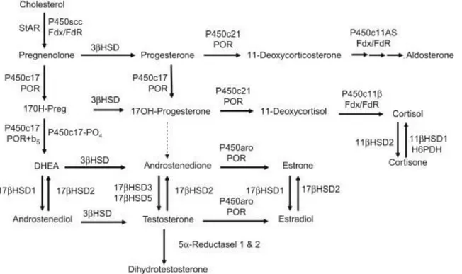
(29) Figure 8 Scheme of the Renin-Angiotensin-Aldosterone system. In case of low blood pressure, the kidney releases Renin that convert angiotensinogen into Angiotensin I that is further transformed into Angiotensin II, its active form. Angiotensin II travels through the blood and signal to the adrenal cortex to synthesize aldosterone inducing water reabsorption and salt retention that create an increase in blood pressure.. Steroid synthesis All steroids are derived from cholesterol. Cholesterol is transported by low-density lipoprotein from the bloodstream to the Outer Mitochondrial Membrane (OMM) and is stored in the OMM until the Steroidogenic acute protein STaR transfers the cholesterol from the outer membrane to the inner membrane of the mitochondria, making the substrate accessible to the required enzyme, CYP11A1, for catalysis (Figure 9) (Miller, 2007; Turcu & Auchus, 2015). SF1 (NR5A1) is known as the master regulator of steroidogenesis in the adrenal gland. SF1 is activating transcription of the cholesterol side chain cleavage enzyme CYP11A1 that initiates the first step of steroid synthesis, the conversion of cholesterol to pregnenolone, and maintains the differentiated state of the steroidogenic cells (Turcu & Auchus, 2015; Yamazaki, 2014). In order to synthesize aldosterone from pregnenolone, the zG requires the consecutive action of HSD3β2, CYP21A2 and CYP11B2 (Aldosterone Synthase, AS) (Figure 9). In contrast to the zG, the zF needs CYP17A1 to catalyze 17OH-hydroxylation of pregnenolone and progesterone that respectively serve as substrate to HSD3β2 and CYP21A2 before CYP11B1 catalyzes the last reaction leading to cortisol synthesis. CYP17A1 also further cleaves 17-OH-pregnenolone into dehydroepiandrosterone (DHEA) (Figure 9). As mouse zF lacks the CYP17A1 enzyme, they consequently do not produce cortisol, but instead display high levels of corticosterone that is produced from deoxycorticosterone by the 11β-hydroxylase (CYP11B1) (Bremer & Miller, 2014; Miller, 2007; Mullins et al., 2009; Turcu & Auchus, 2015). In the human zona Reticularis,. 26.
(30) adrenal androgens are synthetized by the CYP17 enzyme (Figure 9). In the murine X-zone, Cyp17 is not expressed and the murine adrenal does not produce any androgens. However, the X-zone possesses the 20αHSD enzyme, a progesterone-metabolizing enzyme (Hershkovitz et al., 2007). The functional relevance of this expression is unknown.. Disease of the adrenal cortex. Figure 9 Enzymatic reactions involved in steroid hormone synthesis from cholesterol in human. From cholesterol, StAR initiates cholesterol metabolism by transferring cholesterol from the outer membrane to the inner membrane of the mitochondria where enzymes can access it for catalysis. In the zona glomerulosa, cholesterol is then metabolized into aldosterone, or in the zona fasciculata into corticosterone or cortisol. In human, conversion into androgens occurs in the zona reticularis. Further catalysis is occurring in the gonads, where different enzymes are expressed to produce sex hormones (Bremer & Miller, 2014).. Congenital adrenal hyperplasia As is the case for any tissue, many dysfunctions and deregulations of the adrenal cortex can lead to disease development. Congenital adrenal hyperplasia (CAH) is one of them and is the result of mutation(s) in genes coding for steroidogenic enzyme, such as CYP21A2 encoding for the 21-hydroxylase enzyme in 90% of cases. In about 8% of the cases, the mutation is found in the CYP11B1 gene. In both cases, glucocorticoid synthesis is altered and results in high levels of ACTH and CRH, uncontrolled by the glucocorticoid negative feedback on the hypothalamuspituitary axis, as well as a hyperplasia of the adrenal cortex (Mullins et al., 2009; Turcu & 27.
(31) Auchus, 2015). This increase in ACTH leads to an over-synthesis of pregnenolone and subsequently progesterone and adrenal androgens in human resulting in early puberty in boys and ambiguous genitalia in girls. Additional symptoms include facial hair growth, infertility, irregular period in girls and women. CAH are classified in two different forms, the most severe form is affecting about 1:15000 births worldwide while the milder form affects 1:1000 births (Merke & Kabbani, 2001).. Addison’s disease Addison’s disease, also called adrenal insufficiency or hypoadrenocorticism, is a condition in which the adrenal cortex does not produce enough of the mineralocorticoids and glucocorticoids. In Europe, Addison’s disease affects 1 in 10,000 people and patients show sign of fatigue, nausea, weight lost, increased pigmentation, vitiligo and more. The most predominant cause of Addison’s disease is tuberculosis though, in developed countries, autoimmune diseases represent the commonest underlying reason (Burton, Cottrell, & Edwards, 2015; Vaidya, Chakera, & Dick, 2009).. Cushing’s syndrome Cushing’s syndrome is another disease of the adrenal cortex. It results from overexposure to circulating glucocorticoids, either endogenous (pituitary or adrenal tumor) or exogenous (from steroid medication), for an extended period of time leading to adrenal hyperplasia, as well as metabolic disorders, cardiovascular diseases, osteoporosis, psychiatric disorders and overall, a shorter lifespan (Buliman, Tataranu, Paun, Mirica, & Dumitrache, 2016; Newell-Price, Bertagna, Grossman, & Nieman, 2006). Cushing’s diseases are a subtype of Cushing’s syndromes and represent up to 80% of Cushing’s syndromes. They are fairly rare with 40:1,000,000 people being affected. They are the result of an over secretion of ACTH caused by an ACTH-producing tumors (often benign) in the pituitary, increasing glucocorticoid secretion by the adrenal cortex (Buliman et al., 2016; Lonser, Nieman, & Oldfield, 2017).. Adrenocortical tumors While adrenocortical adenoma - benign tumors of the adrenal cortex - are commonly found in the population (1% to 10%), adrenocortical carcinoma (ACC) are rare, but severe, malignancies affecting between 0.7 and 2 millions patients per year (Libé, 2015). Patients generally present with excessive steroid hormones (40-60%) or secondary effects resulting from the pressure of the growing mass on surrounding tissues (30%), but about 15-20% of patients are accidentally 28.
(32) diagnosed. No effective drugs are currently available on the market and the primary treatment remains surgical removal of the tumor, resulting in a 5-year survival rate lower than 35% (Assié et al., 2014). Surgery can be followed with Mitotane treatment to prevent recurrence of the cancer in cases of metastatic diseases. ACC development is attributed to germline or somatic mutations. 3% to 7% of adult ACCs and 50% to 80% of pediatric ACCs are attributed to LiFraumeni Syndrome, patients presenting with germline mutation of the TP53 gene (Libé, 2015). In addition, mutation in the IGF2 gene leading to its overexpression is encountered in 85% of the adrenal carcinomas (De Fraipont et al., 2005). Exome sequencing further identified altered genes in somatic cells. ZNRF3, a negative regulator of the WNT signaling pathway, was mutated in 21% of ACCs while CTNNB1, a downstream effector of the same pathway was found to be mutated in 16% of ACC with ZNRF3 and CTNNB1 mutations being mutually exclusive. TP53 and CDKN2A, both part of the P53/RB signaling were respectively inactivated in 16% and 11% of the ACC analyzed in the study. Finally, with a lesser occurrence, genes involved in chromatin remodeling were also found to be altered (Assié et al., 2014; Zheng et al., 2016).. Homeostasis of the adrenal cortex Adrenal cortex homeostasis model The different cell types corresponding to each zone are easily identified by their shape and secrete specific hormones as described in “Hormonal secretions”. Surrounding the capsule, a single cell layer expresses Wt1 as well as Rspo1 that is continuous with the mesothelial lining of the abdominal cavity. The capsular cells are spindle shaped and organized in 2 to 3 layers around the cortex. Just below the capsule, the zG cells are clustered in rosette-like structures characterized by a central focal point where all cells are in contact. The zG specifically expresses Cyp11b2 (aldosterone synthase) required for aldosterone production as well as Disabled2 (Dab2), a protein involved in aldosterone secretion, Axin2 and Wnt4, both downstream of the WNT/β-catenin signaling pathway and Shh (Romero et al., 2007). The zF cells are organized in columns and express Cyp11b1 that catalysis the final step of glucocorticoids synthesis. In human, the zR produces androgens but mice lack the zR and present instead an X-zone that is a remnant of the fetal adrenal (see “Adrenal Development”). The X-zone lacks the 17α-hydroxylase (CYP17) enzyme and consequently does not produce androgens. Its physiological function is so far unknown, but the X-zone can be identified by. 29.
(33) the expression of 20α-Hsd, encoding an enzyme catalyzing the reduction of progesterone and 11- deoxycorticosterone (Hershkovitz et al., 2007). The adult adrenal cortex undergoes constant cellular renewal. Several studies have now shown the existence of progenitor-like cells in the outer adrenal cortex that maintain a certain degree of undifferentiated state and are proliferating to give rise to steroidogenic cells of the zG (Figure 10)(Kim et al., 2009). As they centripetally migrate, the steroidogenic cells transdifferentiate into zF cells and start to produce zF specific enzymes such as CYP11B1 and AKR (Figure 10). As they reach the X-zone (nulliparous females) or the medulla (males and parous females), the cortical cells undergo apoptosis and are cleared from the adrenal cortex (Figure 10). Although this has never been evaluated in detail, proliferation and apoptosis appear to be in perfect equilibrium maintaining a constant adrenal cortex size during the adult stage. Specific signaling pathways tightly regulate the homeostasis of the adrenal cortex. In particular, SHH, WNT and PKA signaling are the most important pathways involved in maintaining a functioning adrenal cortex (Walczak & Hammer, 2015).. Figure 10 Model of the adrenal cortex homeostasis. A progenitor population resides in the zG and proliferates to give rise to new steroidogenic cells that convert into zona glomerulosa steroid producing cells. As the cells migrate centripetally, they transdifferentiate into zona fasciculata cells. Finally, once they reach the medulla, the steroidogenic cells undergo apoptosis. Balance between the proliferation in the outer cortex and apoptosis in the inner cortex maintains the steroidogenic cortex.. Signaling pathways involved in formation and renewal of the adult cortex Sonic Hedgehog signaling 30.
(34) Hedgehog proteins are a family of highly conserved secreted proteins including Sonic (SHH), Desert (DHH) and Indian (IHH) Hedgehog. They are essential signaling molecules for embryonic development, cell proliferation, differentiation and polarization as well as adult stem cell maintenance. Considering their important role in stem cell and proliferation, it is not surprising that alterations of the hedgehog pathway are linked to tumorigenesis. Upon binding of SHH to the Patched1 (PTCH1) receptor and co-receptor CDO at the plasma membrane, Smoothened (SMO) is released from the inhibitory effect of PTCH1 and activates downstream effectors, the transcription factors of the GLI family (Figure 11A)(Choudhry et al., 2014; Huang et al., 2010). Shh expression is restricted to steroidogenic cells of the outer adrenal cortex in the adult. Its expression is first observed at E11,5 in a subset of steroidogenic cells in the fetal adrenal (Figure 11B). Ptch1, Smo and Gli1 are specifically expressed in the non-steroidogenic capsular cells during development as well as in adult, suggesting that the outer cortex signals to the capsule (Figure 11B). In absence of Shh (Sf1-Cre;Shhflox/-), the adrenal cortex fails to develop normally and is of a dramatically decreased thickness (Figure 11C) (Huang et al., 2010; King, Paul, & Laufer, 2009). Further experiments showed a decrease in proliferation in the non-steroidogenic capsule that also resulted in a thinning of the capsule at the two time- points analyzed: E16,5 and P5. Lineage tracing experiments in embryos of Gli1-CreEERT2 mice additionally identified the GLI1+ as progenitors of the steroidogenic lineages as during development they self-renew and are recruited to the steroidogenic cortex (Huang et al., 2010; King et al., 2009). While GLI1+ cells give rise to the steroidogenic lineages that form the mature cortex during development, their recruitment is thought to be limited postnatally. Instead, lineage-tracing experiments using the Shh-CreERT2 evidenced the presence of a SHH+ progenitor population active in the adult that self-renew and constantly replenish the steroidogenic compartment. Renewal of the adult adrenal cortex is thus thought to rely on SHH+ progenitors located within the zG that are able to self-replicate and give rise to new steroidogenic cells of the zG and the zF (King et al., 2009).. 31.
(35) Sonic hedgehog signaling plays a major role in the adrenal cortex during development to ensure the formation of the mature cortex containing SHH+ progenitors that maintain constant cellular renewal of the adult steroidogenic cortex.. Figure 11 Sonic Hedgehog signaling in the adrenal gland. A) Binding of SHH to the Patched receptor represses its inhibitory action on Smoothened and activates GLI proteins transcriptional activity. (Owens & Watt, 2003) B) In situ hybridization of Sf1 (Top left), Shh (Top right), Ptch1 (bottom left) and Gli1 (King et al., 2009) C) whole mount (top) and H&E staining of adrenal section (bottom) of P5 control and Shh conditional KO animals show significant decrease in adrenal size (Huang et al., 2010). SHH: Sonic Hedgehog, PTCH: Patched, SMO: Smoothened, H&E: Hematoxylin and Eosin coloration.. 32.
(36) Canonical WNT signaling Canonical WNT signaling. The WNT signaling pathway has been studied for more than 37 years and has been shown to play essential roles in developmental and adult organ maintenance processes. Consequently, deregulation of WNT signaling is also associated with many diseases. WNT signaling can act through distinct intracellular signaling pathways including the canonical or WNT/β-catenin signaling pathway, the planar cell polarity pathways and the WNT/Ca2+ pathway, the two latest being β-catenin independent. In this section, I will focus on canonical WNT signaling (Komiya & Habas, 2008). In human, 19 WNT genes have been identified and encode WNT proteins of about 40 kDa (Clevers & Nusse, 2012; Tanaka, Kitagawa, & Kadowaki, 2002). Highly conserved, they are secreted proteins containing a lipid chain and several conserved cysteines. WNT proteins bind to heterodimers of Frizzled receptors (FZD) and the low-density lipoprotein related protein 5/6 (LRP5/6) cofactor. The frizzled receptors are composed of 7 transmembrane domains and the WNT proteins have high affinity for their extracellular cysteine rich domain (Clevers & Nusse, 2012). In the absence of WNT ligand, the destruction complex, located in the cytoplasm, is targeting β-catenin for proteasomal degradation (Figure 12 Right). The destruction complex is formed by APC (adenomatous polyposis coli, a tumor suppressor), AXIN (a scaffolding protein), and the two kinases casein kinase 1 (CDK1) and glycogen synthase kinase 3 (GSK3) (Clevers & Nusse, 2012; Komiya & Habas, 2008; MacDonald, Tamai, & He, 2009; Nusse & Clevers, 2017). GSK3 and CDK1 both phosphorylate β-catenin that is then targeted by the E3 ubiquitineligase β-TRCP for ubiquitination and subsequent proteasomal degradation (MacDonald et al., 2009). Because it is phosphorylated and degraded, β-catenin is prevented from translocating into the nucleus and from activating target genes. Indeed, in the absence of -catenin, the Tcell factor (TCF) and the lymphoid enhancer factor (LEF) repress downstream targets (Figure 12 Right). Upon binding of a WNT ligand to the FZD/LRP5-6 heterodimer, the receptors undergo conformational changes (Figure 12 Left). AXIN then interacts with the cytoplasmic tail of the LRP receptor that is then phosphorylated by the CDK1 and GSK3 kinases (Figure 12 Left) (Clevers & Nusse, 2012). In addition, the co-receptor FZD interacts with Dishevelled (DSH) through its cytoplasmic part and DSH then helps to stabilize the interaction of AXIN to LRP5-6 (Figure 12 Left). The complete role of DSH in canonical WNT signaling is not yet fully resolved, but DSH phosphorylation in the “On” pathway has been suggested to inhibit β-. 33.
(37) catenin phosphorylation by the GSK3 kinase to prevent its degradation. The recruitment of the destruction complex to the membrane results in inhibition of its phosphorylating action on βcatenin, blocking β-catenin degradation and inducing its accumulation in the cytoplasm. Translocation of β-catenin into the nucleus permits interaction with TCF/LEF proteins, which converts this proteins into activators thus inducing transcription of downstream targets (Figure 12 Left) (Clevers & Nusse, 2012; Komiya & Habas, 2008; MacDonald et al., 2009; Nusse & Clevers, 2017).. Figure 12 “On” and “Off” state of the WNT signaling pathway. In the “On” state, the WNT ligand bind to the LPR/Frizzled receptor and recruit the destruction complex composed of AXIN, APX, CK1 and GSK3β inactivating the destruction complex and inhibiting phosphorylation of βcatenin, allowing for cytoplasmic accumulation of β-catenin and its translocation into the nucleus to activate TCF/LEF transcription factors and trigger WNT-target genes transcription. In the “Off” state, the destruction complex is active and binds to cytoplasmic β-catenin that is phosphorylated by GSK3β and CK1, further ubiquitinilated and targeted for proteasomal degradation inhibiting its translocation into the nucleus and the targeted genes transcription. (Pai et al., 2017).. 34.
(38) The R-spondin protein family. The R-spondin (roof plate-specific spondin, from the original identification of Rspo1 expression in the neural tube roof plate during embryogenesis) proteins are a family of secreted proteins that can potentiate WNT signaling (Jin & Yoon, 2012; Lebensohn & Rohatgi, 2018). R-spondins (RSPO) homologs have been identified in all vertebrates and four different Rspondins from RSPO1-4 have been found in mice and human. In human, the four RSPOs are approximately 35 kDa and share between 40 and 60% amino acid sequence identity (Figure 13) (de Lau, Snel, & Clevers, 2012; Jin & Yoon, 2012). The R-spondins are between 234 and 272 amino acids long and contain on the N-terminal an hydrophobic signal peptide (SP) required for secretion, two cysteine-rich furin-like domains (FU1 and FU2), a thrombospondin type 1 repeat domain (TSR) and a basic amino acid rich region (BR) domain of varying length on the C-terminal (Figure 13) (de Lau et al., 2012; Jin & Yoon, 2012).The Furin-like motifs have been found to be essential for activation of the WNT pathway and the TSR and BR domains help to increase the activation efficiency of the WNT/β-catenin signaling through binding to heparan sulfate proteoglycans (HSPGs) (Lebensohn & Rohatgi, 2018).. Figure 13 Scheme of the four R-spondin proteins found in human. The four proteins are composed of a signal peptide essential for secretion, two cysteine-rich furin-like repeats including 15 highly conserved cysteines, a thrombospondin domain with 6 highly conserved cysteines and a basic amino acid rich domain (de Lau et al., 2012).. The leucine-rich repeat-containing G-protein-coupled receptors (LGRs) 4 to 6 have been identified as receptors for the RSPO proteins. In vitro, all four RSPOs can bind to all three LGRs through their FU2 domain (de Lau et al., 2012; Lebensohn & Rohatgi, 2018). In addition, LGR5 and LGR6 have both been identified as adult stem cell markers in the intestine, hair follicle and stomach and skin and sebaceous gland (Leushacke & Barker, 2012). As RSPOs 35.
(39) bind with the LGRs located at the plasma membrane, they can simultaneously interact through their FU1 domain with the two homologous transmembrane E3 ubiquitine ligases, the zinc and ring finger 3 (ZNRF3) and the ring finger 43 (RNF43) (Jin & Yoon, 2012; Lebensohn & Rohatgi, 2018). ZNRF3 and RNF43 are two target genes of β-catenin that act as negative regulators of canonical WNT signaling through ubiquitination, internalization and degradation of the FZD and LRP6 receptors (Figure 14 Left) (Hao, Jiang, & Cong, 2016; Hao et al., 2012; Koo et al., 2012). When RSPO binds to the LGR and ZNRF3/RNF43 together, it induces autoubiquitination of ZNRF3 and RN43 and subsequent membrane clearance of the two E3 ubiquitine ligases, which increases the FZD receptor availability at the plasma membrane enhancing the ability of WNT ligands to bind to their receptor to induce WNT signaling (Figure 14 Right) (Hao et al., 2016).. In a recent study, R-spondins 3 and 2, but not R-spondins 1 and 4, have been shown to be able. Figure 14 Role of R-spondin in potentiating WNT signaling. In absence of RSPO (Left panel), ZNRF3/RNF43, two E3 ubiquitin ligase transmembrane proteins induce turn over of the frizzled receptor, down regulating WNT signaling. R-spondins can bind to their receptor LGR and to ZNRF3/ZNRF3 (Right panel) inducing their auto-ubiquitinilation and subsequent membrane clearance, increasing FZD availability and WNT signaling (Nusse & Clevers, 2017).. to potentiate WNT/β-catenin without binding to LGRs (Lebensohn & Rohatgi, 2018). More specifically, this study highlights that in R-spondins 3 and 2, the FU2 domain is not required to inactivate ZNRF3/RNF43, but the FU1 is strictly necessary (Figure 15). Additionally, when the. 36.
(40) FU1 domain of RSPO3 was transplanted to RSPO1, this was enough to enable WNT potentiation without LGR interaction. However, the BR and TSP domains, that were previously thought to be non-essential for ZNRF3/RN43 membrane clearance, seem, in the case of LGRindependent potentiation, to be strictly required to enhance WNT/β-catenin signaling (Lebensohn & Rohatgi, 2018).. Figure 15 LGR-dependent and independent signalings of RSPOs. RSPO1 to 4 can potentiate WNT signaling by binding to their receptors, LGR4/5/6, in order to induce membrane clearance of ZNRF3/RN43. In addition, RSPO2 and 3 only can inactivate ZNRF3/RNF43 without the need to bind to the LGR receptors, but the TSP/BR binding to HSPG is required. (Lebensohn & WNT/β-catenin signaling in the adrenal cortex. In the adrenal cortex, different factors of the WNT/β-catenin signaling pathway have been identified. RSPO1 is an essential component of the female gonadal differentiation and mutation in its gene result in a female-to-male sex reversal phenotype (Takase, Kanai-Azuma, & Kanai, 2018). In the adrenal, Rspo1 is exclusively expressed in the WT1+ mesothelial cells but its role in the adrenal is not known yet as Rspo1 knock out mice survived and did not show any significant abnormalities in adrenal development, zonation or tissue renewal (Vidal et al., 2016). The other RSPO found in the adrenal is Rspo3 and is expressed specifically in GLI1+ capsular cells. They are both expressed during development and maintained expression into adulthood. In contrast, the expression of Axin2, Lgr4 and Lgr5 has been observed in the zG suggesting that the capsular cells signal to the zG cells (de Lau et al., 2012; Vidal et al., 2016; our data not shown). In addition, Wnt4, encoding a WNT ligand, is expressed in the zG from development to adulthood (Heikkilä et al., 2002; our data). Wnt4-/- mice show decreased expression of zG specific enzymes (such as Cyp11b2) and as a consequence exhibit decreased production of aldosterone (Heikkilä et al., 2002). The role of RSPO3 in development and adult. 37.
(41) tissue maintenance has previously been evidenced exposed by our lab using an inducible deletion of Rspo3 in mice (cCAG-CreERT;Rspo3Floxed/Floxed or Gli1-CreERT2;Rspo3Floxed/Floxed) (Vidal et al., 2016). Rspo3 deletion during development resulted in smaller adrenals with a thinning of the cortex and a decreased number of steroidogenic cells at E16.5. This was accompanied by a decrease in canonical WNT signaling as highlighted by reduced Axin2 and Wnt4 expression (two direct downstream targets), but also by a decrease in Shh expression in the zG and in Gli1 expression in the capsule (Vidal et al., 2016). In addition, the adrenal cortex lacking RSPO3 showed an almost complete loss of proliferation and a disappearance of the zG, as marked by absence of DAB2 labeling and the expression of the zF marker AKR1B7+ reaching to the capsule. Deletion of Rspo3 in adults also showed a decrease in adrenal cortex thickness, cellular proliferation, and loss of zonation with a decrease in zG markers including Shh, Dab2, Wnt4 and Cyp11b2, yet no changes in apoptosis were detected. As well as an important role during development, this study also showed the importance of RSPO3 in adult adrenal cortex maintenance, as loss of Rspo3 resulted in decrease progenitor proliferation and loss of the zG. In addition, it also brought a new model of double paracrine signaling between the capsule and the zG with the capsule signaling RSPO3 to activate WNT signaling within the zG, essential for proliferation and zonation. In turn, the zG secretes SHH that acts on the capsular cells to activate GLI1 co-transcriptional activity (Figure 16) (Vidal et al., 2016).. Figure 16 Paracrine signaling in adult adrenal cortex homeostasis. The capsular cells secrete RSPO3 that activate WNT signaling in the cells of the outer cortex inducing zona glomerulosa specification and proliferation. The cells of the zona glomerulosa secrete SHH that is received by the capsular cells and activate the downstream transcription factor GLI1.. 38.
(42) Finally, as previously mentioned, WNT/β-catenin signaling has been associated to adrenal carcinomas. A conditional mouse mutant carrying a mutation in Exon3 of Ctnnb1, where the phosphorylation sites by the destruction complex are located, prevents degradation of β-catenin and results in constitutive activation of β-catenin. Activation of this mutant within stereoidogenic cells (Sf1+) caused an extension of the zG that resulted in over production of aldosterone and evidenced the inhibitory action of WNT/β-catenin signaling on the zF. These mice also developed cortical hyperplasia through increased proliferation and at later stages, adrenocortical carcinoma (Berthon et al., 2010). These results confirmed the important role of WNT/β-catenin in maintaining adult adrenal cortex homeostasis in term of proliferation and zonation. The data also showed that under hyperactive canonical WNT/β-catenin signaling mice were prone to develop malignant tumors, as has been observed in the human pathology (Assié et al., 2014; Berthon et al., 2010) . cAMP/PKA signaling In the adrenal, binding of ACTH to its receptor the melanocortin receptor 2 (MC2R), a G protein-coupled transmembrane receptor induces adenylate synthase to synthetize cyclic adenosine monophosphate (cAMP). Increase of cytoplasmic cAMP activates the cAMPdependent protein kinase (PKA). PKA is formed by two regulatory subunits, including PRKAR1A, and two inactive catalytic subunits forming a tretramer. Upon binding of cAMP to the regulatory subunits, the catalytic subunits are released from the tetramer and phosphorylate the downstream effectors including the transcription factor cAMP response element binding protein (CREB) (Zilbermint & Stratakis, 2015). As WNT signaling has been shown to trigger zG differentiation, PKA signaling has a major role in the specification and function of the zF. PKA has been evidenced to trigger expression of StAR and Akr1b7, the later being a specific marker of the zF. Overactivation of the PKA pathway can be achieved by ACTH treatment or in a transgenic mouse model with loss of function of the regulatory subunit Prkar1a. In both cases, the zF was found to be extended which was accompanied with excess glucocorticoids production and, in the case of the transgenic model, in the development of ACTH-independent Cushing’s syndrome (Dumontet et al., 2018). In addition, the zG lost its specific markers such as CYP11B2 and a significant decrease in the canonical WNT target genes Axin2 and Lef1 was observed suggesting a repressive action of PKA on WNT signaling and on the zG (Drelon et al., 2016). Furthermore, complementary experiments suggested a mechanism through which PKA signaling downregulates Wnt4, the principal WNT ligand in the zG, which in turn inactivates the canonical WNT pathway and leads to β-catenin phosphorylation, targeting it for. 39.
(43) degradation and preventing it to translocate into the nucleus to activate expression of downstream genes (Drelon et al., 2016). In conclusion, WNT and PKA signaling are required for zonation of the zG and zF, respectively. In addition, their antagonistic actions are essential to maintain the proper adrenal homeostasis and deregulation of one or the other pathway results in loss of the zG or the zF and development of a hypoplastic or hyperplastic adrenal cortex. Figure 17 Signaling pathways in the adult adrenal cortex. SHH signaling acts from the zona glomerulosa to the capsule. In the reverse direction, WNT signaling is activated in the zona glomerulosa through RSPO3 secretion by the capsule and is further enhanced locally by WNT4, secreted by the zona glomerulosa. While WNT signaling is strictly required for the zonation of the zona glomerulosa, so is PKA signaling for the zona fasciculata. WNT and PKA signalings have inhibitory effect on each other and maintain proper zonation and hormonal secretion of the adrenal cortex.. (Figure 17). Hippo signaling The Hippo signaling pathway, as for WNT and SHH signaling, has important functions in development, tissue homeostasis and diseases, but is particularly known for its important role in regulating organ growth and size. Yes-associated protein and transcriptional co-activator with a PDZ-binding domain (YAP and TAZ) are two downstream effectors of the Hippo pathway. When the pathway is “OFF”, YAP and TAZ are unphosphorylated and translocate to the nucleus where they activate gene transcription through binding to TEAD-family transcription factors. When the pathway is “ON”, a cascade of kinase phosphorylation induces YAP/TAZ phosphorylation that results in YAP/TAZ inhibition through sequestration in the cytoplasm and degradation. (Meng, Moroishi, & Guan, 2016; Piccolo, Dupont, & Cordenonsi, 2014).. 40.
(44) Little has been published with regards to Hippo signaling in the adrenal cortex. In fact, to this date only one paper studied the role of YAP/TAZ using a double knock out strategy in steroidogenic cells (Sf1-Cre). YAP is expressed in the nucleus and cytoplasm of the zG as well as in the nucleus of the zF. TAZ is only found in the nuclei of the zG and zF but is also observed in the cytoplasm in the female X-zone. After homozygous deletion of Yap and Taz (as well as in homozygous deletion of Yap and heterozygous deletion of Taz together), degeneration of the adrenal was observed from 10 weeks of age onwards, featuring hypertrophy and presence of vacuolated multinucleated cells in the zF. At a later stage, the adrenal cortex looked highly abnormal with accumulation of vacuolated cells and disturbed zG as highlighted by uneven βcatenin labeling in the zG and unexpected β-catenin labeling in hypertrophic cells, suggesting a generalized degeneration of the cortex affecting all the zones by 30 weeks of age (Figure 18). Further analysis identified apoptosis within the zF, but no decrease in proliferation, to initiate the degenerative phenotype (Levasseur, St-Jean, Paquet, Boerboom, & Boyer, 2017).. Figure 18 Effect of Yap/Taz knockout on the adrenal cortex. β-catenin immunohistochemistry in adrenal section of 30 weeks of age control mouse (Left) or Sf1Cre/+;Yapflox/flox;Tazflox/flox (Right). The mutant cortex is hypertophic and presents vacuolated cells as well as multinucleated structures in the zona fasciculata accompanied with a disrupted distribution of β-catenin (Levasseur et al., 2017).. 41.
Figure


+7


Documents relatifs
Cependant, cette association entre les activités agricoles et les activités pastorales dans les unités domestiques des éleveurs constitue une contrainte majeure pour la
Ce consensus entre les usagers des ressources est un élément indispensable dans la mise en œuvre d’un processus de négociation et de concertation pour la gestion efficace
chological Abstracts (PA), PsycINFO, PsycLit, Higher Education Abstracts, Sage Race Relations Abstracts, Special Educational Needs Abstracts, Current Cites, Studies on
organic carbon stocks using CALIOP near nadir measurements, where the ocean surface backscatter was estimated from co-located Advanced Microwave Scanning Radiometer-EOS (AMSR-E)
L’accès à ce site Web et l’utilisation de son contenu sont assujettis aux conditions présentées dans le site LISEZ CES CONDITIONS ATTENTIVEMENT AVANT D’UTILISER CE SITE WEB..
Also, and for the smallest volume fraction (15%), the characteris- tic time is relatively close to that measured in the bare polymer solution for high enough concentrations; the
Car- ducci si è posto volontariamente all’intersezione di almeno cinque ruo- li: quello di professore di letteratura a Bologna, che gli permette di produrre discorsi e pubblicare
COST OF FUNDS - The cost of funds to the project specific entity raising funds through tax exempt bonds typically reflects market levels less the benefit
