The Applications of Comb Polymer to the Study of
Liver Cell Adhesion and Signaling
by David Yin
B.S. Chemical Engineering
Massachusetts Institute of Technology, 2003 Submitted to the Division of Bioengineering in Partial
Fulfillment of the Requirements for the Degree of Masters of Engineering in Biomedical Engineering
At the
Massachusetts Institute of Technology June 2004
© 2004 Massachusetts Institute of Technology. All Rights Reserved.
Signature of Author:
Divisi iolical Engineering ~i~ ~May 24, 2004
Certified by:
/7
Jy
Linda G. Griffith Professor of Biological a Mechanical Engineering ThesisSupervisor-Accepted by:
Roger D. Kamm Professor of Biological and Mechanical Engineering MEBE Program Director
Accepted by:___
Ae / //Do a A. Lauffenburger Professor of Biological ad Chemical Engineerinr
MASSACHUSETS INSTITt~E
OF TECHNOLOGY
JUL 2 2 2004
Chair of BE Graduate Committee I
---The Applications of Comb Polymer to the Study of Liver
Cell Adhesion and Signaling
By David Yin
Submitted to the Division of Biological Engineering on May 24th, 2004
in Partial Fulfillment of the Requirements for the degree of Masters of Engineering in Biomedical Engineering
Abstract
Comb polymer, which consists of a hydrophobic poly(methyl methacrylate) (PMMA) backbone with hydrophilic hydroxy-poly(ethylene oxide) (HPOEM) side chains, is a tool that has many possible applications for the study of liver cell adhesion and signaling. This polymer has the unique properties of being cell resistant and chemically versatile such that various cell ligands
can be coupled to its side chains. These properties allow adhesion through specific cell receptors to be studied without the effect of background adhesion to adsorbed proteins. By taking
advantage of the ability to target specific receptors the comb polymer could be used as a powerful sorting tool. Sorting could be accomplished by finding cell type specific adhesion
ligands. Several possible such ligands were screened. A ligand containing the tripeptide sequence RGD was found to elicit a strong cell adhesion response. However, this ligand is adherent to many cell types of the liver and would not be suitable for sorting purposes. Other cell type specific ligands tested showed little to no affinity for liver cell adhesion.
Additionally, the comb was utilized to study as530 integrin-specific hepatocyte adhesion and the effect of Epidermal Growth Factor on adhesion. as31 integrin adhesion was mediated using a novel branched peptide, SynKRGD. This peptide consists of a linear peptide sequence
containing RGDSP and the synergy site sequence PHSRN connected by the sequence
GGKGGG. By utilizing the amine side group of Lysine a GGC branch was added. The terminal cysteine was used to conjugate SynKRGD to comb polymer surfaces using
N-(p-Maleimidophenyl) isocyanate (PMPI) chemistry. EGF has a great potential to benefit the field of tissue engineering due to its influence on cell proliferation, migration, and differentiation. EGF is also known to have a de-adhesive effect in some cell types. Hepatocytes were studied on comb surfaces of variable SynKRGD densities with and without the presence of EGF in the media. Distinct morphological differences were observed for hepatocytes on substrates of varying adhesivity with and without the presence of EGF. EGF was found to have a de-adhesive effect on a53Bi integrin adhesion in hepatocytes. This effect became more pronounced as
substrate adhesiveness increased. Thesis Supervisor: Linda G. Griffith Title: Professor of Biological Engineering
2
Acknowledgements
I want to thank Professor Linda G. Griffith for giving me the opportunity to work on the
interesting and challenging problems involving liver cells and comb polymer. My experience in her lab has been invaluable.
I must thank Maria L. Ufret PhD. and Llewellyn "Ley" B. Richardson III who have been key to my success during this Masters experience. When things were rough and my morale was low they picked me up, brushed me off, and gave my project new life. They have not only been great academic support, but wonderful friends as well. I will always be in their debt and will never forget them.
Next I'd like to thank Ada Au, Albert Hwa, and Will Kuhlman for all their support throughout my project. Their input, patience, and help have been tremendously important to my work. They have also been great friends that made me laugh more times than I can remember. I'd also
like to thank Eugene Chan for helping me get started when I first came to the lab. Special thanks to Lily Koo PhD. for all her kind words and her endless encouragement.
Additionally, I'd like to thank Emily and Megan for all their hard work on the perfusions, Nate for helping me find different ways to solve problems, Katy for being a wealth of information on the NPC fraction, Lisa for teaching me how to use the microscope, Alexandria and Joe for their knowledge of hepatocytes, Nick for the stimulating discussions and endless jokes, Christina for all the great conversations about life and the support she provides the lab, and all the other members of the Griffith/Lauffenburger lab who provided me insight, advice, and laughter. I'd like to thank my parents, Tommy and Helen Yin, for all their support throughout the years and providing me with the means to attend MIT. I'd also like to thank my sister, Mary Yin, for being the best sister ever. Her words of love and encouragement have kept me going through all the difficult obstacles MIT has thrown at me.
I would like to thank all my friends from for always telling me that I can do whatever I put my mind to and being there when things were tough. I'd like to thank the MIT men's and women's gymnastics teams for being great friends and putting up with my foul moods at practice. Special thanks to the men's gymnastics coach, Noah Riskin. He has been an incredible mentor to me during my time at MIT. I really appreciate all the times he listened to my thoughts and provided me perspective, so I could keep pushing towards the person I am to become.
Furthermore, I want to thank Shulamit Levenberg PhD. for taking me in as a freshman and introducing me to biological engineering research. She taught me many of the skills I've used to complete my masters work.
Table of Contents:
List of Figures ... 6
List of Tables ... 6
Chapter 1. Introduction and Background ... 7
1.1 O bjective ... 7
1.2 Liver ... 7
1.2.1 Significance ... 7
1.2.2 Structure and Function ... 7
1.2.3 Cell Types ... 9
1.3 Comb Polymer ... 10
1.3.1 Structure and Protein Resistant Properties ... 10
1.3.2 Surface Versatility ... 11
1.4 Integrin Adhesion ... 12
1.5 Applications of Comb Polymer to Liver Cell Adhesion and Signaling ... 13
1.5.1 Liver Cell Sorting ... 13
1.5.2 Effects of EGF Signaling on Hepatocyte Adhesion ... 14
Chapter 2. Cell Sorting by Adhesion ... 15
2.1 Introduction ... 15
2.1.1 Why Sort Cells . ... 15
2.1.2 Previous Sorting Techniques ... 16
2.1.3 Proposed Sorting Technique ... 15
2.1.4 Targeted Receptors for Functional Sorting ... 17
2.2 Materials and Methods . ... 19
2.2.1 Liver Cell Isolation ... 19
2.2.2 Cell Culture . ... 19 2.2.3 Polymer Synthesi . s ... 19 2.2.4 Polymer Activation (NPC) ... 19 2.2.5 Surface Preparation ... 20 2.2.6 Peptides ... 21 2.2.7 Carbohydrates ... 21 2.2.8 Lectins ... 21
2.2.9 Selection Ligand Screening ... 22
2.2.10 Microscopy ... 22
2.3 Results and Discussion ... 22
2.3.1 Results ... 22
2.3.2 Discussion ... 2... 25
Chapter 3. The Effects of EGF Signaling on Hepatocyte Adhesion . ... 28
3.1 Introduction ... 28
3.1.1 EGF and the EGF Receptor ... 28
3.1.2 Previous Work ... 30
3.1.3 The as5p1 Integrin and Ligand ... 31
3.2 M aterials and M ethods ... 32
3.2.1 Liver Cell Isolation ... 32
3.2.2 Cell Culture ... 32
3.2.3 Peptide Synthesis ... 32
3.2.4 Polym er Synthesis ... 34
3.2.5 Polym er activation (PM PI) ... 34
3.2.6 Surface Preparation ... ... 34
3.2.7 Spreading Experim ents ... 35
3.2.8 M icroscopy ... 35
3.2.9 Im age analysis ... 36
3.3 Results and Discussion ... 37
3.3.1 Results ... 37
3.3.2 Discussion ... 44
Chapter 4. Conclusions and Future Work ... 45
4.1 Conclusions and Future W ork ... 45
4.1.1 Liver Cell Sorting ... 45
4.1.2 Effects of EGF Signaling on Hepatocyte Adhesion ... 45
Appendices ... 47
Appendix 1. Perfusion Protocol ... 48
Appendix 2. Hepatocyte Growth Medium (HGM) Preparation ... 49
Appendix 3. Com b Polym er Synthesis ... 51
Appendix 4. Com b Polym er Analysis Protocols ... 54
Appendix 5. Coverslip Silanization ... 55
Appendix 6. NPC Activation ... 56
Appendix 7. Coverslip Coating ... 58
Appendix 8. Cell Resistance Testing ... 59
Appendix 9. PM PI Activation ... 60
Appendix 10. Spreading Experim ent Protocol ... 61
Appendix 11. Selection Ligand Screening Protocol ... 62
Appendix 12. Peptide Synthesis ... 63
Appendix 13. Peptide Cleavage ... 65
Appendix 14. NPC Coupling ... 66
Appendix 15. PM PI Coupling ... 67
Appendix 16. Image Analysis ... 68
Appendix 17. Percoll Endothelial Cell Purification Protocol . ... 69
List of Figures
Figure 1. Structure of the Liver Lobule ... 8
Figure 2. Types of the Liver and Their Physical Relationship ... 9
Figure 3. Structure of Comb Polymer ... 11
Figure 4. Different Comb Polymer Activation Chemistries ... 12
Figure 5. Proposed Adhesion Based Sorting Process ... 17
Figure 6. Structure of NPC ... 20
Figure 7. Hepatocytes Adhered to RGD surfaces ... 23
Figure 8. Hepatocyte Adhesion to PLAEIDGIELTY Surfaces ... 23
Figure 9. Unidentified NPC on REDV Surfaces ... 24
Figure 10. General Scheme of Receptor Tyrosine Kinase Signaling Cascade ... 29
Figure 11. Summary of Trafficking and Signaling of EGFR ... 30
Figure 12. Structure of SynKRGD ... 32
Figure 13. Schematic of Solid Phase Peptide Synthesis ... ... 33
Figure 14. Structure of PMPI ... 34
Figure 15. Examples of Image Analysis ... 36
Figure 16. Comparison of Hepatocyte Adhesion on SynKRGD and Linear RGD with Synergy Site Surfaces ... 37
Figure 17. Hepatocytes on Inactive Comb Absorbed with SynKRGD Peptide ... 38
Figure 18. Examples of Cell Spreading Under the Various Experimental Conditions ... 39
Figure 19. Non-hepatocyte Cells Adhered to SynKRGD Surfaces ... 40
Figure 20. Data from Hepatocyte Adhesion Experiment 1 ... 40
Figure 21. Data from Hepatocyte Adhesion Experiment 2 ... 41
Figure 22. Data from Hepatocyte Adhesion Experiment 3 ... 41
Figure 23. Total Result over All Three Experiments ... 43
List of Tables
Table 1. Probability Values for Each Experiment ... 42Table 2. Probability Values over All Three Experiments ... 43
Chapter 1. Introduction and Background
1.1 Objective
The primary goal of this thesis was to elucidate adhesion and signaling properties in various types of liver cells through application of the comb polymer system.
1.2 liver
1.2.1 Significance
The liver is an important and complex organ. It is located in the right upper quadrant of the abdomen and is central to the processes of metabolism, digestion, detoxification, and elimination of substances from the body. Because the liver is vital to so many of the body's key processes, death is often the end result of liver malfunctions. While the liver is highly regenerative and has the capacity to recuperate from moderate levels of trauma or toxic shock there are still many
diseases of liver function which plague patients all over the world. In 2001 chronic liver
diseases and cirrhosis were the 12th highest cause of death in the United States accounting for 9.4
deaths per 100,000 people (2003). Studying the liver to gain understanding of how its processes are carried out on a cellular level could lead to the preservation of countless lives.
1.2.2 Structure and Function
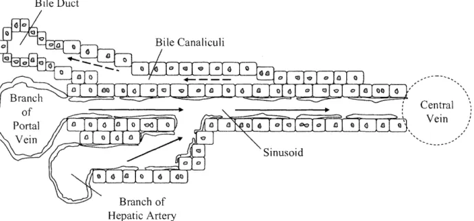
The vasculature of the liver is complex and unique. The liver is divided into many lobules. At the center of each lobule is a central vein. Blood enters the lobule from sinusoids at the periphery. These sinusoids draw blood from two sources, the portal vein and hepatic artery. Blood flows down the sinusoids towards the central vein between plates of hepatocytes that are one to two cells thick. The endothelial cells that line the sinusoids are distinctive in that they contain large fenestrations, or holes, that allow direct contact between the blood and the hepatocytes. The liver's ability to effectively clear blood of many classes of compounds depends on the hepatocyte surface exposure to sinusoidal blood. Within the hepatic plates, between adjacent hepatocytes, lie biliary canaliculi, which drain opposite to the blood flow into bile ducts at the periphery of the lobule. See Figure 1.
Bile Duct Bile Canaliculi .-- 0 0 0 |00 -4-- 0s '---1 0 0
aI
a IO Ic
e
I
00cI Central Vein Hepatic ArteryFigure 1. Structure of the Liver Lobule. Blood flows from the portal vein and the hepatic artery towards the central vein. Bile flows in the opposite direction from blood down the bile
canaliculi.
The liver is a highly studied organ in the field of biotechnology due to its broad range of
functions. The main contribution of the liver to digestion is bile secretion. Bile emulsifies lipids and is the only mechanism for excreting most heavy metals. Liver also regulates the metabolism of carbohydrates, lipids, and proteins. It is one of the two major storage sites for glycogen and is also the major site for gluconeogenisis, the conversion of amino acids, lipids and simple
carbohydrates to glucose. When proteins are metabolized the amino acids become deaminated forming ammonia. Ammonia cannot be metabolized by most tissues and quickly becomes toxic to cells. However, ammonia is removed from the system by conversion to urea, which also occurs mainly in the liver. Synthesis is another key role of the liver. All the nonessential amino acids are synthesized by the liver as well as many plasma proteins such as albumins, globulins, and fibrinogens. In addition to metabolism and synthesis the liver is a vital storage site for iron and vitamins A, D, and B 12. Finally, the liver plays crucial roles in hormone degradation, drug metabolism and toxin removal (Berne 1993).
8
1.2.3 Cell Types
There are several different cell types in the liver each with its own distinct and important functions. The following are brief descriptions of the major cell types (Michalopoulos and DeFrances 1997; Kimiec 2001). See Figure 2.
Kupffer Cell
, Endoihelial Cells
1
Sinusoid
}
Space of Disse ECM\Hepatocytes/
Figure 2. Various Cell Types of the Liver and Their Physical Relationship. Stellate, Kupffer, and endothelial cells reside in-between the hepatocyte plates. Many cell-cell contacts between various cell types are important for liver function.
Hepatocytes are the main functional cells of the liver. Most of the activity of the liver can be attributed to these cells. Loss of hepatocyte function due to injury by biological or chemical agents leads to acute or chronic liver disease. Hepatocytes make up about 60% of the liver in terms of cell number.
Sinusoidal endothelial cells are unique in many structural and functional characteristics from other endothelial cells of the body. They lack the typical basement membrane and are often in complexes with stellate cells. Additionally, these cells contain large cytoplasmic gaps called fenestrations which allow direct contact between the blood and hepatocytes. In terms of cell number, sinusoidal endothelial cells make up about 19% of the liver.
Kupffer cells are the macrophages of the liver. They clear the blood of gut-derived bacteria and bacterial toxins such as endotoxins or peptidoglycans. Additionally, these cells secrete many paracrine factors that influence hepatocytes and stellate cells. In terms of cell number, Kupffer cells make up about 15% of the liver.
Stellate or Ito cells are a fibroblast like cell that are unique to the liver. They have a distinctive morphology and surround hepatocytes with long processes. Stellate cells have several functions consisting of vitamin A storage, synthesis of connective tissue proteins, and secretion of several growth factors. In terms of cell number Stellate cells make up about 6% of the liver.
1.3 Comb Polymer
1.3.1 Structure and Protein Resistant Properties
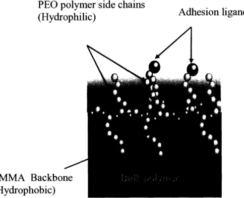
Studying specific receptor-ligand interactions of cell adhesion can be difficult due to high levels of nonspecific protein absorption to surfaces. Adsorbed protein can lead to uncontrolled cell adhesion through many different receptor-ligand systems. A comb polymer was utilized in order to study liver cell adhesion in a specific and controlled manner. The comb polymer consists of a hydrophobic poly(methyl methacrylate) (PMMA) backbone with hydrophilic
hydroxy-poly(ethylene oxide) (HPOEM) side chains. When coated onto a surface and introduced to an aqueous environment the backbone and side chains segregate at the liquid-substrate interface (see Figure 3). Through hydrophobic interactions the backbone is attracted to the substrate surface, while the hydrophilic side chains reach out towards the bulk liquid. This segregation of polymer components creates a PEO brush on the substrate surface. The hydrophilic side chains of the brush are mobile due to the free energy of the system. Because of the constant side chain motion proteins are unable to reach the substrate surface and therefore unable to adsorb, creating a protein free surface environment. A protein free surface is ideal in that it is resistant to cell
adhesion. To maintain this cell resistant property the polymer must consist of 30-35% HPOEM. This percentage ensures that there are enough side chains to effectively resist proteins, while maintaining a high enough hydrophobic polymer content such that the bulk polymer is not water soluble. The synthesis of this polymer has been previously described by (Irvine, Mayes et al. 2001).
PEO polymer side chains
(Hydrophilic)ands
(ri/1
PMMA Backbone
(Hydrophobic)
Figure 3. Structure of Comb Polymer (Koo, Irvine et al. 2002). The hydrophilic side chains move freely in the aqueous environment and prevent protein adsorption to the surface.
1.3.2 Surface Versatility
The hydrophilic side chains of the comb polymer are hydroxy-terminated. These hydroxyl groups can be exploited as a way to conjugate a variety of small molecules such as short peptide sequences. Conjugation to comb polymer can be accomplished through a variety of chemistries such as 2,2,2-trifluoroethanesulfonyl chloride (tresyl chloride) activation, 4-nitrophenyl
chloroformate (NPC) activation, and N-(p-Maleimidophenyl) isocyanate (PMPI) activation. See Figure 4.
0, 0 N 0o S tooN ~OHO O Comb cSvCF3 Polymer 0 s -- [3I A , X HS _ H H HH PMPI O ,NNO2 NPC H2N- i,._ C]l o S C F3 H H Tresyl Chloride
Figure 4. Different Comb Polymer Activation Chemistries (Courtesy of Maria L. Ufret Ph.D.). These activation chemistries are commonly used to conjugate various peptides to comb polymer.
1.4 Integrin Adhesion
Integrins are the major class of cell adhesion receptors that mediate cell-matrix interactions in metazoans (Hynes 2002). Integrins are heterodimeric cell adhesion molecules that consist of an
a and a 3 subunit. These subunits contain both extracellular and intracellular domains though the intracellular domains are typically small (30-50 amino acids). In humans there are 18 a and 8 P subunits. The different combinations of a and 3 subunits result in 24 specific integrins with nonredundant functions. Most integrins recognize and bind to relatively short peptide sequences. For example, a subset of integrins recognize the tripeptide sequence Arginine-Glycine-Aspartic Acid (RGD) which can be found in many extracellular matrix (ECM) molecules such a as
fibronectin, vitronectin, and fibrinogen (Koivunen, Wang et al. 1995). The ability of integrins to bind ECM requires the presence of Mg+ 2 in the cellular environment. This necessity is due to a characteristic metal ion dependent adhesion site motif (MIDAS) found in the integrin structure (Plows 2000). While the diversity of integrins increases as organisms become more complex the structure and function is conserved from sponges to humans (Hynes and Zhao 2000). Integrins
12 - L_OH /I __'_N---~ %. ~ ~ ~ ~ ~ Peptide
= E
I..·~
O
are expressed by a variety of cells and each cell type expresses several integrins allowing cells to bind several matrix molecules. Integrins not only adhere cells to surfaces but are transmembrane
mechanical connections of the extracellular environment to the cytoskeletal intracellular
structure. After integrins adhere to their ligands, they cluster and recruit various cytoskeletal and cytoplasmic proteins (Miyamoto 1995), which eventually lead to the formation of specialized adhesive structures called focal adhesions. Because of their interactions with the intracellular environment, integrins play an important role in triggering various cell processes such as proliferation, differentiation, apoptosis, and cell migration (Flier 2001). Integrins are key to the phenomena of anchorage dependent cell survival. Integrins generally exhibit low ligand
affinities (KD equals 10-6 -10-8mol/liter) compared to the affinities of cell surface hormone receptors (KD equals 10-9 -10-' l mol/liter) (Lodish 2000). However, each cell creates hundreds of
thousands of integrin interactions with extracellular matrix allowing them to remain attached to the ECM. These weaker interactions are beneficial to behaviors such as cell migration where the ability to break contacts with the extracellular matrix would be essential.
1.5 Applications of Comb Polymer to Liver Cell Adhesion and Signaling
As stated above, most integrins recognize and bind to relatively short peptide sequences. Many such peptide sequences have been identified in the literature and are easily synthesized. Once obtained, these peptides can be coupled to comb polymer surfaces through one of the many conjugation chemistries. Because the comb polymer is inherently cell resistant when integrin specific peptides are coupled to the surface, cells should only adhere via the desired integrin of study. Additionally, comb polymer is ideal for the presentation of integrin ligands because surface clustering can be achieved (Koo, Irvine et al. 2002). It is well characterized in the literature that integrin clustering allows cells to adhere in a more effective manner (Maheshwari, Brown et al. 2000).
1.5.1 Liver Cell Sorting
Different cell types in the liver express varying levels and kinds of integrins. In order to sort cells of similar size and density these differences in integrin expression can be exploited. It is well known that cell substrate interactions can be used to separate mixed populations of cells into
subpopulations by taking advantage of varying adhesivities (Wysoki 1978; Hammer 1987). This thesis explores possible adhesion ligands and their effectiveness for sorting cells of the liver.
1.5.2 Effects of EGF Signaling on Hepatocyte Adhesion
Epidermal Growth Factor (EGF) affects many cell types including epithelial and mesenchymal lineages. EGF can elicit a wide range of cellular responses depending on cell type such as mitogenisis, apoptosis, migration, protein secretion, differentiation or dedifferentiation (Wells
1999). Because EGF can stimulate proliferation, migration, and differentiation it has been highly studied in the field of tissue engineering and has the potential for many clinical applications. In addition to these cellular processes, EGF is also known to have a de-adhesive influence (Xie, Pallero et al. 1998; Glading, Chang et al. 2000). The crosstalk between EGF receptor signaling and integrins is an important phenomena to understand for bioengineers because the use of EGF as a mitogen would then effect the cellular interaction with biomaterials perhaps leading to undesired results. The de-adhesive effect of EGF on hepatocytes in culture has been observed in the literature (Kuhl and Griffith-Cima 1996). However, through the use of comb polymer the interaction between EGF signaling and specific integrins can now be elucidated.
Chapter 2. Cell Sorting by Adhesion
2.1 Introduction
2.1.1 Why Sort Cells?
Rat livers are typically perfused with collagenase and then the cells are purified through a series of centrifugation steps to yield a parenchymal and a nonparenchymal cell (NPC) fractions (Seglen 1976; Powers and Griffith-Cima 1996), yielding liver cells for study. The parenchymal fraction contains about 95% hepatocytes and 5% other liver cell types (Powers, Janigian et al. 2002). The NPC fraction however is not well characterized and the percentages of the various cell types are unknown. Information about the remaining 5% of the parenchymal cell fraction and the total break down of the NPC fraction would be invaluable to tissue engineers. One motivation for sorting and identifying cells efficiently is this lack of data. Another motivation for cell sorting would be to purify small hepatocytes from the NPC fraction. It has been
hypothesized that these smaller hepatocytes might have a higher proliferative potential in which case they would be a better target for tissue engineering use. Finally, due to the high level of cell cooperativity in the liver, effective in vitro study would require that all the different cell types be present in the chosen culture system (Bhatia, Balis et al. 1999). Thus, the ability to sort cells and
add them back to the culture system in known quantities would be critical.
2.1.2 Previous Sorting Techniques
There are many sorting techniques that are currently used to separate liver cell types. The following is a brief description of methods that are widely used and their drawbacks.
Percoll is a commercially available gradient material that consists of a colloidal suspension of silica particles coated with polyvinyl pyrrolidine (Alpini, Phillips et al. 1994). Centrifuging cells in the presence of a Percoll gradient allows cells to be separated by size and density (Leo, Mak et al. 1985; Smedsrod, Pertoft et al. 1985). However, no method based exclusively on size and density can yield a cell population of high purity from complex mixtures of hepatic cells (Alpini, Phillips et al. 1994). This limitation is due to the overlap in size and density of many cell types. This method is fairly effective but has the drawback of being time consuming. Liver cells
require signals from substrate adhesion to survive thus the long periods spent in suspension cause cell viability to drop dramatically.
Elutriation is a process where fluid flow is forced counter to the force of centrifugation. This allows futher separation of particles by size and density. Elutriation has been used through out the literature to separate liver cells (Alpini, Lenzi et al. 1989; Janousek, Strmen et al. 1993; Valatas, Xidakis et al. 2003). However, this process like density gradient separation is time consuming. Additionally, the elutriation process can lead to physical damage of cells resulting in cell death.
Florescence activated cell sorting (FACS) is a commonly used method for sorting cells. FACS takes advantage of cell differences that can be detected by fluorescent fluorescently labeled antibodies. Cells are labeled and then sent individually through a florescence detector which then statically charges cells based on their fluorescent intensity and color. While this method is effective it is not widely used to isolate specific liver cell subpopulations. FACS has a relatively low cell yield and has a slow rate of sorting (107 cells/hr). Additionally, FACS instruments are extremely expensive and require highly trained personnel (Shapiro 1983; Alpini, Phillips et al. 1994). Furthermore, the FACS process is generally very species specific because antibodies are typically utilized as the fluorescent label. Thus, if the process were optimized for sorting rat liver cells whole new sets of antibodies would have to be generated for human liver cell sorting. Generating new antibodies would difficult, time consuming, and expensive.
2.1.3 Proposed Sorting Technique
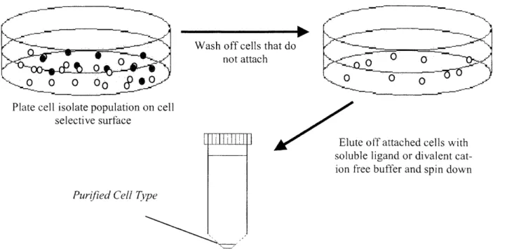
By utilizing the comb polymer and its ligand conjugation versatility it is possible to generate a cell selective surface. Specific cells could be selected from a mixed population of cells based on their adhesive properties. Once non-adherent cells are washed away, selected cells could then be removed from the surface through receptor competition using soluble ligand (see Figure 5). There are several benefits to sorting cells in this way. Sorting cells by adhesion would not require that cells remain in suspension for long periods of time, thus increasing cell viability. Moreover, this method would not depend on size and density differences leading to higher cell type resolution. If integrin adhesion was used to sort cells, as opposed to antibodies, the process could easily be applied to different species due to the evolutionary conservation of integrin structure and function.
Wash off cells that do not attach
Plate cell isolate population on cell selective surface
Putrifized C~ell Type Purijied Cell Tyvpe
]I
/
Elute off attached cells with soluble ligand or divalent cat-ion fee buffer and spin downFigure 5. Proposed Adhesion Based Sorting Process. Sorting by integrin adhesion would require that cells spend less time in suspension and could be readily applied to different species.
2.1.4 Targeted Receptors br Functional Sorting
In order to create a cell selective surface the literature was reviewed for possible ligand
candidates that might be specific to a particular cell type. While the focus of the project was on integrin adhesion other adhesion ligand candidates were also tested.
Integrin Candidates
at93 integrin is a candidate for hepatocyte selection. The act9 integrin is only expressed by the
hepatocytes of the liver (Palmer, Ruegg et al. 1993). While not much is known about this integrin, its exclusive hepatocyte expression makes a good possible candidate for selection. Several short peptide sequences have been described in the literature as having Cal9p specificity. The ones studied in this thesis are PLAEIDGIELTY (Schneider, Harbottle et al. 1998; Yokosaki, Matsuura et al. 1998) and SVVYGLR (Yokosaki, Matsuura et al. 1999).
a4lI integrin is a candidate for endothelial cell selection. In the literature a ligand for 4 1
integrins, REDV was found to be endothelial cell specific (Hubbell, Massia et al. 1991; Massia
i
and Hubbell 1992). Another 413 integrin specific ligand, IDAPS, (Mould and Humphries 1991) was also tested.
Nonintegrins Candidates
Asialoglycoprotein Receptor (ASGP-R) is a candidate for hepatocyte selection. The ASGP-R has been highly characterized in the literature. This receptor is uniquely expressed in
hepatocytes and binds to galactose terminal oligosaccharides. The physiological function of the ASGP-R is to remove damaged proteins from the blood. It has also been shown in previous studies that selective immobilization of hepatocytes using the ASGP-R is possible (Weigel, Schmell et al. 1978; Oka and Weigel 1986; Lopina, Wu et al. 1996). Galactose molecules coupled to the ends of the HPOEM side chains of comb polymer could mimic the structure of galactose terminal oligosaccharides.
Neural Cell Adhesion Molecule (NCAM) is a candidate for stellate cell selection. NCAM is found in most nerve tissue and some non-neural tissues. In the literature activated stellate cells have been shown to exclusively express NCAM in the liver (Knittel, Aurisch et al. 1996). NCAM is typically involved in homophilic binding. However, a peptide sequence that shows NCAM specific adhesion, ASKKPKRNIKA, has been reported in the literature (Knittel, Aurisch et al. 1996).
Lectins are candidates for endothelial cell selection. Lectins are plant derived molecules that recognize carbohydrate moieties in glycoproteins, many of which are displayed on cell surfaces. These molecules have been used much like antibodies to identify and sort cells (Alpini, Phillips
et al. 1994; Marelli-Berg, Peek et al. 2000; Ismail, Poppa et al. 2003). Several lectins have been known to display endothelial cell sensitivity. In the literature, certain lectins have been reported to have rat endothelial cell sensitivity such as Concanavalin A (ConA) and Lens culinaris (LCA) (Smolkova, Zavadka et al. 2001).
2.2 Materials and Methods
2.2.1 Liver Cell Isolation
Liver cell isolations were conducted by Emily Larson and Megan Whittemore. Cells were isolated using a modified two-step collagenase perfusion method from 150 to 230g male Fischer rats (Seglen 1976; Powers and Griffith-Cima 1996). Once isolated the cells are spun down at 50G for 3 minutes, 3 times. The pellets are about 95% hepatocytes and 5% NPC. The supernatants containing mostly NPC's are decanted or aspirated. Cells from the pellets were used for hepatocyte studies, and cells from the supernatant were used for NPC studies. See Appendix 1 for a more detailed perfusion protocol.
2.2.2 Cell Culture
Hepatocytes in all experiments were cultured using modified Hepatocyte Growth Medium (HGM) (Block, Locker et al. 1996). For full HGM preparation see Appendix 2.
NPC in all experiments were cultured in Endothelial Cell Growth Medium 2 (EGM-2) purchased from Cambrex (catalog #CC-3162).
2.2.3 Polymer Synthesis
The comb polymer used in all studies was a two component polymer consisting of PMMA with 10 mer HPOEM side chains of 526 molecular weight. These side chains are about 3.5nm in length. The same batch of polymer was used for all studies (Large Batch 003) and synthesized by Dan Pregibon (Summer 2003). NMR analysis indicated that this batch of polymer was 33% HPOEM, which is within the range for cell resistance and water insolubility. The synthesis of comb polymer has been previously described (Irvine, Mayes et al. 2001). For a detailed synthesis protocol see Appendix 3. Polymer composition and properties were analyzed using techniques outlined in Appendix 4. Before use in experiments cell resistance properties of polymer were tested using the protocol available in Appendix 8.
2.2.4 Polymer activation (NPC)
Evaluation of cell sorting ligands was carried out using NPC activated comb polymer. NPC activation allows ligands to be coupled through terminal amines (Veronese, Largajolli et al.
1985; Jo, Shin et al. 2001). See Appendix 6 for a detailed NPC activation protocol. NMR analysis indicated that NPC activation yielded 50% activated groups. For the chemical structure of NPC see Figure 6.
-° /N0 2
Figure 6. Structure of NPC (Courtesy of Maria L. Ufret Ph.D.). NPC is conjugated to the comb polymer through the chloroformate. The p-phenoxy then becomes a leaving group for peptide conjugation.
2.2.5 Surface Preparation
Substrates were prepared on 12mm diameter circular glass coverslips. In order to increase polymer affinity for the glass surface and reduce polymer delamination the coverslips were all silanized using 4% metacryloxypropyl-trimethoxysilane (MPTS) (Gelest Inc, cat #SIM6487.4), which increased surface hydrophobicity. See Appendix 5 for detailed coverslip silanization protocol.
Treated coverslips were spin coated with 20mg/mL comb polymer in methyl ethyl ketone (MEK). See Appendix 7 for spin coating protocol. The comb polymer used for coating is a 1 to 3 blend of NPC activated comb polymer to inactive comb polymer. Blending is done to obtain ligand clustering and to increase the efficiency of the conjugation reaction. Work by Ada Au (Griffith Lab, Massachusetts Institute of Technology) indicates that using only activated polymer yields a lower NPC conjugation reaction efficiency. This lower efficiency could be due to higher local concentrations of side product produced. Spin coated coverslips were left overnight in a vacuum oven before use.
Peptide coupling was done by leaving NPC activated comb surfaces for four hours covered with lmg/mL peptide in coupling solution. Coupling coverslips were kept in a sealed humidified box at room temperature. After coupling coverslips were washed 3 times with coupling solution (0. IM sodium bicarbonate) and then covered with blocking solution (1 to 1, 0.5M sodium bicarbonate and 0. IM ethanolamine). Blocking solution was left on over night. While blocking, coverslips were left in a sealed humidified box. Blocking is done to deactivate any remaining
NPC groups. For a detailed NPC coupling protocol see Appendix 14. Surfaces are ready to use once blocked.
NPC activated surfaces were coupled with radiolabel RGD and quantified (Ada Au, unpublished data). Surfaces prepared from one to four blends of NPC comb and inactive comb were found to have about 14,000 RGD groups per square micron. This result indicates peptide coupling does occur. Because radioactive surface quantification is a time consuming and highly regulated process each peptide tested was not quantified. However, because peptide coupling has been validated by many members of the lab using different peptide sequences it is assumed with confidence that coupling has occurred.
2.2.6 Peptides
All peptides were ordered from either MIT Biopolymers Laboratory or Tufts University Core Facility. Exact sequences ordered were PLAEIDGIELTY, SVVYGLR, GREDVY, GIDAPSY, ASKKPKRNIKA, and GRGDSPY.
2.2.7 Carbohydrates
Amino terminal carbohydrate ligands were ordered so they could be coupled to be comb polymer using the same method as peptides. 1-amino-1-deooxy-,f-D-galactose was ordered from Sigma (catalog #A-2267). A negative control carbohydrate 1-amino-l -deooxy-P-D-glucose was ordered from Indofine (Catalog #04-268).
2.2.8 Lectins
Fluorescently labeled Concanavalin A (catalog #C7642) and Lens culinaris (catalog # L9262) were ordered from Sigma to test endothelial cell specificity. Before attempting to conjugate lectins to comb surfaces, a live staining using fluorescently labeled lectins was conducted. Sinusoidal endothelial cells were purified using a percoll gradient (purifications were done by Albert Hwa, see Appendix 17) and then seeded onto collagen treated tissue culture plastic. Cells were allowed to spread overnight. Lectins were then dissolved in EGM-2 media (0.1, 1 and 5 mg/mL) and incubated on cells for 1 hour.
2.2.9 Selection Ligand Screening
To test cell adhesion substrates were placed in 24 well plates and then sealed down by silicone sealing rings. These rings cover some of the substrate surface reducing it from 12mm in diameter to 7mm. Hepatocytes and NPC were seeded at 15,000 cells per substrate in 150 uL of media. This concentration was selected such that there would be enough cells to adhere without overcrowding the surface. It was found that when hepatocytes were seeded on inactive comb substrates at concentrations above 50,000 cells per substrate, cells displayed nonspecific surface adhesion in large rounded clumps. This behavior could be attributed to an upper limit of comb polymer protein resistance. Cells were counted using a hemocytometer and trypan blue exclusion. Cells were incubated on surfaces for 24 hours before observation. All substrate conditions were done in triplicate and the experiment was repeated three times. Substrates prepared with only inactive comb were used as negative controls. For a detailed selection ligand screening protocol see Appendix 11.
2.2.10 Microscopy
All microscopy was done with an Axiovert 100. Pictures were taken with a Zeiss Axiocam (#412-312) and acquired using Open Lab 3.0.4 software.
2.3 Results and Discussion
2.3.1 Results
Hepatocytes are known to express integrins which have affinity for the tripeptide sequence RGD. Thus, surfaces conjugated to RGD were tested for hepatocyte adhesion. RGD induced a
significant level of cell adhesion and spreading. See Figure 7.
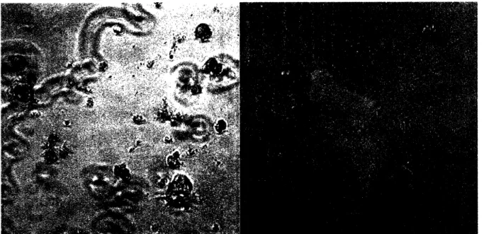
22
Figure 7. Hepatocytes Adhered to RGD surfaces. Hepatocytes adhere and spread well on RGD conjugated surfaces. This result was consistently reproducible.
The a9313 integrin ligands tested, PLAEIDGIELTY and SVVYGLR, were not found to be good candidates for hepatocyte sorting use. Experiments were carried out using cells from the
hepatocyte fraction. The sequence PLAEIDGIELTY showed some cell adhesion, but most cells were rounded and not well adhered. The sequence SVVYGLR showed little to no hepatocyte adhesion. Cells immobilized on the surface were all rounded. See Figure 8.
Figure 8. Hepatocyte Adhesion to PLAEIDGIELTY Surfaces. Few hepatocytes spread on PLAEIDGIELTY making it unsuitable for sorting purposes.
The a4131integrin ligands tested, REDV and IDAPS, were found not to be good candidates for
endothelial cell sorting use. The REDV sequence showed some cell adhesion, but the results varied from isolation to isolation. Additionally, cells in these experiments were stained with fluorescent low density lipoproteins (DiLDL) to verify cell type (Biomed Tech, cat#BT-904). DiLDL specifically stains endothelial cells and Kupffer cells. Staining results indicated that adhered cells were neither endothelial cells nor Kupffer cells. Thus, ligand specificity was not achieved. It is thought that these unidentified cells could be small hepatocytes (see Figure 9). The IDAPS showed no significant cell adhesion for any experiments. All experiments were seeded with NPC fraction cells.
Figure 9. Unidentified NPC on REDV Surfaces. These cells are thought to be small hepatocytes present in the NPC fraction.
Previous literature has indicated that the asialoglycoprotein receptor was a probable ligand for hepatocyte specific adhesion. To target the asialoglycoprotien receptor 1-amino-l-deoxy-P-D-galactose was coupled to comb substrates using the same protocol as peptide coupling. All experiments were seeded with hepatocytes. None of these experiments exhibited hepatocyte adhesion to galactose conjugated comb.
Neural Cell Adhesion Molecule
Activated stellate cells are known to uniquely express NCAM in the liver. The peptide sequence ASKKPKRNIKA was used to prepare comb surfaces to target NCAM and encourage specific
stellate cell adhesion. All experiments were seeded with NPC fraction cells. However, these peptide surfaces showed no cell adhesion.
Lectins
Endothelial cells were plated overnight and then stained with Concanavalin A (Con A) and Lens culinaris (LCA). Both lectins showed endothelial cell staining when compared to DiLDL staining.
2.3.2 Discussion
None of the ligands screened for a931 integrin selection were deemed suitable for use in an adhesion based sorting procedure. The ligand PLAEIDGIELTY, specific for the ag,31 integrin, showed levels of adhesion to low too be used for sorting purposes. The ligand SVVYGLR, also for the aCo,1 integrin displayed no visually detectable levels of cell adhesion. There are several reasons for which these ligands were not suitable. The level of a13 1integrin expression on
hepatocytes is unknown and thus there may not be high enough expression to maintain cell adhesion via this integrin alone. Another possible reason is that the affinity for the a9131 integrin to these particular ligands may be too low to support cell adhesion. Additionally, in has been found in the literature that a91, integrin appears to oppose cell spreading and stimulate cell migration (Liu, Slepak et al. 2001). This function of inducing migration could explain the poor levels of cell adhesion observed.
The ligands tested for a413l integrin adhesion specificity, REDV and IDAPS, were not found to be suitable for an adhesion based sorting method. While REDV showed some cell adhesion the results were inconsistent from isolation to isolation and it was fount that adhered cells were not the desired endothelial cell type. These cells are thought to be small hepatocytes left in the NPC fraction. IDAPS was not found to display any visually detectable cell adhesion properties. As with the a1,31 integrin, the level of a431integrin expression of sinusoidal endothelial cells is
yielded positive endothelial cell adhesion to REDV substrates were done with a different endothelial cell type.
While other systems were able to use the asialoglycoprotein receptor to target hepatocyte specific adhesion there was no hepatocyte adhesion observed in these experiments. There are several possible explanations for this lack of adhesion. The coupling protocol used was directly adopted from peptide coupling. Thus, there is the possibility that no carbohydrates coupled to the surface. The literature was reviewed for a way to quantify the amount of carbohydrate on the surface. A possible method would be to use tritium labeled sugars. Furthermore, the
asialoglycoprotein receptor binding affinity for galactose terminal oligosaccharides increases with increasing ligand valency with highest affinity occurring for a tribranched ligand (Lopina, Wu et al. 1996). The galactose presentation on the comb surface may have been too sparse to mimic this tribranched conformation.
NCAM coupled surfaces displayed no cell adhesion. NCAM is specifically expressed by
activated stellate cells of the liver. The majority of stellate cells in the liver are quiescent. Thus, the number of activated stellate cells may be so small that the odds of capturing many using this type of surface sorting could be very low due to the low seeding density. If this process could be scaled and optimized the ability to characterize the number of activated stellate cells in a freshly isolated liver could be scientifically useful and give further insight into liver function on a cellular level.
Though lectins did stain the endothelial cells of the liver there was a high level of nonspecific background staining which could lead to nonspecific cell staining. Furthermore, the lectins tested were used in the literature as a stain on fixed cells. When used as a live cell stain they appeared to be toxic to cells. Due to this cell toxicity further lectin testing was not pursued.
When testing surfaces using NPC, experimental results were variable from isolation to isolation. There are several possible explanations for this phenomenon. During liver cell isolations the NPC are highly sensitive to the flow rate used to perfuse the liver and can cause variable cell viability. Further, cell death appeared to be sensitive to the seeding concentration. At higher cell
densities more cells appeared to die. This effect could be due to dying cells signaling
surrounding cells to apoptose as well. Though cells were counted before seeding, it was difficult to maintain a constant seeding density. As a result of the isolation there is a significant amount of cell debris present when counting cells. The debris size can often be as large as cells making counting difficult and inaccurate. In order to more effectively utilize the NPC fraction without further purification a better method of counting cells must be developed.
Chapter 3. The Effects of EGF Signaling on Hepatocyte Adhesion
3.1 Introduction
3.1.1 EGF and the EGF Receptor
Many different growth factors have been discovered over the last few decades. Growth factors are generally proteins that stimulate a multitude of cell functions such as proliferation, migration, and differentiation. Epidermal Growth Factor (EGF) is a well characterized growth factor. It was first isolated from the submaxillary glands of adult male mice and has been found to stimulate proliferation, in vivo and in vitro, of many epithelial tissues (Ogiso, Ishitani et al. 2002). EGF has been shown to elicit a wide range of cellular responses depending on cell type such as mitogenisis, apoptosis, migration, protein secretion, de-adhesion, differentiation or
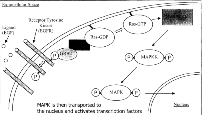
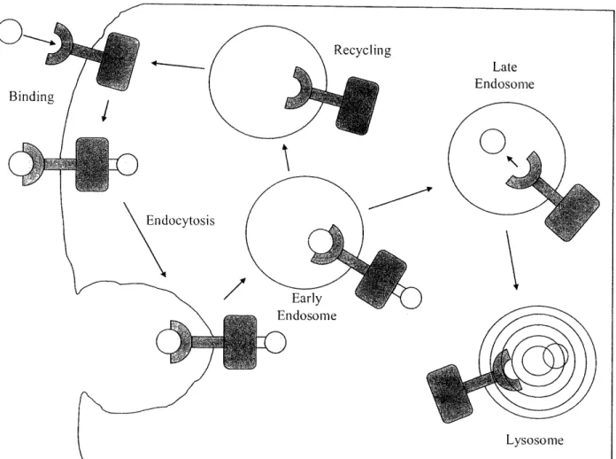
dedifferentiation (Wells 1999). Human epidermal growth factor is a single chain polypeptide that is 53 amino acids long and contains three internal disulfide bonds (Lu, Chai et al. 2001; Ogiso, Ishitani et al. 2002). The structure and function of the EGF receptor is evolutionarily conserved from nematodes to humans (Burke, Schooler et al. 2001). The EGF receptor is a transmembrane glycoprotein that consists of 1186 amino acids. The EGF receptor is part of a family of receptors called Receptor Tyrosine Kinases. Each of these types of receptors binds a single ligand. Then these receptor-ligand complexes dimerize. Once dimerized the receptors phosphorylate each other in their cytoplasmic domains allowing them to then phosphorylate other proteins, beginning a complex signaling cascade resulting in phosphorylation of MAP Kinase and transcriptional modulation (see Figure 10) (Ogiso, Ishitani et al. 2002). Generally, once the EGF receptor is activated it is quickly internalized through coated pits into early endosomes and eventually transported to lysosomes where the receptor ligand complexes become degraded (see Figure 11). While the EGF receptor is known to signal at the cell surface there is data in the literature that indicates signaling from the endosomes as well (Wang, Pennock et al. 2002). The EGF receptor does not always follow the path to immediate degradation. Many times the receptor is recycled to the cell surface three to five times before it its ultimately
degraded.(Clague and Urbe 2001)
Extracellular Space
Figure 10. General Scheme of Receptor Tyrosine Kinase Signaling Cascade. EGF activates a complicated signally cascade that results in transcriptional modulation.
Receptor Tyrosine Kinase Ligand (EGF) p APKK p MAPK p
MAPK is then transported to
the nucleus and activates transcription factors
Z ... I e V
(
Figure 11. Summary of Trafficking and Signaling of EGFR. EGF signaling can continue after the EGF receptor complex has been endocytosed.
3.1.2 Previous Work
The effect of EGF on hepatocytes has been explored in the literature (Moriarity and Savage 1980; Gladhaug and Christoffersen 1987). (Kuhl and Griffith-Cima 1996) studied the effects of EGF on hepatocytes using both soluble and tethered presentations. In their studies hepatocytes were seeded on substrates that had been coated with polyethylene oxide (PEO) stars. However, the PEO stars utilized were relatively large and poorly packed and thus inefficient inhibitors of protein absorption. Because all of their experiments were done in serum free media their
substrates adsorbed with 1:1 Type I Collagen and Cell Tak. Consequently, all cell spreading and adhesion was due to these adsorbed adhesion proteins. Soluble EGF at a concentration of 10 ng/mL was shown to completely inhibit the spreading of hepatocytes on these substrates.
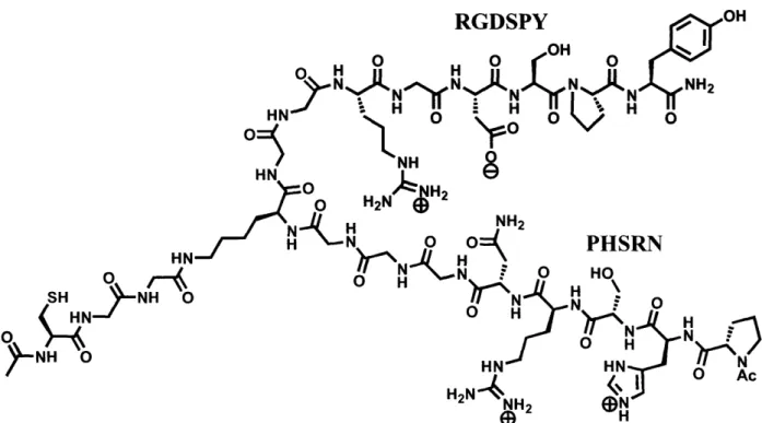
3.1.3 The a-i/lJ Integrin and Ligand
a53Pl integrin is part of the subgroup of integrins that recognizes the tripeptide sequence Arginine-Glycine-Aspartic Acid (RGD) (Hynes 2002). This integrin has been shown to be important to hepatocyte adhesion to the extracellular matrix (Stamatoglou, Sullivan et al. 1990; Schaffert, Sorrell et al. 2001). Adhesion of hepatocytes to RGD substrates has been studied in the literature (Bhadriraju and Hansen 2000). However, it is known that RGD is only a minimal recognition motif for a53BI. Higher affinity to t5f31can be achieved through the simultaneous presentation of
RGD and a synergy site sequence, PHSRN, derived from 9th type III repeating unit of fibronectin
(Dillow, Ochsenhirt et al. 2001). Peptides that contained both the RGD and PHSRN sequences were synthesized in order to study hepatocyte adhesion through the a53p integrin. Peptides that contain both these sequences have been used throughout the literature. However, these have typically been incorporated into single linear peptide sequences (Kao and Lee 2001; Kao, Lee et al. 2001; Kim, Jang et al. 2002). In order for there to be synergistic activity of these sequences they must be correctly spaced. It has been noted in the literature that a spacer of six glycines between sequences results in a higher cell adhesion response than other glycine spacer lengths (Kao and Lee 2001). This work indicates that there are steric limitations to the function of activity of RGD with PHSRN. In order to further overcome these limitations Maria L. Ufret Ph.D. (Griffith Lab, Massachusetts Institute of Technology) designed a novel branched RGD-PHSRN peptide that would allow additional independent freedom of movement for each sequence, while approximately maintaining the six glycine spacer length. The branch peptide consists of the linear sequence PHSRNGGGKGGRGDSPY with a branch emanating from the lysine residue consisting of GGC (see Figure 12). This peptide will be referred to as SynKRGD. SynKRGD was tethered to PMPI activated comb polymer surfaces through the cysteine of the lysine branch. Surface tethering will be further discussed in the materials and methods section below.
RGDSPY HN
0J
HN )0 HN °RX d-< -NH-O
O AcFigure 12. Structure of SynKRGD (Courtesy of Maria L. Ufret Ph.D.). The branching of SynKRGD provides freedom of movement such that each arm can find its optimal binding position.
3.2 Materials and Methods
3.2.1 Liver Cell Isolation
See liver cell isolation in Chapter 2, Materials and Methods, 2.2.1.
3.2.2 Cell Culture
All experiments were cultured using modified Hepatocyte Growth Medium (HGM) from (Block, Locker et al. 1996). For full HGM preparation see Appendix 2. EGF free media was also
prepared and was otherwise identical to complete HGM.
3.2.3 Peptide Synthesis
The linear portion of the SynKRGD peptide and linear RGD with synergy site peptide (CPHSRNGGGGGGRGDSPY) were synthesized using an Advanced ChemTech 396Q and standard 9-fluorenylmethyloxycarbonyl (FMOC) chemistry. Benzotriazole-1-yl-oxy-tris-pyrrolidino-phosphonium hexafluorophosphate (PyBOP) and N-hydroxybenzotriazole (HOBt)
were used as activating agents. NovaSyn® TGR resin (catalog #01-64-0060) was used and purchased from Novabiochem (http://www.emdbiosciences.com). All amino acids were also purchased from Novabiochem. The additional branch of SynKRGD was added by hand. The methoxytrityl (Mtt) protecting group of the lysine was removed using 1% trifluoroacetic acid (TFA), resulting in a free amine. This amine was utilized to add the GGC the branch of SynKRGD using FMOC chemistry. The amino terminus was capped using acetic anhydride. Solid phase peptide synthesis is detailed in Figure 13 below. Once the peptide was synthesized it was cleaved from the resin using TFA:triisopropylsilane (TIS):H20:Ethanedithiol (EDT)
(92.5:2.5:2.5:2.5) and then precipitated using ice cold ether, spun down, and resuspended in ice cold ether several times. The peptide was then lyophilized overnight and subsequently purified by High Pressure Liquid Chromatography (HPLC). See Appendix 12 and 13 for detailed protocols on peptide synthesis and cleavage from resin.
Side chain protecting group
N-a protecting R ro Activating
group R 1 Group Ri 0 NI--O- [2 -o I+
,--__
__Deprotect
T
VI
HCIN
C
HI
I
II
H2N C-- H H H NHc-OH H2Nc--H H nCleave
1\ / n-I ICouple and
deprotect
-in fimi c -- 11 1111i., I R1 lI 1°l~ I l Ll1Ai Ii I II I I II 1 H2N- C N---C--N-- C N-- C--- L e H H H n-IFigure 13. Schematic of Solid Phase Peptide Synthesis (Courtesy of Maria L. Ufret Ph.D.). Coupling and deprotecting is repeated for each amino acid added to the peptide. After peptides are cleaved from the resin they are purified using HPLC.
3.2.4 Polymer Synthesis
See Chapter 2, Materials and Methods, 2.2.3.
3.2.5 Polymer activation (PMPI)
Studying the effect of EGF on a531integrin adhesion was carried out using PMPI activated comb
polymer. PMPI activation allows ligands to be coupled through cysteine residues (Annunziato, Patel et al. 1993). See Appendix 9 for a detailed PMPI activation protocol. NMR analysis indicated that PMPI activation yielded about 25% activated groups. For the chemical structure of PMPI see Figure 14.
0, 0
Figure 14. Structure of PMPI (Courtesy of Maria L. Ufret Ph.D.). PMPI is conjugated to comb polymer through the isocyanate group. The cysteines of peptides then bind to the maleimide during conjugation.
3.2.6 Surface Preparation
Substrates were prepared on 10mm diameter circular glass coverslips. In order to increase polymer affinity for the glass surface and reduce polymer delamination the coverslips were all silanized using 4% metacryloxypropyl-trimethoxysilane (MPTS) (Gelest Inc, cat #SIM6487.4). This treatment increases surface hydrophobicity. See Appendix 5 for detailed coverslip
silanization protocol.
Treated coverslips were spin coated with 20mg/mL comb polymer in methyl ethyl ketone (MEK). See Appendix 7 for spin coating protocol. The comb polymer used for coating was a blend of PMPI activated and non-activated. Surfaces made were either 10% or 25% PMPI activated comb polymer. Blending is done to obtain ligand clustering and control surface
concentration of ligand. Spin coated coverslips were left overnight in a vacuum oven before use.
Peptide coupling was done by leaving PMPI activated comb surfaces for four hours covered with 125 tiM peptide in 7.4 pH phosphate buffer. While coupling, coverslips were kept in a sealed humidified box at room temperature. After coupling, coverslips were washed 3 times with pH 7.4 PBS. For a detailed PMPI coupling protocol see AppendixlS 5. Surfaces are ready to use at the end of the coupling process.
Coupled peptides were quantified using radiolabeled SynKRGD and it was determined that for a 4 hour coupling 10% SynKRGD surfaces displayed 228,000 peptides/jlm2, while 25%
SynKRGD surfaces displayed 577,000 peptides/gm2(Ley Richardson, Griffith Lab, Massachusetts Institute of Technology unpublished data).
3.2.7 Spreading Experiments
Spreading area was used as a measurement of cell adhesion. It is assumed that the larger the spread cell area the higher the affinity of the cell for the substrate. To test cell adhesion to 10% and 25% SynKRGD substrates with and without the presence of EGF, substrates were placed in 24 well tissue culture plates. Hepatocytes were seeded at 15,000 per substrate in 500 uL of media. The seeding density was selected such that there would be enough cells to adhere without overcrowding the surface such that cell spreading area could be more easily calculated. Cells were counted using a hemacytometer and trypan blue exclusion. Once seeded, cells were incubated for 27 hours with or without EGF before analysis. At 27 hours, live cells were fluorescently stained with 5,ll/ml Vybrant Dil (Molecular Probes) and 1 l/m L Hoechst for plasma membrane and nuclei, respectively. After staining, nine different fields were taken for each coverslip. Each field was photographed three times for a bright field, florescent spread area, and fluorescent nuclei. All substrate conditions were done in triplicate and each experiment was repeated at least once. For a detailed spreading experiment protocol see Appendix 10.
3.2.8 AIicroscopy
All microscopy was done with an Axiovert 135. Photos were taken with Hamamatsu Digital Camera (#C4742-95) and saved using Open Lab 2.2.5 software. All images were taken at 10x.
3.2.9 Image analysis
Images for spreading were analyzed using the Scion Image software (version Beta 4.0.2)
obtained from www.scioncorp.com. Scion Image was used to calculate the total spread cell area in m2per image field. The conversion used was 0.745 pixels per micrometer. The number of nuclei per field are also counted. For each field the total spread cell area is then divided by the number of nuclei. The areas/nuclei for all the fields of each condition are then averaged to obtain an average area/nuclei. See Figure 15 below for examples of field images. See Appendix 16 for a detailed Image Analysis Protocol.
Figure 15. Examples of Image Analysis. A) Bright field. B) Nuclear stain. C) Cell area stain. D) Image analyzed using Scion Image with traced cell area.
36 .- ;.. ,· i · · · :i;·r· ;r::;:: I ..·(L, ii-i ;i·;··-..; ·` B ·.i :--···:· ;:,·;.I I:i_ : i·; ··· ;t; I:··:.: ·? ::i: ·····-- ·· ···.r;·: .·:.··:·-·:-- ; ; ; ,·;·
i··;:· c.i·.·r-i
I···.i i·
-·. : ii·
3.3 Results and Discussion
3.3.3. 1 Results
To test the adhesivity of SynKRDG versus linear RGD with synergy site, 100% PMPI polymer surfaces were coupled with each peptide and seeded with 20,000 hepatocytes per substrate (see Figure 16). Greater spreading of hepatocytes was observed on SynKRGD than linear RGD peptide with synergy site.
I'..
-·
,1 Q)
- ; i%
Figure 16. Comparison of Hepatocyte Adhesion on SynKRGD and Linear RGD with Synergy Site Surfaces. Peptides were coupled to 100% PMPI surfaces. Hepatocytes were seeded at 20,000 cells per substrate in HGM. A) Hepatocytes spread on SynKRGD. B) Hepatocytes spread on linear RGD with synergy site.
Experiments conducted compared the adhesion of hepatocytes on two concentrations of SynKRGD and then with and without the presence of 20 ng/mL of EGF. To ensure that the adhesive properties of the SynKRGD surfaces was due to coupled ligand and not nonspecifically adsorbed peptide, surfaces prepared from inactive comb were carried through the coupling procedure. Hepatocytes were seeded on the peptide adsorbed surfaces for 24 hours and no adhesion was observed (see Figure 17).
,. Jr
i:: ·:: j I:; :' ·· ·-· :::::;. t. il: i :r·:::::":':::I :: I :1( :::I '; '" - : ,··;-:t:· i :_:·P i I :: ·· ;:: · ;·· ...:, I;·.·i ? i:-:::
Figure 17. Hepatocytes on Inactive Comb Absorbed with SynKRGD Peptide. Hepatocytes showed no adherence or spreading on SynKRGD surfaces.
The effect of EGF on hepatocyte spreading on 10% and 25% SynKRGD was studied over three experiments. Distinct morphological differences of hepatocytes could be observed as surface ligand concentration decreased and in the presence or absence of EGF. As surface ligand density decreases the spread cell area also decreases. Additionally, cell shape changes from flat and circular to a more amorphous morphology. Cells in the presence of EGF are much less spread than cells in the absence of EGF. There are a higher number of rounded unspread cells attached to surfaces in the presence of EGF. Furthermore, in the presence of EGF many cells also take on a long slender morphology (see Figure 18).
38
10%
SynKRGD
25%
SynKRGD
N~~~P
EGF
<K>
;
j
~Ly'0t-o
l
*v~ ~ ~ ~ , ,,, 1 ,,8~o -,·, ' ,EGF~~~~~~~:
00IWith
EGF
Figure 18. Examples of Cell Spreading Under the Various Experimental Conditions. Cells are most spread on 25% SynKRGD without EGF and least spread on 10% SynKRGD with EGF.
While studying hepatocyte adhesion, occasionally cells of distinctly different morphologies would be observed. These cells were thought to be NPC that had not been purified from the hepatocytes during isolation. The morphology of these unidentified cells is similar to that of hepatic stellate cells, due to their long thin extensions. Stellate cells are the fibroblast like cells of the liver and are known to express as5p integrins. Thus, these cells would adhere to the SynKRGD surfaces (see Figure 19).
Figure 19. Non-hepatocyte Cells Adhered to SynKRGD Surfaces. These cells are thought to be stellate cells due to their morphology. A) Cell membrane stained with DiI. B) Bright field image of same cell.
Hepatocytes were seeded on 10% and 25% SynKRGD surfaces with and without the presence of EGF in the media. The following three Figures (20, 21, and 22) are the results from each of the three experiments. A similar trend was observed over three experiments. All error bars are the mean standard error, standard deviation divided by the square root of the number of fields taken for each condition.
Hepatocyte Spreading on SynKRGD: Experiment 1
r
- 0 E*·M No EGF and 10% SynKRGD* No EGF and 25% SynKRGD
Ow/EGF and 10% SynKRGD
[]w/EGF and 25% SynKRGD
. T .
.
Figure 20. Data from Hepatocyte Adhesion Experiment 1. Hepatocytes were seeded at 15,000 cells per substrate in HGM and incubated for 27 hours before staining.
40 I'+VV 1200 -" 1000 = 800-z () < 600 (U > 400-200 n--·-·-I·i; -·---- ·---- 11·----c · ,· I-c--I--v I I '', I
Hepatocyte Spreading on SynKRGD: Experiment 2 1200 -0 z 0 a0, L On I: 800 600 400 200 -0
C No EGF and 10% SynKRGD E No EGF and 25% SynKRGD O w/EGF and 10% SynKRGD
Ow/EGF and 25% SynKRGD
Figure 2 1. Data from Hepatocyte Adhesion Experiment 2. Hepatocytes were seeded at 15,000 cells per substrate in HGM and incubated for 27 hours before staining.
Hepatocyte Spreading on SynKRGD : Experiment 3
1800 1600 1400 E 1200 z 1000 z ' 800 0) X 600 < 400 200 0
Figure 22. Data from Hepatocyte Adhesion Experiment 3. Hepatocytes were seeded at 15,000 cells per substrate in HGM and incubated for 27 hours before staining.
I I I I
I vvv