HAL Id: inserm-00991192
https://www.hal.inserm.fr/inserm-00991192
Submitted on 14 May 2014
HAL is a multi-disciplinary open access
archive for the deposit and dissemination of
sci-entific research documents, whether they are
pub-lished or not. The documents may come from
teaching and research institutions in France or
abroad, or from public or private research centers.
L’archive ouverte pluridisciplinaire HAL, est
destinée au dépôt et à la diffusion de documents
scientifiques de niveau recherche, publiés ou non,
émanant des établissements d’enseignement et de
recherche français ou étrangers, des laboratoires
publics ou privés.
associated lymphoid tissue
Kim Martinet, Stéphane Bloquet, Christine Bourgeois
To cite this version:
Kim Martinet, Stéphane Bloquet, Christine Bourgeois. Ageing combines CD4 T cell lymphopenia in
secondary lymphoid organs and T cell accumulation in gut associated lymphoid tissue. Immunity &
Ageing, 2014, 11 (1), pp.8. �inserm-00991192�
R E S E A R C H
Open Access
Ageing combines CD4 T cell lymphopenia in
secondary lymphoid organs and T cell
accumulation in gut associated lymphoid tissue
Kim Zita Martinet
1,2, Stéphane Bloquet
2,3and Christine Bourgeois
1,2*Abstract
Background: CD4 T cell lymphopenia is an important T cell defect associated to ageing. Higher susceptibility to infections, cancer, or autoimmune pathologies described in aged individuals is thought to partly rely on T cell lymphopenia. We hypothesize that such diverse effects may reflect anatomical heterogeneity of age related T cell lymphopenia. Indeed, no data are currently available on the impact of ageing on T cell pool recovered from gut associated lymphoid tissue (GALT), a crucial site of CD4 T cell accumulation.
Results: Primary, secondary and tertiary lymphoid organs of C57BL/6 animals were analysed at three intervals of ages: 2 to 6 months (young), 10 to 14 months (middle-aged) and 22 to 26 months (old). We confirmed that ageing preferentially impacted CD4 T cell compartment in secondary lymphoid organs. Importantly, a different picture emerged from gut associated mucosal sites: during ageing, CD4 T cell accumulation was progressively developing in colon and small intestine lamina propria and Peyer’s patches. Similar trend was also observed in middle-aged SJL/B6 F1 mice. Interestingly, an inverse correlation was detected between CD4 T cell numbers in secondary lymphoid organs and colonic lamina propria of C57BL/6 mice whereas no increase in proliferation rate of GALT CD4 T cells was detected. In contrast to GALT, no CD4 T cell accumulation was detected in lungs and liver in middle-aged animals. Finally, the concomitant accumulation of CD4 T cell in GALT and depletion in secondary lymphoid organs during ageing was detected both in male and female animals.
Conclusions: Our data thus demonstrate that T cell lymphopenia in secondary lymphoid organs currently associated to ageing is not sustained in gut or lung mucosa associated lymphoid tissues or non-lymphoid sites such as the liver. The inverse correlation between CD4 T cell numbers in secondary lymphoid organs and colonic lamina propria and the absence of overt proliferation in GALT suggest that marked CD4 T cell decay in secondary lymphoid organs during ageing reflect redistribution of CD4 T cells rather than generalized CD4 T cell decay. Such anatomical heterogeneity may provide an important rationale for the diversity of immune defects observed during ageing.
Keywords: Immunology, T cell, T cell lymphopenia, T cell homeostasis, Immunosenescence, Ageing, Aging, Age, GALT, MALT, CD4 T cell lymphopenia, CD4 T cell homeostasis
Background
Increased susceptibility to infections, cancer [1,2] or some auto-immune pathologies [3,4] are immune defects commonly associated to ageing [5]. The impact of ageing on T cell compartment is commonly defined by reduction of thymic production, T cell lymphopenia, increased of
effector/memory T cells subsets [6,7]. Additionally, in-trinsic defects affecting T cell responsiveness have been described [8]: altered cell membrane fluidity [9], alteration of cell surface expression of co-stimulatory molecules and cytokine receptors, molecular and transcriptional changes [10]. T cell lymphopenia, i.e. decrease in T cell number, has been extensively described during human [11-13] and mice ageing [14,15] and is considered as an important cofactor of age related immune defects. However, the mechanisms by which T cell lymphopenia contribute both to defective and/or exacerbated immune responses in
* Correspondence:christine.bourgeois@u-psud.fr
1INSERM U1012, Faculté de Médecine Paris-Sud, 63 rue Gabriel Péri, 94276 Le
Kremlin-Bicêtre, France
2Univ Paris-Sud, UMR-S1012, Le Kremlin-Bicêtre, France
Full list of author information is available at the end of the article
© 2014 Martinet et al.; licensee BioMed Central Ltd. This is an Open Access article distributed under the terms of the Creative Commons Attribution License (http://creativecommons.org/licenses/by/2.0), which permits unrestricted use, distribution, and reproduction in any medium, provided the original work is properly credited. The Creative Commons Public Domain Dedication waiver (http://creativecommons.org/publicdomain/zero/1.0/) applies to the data made available in this article, unless otherwise stated.
context of infection or autoimmunity respectively remain uncertain. Two opposing immune alterations have been essentially associated to T cell lymphopenia so far. A first mechanism is the loss of clonal diversity [16-20] presum-ably leading to the inability of elderly to respond to certain pathogens. It is commonly related to progressive decrease in thymic export [21-23] although such mechanism is probably not exclusive [24-27]. Importantly, loss of diver-sity is not consistently described in human analyses and remains to be fully characterized [28]. The second alter-ation commonly related to age is the higher proportion of effector/memory cells among conventional T cells in aged individuals [29]. The age related skewing of naïve T cells towards effector and/or memory T cells is thought to reflect both loss of naïve T cell production and lympho-penia induced proliferation (LIP) of naïve T cells [30,31]. It has been claimed that such exacerbated immune responses may lead to auto-immune pathologies [3,4]. However, LIP essentially occurred in context of severe lymphopenia (such as constitutively lymphopenic hosts) and may not develop in context of partial lymphopenia such as observed in ageing [14,32]. Thus, the exact relationship between diversity of age-related immune defects and T cell lymphopenia remains under debate. We hypothesize that age-related T cell lymphopenia develops differently in sec-ondary lymphoid organs compared to tertiary lymphoid organs and non-lymphoid tissues. Indeed, T cell lympho-penia has been essentially described in secondary lymph-oid organs and blood. However, very little information is currently available on T cell compartment in mucosa associated lymphoid tissues (MALT) such as gut or lung associated lymphoid tissue or non-lymphoid tissues. We focused on gut associated lymphoid tissue (GALT) that is presumably highly relevant to CD4 T cell homeo-stasis [33] and readdressed T cell lymphopenia during ageing by performing an exhaustive analysis of the impact of ageing on T cell compartment integrating anatomical location. We analysed CD4 and CD8 T cell compartments in secondary (i.e. mesenteric lymph nodes, superficial lymph nodes, and spleen either globally or separately), tertiary lymphoid organs (as MALT) and non-lymphoid tissue (liver) for different ages ranges (young (2–6 months old), middle-aged (10–14 months old) and old C57BL/6 animals (22–26 months old)).
We confirmed that ageing preferentially affected CD4 T cell compartment in secondary lymphoid organs. We demonstrated that ageing induces concomitantly CD4 T cell decay in secondary lymphoid organs (and notably superficial lymph nodes) and increase of CD4 T cell numbers in the lamina propria of colon and small intes-tine and Peyer’s patches. Such aged related CD4 T cell accumulation was locally restricted since liver or lungs did not show any difference with age in CD4 T cell absolute numbers. Interestingly, an inverse association
was detected between CD4 T cell numbers recovered in secondary lymphoid organs and in colonic lamina pro-pria. Secondly, we demonstrated that T cell accumula-tion in GALT was not related to higher proliferaaccumula-tion of GALT CD4 T cells during ageing. Collectively these data suggest that CD4 T cell lymphopenia commonly described in secondary lymphoid organs (and blood) during ageing may reflect altered redistribution of CD4 T cells rather than extensive depletion. Such anatomical heterogeneity may provide an important rationale for the diversity of immune defects observed during ageing and lead to drastically re-evaluate the notion of age related T cell lymphopenia.
Results
Differential CD4 and CD8 decay in lymphoid organs depending on age
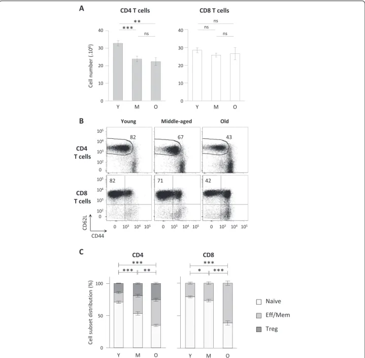
C57BL/6 animals were analysed at three intervals of ages: 2 to 6 months (young), 10 to 14 months (middle-aged) and 22 to 26 months (old). We first determined the absolute number of CD45+ TCRβ+ CD4+ (CD4 T cells) or CD45+ TCRβ+ CD8α+ (CD8 T cells) lymphocytes recovered from secondary lymphoid organs (spleen and lymph nodes) at different ages (Figure 1A). We observed a significant decrease in the number of CD4 T cells in lymphoid organs in “middle-aged” and “old” mice com-pared to “young” animals. No significant decay was further detected between 10–14 months and 22–26 months old mice. In contrast, we did not detect variations in CD8 T cell numbers whatever the age interval considered. We thus confirmed a differential behaviour of CD4 and CD8 T cells depending on the age [15,34-36]. Ageing is com-monly associated to a shift of naïve and effector/memory T cell subsets proportion towards effector/memory T cells [25,29,37,38]. We thus evaluated whether the proportion of naïve and effector/memory cell pools were equally affected in CD4 and CD8 T cell subsets during ageing. Analysing L-selectin (CD62L), a lymphoid homing mol-ecule, and CD44 expression, we identified naïve and effector memory T cell subsets in secondary lymphoid organs, as exemplified on splenic T cells in Figure 1B. Analyses on CD4 T cells were performed on FOXP3-cells to exclude regulatory T FOXP3-cells (FOXP3+) that may exhibit different homeostasis compared to the so-called conventional FOXP3- CD4 T cells. Increasing frequencies of both effector/memory CD4 and CD8 T cells were already detectable when comparing middle-aged to young animals (Figure 1C). However the amplitude of skewing differed when considering CD4 and CD8 T cell subsets: effector/memory CD4 T shift was progressively developing from young to old animals, whereas effector/memory CD8 T shift was mildly increased in middle-aged animals, and became more intense from middle-aged to old ani-mals. Thus, whereas CD4 and CD8 T cell numbers are
differently affected during ageing, increasing proportion of effector/memory T cells are detected in both CD4 and CD8 T cells. We further investigated this apparent dis-crepancy by analysing naïve and effector/memory T cell
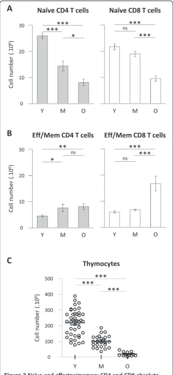
numbers. Naïve CD4 T cell numbers were significantly different between young and middle-aged mice whereas naïve CD8 T cell numbers remain statistically stable in the same interval (Figure 2A). However, both naive CD4 and
Figure 1CD4 but not CD8 T cell decay in secondary lymphoid organs during ageing. (A) Total CD4 and CD8 T cell numbers recovered from pooled spleen and lymph nodes. Numeration and FACS analyses of secondary lymphoid organs were performed on young (2–6 months (Y); n = 30), middle-aged (10–14 months (M); n = 20) and old (22–26 months (O); n = 12) C57BL/6 mice. (B) Representative dot plots of CD62L and CD44 expression on FOXP3- CD4 T cells (conventional CD4 T cells) and CD8 T cells recovered from spleen of young, middle-aged and old C57BL/ 6 mice. (C) Naïve, effector/memory or regulatory of CD4 and CD8 T cell distributions in pooled spleen and lymph nodes from young (n = 20), middle-aged (n = 10) and old (n = 10) C57BL/6 mice. Naïve CD4 T cells were identified as CD45+ TCRβ+ CD4+ FOXP3- CD62L+ CD44low cells, naïve CD8 T cells as CD45+ TCRβ+ CD8α+ CD62L+ CD44- cells, effector/memory CD4 T cells as CD45+ TCRβ+ CD4+ FOXP3- CD62L- CD44high, effector/memory CD8 T cells as CD45+ TCRβ+ CD8α+ non CD62L+CD44- and regulatory CD4 T cells (Treg) as CD45+ TCRβ+ CD4+ FOXP3+. Cumulative results show the mean ± SEM of absolute numbers. P values indicate statistical difference between effector/memory proportion depending on age as followed: ns, non-significant; *, p < 0.05; **, p < 0.01; ***, p < 0.001 (Student’s t test).
CD8 T cell numbers were significantly decreased in old mice compared to young animals. We thus observed a dif-ferent impact of ageing on CD4 and CD8 T cells among naïve T cell compartment: naïve CD4 T cell compartment being affected more rapidly than naïve CD8 T cell pool. Reduction in naïve CD4 T cell numbers in lymphoid organs essentially corroborated the decay observed in total CD4 T cells. However, the drastic decay observed in naïve CD8 T cells in old mice was not reflected in total CD8 T cell numbers. We next considered the evolution of ef-fector/memory T cell numbers: the number of effector/ memory CD4 T cells (TCRβ+ CD4+ FOXP3- CD62L-CD44high cells) significantly increased in middle-aged and old mice, albeit with low amplitude, compared to young an-imals (Figure 2B). Evolution of regulatory FOXP3+ CD4+ T cells (Treg) numbers exhibited a similar profile than effector/memory CD4 T cells numbers (not shown). Re-garding effector/memory CD8 T cell numbers (TCRβ+ CD8α+ non CD62L+CD44- cells) recovered from lymph-oid organs, no significant difference was detected between young and middle-aged animals. However, a significant increase was detected when comparing old animals to young or middle-aged groups (Figure 2B). As a control for ageing, we finally determined thymocyte numbers in the three groups of age. As expected, reduction in thymocyte numbers, a clear hallmark of T cell ageing, was signifi-cantly detected in middle-aged mice and further aggra-vated in old mice (Figure 2C).
Collectively, analysing naïve and effector/memory absolute numbers provided interesting insights on the shift of naïve T cells towards effector/memory T cells during ageing. We observed that physiological ageing is not equally affecting CD4 and CD8 T cell pools. Total CD4 T cell decay reflected massive reduction of naïve CD4 T cells occurring in middle-aged animals combined to a mild increase of effector/memory CD4 T cells in old animals. A different timeline emerged when considering CD8 T cell compartment: naïve and effector/memory CD8 T cells numbers were essentially not affected in middle-aged animals in contrast to older animals who exhibited clear naïve CD8 T cell decay and increase in effector/memory CD8 T cells.
T cell decay differed depending on the second lymphoid organs considered
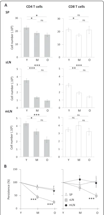
Because some contradictions emerged from data on T cell numbers recovered from lymph nodes and/or spleen [14,39], we next ascertain whether differential behaviour of CD4 and CD8 T cells was homogenous in all secondary lymphoid organs. When considering separately spleen, mesenteric lymph nodes and superficial lymph nodes (i.e. axillary, brachial and inguinal lymph nodes), CD4 T cell decay was detected in all organs when comparing middle-aged or old mice to young animals (Figure 3A left).
Figure 2Naïve and effector/memory CD4 and CD8 absolute numbers in secondary lymphoid organs during ageing. Numeration and FACS analyses were performed on spleen and lymph nodes from young, middle-aged and old C57BL/6 mice as described in Figure 1. (A, B) Absolute numbers of naïve (A) and effector/memory (B) CD4 and CD8 T cells recovered in secondary lymphoid organs. (C) Thymocyte numbers. Numerations were performed on young (n = 10 to 30), middle-aged (n = 10 to 20) and old (n = 10 to 12) C57BL/6 mice. For each experiment, comparison of young animals to middle-aged and/or old animals was simultaneously performed. Cumulative results show the mean ± SEM of absolute numbers. Statistical significance (Student’s t test) is shown: ns, non-significant; *, p < 0.05; **, p < 0.01; ***, p < 0.001.
However, the amplitude differed: CD4 T cells from super-ficial lymph nodes appeared more affected than those in mesenteric lymph nodes and spleen. Because total CD8 T cell numbers were essentially preserved in pooled second-ary lymphoid organs analysis, we were not expecting a major difference in secondary lymphoid organs considered individually. As expected, numbers of CD8 T cells re-covered in the spleen and mesenteric lymph node were essentially not affected, as mice grew older. However, superficial lymph nodes exhibited a different profile revealing a significant decay in the numbers of CD8 (Figure 3A right). In conclusion, T cell distribution was gradually affected depending on the lymphoid organs considered: splenic cells appeared mildly affected; mesen-teric lymph nodes exhibited partial T cell lymphopenia; T cell lymphopenia was more marked in superficial lymph nodes. To directly compare T cell decay in each secondary lymphoid structure considered, we presented the per-centage of residual CD4 and CD8 T cells in middle-aged and old animals compared to young mice (Figure 3B). The percentage of residual CD4 T cells at 10–14 months (middle-age) was significantly lower in the superficial lymph nodes compared to mesenteric lymph nodes and spleen (p < 0.01 and <0.001 respectively). CD4 T cell decay in mesenteric lymph nodes resembled those observed in spleen. Stronger decay in superficial lymph nodes com-pared to mesenteric lymph nodes or spleen was also found in 22–26 months old mice (p < 0.05 and <0.001 respect-ively). Similarly, the specific impact of ageing in superficial lymph nodes was detected among CD8 T cells, although detectable solely in old animals (p < 0.05 and p < 0.001 when comparing to mLN and SP respectively). We thus observed that age related T cell lymphopenia is exa-cerbated in superficial lymph nodes, whereas T cells in mesenteric lymph nodes and spleen are better preserved.
Age associated T cell accumulation in gut associated lymphoid tissue during ageing
The obvious dichotomy between superficial and mesen-teric lymph nodes led us to further investigate gut asso-ciated lymphoid structure. Because CD4 T cells in the gut have been essentially located in the lamina propria, we isolated T lymphocytes from colonic lamina propria (cLP), small intestine lamina propria (siLP) and associated lymphoid structures: Peyer’s patches. The gating strategy is shown in Figure 4A. We studied the dynamics of T cell numbers in these structures depending on the age. In Peyer’s patches, CD4 T cell numbers increased with age but absolute CD8 T cell numbers remained constant through ageing. In siLP, both CD4 and CD8 absolute numbers progressively increased from young to middle-aged or old mice (Figure 4B). In cLP, we observed a differ-ent dynamic: increase in CD4 and CD8 T cell numbers was predominantly observed between 10–14 months and
Figure 3Age related T cell lymphopenia differently affects secondary lymphoid organs. (A) CD4 and CD8 T cell numbers recovered from spleen (SP), superficial (inguinal, brachial and axillary lymph nodes (sLN)) and mesenteric (mLN) lymph nodes. Numeration and FACS analyses of each secondary lymphoid organs were performed from young (Y) (n = 30), middle-aged (M) (n = 20) and old (O) (n = 12) C57BL/6 mice. (B) CD4 and CD8 T cell persistence in each secondary lymphoid organ. Age related persistence was expressed as the percentage of residual T cells recovered in either middle-aged or old mice compared to values recovered from young animals [older absolute number / young absolute number x 100]. Graphs show mean ± SEM of absolute numbers of independent experiments comparing young animals to middle-aged and/or old animals. Statistical difference between sLN and SP T cell recovery depending of age is shown (Student’s t test): ns, non-significant; *, p < 0.05; **, p < 0.01; ***, p < 0.001.
22–26 months (Figure 4B). We performed a similar set of experiment in a different strain background using B6/SJL F1 animals. Comparing “young” and “inter-mediate” B6/SJL F1 animals, we detected decreased thy-mocytes (157.9 +/−13.5 106 versus 15.1 +/−5.6 106 thymocytes in young and middle-aged animals respect-ively; <0.0001). Regarding gut associated lymphoid tissue (GALT), an increase in CD4 T cell numbers in Peyer’s patches was detected (0.35 +/−0.09 106versus 0.85 +/−0.15 106 CD4 T cells in young and middle-aged animals res-pectively; p 0.0128) and preserved CD4 T cell counts were observed in cLP and siLP. Thus we confirmed in a different strain model that T cell lymphopenia developing in lymph-oid organs was not recapitulated in GALT and even associ-ated to CD4 T cell accumulation at some sites.
Association between CD4 T cell lymphopenia in secondary lymphoid organs and accumulation in gut associated lymphoid tissue
Simultaneous accumulation of CD4 T cell at mucosal sites and the development of T cell lymphopenia at lymphoid sites raised the question of a direct association between these two phenomena during ageing. Indeed, a strong inverse association was observed between CD4 T cell numbers in colonic lamina propria (cLP) sites and lymphoid sites (Figure 4C). To note, the association was not detected when considering small intestine lamina propria (siLP) or Peyer’s patches (PP) presumably due to the limited variation in numbers observed in these organs. Indeed, ageing induced a 6-fold increase in CD4 T cells in cLP but a 2-fold increase in siLP and PP.
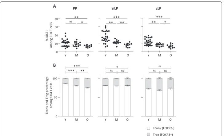
To evaluate whether such accumulation in GALT is reflecting local proliferation or, instead, recruitment to the site, we determined Ki67 expression in CD4 T cells recovered from GALT. We could not detect any increase in Ki67 expression in any of the three gut associated lymphoid structures during ageing. A significant reduc-tion of Ki67 positive CD4 T cells was even detected in all three organs at old ages. Reduced proliferation was already detectable at middle age in siLP and cLP, sug-gesting local proliferation was not responsible for such accumulation (Figure 5A). As a control, Ki67 expression was also assessed in secondary lymphoid organs, which
exhibited significant increase in proliferative fraction (data not shown). Because GALT has been described as a favourable environment for regulatory T cells (Treg) induction, we finally ascertain that CD4 T cell accumu-lation was not directly reflecting specific accumuaccumu-lation or production of FOXP3 expressing Treg. No or mild in-crease in Treg percentages was detected in lamina propria (siLP or cLP) and Peyer’s patches respectively. We also examined the percentages of γ-IFN, IL-4 and IL-17 produ-cing CD4 T cells in secondary lymphoid organs and GALT. Because no deliberate immune response was in-duced in these animals, percentages of cytokine pro-ducing CD4 T cells were relatively low (< 5% for IL-4 and IL-17; < 10% for γ-IFN) confirming that CD4 T cell accumulation in GALT was not related to on-going immune responses. We detected increased percentages of γ-IFN and IL-4 producing CD4 T cells in secondary lymphoid organs as previously described but not among GALT CD4 T cells recovered from old animals (data not shown).
Collectively, our data suggest that CD4 T cell increase observed in the GALT reflect progressive recruitment and accumulation of CD4 T cells from lymphoid sites rather than peripheral expansion of gut resident CD4 T cells.
Age dependent CD4 T cell accumulation in gut associated lymphoid tissue but not in lungs or liver
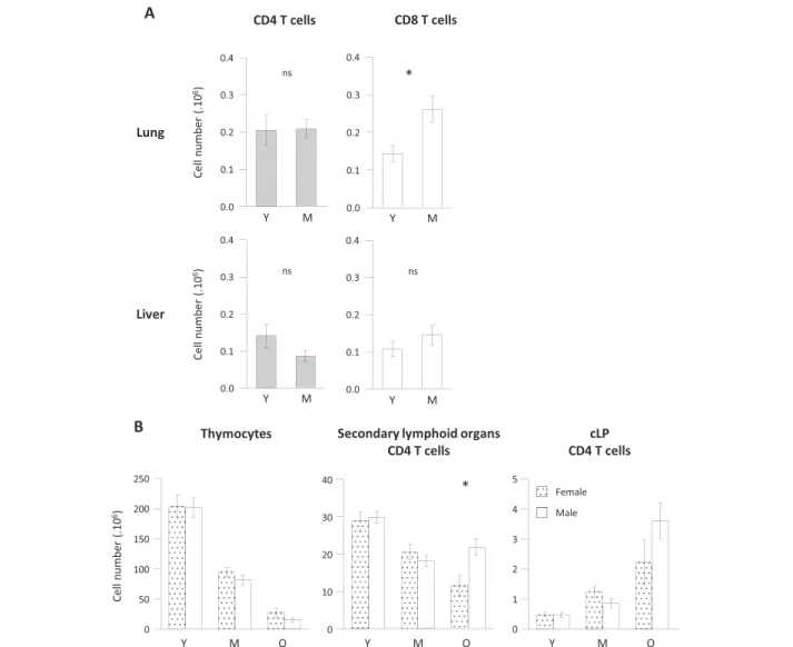
We next questioned whether increase in CD4 T cell numbers observed in gut associated lymphoid tissue (GALT) was a common feature of all tertiary lymphoid organs and even of non-lymphoid tissues in middle-aged animals. We thus analysed two additional sites: the mu-cosa associated lymphoid tissues (MALT) in the lungs and a non-lymphoid tissue: the liver which has been described to comprise important T cell numbers, espe-cially double negative CD4- CD8- T cells [40,41]. To note, T cell recovery in young animals confirmed that GALT represented a major site of CD4 T cell accumulation: CD4 T cell recovery in lungs and liver were approxi-mately 15-fold lower than numbers recovered from small intestine lamina propria (Figure 6A). In the lungs, CD4 T cells were preserved at middle age, whereas CD8 T cell
(See figure on previous page.)
Figure 4CD4 and CD8 T cell absolute numbers increased during ageing in gut associated lymphoid tissues. (A) Gating strategy to identify CD4 and CD8 T cells. Dot plots shows cells isolated from small intestine lamina propria of a 23 months old C57BL/6 mouse. CD45 expressing cells were first selected, then lymphocytes gate was determined to finally discriminate TCRβ+ CD4+ cells and TCRβ+ CD8α+ cells. (B) Graphs show total CD4 (left) and CD8 (right) T cell numbers recovered from Peyer’s patches (PP), small intestine lamina propria (siLP) and colonic lamina propria (cLP) during ageing. Numeration and FACS analysis were performed on PP, siLP and cLP from young (Y) (n = 20), middle-aged (M) (n = 15) and old (O) (n = 10) C57BL/6 mice. Cumulative results show the mean ± SEM of absolute numbers recovered from independent experiment comparing young, middle-aged and/or old animals. Statistical significance (Student’s t test) is shown: ns, non-significant; *, p < 0.05; **, p < 0.01; ***, p < 0.001. (C) Association between mucosal CD4 T cell numbers (PP, siLP and cLP respectively) and secondary lymphoid organs (pooled mesenteric, axillary, brachial and inguinal lymph nodes and spleen) CD4 T cell numbers was evaluated using Spearman test. Spearman r and p value are indicated in each graph.
absolute numbers increased, thus demonstrating that lungs MALT differed from GALT. In the liver, no differ-ence among CD4 or CD8 T cell absolute numbers was de-tected between middle-aged and young mice. Collectively, we demonstrated that CD4 T cell accumulation was essen-tially restricted to the gut associated lymphoid structures.
No major influence of sex on CD4 T cell distribution during ageing
Sex of animals and sex hormones may differentially regulate CD4 homeostasis [42]. To ascertain whether CD4 T cell redistribution in cLP occurs equally in male and female animals, we analysed CD4 T cell distribution based on the sex of the animals. As shown in Figure 6B, sex had only minor influence on CD4 T cell lymphope-nia at lymphoid sites and CD4 T cell accumulation at mucosal sites. Comparing young, intermediate and old animals, we observed an equal decay of thymocytes in male and female animals. In pooled secondary lymphoid organs, CD4 T cells decay was observed in both groups, although significantly more exacerbated in female ani-mals (p 0.0156 when comparing old groups). Conversely,
CD4 T cell numbers recovered in cLP were equally in-creased in male and female animals. These data demon-strate that CD4 T cell accumulation in gut associated lymphoid tissue associated to CD4 T cell lymphopenia during ageing occurs both in male and female animals, al-though the amplitude of CD4 T cell depletion in lymphoid organs appeared to be more drastic among female mice.
Discussion
Ageing is a multifactorial process [24,25] integrating general metabolic changes and immune specific alter-ations such as progressive decrease in thymic export [21-23]. An expected consequence of declining T cell production is the development of progressive T cell lym-phopenia. The incidence of T cell lymphopenia during human ageing is extensively reported in blood samples [11-13] but a more controversial picture emerges from lymphoid organs in murine studies. T cell numbers recovered from spleen or pooled secondary lymphoid organs are reported to be unchanged [24,39] or reduced [14,15] depending notably on the organs considered. In that respect, our current analysis in C57BL/6 mice
Figure 5No local proliferation, nor preferential Treg induction in ageing GALT. (A) Proliferative status of CD4 T cells in the GALT. Percentage of Ki67 expressing cells were determined on CD4 T cells recovered from PP, siLP and cLP from young (Y) (n = 24), middle-aged (M) (n = 12) and old (O) (n = 10) C57BL/6 mice. (B) Proportion of FOXP3 non-expressing (Tconv) and FOXP3 expressing (Treg) CD4 T cells in GALT from young (n = 25), middle-aged (n = 15) and old (n = 10) C57BL/6 mice. Cumulative results show the mean ± SEM of absolute numbers recovered from independent experiments comparing young, middle-aged and aged animals. Statistical significance (Student’s t test) is shown: ns, non-significant; *, p < 0.05; **, p < 0.01.
reconciles this apparent contradiction regarding cell numbers recovered from secondary lymphoid organs: mesenteric lymph nodes and spleen being essentially preserved, whereas superficial lymph nodes studied ex-hibited significant T cell depletion affecting primarily CD4 T cell in middle-aged animals but also CD8 T cells at older ages. A second common observation regarding immune ageing is the differential behaviour of CD4 and CD8 T cell compartments when ageing developed [43]. Evidence of T cell lymphopenia differently affecting CD4 and CD8 T cell compartments is also described in bone marrow transplantation protocols or in a model of genetic thymectomy [15,34,35]. These differential behaviours of CD4 and CD8 T cells are also substantiated by phenotypic
and molecular analyses [28,36,44]. Numerous publications attribute such differential behaviour to specific age related oligoclonal expansion of CD8 T cells both in mice and humans [45-48]. Accordingly, we observed that CD4 T cell numbers recovered from secondary lymphoid organs were rapidly affected: a significant decay was detected in middle-aged animals, whereas CD8 T cells were essen-tially preserved. However, we demonstrated here that differ-ential behaviour of CD4 and CD8 T cells also arise among their naïve T cell compartments. The size of the naïve T cell compartment theoretically results from the combined effect of thymic production, homeostatic control and peripheral activation leading to recruitment of T cells from the naïve pool. Because loss of thymic production observed during
Figure 6No T cell lymphopenia in lungs and liver during ageing and no impact of sex on GALT CD4 T cell accumulation. (A) Lungs and liver CD4 and CD8 T cells were recovered from young (Y) (n = 7), middle-aged (M) (n = 7) C57BL/6 mice. Numeration and FACS staining were performed as previously described. (B) Sex dependent analyses of thymocytes (left), pooled secondary lymphoid organs (middle) and colonic lamina propria (right) CD4 T cell numbers. Female animals are represented in dotted columns, male in plain white columns. Cumulative results show the mean ± SEM of absolute numbers recovered from independent experiments comparing young and middle-aged animals. Statistical significance (Student’s t test) is shown: ns, non-significant; *, p < 0.05; **, p < 0.01.
ageing affects equally single positive CD4 or CD8 thymo-cytes production [21], our results suggest that naïve CD8 T cells are preserved due to poor turn over, whereas naïve CD4 T cell pool appears highly stimulated and directly impacted by partial reduction in thymic production. This faster consumption rate of CD4 T cells may reflect the more pleiotropic function of CD4 T cells compared to CD8 T cells, CD4 T cells being central partners of innate, humoral and cytotoxic responses.
A third classical feature of T cell ageing is a shift from naïve towards effector/memory T cell predominance observed in blood and lymphoid organs [49]. This shift may rely on either naïve T cell decay and/or increase of the effector/memory fraction. By providing naïve and effector/memory absolute numbers, we demonstrated that a different balance between naïve decay and increase in effector/memory cells seems to develop among CD4 and CD8 T cells. In middle-aged animals, naïve CD4 T cell decay was prominent whereas effector/memory CD4 T cell numbers were, albeit significantly, only mildly in-creased. To note, we excluded regulatory T cells, a particular subset of CD4 T cells exhibiting suppressive activity, from naïve and effector/memory CD4 T cell pool analysis. In contrast, CD8 T cell numbers were es-sentially preserved in middle-aged animals: no significant difference was detected when considering total, naïve or effector/memory CD8 T cell numbers separately. In old animals, a different picture emerged: a prominent increase in effector/memory CD8 but not CD4 T cells was detec-ted. Our data suggest that changes in activation profile is essentially related to decrease in naïve T cell numbers when considering CD4 T cell compartment, whereas oligoclonal CD8 expansion occurring in older mice also contributes to naïve towards effector/memory shift among CD8 T cell compartment. Collectively, we demonstrated the existence of an unparalleled impact of ageing on naïve CD4 and effector/memory CD8 T cells.
More importantly, we provided in this report the first evaluation of the impact of ageing on CD4 and CD8 T cells residing in secondary and tertiary lymphoid organs and in non-lymphoid tissues. In striking contrast to sec-ondary lymphoid organs, we detected a progressive accu-mulation of CD4 T cells in all gut associated lymphoid tissues (GALT) considered: Peyer’s patches, lamina propria from the small intestine and the colon. Such increase was not detected for CD8 T cells in Peyer’s patches but was observed at a lesser amplitude in lamina propria. CD4 T cell numbers recovered from lamina propria were consist-ently up to 6-fold higher than CD8 T cell numbers recov-ered. Thus, although both CD4 and CD8 T cells appear to accumulate in the lamina propria with age, such effect may be particularly crucial for the CD4 T cell compart-ment. As a control, we next analysed CD4 and CD8 T cell accumulation in lungs and liver. CD8 but not CD4 T cell
accumulation was detected in the lungs mucosa associated lymphoid tissues suggesting CD4 T cell accumulation in the intestine was a specific feature of the GALT. Analyses of T cell recovery in the liver revealed essentially preserved T cell numbers although later time points may be required to fully ascertain the impact of ageing on liver resident T cells. This observation is in accordance with previous publications demonstrating a high increase of DN T cell rather than CD4 or CD8 T cells in aged liver [40,41]. Thus, CD4 and CD8 T cell distribution/accumulation during ageing appears highly differing depending on the tissues considered. The mechanisms responsible for such predominant age dependent accumulation of CD4 T cells in the GALT remain to be further investigated. Two main hypotheses can be formulated: CD4 T cell accumulation in the intestine may reflect age related skewing of CD4 T cell distribution from secondary lymphoid organs to the GALT or specific proliferative activity of CD4 T cells res-iding in the gut ensuring local CD4 T cell production. Evaluating Ki67 expression in GALT CD4 T cells provided insight showing that local proliferation is not significantly increased but even reduced during ageing. This obser-vation suggests that change in T cell numbers in the gut are predominantly induced by recruitment rather than local proliferation. Accordingly, preserved proportion of FOXP3+ regulatory T cells suggests that CD4 T cell accu-mulation applies to both conventional and regulatory T cells. Additionally, we detected an association between the severity of T cell lymphopenia at lymphoid sites and CD4 T cell accumulation in colonic lamina propria (cLP). The absence of association with small intestine lamina propria (siLP) may reflect either the lower fold of CD4 T cell mulation in siLP although different mechanisms of accu-mulation developing in cLP and siLP respectively cannot be excluded. Increasing numbers of reports demonstrated the crucial role of microbial colonization of mucosal site and mucosal immunity on immune responses [50,51]. It is tempting to speculate that mucosal immunity may directly influence the severity of lymphoid T cell lymphopenia. However, we cannot strictly discriminate in our experi-mental settings the causal or consequential link between lymphoid T cell lymphopenia and mucosal accumulation. This question will require further investigations.
Collectively, analyses of T cell recovery in multiple sites indicate that T cell lymphopenia is not a consistent feature of ageing: T cell lymphopenia was essentially re-stricted to the CD4 subset in some secondary lymphoid organs. Such heterogeneity questions the exact incidence of T cell lymphopenia developing during ageing. One may consider that redistribution of CD4 T cells rather than T cell lymphopenia is the crucial phenomena occur-ring duoccur-ring ageing.
Finally, these observations are also highly relevant in context of accelerated ageing. For instance, HIV infection
is frequently defined as accelerated ageing, due to persist-ent T cell lymphopenia and skewed naïve to effector/ memory phenotype. However, HIV infection is associated to drastic CD4 T cell depletion in the gut prior to the progressive depletion detected in the blood [52]. Such observation constitutes a major drawback to the characterization of HIV infection as an accelerated ageing process. One may question the long-term thread that local CD4 T cell depletion in the gut may induce in old HIV infected patients.
Conclusion
Our results demonstrate that T cell lymphopenia com-monly associated to secondary lymphoid organs is surpris-ingly restricted to these specific sites, and is not applicable to important sites of T cell accumulation such as gut associated lymphoid tissue (GALT). These results are in accordance with a high degree of compartmentalization among organs previously described [53,54]. Ageing ap-pears to induce tissue-specific modulation of T cell com-partments: ageing was associated with significant increase in CD8 T cells in lungs, mixed CD4 and CD8 T cell accu-mulation in GALT, and no change in the liver. Marked decay of CD4 T cells in secondary lymphoid organs during ageing may rely on different sites of CD4 T cell accu-mulation rather than generalized CD4 T cell decay. Such observation refreshes the concept of age related T cell lymphopenia by favouring a model of age related change in T cell distribution among secondary, tertiary and non-lymphoid sites. The concept of tissular heterogeneity of ageing may also provide an interesting rationale for the diverse effects of ageing on immune responses.
Methods
Mice
6 to 8 weeks old C57BL/6 mice were purchased from Janvier Laboratories, and mated in our facilities. Mice were sacrificed at 2 to 6 months of age, 10 to 14 months of age and at 22 to 26 months old respectively named “young”, “middle-aged” and “old” animals. Mice were maintained under pathogen free conditions at the central animal facility of Paris-Sud Faculty of Medicine. As control, young (2 months, n = 6) and middle aged B6-SJL F1 animals (11 months, n = 6) were also analysed. All pro-tocols were conducted in compliance with French and European animal welfare regulations (agreement B-94-043-12 and license 94–440, delivered by the French veter-inary authorities) and validated by the Ethics Committee of University Paris-Sud (CEEA27).
Lymphoid organs cell purification Standard procedures
Single cell suspensions were prepared from lymph nodes (mesenteric and pooled inguinal, brachial, axillary), spleens,
thymus and Peyer’s Patches in DMEM High Glucose containing 10% FCS and 20 mM Hepes buffer (PAA Laboratories GmbH) by dilaceration on 70-μm-mesh cell strainer (BD biosciences).
Cell isolation from small intestine and colon lamina propria
Intestines were treated following standard procedures [55]. Intestines were washed with PBS 1X, mesentery and fat were removed. Peyer’s patches from small intestine were excised and treated as described above. Intestines were then opened longitudinally, cut into 1 cm pieces, and washed 5 times for 10 min at 37°C in pre-warmed PBS 1X containing 3 mM EDTA. Intestine pieces were then washed for 10 min at 37°C in pre-warmed PBS 1X. The tissues were digested in collagenase 100 U/mL (Sigma) and DNase 50 U/mL (Sigma) in pre-warmed RPMI 1640 containing 10% FBS and 20 mM Hepes buffer at 37°C for 45 min. Aspirating 10–15 times the suspension using a 10 mL syringe completed the digestion. All incubations were done under magnetically agitation. Cell suspensions were washed twice in DMEM High Glucose containing 10% FCS and 20 mM Hepes buffer before flow cytometry staining.
Lymphocyte isolation from liver and lungs
Liver single cell suspensions were collected in DMEM High Glucose containing 10% FCS and 20 mM Hepes buffer (PAA Laboratories GmbH), following dilaceration on 100 μm mesh cell strainer. Lungs were washed with PBS 1X, finely minced and stirred in PBS 1X with 400 μg/mL Liberase (Roche) during 20 minutes at 37°C under 250 rpm agitation; suspensions were subsequently filtered and dilacerated on 70 μm mesh cell strainer. Finally, Ficoll centrifugation at 2500 rpm was performed for 20 minutes to isolate lymphocytes. These lymphocyte suspensions were washed twice in DMEM High Glucose containing 10% FCS and 20 mM Hepes buffer before flow cytometry staining.
Absolute numbers determination
Cells counts were performed in duplicates after addition of Trypan blue dye using Malassez haemocytometer cell.
Flow cytometry
Extracellular staining was preceded by incubation with purified anti-CD16/32 antibodies (FcγRII/III block, 2.4G2) (eBioscience) to block non-specific staining. Cells were stained with FITC-, PE-, PerCP-Cy5.5-, PE-Cy5-, PE-Cy7-, APC-, and APC-H7- labelled appropriate anti-bodies including: TCRβ (H57-597, eBioscience); CD45 (30-F11, eBioscience and BD Biosciences); CD44 (IM7, eBioscience); CD8α (53–6.7, eBioscience); CD62L (MEL-14, eBioscience); CD4 (GK1.5, BD Biosciences and Miltenyi Biotec); or appropriate isotype Abs. Intranuclear
FOXP3 staining was performed using eBioscience PE- or APC-conjugated FOXP3 staining buffer set (FJK-16 s). Intranuclear Ki-67 staining was performed using BD Phar-mingen Ki-67 (B56) on permeabilized cells. Six-colour flow cytometry was performed with a FACSCanto cyto-meter (BD Biosciences). Data files were analysed using FlowJo software (Tree star Inc.).
Statistical analyses
Statistical analyses were performed using unpaired Student’s t test with Graph Pad Software. Mean and standard error mean of experiments are shown. Associ-ation were tested using a Spearman test.
Competing interests
The authors declare that they have no competing interests. Authors’ contributions
KM performed the experiments, analysed data and contributed to experiments designing and manuscript writing. SB ensured mice welfare during the course of the work. CB designed the experiments, analysed the data and wrote the article. All authors read and approved the final manuscript.
Acknowledgments
This work was supported by the ANRS (Agence Nationale de la Recherche contre le SIDA et les hépatites C), la Fondation pour la Recherche Médicale (FRM) and benefited from donations of the CIC bank (Crédit Industriel et Commercial) and Pericles consulting group. We thank Elisabeth Huc and Laurent Potier who kindly provided SJL/B6 F1 mice. We thank Dr C. Tanchot and Dr F. Simonetta for critical reading, and Prs M. Tardieu and J.F. Delfraissy for their support.
Author details
1INSERM U1012, Faculté de Médecine Paris-Sud, 63 rue Gabriel Péri, 94276 Le
Kremlin-Bicêtre, France.2Univ Paris-Sud, UMR-S1012, Le Kremlin-Bicêtre, France.3Animalerie centrale, Faculté de Médecine Paris-Sud, Univ Paris-Sud,
Le Kremlin-Bicêtre, France.
Received: 21 October 2013 Accepted: 29 April 2014 Published: 8 May 2014
References
1. Derhovanessian E, Solana R, Larbi A, Pawelec G: Immunity, ageing and cancer. Immun Ageing 2008, 5:11.
2. Fulop T, Larbi A, Witkowski JM, Kotb R, Hirokawa K, Pawelec G: Immunosenescence and cancer. Crit Rev Oncog 2013, 18:489–513. 3. Krupica T, Fry TJ, Mackall CL, Krupica T: Autoimmunity during
lymphopenia: a two-hit model. Clin Immunol 2006, 120:121–128. 4. Calzascia T, Pellegrini M, Lin A, Garza KM, Elford AR, Shahinian A, Ohashi PS,
Mak TW: CD4 T cells, lymphopenia, and IL-7 in a multistep pathway to autoimmunity. Proc Natl Acad Sci U S A 2008, 105:2999–3004.
5. Wick G, Jansen-Dürr P, Berger P, Blasko I, Grubeck-Loebenstein B: Diseases of aging. Vaccine 2000, 18:1567–1583.
6. Pawelec G, Larbi A: Immunity and ageing in man: annual review 2006/2007. Exp Gerontol 2008, 43:34–38.
7. Weng N-P: Aging of the immune system: how much can the adaptive immune system adapt? Immunity 2006, 24:495–499.
8. Larbi A, Pawelec G, Wong SC, Goldeck D, Tai JJ-Y, Fulop T: Impact of age on T cell signaling: a general defect or specific alterations? Ageing Res Rev 2011, 10:370–378.
9. Larbi A, Dupuis G, Khalil A, Douziech N, Fortin C, Fülöp T: Differential role of lipid rafts in the functions of CD4+ and CD8+ human T lymphocytes with aging. Cell Signal 2006, 18:1017–1030.
10. Chen G, Lustig A, Weng N-P: T cell aging: a review of the transcriptional changes determined from genome-wide analysis. Front Immunol 2013, 4:121.
11. Proust J, Rosenzweig P, Debouzy C, Moulias R: Lymphopenia induced by acute bacterial infections in the elderly: a sign of age-related immune dysfunction of major prognostic significance. Gerontology 1987, 31:178–185.
12. Rea I, Alexander H, Crockard A, Morris T: CD4 lymphopenia in very elderly people. Lancet 1996, 347:328–329.
13. Sauce D, Larsen M, Fastenackels S, Roux A, Gorochov G, Katlama C, Sidi D, Sibony-Prat J, Appay V: Lymphopenia-driven homeostatic regulation of naive T cells in elderly and thymectomized young adults. J Immunol 2012, 189:5541–5548.
14. Bourgeois C, Stockinger B: CD25 + CD4+ regulatory T cells and memory T cells prevent lymphopenia-induced proliferation of naive T cells in transient states of lymphopenia. J Immunol 2006, 177:4558–4566. 15. Den Braber I, Mugwagwa T, Vrisekoop N, Westera L, Mögling R, de Boer AB,
Willems N, Schrijver EHR, Spierenburg G, Gaiser K, Mul E, Otto SA, Ruiter AFC, Ackermans MT, Miedema F, Borghans JAM, de Boer RJ, Tesselaar K: Maintenance of peripheral naive T cells is sustained by thymus output in mice but not humans. Immunity 2012, 36:288–297.
16. Mackall C, Fleisher T, Brown M, Andrich M, Chen C, Feuerstein I, Horowitz M, Magrath I, Shad A, Steinberg S, Leonard H, Gress R: Age, thymopoiesis, and CD4+ T-lymphocyte regeneration after intensive chemotherapy. N Engl J Med 1995, 332:143–149.
17. Mosley R, Koker M, Miller R: Idiosyncratic alterations of TCR size distributions affecting both CD4 and CD8 T cell subsets in aging mice. Cell Immunol 1998, 189:10–18.
18. Hall MA, Reid JL, Lanchbury JS: The distribution of human TCR junctional region lengths shifts with age in both CD4 and CD8 T cells. Int Immunol 1998, 10:1407–1419.
19. Buchholz VR, Neuenhahn M, Busch DH: CD8+ T cell differentiation in the aging immune system: until the last clone standing. Curr Opin Immunol 2011, 23:549–554.
20. LeMaoult J, Messaoudi I, Manavalan JS, Potvin H, Nikolich-Zugich D, Dyall R, Szabo P, Weksler ME, Nikolich-Zugich J: Age-related dysregulation in CD8 T cell homeostasis: kinetics of a diversity loss. J Immunol 2000, 165(5):2367–2373.
21. Aspinall R: Age-associated thymic atrophy in the mouse is due to a deficiency affecting rearrangement of the TCR during intrathymic T cell development. J Immunol 1997, 158:3037–3045.
22. Mackall CL, Punt JA, Morgan P, Farr AG, Gress RE: Thymic function in young/old chimeras: substantial thymic T cell regenerative capacity despite irreversible age-associated thymic involution. Eur J Immunol 1998, 28:1886–1893.
23. Aspinall R, Pitts D, Lapenna A, Mitchell W: Immunity in the elderly: the role of the thymus. J Comp Pathol 2010, 142(Suppl):S111–S115.
24. Linton PJ, Dorshkind K: Age-related changes in lymphocyte development and function. Nat Immunol 2004, 5:133–139.
25. Miller RA: The aging immune system: primer and prospectus. Science 1996, 273:70–74.
26. Lefebvre JS, Maue AC, Eaton SM, Lanthier PA, Tighe M, Haynes L: The aged microenvironment contributes to the age-related functional defects of CD4 T cells in mice. Aging Cell 2012, 11:732–740.
27. Zediak VP, Maillard I, Bhandoola A: Multiple prethymic defects underlie age-related loss of T progenitor competence. Blood 2007, 110:1161–1167.
28. Czesnikiewicz-Guzik M, Lee W-W, Cui D, Hiruma Y, Lamar DL, Yang Z-Z, Ouslander JG, Weyand CM, Goronzy JJ: T cell subset-specific susceptibility to aging. Clin Immunol 2008, 127:107–118.
29. Lerner A, Yamada T, Miller RA: Pgp-1hi T lymphocytes accumulate with age in mice and respond poorly to concanavalin A. Eur J Immunol 1989, 19:977–982.
30. Bell EB, Sparshott SM: The peripheral T-cell pool: regulation by non-antigen induced proliferation? Semin Immunol 1997, 9:347–353. 31. Kieper WC, Jameson SC: Homeostatic expansion and phenotypic
conversion of naive T cells in response to self peptide/MHC ligands. Proc Natl Acad Sci U S A 1999, 96:13306–13311.
32. Bourgeois C, Stockinger B: T cell homeostasis in steady state and lymphopenic conditions. Immunol Lett 2006, 107:89–92.
33. Ganusov VV, De Boer RJ: Do most lymphocytes in humans really reside in the gut? Trends Immunol 2007, 28:514–518.
34. Mackall CL, Granger L, Sheard MA, Cepeda R, Gress RE: T-cell regeneration after bone marrow transplantation: differential CD45 isoform expression
on thymic-derived versus thymic-independent progeny. Blood 1993, 82:2585–2594.
35. Bourgeois C, Hao Z, Rajewsky K, Potocnik AJ, Stockinger B: Ablation of thymic export causes accelerated decay of naive CD4 T cells in the periphery because of activation by environmental antigen. Proc Natl Acad Sci U S A 2008, 105:8691–8696.
36. Jankovic V, Messaoudi I, Nikolich-Zugich J: Phenotypic and functional T-cell aging in rhesus macaques (Macaca mulatta): differential behavior of CD4 and CD8 subsets. Blood 2013, 102:3244–3251.
37. Tanchot C, Rocha B: The organization of mature T-cell pools. Immunol Today 1998, 19:575–579.
38. Ma A, Koka R, Burkett P: Diverse functions of IL-2, IL-15, and IL-7 in lymphoid homeostasis. Annu Rev Immunol 2006, 24:657–679.
39. Utsuyama M, Hirokawa K: Age-related changes of splenic T cells in mice–a flow cytometric analysis. Mech Ageing Dev 1987, 40:89–102.
40. Stienen A, Feyen O, Niehues T: Effect of age on homeostasis of lymphocytes in an interleukin-7-deficient mouse model. Eur Cytokine Netw 2011, 22:63–72.
41. Ishimoto Y, Tomiyama-Miyaji C, Watanabe H, Yokoyama H, Ebe K, Tsubata S, Aoyagi Y, Abo T: Age-dependent variation in the proportion and number of intestinal lymphocyte subsets, especially natural killer T cells, double-positive CD4+ CD8+ cells and B220+ T cells, in mice. Immunology 2004, 113:371–377.
42. Menzies FM, Henriquez FL: Immunomodulation by the Female Sex Hormones. Open Infect Dis J 2009, 3:61–72.
43. Strindhall J, Skog M, Ernerudh J, Bengner M, Löfgren S, Matussek A, Nilsson BO, Wikby A: The inverted CD4/CD8 ratio and associated parameters in 66-year-old individuals: the Swedish HEXA immune study. Age (Dordr) 2013, 35:985–991.
44. Gupta S, Su H, Bi R, Agrawal S, Gollapudi S: Life and death of lymphocytes: a role in immunesenescence. Immun ageing I A 2005, 2:12.
45. Posnett DN, Sinha R, Kabak S, Russo C: Clonal populations of T cells in normal elderly humans: the T cell equivalent to “benign monoclonal gammapathy”. J Exp Med 1994, 179:609–618.
46. Hingorani R, Choi I, Akolkar P, Gulwani-akolkar B, Pergolizzi R, Silver J, Gregersen PK: Clonal Predominance of T Cell Receptors Within the CD8+ CD45RO + Subset in Normal Human Subjects. J Immunol 1993, 151(10):5762–5769.
47. Ahmed M, Lanzer KG, Yager EJ, Adams PS, Johnson LL, Blackman MA: Clonal expansions and loss of receptor diversity in the naive CD8 T cell repertoire of aged mice. J Immunol 2009, 182:784–792.
48. Callahan JE, Kappler JW, Marrack P: Unexpected expansions of CD8-bearing cells in old mice. J Immunol 1993, 151:6657–6669. 49. Gruver AL, Hudson LL, Sempowski GD: Immunosenescence of ageing.
2007, 211(2):144–156.
50. Honda K, Littman DR: The microbiome in infectious disease and inflammation. Annu Rev Immunol 2012, 30:759–795.
51. Kamada N, Seo S-U, Chen GY, Núñez G: Role of the gut microbiota in immunity and inflammatory disease. Nat Rev Immunol 2013, 13:321–335. 52. Veazey RS, DeMaria M, Chalifoux LV, Shvetz DE, Pauley DR, Knight HL,
Rosenzweig M, Johnson RP, Desrosiers RC, Lackner AA: Gastrointestinal tract as a major site of CD4+ T cell depletion and viral replication in SIV infection. Science 1998, 280:427–431.
53. Sheridan BS, Lefrançois L: Regional and mucosal memory T cells. Nat Immunol 2011, 131:485–491.
54. Sathaliyawala T, Kubota M, Yudanin N, Turner D, Camp P, Thome JJC, Bickham KL, Lerner H, Goldstein M, Sykes M, Kato T, Farber DL: Distribution and compartmentalization of human circulating and tissue-resident memory T cell subsets. Immunity 2013, 38:187–197.
55. Schulthess J, Meresse B, Ramiro-Puig E, Montcuquet N, Darche S, Bègue B, Ruemmele F, Combadière C, Di Santo JP, Buzoni-Gatel D, Cerf-Bensussan N: Interleukin-15-dependent NKp46+ innate lymphoid cells control intestinal inflammation by recruiting inflammatory monocytes. Immunity 2012, 37:108–121.
doi:10.1186/1742-4933-11-8
Cite this article as: Martinet et al.: Ageing combines CD4 T cell lymphopenia in secondary lymphoid organs and T cell accumulation in gut associated lymphoid tissue. Immunity & Ageing 2014 11:8.
Submit your next manuscript to BioMed Central and take full advantage of:
• Convenient online submission
• Thorough peer review
• No space constraints or color figure charges
• Immediate publication on acceptance
• Inclusion in PubMed, CAS, Scopus and Google Scholar
• Research which is freely available for redistribution
Submit your manuscript at www.biomedcentral.com/submit