HAL Id: hal-01446879
https://hal-univ-rennes1.archives-ouvertes.fr/hal-01446879
Submitted on 10 Apr 2017
HAL is a multi-disciplinary open access
archive for the deposit and dissemination of sci-entific research documents, whether they are pub-lished or not. The documents may come from teaching and research institutions in France or abroad, or from public or private research centers.
L’archive ouverte pluridisciplinaire HAL, est destinée au dépôt et à la diffusion de documents scientifiques de niveau recherche, publiés ou non, émanant des établissements d’enseignement et de recherche français ou étrangers, des laboratoires publics ou privés.
enrichment (BD Bactec™) for rapid diagnosis of bone
and joint infections
Elise Lallemand, Cédric Arvieux, Guillaume Coiffier, Jean-Louis Polard,
Jean-David Albert, Pascal Guggenbuhl, Anne Jolivet-Gougeon
To cite this version:
Elise Lallemand, Cédric Arvieux, Guillaume Coiffier, Jean-Louis Polard, Jean-David Albert, et al.. Use of MALDI-TOF mass spectrometry after liquid enrichment (BD Bactec™) for rapid diagno-sis of bone and joint infections. Research in Microbiology, Elsevier, 2017, 168 (2), pp.122-129. �10.1016/j.resmic.2016.09.005�. �hal-01446879�
Use of MALDI-TOF mass spectrometry after liquid enrichment (BD Bactec™) for rapid diagnosis of bone and joint infections
Elise Lallemand, Cédric Arvieux, Guillaume Coiffier, Jean-Louis Polard, Jean-David Albert, Pascal Guggenbuhl, Anne Jolivet-Gougeon
PII: S0923-2508(16)30115-2
DOI: 10.1016/j.resmic.2016.09.005
Reference: RESMIC 3540
To appear in: Research in Microbiology
Received Date: 6 February 2016 Revised Date: 19 August 2016 Accepted Date: 16 September 2016
Please cite this article as: E. Lallemand, C. Arvieux, G. Coiffier, J.-L. Polard, J.-D. Albert, P.
Guggenbuhl, A. Jolivet-Gougeon, Use of MALDI-TOF mass spectrometry after liquid enrichment (BD Bactec™) for rapid diagnosis of bone and joint infections, Research in Microbiologoy (2016), doi: 10.1016/j.resmic.2016.09.005.
This is a PDF file of an unedited manuscript that has been accepted for publication. As a service to our customers we are providing this early version of the manuscript. The manuscript will undergo copyediting, typesetting, and review of the resulting proof before it is published in its final form. Please note that during the production process errors may be discovered which could affect the content, and all legal disclaimers that apply to the journal pertain.
M
AN
US
CR
IP
T
AC
CE
PT
ED
For publication
1Use of MALDI-TOF mass spectrometry after liquid enrichment (BD
2Bactec™) for rapid diagnosis of bone and joint infections
34
Elise Lallemanda,b, Cédric Arvieuxc,d, Guillaume Coiffierd,e,f, Jean-Louis Polardd,g, Jean-David 5
Albertd,e,f, Pascal Guggenbuhld,e,f,h, Anne Jolivet-Gougeona,b,d,h* 6
7
a
EA 1254 Microbiologie, Université de Rennes 1, 2, avenue du Professeur Léon Bernard, 35043
8
Rennes, France
9
b
Pole Biologie Rennes University Hospital, 35043, Rennes, France
10
c
Service des Maladies infectieuses, Rennes University Hospital, 2 rue Henri Le Guilloux, 35043,
11
Rennes, France
12
d
Centre de Référence en Infections Ostéo-Articulaires du Grand Ouest (CRIOGO)
13
e
Service de Rhumatologie, Hôpital Sud, CHU F- 35000 Rennes, France
14
f
INSERM UMR U991 F-35000 Rennes, France
15
g
Service de Chirurgie orthopédique, Rennes University Hospital, 2 rue Henri Le Guilloux, 35043,
16
Rennes, France
17
h
Université de Rennes 1 F- 35000 Rennes, France
18 19 20 21 22 23 24 25 Corresponding author* 26
Anne Jolivet-Gougeon, Equipe de Microbiologie, EA 1254, Université de Rennes 1, 2, avenue du Professeur Léon Bernard, 27 35043 Rennes, France 28 Phone: (33) 2 23 23 43 05 – Fax: (33) 2 23 23 49 13 29 E-mail: anne.gougeon@univ-rennes1.fr 30 31
M
AN
US
CR
IP
T
AC
CE
PT
ED
Abstract 32Advantages of MALDI-TOF MS (MS) were evaluated for diagnosis of bone and joint 33
infections after enrichment of synovial fluid (SF) or crushed osteoarticular samples (CSs). MS 34
was performed after enrichment of SF or crushed osteoarticular samples CS (n=108) in both 35
aerobic and anaerobic vials. Extraction was performed on 113 vials (SF: n=47; CS: n=66), 36
using the Sepsityper® kit prior identification by MS. The performances of MS, score and 37
reproducibility results on bacterial colonies from blood agar and on pellets after enrichment in 38
vials, were compared. MS analysis of the vial resulted in correct identification of bacteria at a 39
species and genus level (80.5% and 92% of cases, respectively). The reproducibility was 40
superior for aerobic Gram-positive bacteria (Staphylococci and Gram-positive bacilli: 100% 41
colonies), as compared to aerobic Gram-negative bacilli (89.7%), anaerobes (83.3%) and 42
Streptococcus/Enterococcus (58.8%). MS performance was significantly better for 43
staphylococci than for streptococci on all identification parameters. For polymicrobial 44
cultures, identification (score>1.5) of two species by MS was acceptable in 92.8% of cases. 45
Use of MS on enrichment pellets of bone samples is an accurate, rapid and robust method for 46
bacterial identification of clinical isolates from osteoarticular infections, except for 47
streptococci, whose identification to species level remains difficult. 48
49 50 51 52
Keywords: MALDI-TOF mass spectrometry; Osteoarticular infection; Sepsityper® kit; Time 53
of detection; Beadmill processing; Polymicrobial samples. 54
M
AN
US
CR
IP
T
AC
CE
PT
ED
1. Introduction 56Direct examination is an unreliable method for the diagnosis of bone infections [1], with 57
a sensitivity threshold assessed at an inoculum of approximately 104 UFC/mL. Achieving an 58
enrichment step in a liquid medium with prolonged incubation of at least 14 days is essential 59
[2] for correct diagnosis. This time is required to observe the growth of "small colony 60
variants" or fastidious bacteria and to dilute any antibiotic potentially present in the synovial 61
fluid (SF) or crushed bone samples (CSs). A biopsy beadmill processing step [3, 4] or a step 62
of sonication [5] on prosthetic samples provides improvement of culture performances. This 63
is particularly true in the case of bacterial biofilms [6], chronic or complicated infections 64
associated with prosthetic material. Infections on osteosynthesis material may be 65
polymicrobial (10 to 15%) [7], and diagnosis of these infections remains difficult and often 66
fails to identify all these bacterial species. 67
Universal gene amplification techniques (eg. 16SrDNA, sodA) are a diagnostic option, 68
particularly in case of prior antibiotic treatment, but the time consumed (due to the necessary 69
secondary sequencing of the amplified product), the cost of this test and its low sensitivity are 70
major disadvantages to its use [4,8]. Specific polymerase chain reactions (PCRs) (Borrelia, K. 71
kingae, Tropheryma whipplei, etc.) are more sensitive and specific tests, but the procedure 72
requires targeting a single gene with a known sequence. This is a limit to its use in the 73
context of bone and joint infections, where the pathogen is often unknown; accurate diagnosis 74
may require laboratories to perform several specific PCRs. 75
Matrix-assisted laser desorption/ionization-time of flight mass spectrometry, or 76
MALDI-TOF MS (MS), is frequently used for identification of a single colony (isolated on 77
agar media) from clinical and environmental samples [9-11]. The MS system provides rapid 78
and high-confidence identification of bacteria, yeasts and fungi, based on proteomic 79
fingerprinting using high-throughput MALDI-TOF mass spectrometry. Its use has recently 80
M
AN
US
CR
IP
T
AC
CE
PT
ED
been extended to clinical diagnosis, either directly from positive blood culture vials [12] or 81
from samples such as urine [13]. Research suggests that this technique is relevant for 82
microorganism identification, with functionality comparable to routine methods used in the 83
clinical microbiology laboratory [14]. In the case of blood culture vials, bacterial 84
identification by MS directly on the vial pellets optimizes the rendering time result with a 85
time-saving of 1 to 24 h over conventional methods depending on the extraction technique 86
[15, 16]. Results quickly available contribute to reducing morbidity [17] and mortality in 87
addition to lower cost of treatment and length of hospital stay. 88
This study evaluated the usefulness of MS for rapid diagnosis of bone and joint 89
infections. Synovial fluid (SF) or crushed osteoarticular samples (CSs) were enriched in 90
aerobic and anaerobic blood vials before harvesting bacteria (from positive vial cultures), 91
which were then rapidly identified by MALDI-TOF. To assess the performance of MS, score 92
and reproducibility results on bacterial colonies, directly seeded on blood agar from the 93
sample and on pellets after enrichment in blood vials, were compared. Additionally, we 94
defined the detection rate of culture for SF and CS by bacterial species after enrichment in 95
aerobic and anaerobic blood vials. 96
97
2. Material and methods 98
2.1. Samples - Scheme of the study
99
This was a prospective single-center study conducted at the University Hospital of 100
Rennes (Reference Centre for Complex Osteoarticular Infections for the West of France) from 101
January to October 2013. Osteoarticular samples (OASs) were collected and analyzed at the 102
Laboratory of Bacteriology within 2 h of receipt after possible storage at room temperature. 103
Synovial fluids (SFs) were collected in a sterile tube (Falcon ®) and bone samples in a sterile 104
M
AN
US
CR
IP
T
AC
CE
PT
ED
jar (30 mL, HDPE Nalgen). The articular and bone samples were included prospectively, 105
except for laboratory closing hours (21:00-7:30). 106
107
2.2. Bacteriological studies
108
SF and CSs were treated according to microbiological routine techniques. Bone samples 109
were crushed using a bead mill (Retsch® MM400 crusher: frequency 30.0 / s, for two min 110
and 30 s). Tubes containing 10 sterile stainless steel beads (4 mm diameter) (AISI 304 Grade 111
1000; AFBMA; Hammer & Lemarié, France) in 10 mL of molecular biology grade distilled 112
water were prepared, sterilized, tested and stored at room temperature for a maximum of 3 113
months in the laboratory. Following all safety protocols, contents of one tube was poured into 114
each sterile container (HDPE) containing the OAS and grinded [4]. 115
To ensure proper identification of cultures on solid media by MS, 50 µL of SF or CS 116
were plated on Columbia agar supplemented with horse blood (5%) (Oxoid®), chocolate agar 117
(Oxoid®) in atmosphere enriched with 5% CO2 for 72 h and Columbia agar supplemented
118
with horse blood (5%) in an anaerobic atmosphere for 5 days at 37°C.[18,19] 119
Each sample (n=108) was enriched by inoculating 1 mL (minimum volume obtained for 120
some joints) in an aerobic blood culture vial (BD BACTEC™ Plus Aerobic/F) and in an 121
anaerobic blood culture vial (BD BACTEC™ Lytic/10 Anaerobic/F), incubated in automatic 122
chambers for 14 days. Aerobic blood culture vial (BD BACTEC™ Plus Aerobic/F) and 123
anaerobic blood culture vial (BD BACTEC™ Lytic/10 Anaerobic/F) were used because they 124
proved to be the most efficient pair of blood aerobic/anaerobic culture media [20]. 125
After extraction performed according to manufacturer’s recommendations (Sepsityper® 126
kit; Bruker), identification of bacterial species was performed using the MS technique 127
(Microflex LT/SH mass spectrometer Biotyper, Bruker) either on a single colony from agar 128
media (routine use) [21] or on extracted enriched vial pellets (Sepsityper® kit), placed onto 129
M
AN
US
CR
IP
T
AC
CE
PT
ED
the polished steel target plate for rapid identification by MALDI-TOF. Once a positive vial 130
was automatically detected, 1 ml of broth was extracted without delay (< 2 h, to preserve 131
spectra) with formic acid overlay [18] and analyzed via the same method as for colonies. 132
Criteria for interpretation of results were based on the manufacturer’s recommendations 133
(Bruker®). Identification was established through biostatistics reliability levels on the basis 134
of a correlation between the acquired spectrum and the reference spectra. The spectrum of the 135
unknown test organism, acquired through MALDI Biotyper CA System Software®, was 136
electronically transformed into the peak list. Using a biostatistical algorithm, this peak list was 137
compared to reference peak lists of organisms in the reference database, and a log(score) 138
value between 0.00 and 3.00 was calculated. The higher the log(score) value, the more 139
reliable the degree of similarity (to a given organism in the reference FDA-cleared database). 140
A log(score) value of ≥ 2.00 indicated an excellent probability for test organism identification 141
at the species level. The interpretation considered two independent parameters: the value of 142
the homology score and the reproducibility of identification obtained (on 10 measurements 143
carried out after laser impacts, the same bacterial species must be found at least three times 144
with the highest scores, particularly in cases with a score <1.7). Identification with a score ≥ 2 145
was considered reliable to the species; identification with a score ≥ 1.7 was considered 146
reliable to the genus. An identification score of 1.5 was also examined in light of several 147
prior studies suggesting that it adequately identified the bacterial genus [22-24]. Identification 148
was considered unacceptable when the score (< 1.7) and reproducibility were insufficient, and 149
incorrect when the score or reproducibility was acceptable, with poor identification to the 150
species level. If necessary, 16SrDNA PCR was performed to confirm bacterially uncertain 151
identifications, as previously described [4]. To assess the performance of MS in diagnosing 152
bone and joint infections, we compared score and reproducibility results on bacterial 153
colonies from blood agar and on pellets after enrichment in blood vials. 154
M
AN
US
CR
IP
T
AC
CE
PT
ED
155 2.3. Statistical analysis 156Means were compared using the Student test and percentages using the chi-square test 157
(or Fisher’s exact test when sample size was less than 5). P values less than 0.05 were 158 considered significant. 159 160 3. Results 161
3.1. Scheme of the study and description of samples
162
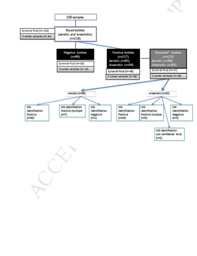
A total of 108 osteoarticular samples (OASs) were collected and 216 enrichment vials 163
(BD BACTEC™ ) were inoculated; 117 were detected positive in automatic chambers 164
(Bactec® 9240, Becton Dickinson) and 113 were analyzed within 2 h following a positive 165
detection rate of culture (for extraction consistent with the Sepsityper® kit manufacturer's 166
recommendations) (Fig. 1). During the incubation period of 336 h, all positive vials were 167
detected before 227 h. 168
After extraction (1 mL sample with the Sepsityper kit), MS identifications were 169
performed on final extracted pellets. Among the aerobic-positive vials (n=58), 50 (86.2%) 170
were considered to be monomicrobial samples, 7 (12%) polymicrobial and 1 (1.8%) negative. 171
In anaerobic vials (n =55), 45 (81.8%) were monomicrobial, 5 (9.1%) were polymicrobial and 172
3 (5.5%) were negative. The list of bacterial isolates obtained from enriched media (aerobic 173
and anaerobic vials, incubated in automatic chambers for 14 days) and from agar media is 174
shown in Table 1. 175
176
3.2. Results of bacterial identification by MS on blood agar (Table 2)
177
According to defined criteria, results of the identification by MS from colonies 178
picked on blood agar (n = 104) (colonies on agar plates obtained from direct spreading of 179
M
AN
US
CR
IP
T
AC
CE
PT
ED
samples or transplanting from enrichment vials) were consistent with species identification in 180
79.8% of cases, with the genus in 90.4% of cases, and unacceptable identification in 1.9% of 181
cases (score and insufficient reproducibility), or incorrect identification in 4.8% (score or 182
acceptable reproducibility, but poor identification at the species level). 183
Aero-anaerobic bacteria species analyzed on blood agar showed highly acceptable 184
identification rates (score>1.7) (100%), with the exception of anaerobes (83.3%) and 185
Streptococcus (70.6%). No relevant misidentifications at the genus level were reported at the 186
log(score) cut-off of 1.6. For Streptococcus, five incorrect identifications were detected. 187
Reproducibility was superior for aerobic Gram-positive bacteria (Staphylococci and Gram 188
positive bacilli: 100% colonies) compared to aerobic Gram-negative bacilli (89.7%), 189
anaerobes (83.3%) and Streptococcus/Enterococcus (58.8%). 190
MS performance was better for staphylococci than for streptococci for all parameters: a 191
high degree of identification (38.5% vs.17.6%, p=0.03), species identification (89.7% vs. 192
58.8%, p=0.001), genus identification (100% vs.70.6%, p<0.001), incorrect identification 193
(0% vs.29.4%, p=0.03) and acceptable reproducibility (100% vs.58.8%, p<0.001). 194
195
3.3. Comparison of MS score results from pellets after enrichment in blood vials from blood 196
agar (Table 2) 197
198
MS analysis on vial pellets resulted in correct identification of bacterial species at a 199
species and genus level (80.5% and 92% of cases, respectively). There was no significant 200
difference between MS identification on vials containing Gram-negative bacilli and 201
staphylococci regarding the high degree of identification, identification to genus and species, 202
unacceptable identifications, incorrect identifications, absence of identification and 203
reproducibility. Incorrect identifications from vial pellets, as compared to the expected 204
M
AN
US
CR
IP
T
AC
CE
PT
ED
identification (MS from colonies and / or PCR 16SrDNA), were observed in streptococci and 205
related species (S. minor/sinensis; S.oralis/pneumoniae; S.parasanguis/Gemellans 206
haemolysans) and Arthrobacter cumminsii/lipophilic Corynebacterium F1 group). The 207
absence of a peak was observed in four cases: S. oralis (no growth on solid media), 208
S.sanguinis, S. minor and S.haemolyticus (<10 colonies on agar corresponding media) and 209
identification was un-interpretable in two cases (S.parasanguis/Gemella haemolysans and 210
Arthrobacter cumminsii/lipophilic Corynebacterium gp F1). 211
212
3.4. Polymicrobial samples (Table 3)
213
In polymicrobial cultures, identification of the two species by MS was acceptable in 214
92.8% of cases [26/28 identifications (92.9%) with a score >1.5; 2/28 identifications (7.1%) 215
with 1.5<score<1.7)]. Correct identification was obtained in all cases (14/14) of a single 216
bacterial species and in 12/14 (85.7%) for 2 bacterial species; no peak could be detected for 217
2/14 (14.3%) vials (second identification) (Table 3). 218
219
4. Discussion 220
MALDI-TOF MS technology showed superiority in identification of most clinical 221
isolates at the genus and species level [9, 11, 25] compared to conventional phenotypic 222
bacterial identification systems. Moussaoui et al. [23] tested a new protocol for bacterial 223
identification from blood culture broths in hospital routine using collection tubes with 224
separator gels. In 503 samples tested over three months, they found that a score> 1.4 was 225
relevant if the score (at the species level) was reproducible at least four times, providing 226
successive proposals. Some differences in scores were observed in the literature between 227
results found on aerobic and anaerobic vials. Christner et al. [15] described a lower estimated 228
mean identification score in the linear mixed-effect model analysis of study data for S. aureus 229
M
AN
US
CR
IP
T
AC
CE
PT
ED
species from aerobic (1,786) compared to anaerobic vials (2,101). In contrast, no such 230
difference was observed in our study (2.31 and 2.30, respectively). Focusing on differences in 231
performance according to bacterial species, our results are consistent with those found in 232
prior literature on blood vials extracted via different methods: Gram-negative bacilli and S. 233
aureus were better identified than other Gram-positive bacteria [12, 22, 24, 26, 27]. 234
To our knowledge, few studies have evaluated the performance of MS in identifying 235
bacteria directly on vial pellets after enrichment of bone samples. Using the Sepsityper kit on 236
blood pellets, the percentage of correct identification was 92% at the species level; this 237
number increased when decreasing to the threshold of 1.5, retained by some studies [24]. In 238
our work, P. acnes were all correctly identified (score> 2). This finding is in contrast to a 239
study conducted by Stevenson et al. [28] that reported 27.3% of unacceptable identifications 240
(score <1.7) for P. acnes, a result that was previously found by MS directly performed on 241
colonies [29]. However, their study carried out only a series of five 1-to-2 min 242
washing/centrifugation steps (without the lysis step of the Sepsityper kit) to remove red blood 243
cells and proteins from the blood culture broths. In our study, all unidentified bacteria and the 244
majority of incorrect identifications concerned the genus Streptococcus (13.6%), especially 245
the alpha-hemolytic group. This was already demonstrated in many prior studies on blood 246
culture vials [22, 26, 27]. Using the Sepsityper kit, the percentage of high degree of 247
identification (score> 2.3) on enriched bone samples was higher in our study (54.9%) than 248
what was found in blood culture vials by Kok et al. [27], who reported 47.1% for Gram-249
negative bacteria, 9.8% for staphylococci and 22.6% for streptococci (in our study 68.2%, 250
60% and 34.6%, respectively). However, Kok et al. [27] detected more coagulase-negative 251
staphylococci and non-fermenting Gram-negative bacilli, both of which are commonly less 252
well identified, potentially explaining the differences from our work. The percentage of high 253
degree of identification (score> 2.3) was significantly higher on vial pellets than on blood 254
M
AN
US
CR
IP
T
AC
CE
PT
ED
agar in our study. This may be related to the fact that, for identification from vial pellets, 255
Sepsityper extraction was followed by extraction with ethanol/formic acid, increasing 256
efficiency. 257
Using the Bactec FX automated blood culture system, Kok et al. [27] reported 6.1% 258
polymicrobial blood vials, with unidentified (32.3%) or misidentified vials (3.2%) at the 259
species level. In case of multiple identifications, it was possible to take into account the 260
presence of any species with a score and/or acceptable reproducibility. Conversely, the 261
presence of a single bacterial species by MS, after extraction, did not exclude the presence of 262
other species in the sample. A study by Martinez et al. [30] found that none of the tested 263
methods were capable of consistently identifying polymicrobial cultures in their entirety. In 264
most studies, only the predominant species was identified from cultures of polymicrobial 265
clinical specimens, which might be explained by bacterial growth competition, with the 266
elimination of one (or more) species in the liquid medium. Chen et al. [31] demonstrated that, 267
for 21 blood cultures composed of two bacterial species, the Bruker Biotyper® was the only 268
system that generated polymicrobial identification: in five out of the 21 mixed-culture 269
specimens (23.8%), the two species present were identified (with >1.6 confidence scores); in 270
the remaining 16 mixed-culture specimens (76.2%), MS identified only the major species of 271
the mixed cultures. A better result was obtained in our study, with an acceptable score of 272
reproducibility, identifying two species in bone samples in 92.8% of cases. 273
The bacterial inoculum of bone sample introduced into blood vials is another 274
important element to take into account, based on the fact that the threshold proposed by the 275
manufacturer underestimates the proportion of correct identifications, resulting in a lower 276
score (that is an artifact of the sample quality: low inoculum and the presence of background 277
noise), rather than a low degree of correlation between the mass spectrum of the sample and 278
the best profile in the database [15]. Direct detection of bacteria in urine by MS was only 279
M
AN
US
CR
IP
T
AC
CE
PT
ED
possible with an inoculum of at least 103 UFC/mL [32]. Works carried out on blood culture 280
vials showed acceptable identification from 106 CFU/mL. For comparison, the average 281
inoculum was 5 × 10⁸ CFU/mL for detection of bacterial growth by the blood culture system 282
[15]. Several studies also reported detection of lower inocula with Gram-positive bacteria by 283
an automatic chamber, but when the inoculum was < 106 CFU/mL, the analyzed spectra were 284
close to those obtained from sterile vials [15]. 285
In previous works using the Sepsityper kit on blood pellets, identifications at the 286
species level were obtained in less than 2 h [27]. Buchan et al. [33] reported that median times 287
to identification using the MALDI Biotyper/Sepsityper were 23 to 83 h faster than routine 288
methods for Gram-positive isolates, and 34 to 51 h faster for Gram-negative isolates in blood 289
samples. This extraction technique has been standardized and validated in the literature, 290
further reducing completion time [27, 34, 35]. Many other simplified efficient extraction 291
methods have also already been tested on blood culture vials. Several techniques reduced the 292
extraction time by half, for example those methods using saponin,[16] ammonium chloride 293
[26], trifluoroacetic acid or formic acid [22], or even methods composed only of a series of 294
centrifugations.[34] It is also possible to reduce the final cost of testing [36]. However, 295
homemade techniques easily fail to completely respond to standardized criteria required in 296
medical biology, and results are difficult to compare between different studies. 297
298
In conclusion, the use of MALDI-TOF MS on bone and synovial samples in culture 299
vials can be performed for diagnosis and management of oste-oarticular infections. This 300
technique reduces the time to report results to the clinician, with a reduced cost [31]. It may 301
also allow identification of a second bacterial species in case of polymicrobial samples, but 302
identification of streptococci to the species level remains difficult. Further improvements in 303
M
AN
US
CR
IP
T
AC
CE
PT
ED
the technique are possible, including optimization of extraction methods for CS and SF before 304
switching on the MS, and continued enrichment of the MS database. 305
306
Acknowledgments 307
We thank Philippe Gautier for technical assistance for PCR 16SrDNA and Adina Pascu 308
for his help in formatting of the manuscript. This work was supported by the Centre de 309
Reference des Infections Ostéoarticulaires complexes du Grand Ouest (CRIOGO, France). 310
Authors' contributions 311
EL and AJG designed the research, designed experiments, assessed and interpreted the 312
results and prepared the manuscript. CA, GC, JLP, JDA and PG took clinical samples and 313
carried out data analysis. All authors read and approved the final manuscript. 314
Competing interests 315
The authors declare that they have no competing interests. 316
317
References 318
319
[1] Societe de Pathologie Infectieuse de Langue F, College des Universitaires de Maladies 320
Infectieuses et T, Groupe de Pathologie Infectieuse P, Societe Francaise d'Anesthesie et de R, 321
Societe Francaise de Chirurgie Orthopedique et T, Societe Francaise d'Hygiene H, et al. 322
[Recommendations for clinical practice. Osteo-articular infection therapy according to 323
materials used (prosthesis, implants, osteosynthesis)]. Med Mal Infect 2009;39:745-74. 324
[2] Schafer P, Fink B, Sandow D, Margull A, Berger I, Frommelt L. Prolonged bacterial 325
culture to identify late periprosthetic joint infection: a promising strategy. Clin Infect Dis 326
2008;47:1403-9. 327
[3] Roux AL, Sivadon-Tardy V, Bauer T, Lortat-Jacob A, Herrmann JL, Gaillard JL, et al. 328
Diagnosis of prosthetic joint infection by beadmill processing of a periprosthetic specimen. 329
Clin Microbiol Infect 2011;17:447-50. 330
[4] Bemer P, Plouzeau C, Tande D, Leger J, Giraudeau B, Valentin AS, et al. Evaluation 331
of 16S rRNA gene PCR sensitivity and specificity for diagnosis of prosthetic joint infection: a 332
prospective multicenter cross-sectional study. J Clin Microbiol 2014;52:3583-9. 333
M
AN
US
CR
IP
T
AC
CE
PT
ED
[5] Trampuz A, Piper KE, Jacobson MJ, Hanssen AD, Unni KK, Osmon DR, et al. 334
Sonication of removed hip and knee prostheses for diagnosis of infection. N Engl J Med 335
2007;357:654-63. 336
[6] Gomez E, Cazanave C, Cunningham SA, Greenwood-Quaintance KE, Steckelberg 337
JM, Uhl JR, et al. Prosthetic joint infection diagnosis using broad-range PCR of biofilms 338
dislodged from knee and hip arthroplasty surfaces using sonication. J Clin Microbiol 339
2012;50:3501-8. 340
[7] Microbiologie SFd. REMIC: référentiel en microbiologie médicale (bactériologie et 341
mycologie). SFM, Paris 2010;4ème édition. 342
[8] Rogers GB, Carroll MP, Bruce KD. Studying bacterial infections through culture-343
independent approaches. J Med Microbiol 2009;58:1401-18. 344
[9] Bizzini A, Jaton K, Romo D, Bille J, Prod'hom G, Greub G. Matrix-assisted laser 345
desorption ionization-time of flight mass spectrometry as an alternative to 16S rRNA gene 346
sequencing for identification of difficult-to-identify bacterial strains. J Clin Microbiol 347
2011;49:693-6. 348
[10] Croxatto A, Prod'hom G, Greub G. Applications of MALDI-TOF mass spectrometry 349
in clinical diagnostic microbiology. FEMS Microbiol Rev 2012;36:380-407. 350
[11] Eigner U, Holfelder M, Oberdorfer K, Betz-Wild U, Bertsch D, Fahr AM. 351
Performance of a matrix-assisted laser desorption ionization-time-of-flight mass spectrometry 352
system for the identification of bacterial isolates in the clinical routine laboratory. Clin Lab 353
2009;55:289-96. 354
[12] Ferreira L, Sanchez-Juanes F, Porras-Guerra I, Garcia-Garcia MI, Garcia-Sanchez JE, 355
Gonzalez-Buitrago JM, et al. Microorganisms direct identification from blood culture by 356
matrix-assisted laser desorption/ionization time-of-flight mass spectrometry. Clin Microbiol 357
Infect 2011;17:546-51. 358
[13] Ferreira L, Sanchez-Juanes F, Gonzalez-Avila M, Cembrero-Fucinos D, Herrero-359
Hernandez A, Gonzalez-Buitrago JM, et al. Direct identification of urinary tract pathogens 360
from urine samples by matrix-assisted laser desorption ionization-time of flight mass 361
spectrometry. J Clin Microbiol 2010;48:2110-5. 362
[14] Garcia P, Allende F, Legarraga P, Huilcaman M, Solari S. [Bacterial identification 363
based on protein mass spectrometry: A new insight at the microbiology of the 21st century]. 364
Rev Chilena Infectol 2012;29:263-72. 365
[15] Christner M, Rohde H, Wolters M, Sobottka I, Wegscheider K, Aepfelbacher M. 366
Rapid identification of bacteria from positive blood culture bottles by use of matrix-assisted 367
laser desorption-ionization time of flight mass spectrometry fingerprinting. J Clin Microbiol 368
2010;48:1584-91. 369
[16] Meex C, Neuville F, Descy J, Huynen P, Hayette MP, De Mol P, et al. Direct 370
identification of bacteria from BacT/ALERT anaerobic positive blood cultures by MALDI-371
TOF MS: MALDI Sepsityper kit versus an in-house saponin method for bacterial extraction. J 372
Med Microbiol 2012;61:1511-6. 373
[17] Galar A, Leiva J, Espinosa M, Guillen-Grima F, Hernaez S, Yuste JR. Clinical and 374
economic evaluation of the impact of rapid microbiological diagnostic testing. J Infect 375
2012;65:302-9. 376
[18] McElvania Tekippe E, Shuey S, Winkler DW, Butler MA, Burnham CA. Optimizing 377
identification of clinically relevant Gram-positive organisms by use of the Bruker Biotyper 378
matrix-assisted laser desorption ionization-time of flight mass spectrometry system. J Clin 379
Microbiol 2013;51:1421-7. 380
[19] Bémer P, Léger J, Tandé D, Plouzeau C, Valentin AS, Jolivet-Gougeon A, et al. Centre 381
de Référence des Infections Ostéo-articulaires du Grand Ouest (CRIOGO) Study Team. How 382
Many Samples and How Many Culture Media To Diagnose a Prosthetic Joint Infection: a 383
M
AN
US
CR
IP
T
AC
CE
PT
ED
Clinical and Microbiological Prospective Multicenter Study. J Clin Microbiol. 2016 54:385-384
91. 385
[20] Rohner P, Pepey B, Auckenthaler R. Advantage of combining resin with lytic 386
BACTEC blood culture media. J Clin Microbiol 1997;35:2634-8. 387
[21] Schulthess B, Brodner K, Bloemberg GV, Zbinden R, Bottger EC, Hombach M. 388
Identification of Gram-positive cocci by use of matrix-assisted laser desorption ionization-389
time of flight mass spectrometry: comparison of different preparation methods and 390
implementation of a practical algorithm for routine diagnostics. J Clin Microbiol 391
2013;51:1834-40. 392
[22] La Scola B, Raoult D. Direct identification of bacteria in positive blood culture bottles 393
by matrix-assisted laser desorption ionisation time-of-flight mass spectrometry. PLoS One 394
2009;4:e8041. 395
[23] Moussaoui W, Jaulhac B, Hoffmann AM, Ludes B, Kostrzewa M, Riegel P, et al. 396
Matrix-assisted laser desorption ionization time-of-flight mass spectrometry identifies 90% of 397
bacteria directly from blood culture vials. Clin Microbiol Infect 2010;16:1631-8. 398
[24] Nonnemann B, Tvede M, Bjarnsholt T. Identification of pathogenic microorganisms 399
directly from positive blood vials by matrix-assisted laser desorption/ionization time of flight 400
mass spectrometry. APMIS 2013;121:871-7. 401
[25] Biswas S, Rolain JM. Use of MALDI-TOF mass spectrometry for identification of 402
bacteria that are difficult to culture. J Microbiol Methods 2013;92:14-24. 403
[26] Prod'hom G, Bizzini A, Durussel C, Bille J, Greub G. Matrix-assisted laser desorption 404
ionization-time of flight mass spectrometry for direct bacterial identification from positive 405
blood culture pellets. J Clin Microbiol 2010;48:1481-3. 406
[27] Kok J, Thomas LC, Olma T, Chen SC, Iredell JR. Identification of bacteria in blood 407
culture broths using matrix-assisted laser desorption-ionization Sepsityper and time of flight 408
mass spectrometry. PLoS One 2011;6:e23285. 409
[28] Stevenson LG, Drake SK, Murray PR. Rapid identification of bacteria in positive 410
blood culture broths by matrix-assisted laser desorption ionization-time of flight mass 411
spectrometry. J Clin Microbiol 2010;48:444-7. 412
[29] Seng P, Drancourt M, Gouriet F, La Scola B, Fournier PE, Rolain JM, et al. Ongoing 413
revolution in bacteriology: routine identification of bacteria by matrix-assisted laser 414
desorption ionization time-of-flight mass spectrometry. Clin Infect Dis 2009;49:543-51. 415
[30] Martinez RM, Bauerle ER, Fang FC, Butler-Wu SM. Evaluation of three rapid 416
diagnostic methods for direct identification of microorganisms in positive blood cultures. J 417
Clin Microbiol 2014;52:2521-9. 418
[31] Chen JH, Ho PL, Kwan GS, She KK, Siu GK, Cheng VC, et al. Direct bacterial 419
identification in positive blood cultures by use of two commercial matrix-assisted laser 420
desorption ionization-time of flight mass spectrometry systems. J Clin Microbiol 421
2013;51:1733-9. 422
[32] Kohling HL, Bittner A, Muller KD, Buer J, Becker M, Rubben H, et al. Direct 423
identification of bacteria in urine samples by matrix-assisted laser desorption/ionization time-424
of-flight mass spectrometry and relevance of defensins as interfering factors. J Med Microbiol 425
2012;61:339-44. 426
[33] Buchan BW, Riebe KM, Ledeboer NA. Comparison of the MALDI Biotyper system 427
using Sepsityper specimen processing to routine microbiological methods for identification of 428
bacteria from positive blood culture bottles. J Clin Microbiol 2012;50:346-52. 429
[34] Juiz PM, Almela M, Melcion C, Campo I, Esteban C, Pitart C, et al. A comparative 430
study of two different methods of sample preparation for positive blood cultures for the rapid 431
identification of bacteria using MALDI-TOF MS. Eur J Clin Microbiol Infect Dis 432
2012;31:1353-8. 433
M
AN
US
CR
IP
T
AC
CE
PT
ED
[35] Morgenthaler NG, Kostrzewa M. Rapid identification of pathogens in positive blood 434
culture of patients with sepsis: review and meta-analysis of the performance of the sepsityper 435
kit. Int J Microbiol 2015;2015:827416. 436
[36] Martiny D, Dediste A, Vandenberg O. Comparison of an in-house method and the 437
commercial Sepsityper kit for bacterial identification directly from positive blood culture 438
broths by matrix-assisted laser desorption-ionisation time-of-flight mass spectrometry. Eur J 439
Clin Microbiol Infect Dis 2012;31:2269-81. 440
441
M
AN
US
CR
IP
T
AC
CE
PT
ED
443Fig. 1. Scheme of the study for 216 vials and results of MALDI-TOF MS identification after 444
extraction on 113 positive vials. *Vials were extracted with the Sepsityper kit before MS 445 identification. 446 447 448 449 450 451 452 453 454 455 456 457 458 459 460
M
AN
US
CR
IP
T
AC
CE
PT
ED
Table 1. List of bacterial isolates obtained from (1) enriched media (aerobic vial (BD 461
BACTEC Plus Aerobic/F and anaerobic vial (BD BACTEC Lytic/10 Anaerobic/F), 462
incubated in automatic chambers for 14 days (Bactec 9240, Becton Dickinson) and (2) agar 463
media (blood agar). 464
Bacterial species No. of isolates
16SrDNA PCR identification Enrichment broth Standard cultures (blood agar) Aerobic incubation Anaerobic incubation (n=58) (n=55) (n=42) Arthrobacter cumminsii 1 1 0 + Clostridium subterminale 0 2 2 Enterobacter cloacae 3 3 5 Enterococcus faecalis 4 4 3 Escherichia coli 5 5 4 Kingella kingae 1 0 0 + Klebsiella pneumoniae 1 0 0 Listeria monocytogenes 1 1 0 Propionibacterium sp. 0 4 4 Pseudomonas aeruginosa 3 1 3 Staphylococcus aureus 20 19 20 Staphylococcus (negative coagulase) 5 4 Staphylococcus epidermidis 5 3 0 Streptococcus agalactiae 2 2 0 Streptococcus pneumoniae 1 1 1 + Streptococcus 6 5 5 +
Species identified by 16SrDNA PCR are indicated (+). 465
466
Table 2. Results of identification scores obtained with the MALDI-TOF MS technique on 467
each bacterial group, i.e. from bacterial colonies (on agar plates obtained from direct 468
spreading of samples or transplanting from enrichment vials) and from pellets after 469
enrichment in blood vials (aerobic and anaerobic). *Vials were extracted with the 470
Sepsityper® Kit before MS identification. 471
M
AN
US
CR
IP
T
AC
CE
PT
ED
472Table 3. Results of score and reproducibility of extracted bone and articular samples with 473
multiple identifications with MALDI-TOF mass spectrometry (MS) technique. * Vials were 474
extracted with the Sepsityper kit before MS identification. 475
476 477
M
AN
US
CR
IP
T
AC
CE
PT
ED
Table 2. Results of MALDI(TOF MS identification Blood vials (both) (n=113) Blood agar (n=104) Staphylococcus (n=39) Streptococcus Enterococcus (n=17) Gram negative bacilli (n=29) Gram positive bacilli (n=4) Anaerobes (n=12) No of isolates (%)High degree of identification to species
Score > 2.3 62 (54.9) 42 (40.4) 15 (38.5) 3 (17.6) 20 (69) 1 (25) 3 (25) Identification to species Score > 2 91 (80.5) 83 (79.8) 35 (89.7) 10 (58.8) 29 (100) 2 (50) 7 (58.3) Identification to genus Score > 1.7 104 (92) 94 (90.4) 39 (100) 12 (70.6) 29 (100) 4 (100) 10 (83.3) Identification to genus with modified
threshold Score > 1.5 107 (94.7) 94 (90.4) 39 (100) 12 (70.6) 29 (100) 4 (100) 10 (83.3) Unacceptable identification Score < 1.7 2 (1.8) 2 (1.9) 0 0 0 0 2 (16.7) Incorrect identification 2 (1.8) 5 (4.8) 0 5 (29.4) 0 0 0 No identification 4 (3.5) 0 0 0 0 0 0 Acceptable reproducibility 99 (87.6) 89 (85.6) 39 (100) 10 (58.8) 26 (89.7) 4 (100) 10 (83.3)
M
AN
US
CR
IP
T
AC
CE
PT
ED
Sample identification Type of blood vial Bacterial species identified from solidmedia
1st bacterial species identified from vials after extraction by
Sepsis typer kit
MALDI-TOF score*
Reproducibility MALDI-TOF*
2nd bacterial species identified from vials after extraction by
Sepsis typer kit (if polymicrobial) MALDI-TOF score* Reproducibility MALDI-TOF* Total number of different bacterial species identified in the sample Sample
1 Aerobic Staphylococcus aureus
+ Enterobacter cloacae Staphylococcus aureus 2,036 1 Enterobacter cloacae 2,04 8 2 SF**
1 Anaerobic Staphylococcus aureus
+ Enterobacter cloacae Enterobacter cloacae 2,241 9 Staphylococcus aureus 1,84 1 2 SF
2 Aerobic
Arthrobacter cumminsii + Weeksella virosa + Oligella urethralis
Weeksella virosa 2,041 2 Arthrobacter cumminsii 1,701 5 3 CS***
3 Anaerobic
Peptoniphilus harei + Propionibacterium avidum
Propionibacterium avidum 2,127 4 Peptoniphilus harei 1,849 0 0 CS
4 Aerobic Staphylococcus aureus
+ Escherichia coli Staphylococcus aureus 2,235 9 Escherichia coli 1,96 2 2 CS
4 Anaerobic Staphylococcus aureus
+ Escherichia coli Staphylococcus aureus 2,312 10 none 2 CS
5 Aerobic Enterococcus faecalis
+ Staphylococcus aureus Enterococcus faecalis 2,147 5 Staphylococcus aureus 1,92 5 2 CS
5 Anaerobic
Enterococcus faecalis + Staphylococcus aureus + Streptococcus oralis
Staphylococcus aureus 2,099 6 Enterococcus faecalis 1,964 4 3 CS
6 Aerobic
Enterococcus faecalis + Staphylococcus aureus + S oralis
Enterococcus faecalis 2,224 6 Staphylococcus aureus 1,886 3 3 CS
6 Anaerobic
Enterococcus faecalis + Staphylococcus aureus + S oralis
Enterococcus faecalis 2,41 8 none CS
7 Aerobic
Klebsiella pneumoniae + Enterobacter arerogenes + Eikeinella corrodens
M
AN
US
CR
IP
T
AC
CE
PT
ED
7 Anaerobic Klebsiella pneumoniae + Enterobacter arerogenes + Streptococcus anginosus + Actinomyces radingue + Parvimonas micraKlebsiella pneumoniae 2,188 8 Streptococcus anginosus 1,684 1 5 CS
8 Aerobic Pseudomonas aeruginosa + Eikenella corrodens + Actinomyces odontolyticus + Aggregatibacter aphrophilus
Pseudomonas aeruginosa 2,234 6 Streptococcus anginosus 1,597 2 4 CS
8 Anaerobic Pseudomonas aeruginosa + Eikenella corrodens + Aggregatibacter aphrophilus + Staphylococcus epidermidis
Staphylococcus epidermidis 2,017 6 Pseudomonas aeruginosa 1,804 4 4 CS
*on pellets extracted from vial **synovial fluid