HAL Id: hal-02620849
https://hal.inrae.fr/hal-02620849
Submitted on 26 May 2020
HAL is a multi-disciplinary open access
archive for the deposit and dissemination of
sci-entific research documents, whether they are
pub-lished or not. The documents may come from
teaching and research institutions in France or
abroad, or from public or private research centers.
L’archive ouverte pluridisciplinaire HAL, est
destinée au dépôt et à la diffusion de documents
scientifiques de niveau recherche, publiés ou non,
émanant des établissements d’enseignement et de
recherche français ou étrangers, des laboratoires
publics ou privés.
Distributed under a Creative Commons Attribution| 4.0 International License
Hong Sheng Cheng, Jeannie Xue Ting Lee, Walter Wahli, Nguan Soon Tan
To cite this version:
Hong Sheng Cheng, Jeannie Xue Ting Lee, Walter Wahli, Nguan Soon Tan. Exploiting vulnerabilities
of cancer by targeting nuclear receptors of stromal cells in tumor microenvironment. Molecular Cancer,
BioMed Central, 2019, 18, �10.1186/s12943-019-0971-9�. �hal-02620849�
R E V I E W
Open Access
Exploiting vulnerabilities of cancer by
targeting nuclear receptors of stromal cells
in tumor microenvironment
Hong Sheng Cheng
1*†, Jeannie Xue Ting Lee
2†, Walter Wahli
2,3,4and Nguan Soon Tan
1,2*Abstract
The tumor microenvironment is a complex and dynamic cellular community comprising the tumor epithelium and various tumor-supporting cells such as immune cells, fibroblasts, immunosuppressive cells, adipose cells, endothelial cells, and pericytes. The interplay between the tumor microenvironment and tumor cells represents a key contributor to immune evasiveness, physiological hardiness and the local and systemic invasiveness of malignant cells. Nuclear receptors are master regulators of physiological processes and are known to play pro−/anti-oncogenic activities in tumor cells. However, the actions of nuclear receptors in tumor-supporting cells have not been widely studied. Given the excellent druggability and extensive regulatory effects of nuclear receptors, understanding their biological functionality in the tumor microenvironment is of utmost importance. Therefore, the present review aims to summarize recent evidence about the roles of nuclear receptors in tumor-supporting cells and their implications for malignant processes such as tumor proliferation, evasion of immune surveillance, angiogenesis, chemotherapeutic resistance, and metastasis. Based on findings derived mostly from cell culture studies and a few in vivo animal cancer models, the functions of VDR, PPARs, AR, ER and GR in tumor-supporting cells are relatively well-characterized. Evidence for other receptors, such as RARβ, RORγ, and FXR, is limited yet promising. Hence, the nuclear receptor signature in the tumor microenvironment may harbor prognostic value. The clinical prospects of a tumor microenvironment-oriented cancer therapy exploiting the nuclear receptors in different tumor-supporting cells are also encouraging. The major challenge, however, lies in the ability to develop a highly specific drug delivery system to facilitate precision medicine in cancer therapy.
Keywords: Nuclear receptors, Tumor microenvironment, Cancer-associated fibroblast, Myeloid-derived suppressor cells, Tumor-associated macrophage
Background
In human cells, there are 48 nuclear receptors (NRs) that play integral roles in numerous physiological func-tions such as metabolism, cell development, immunity, and stress response. Classically, following direct lipo-philic ligand binding, NRs will recognize and bind to specific DNA motifs across the genome, which are known as NR response elements. The binding of an NR to its response element and transcriptional activation of target genes often require homodimerization of NRs or
heterodimerization with retinoid X receptor (RXR) coupled to the recruitment of coactivator proteins, although certain receptors are functionally active as a monomer [1, 2]. Independent of ligand binding, the activities of NRs can also be modulated by posttrans-lational modifications such as phosphorylation, ubiquiti-nation, and SUMOylation, or indirect recruitment to the genome by other DNA-bound transcription factors via tethering mechanisms [2, 3]. Increasing evidence has also unveiled the pivotal roles of NRs in chromatin remodeling [4]. Furthermore, certain NRs such as pro-gesterone receptor (PR) and peroxisome proliferator-ac-tivated receptor (PPAR)-γ possess different isoforms resulting from alternative splicing. Variations in the tissue expression profile, ligand affinity and target genes
* Correspondence:hscheng@ntu.edu.sg;nstan@ntu.edu.sg
Hong Sheng Cheng and Jeannie Xue Ting Lee share 1stauthorship 1School of Biological Sciences, Nanyang Technological University Singapore,
60 Nanyang Drive, Singapore 637551, Singapore
Full list of author information is available at the end of the article
© The Author(s). 2019 Open Access This article is distributed under the terms of the Creative Commons Attribution 4.0 International License (http://creativecommons.org/licenses/by/4.0/), which permits unrestricted use, distribution, and reproduction in any medium, provided you give appropriate credit to the original author(s) and the source, provide a link to the Creative Commons license, and indicate if changes were made. The Creative Commons Public Domain Dedication waiver (http://creativecommons.org/publicdomain/zero/1.0/) applies to the data made available in this article, unless otherwise stated.
between different isoforms have been reported, further enlarging the scope of the cellular events coordinated by NRs [5, 6] Hence, given the complex and multifaceted regulatory network coordinated by NRs, their impacts on human physiology are undoubtedly highly consequential.
In drug development, NRs are ideal therapeutic targets because their activities can be readily induced or repressed with small molecules that mimic their natural ligands, allowing fine manipulation of the biological functions or pathological processes controlled by the receptors. This possibility is particularly true for endo-crine receptors such as thyroid hormone receptor (THR), vitamin D receptor (VDR), estrogen receptor (ER), androgen receptor (AR), glucocorticoid receptor (GR) and PR, as well as adopted orphan receptors such as farnesoid X receptor (FXR), RAR-related orphan receptor (ROR) and PPARs with well-characterized endogenous ligands. In this context, the involvement of NRs in various types of cancer has been extensively documented [7,8]. Clinically, strategies that aim to block AR and ER, namely, androgen deprivation therapy and selective ER modulators, are widely employed to treat prostate and breast cancer, respectively, strongly sup-porting the practicality of NRs as druggable targets to improve cancer treatment outcomes.
Recently, the tumor microenvironment (TME) has swiftly garnered the attention of the cancer research community and has been accepted as the key contribu-tor to tumor progression. The interplay between TME and the tumor epithelium empowers the aggressiveness of tumor cells by enhancing tumor proliferation, chemoresistance, immune evasion and metastatic ten-dency [9]. Other than cancer cells, TME is populated by highly heterogeneous groups of cells, including cancer-associated fibroblasts (CAFs), tumor-cancer-associated
macro-phages (TAMs), endothelial cells, adipose cells,
myeloid-derived suppressor cells (MDSCs), and other immune and inflammatory cells. All members of the
microenvironment function cooperatively with the
assistance of a vast variety of cytokines, chemokines, growth factors, and other signaling molecules, to com-pose a dynamic and ever-evolving network that offers sharpened stress responses and enhanced survivability to the malignant cells [9].
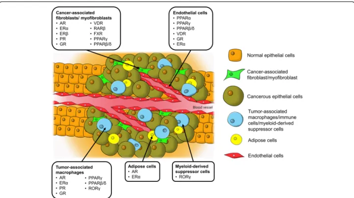
In this context, although NRs in tumor cells have been widely studied, their implications in TME are compa-ratively underappreciated. Given the pro-oncogenic roles of TME as well as the pronounced regulatory effects and excellent druggability of NRs, understanding the roles of these receptors in TME is of great interest. The impli-cated NRs in various tumor-supporting cells in TME presented in this review are illustrated in Fig. 1. Know-ledge of the NR expression profile not only helps to pro-vide a fundamental understanding in the realm of cancer
biology but also harbors enormous clinical value in cancer therapy. Thus, this review aims to highlight key findings of the biological functions of NRs in dif-ferent cell types presented in TME in relation to their pro−/ anti-tumor activities. The empirical findings are also discussed concerning the challenges, limitations and future direction of the current research paradigm with high hopes of developing a new anti-cancer strategy by exploiting NRs in TME.
Cancer-associated fibroblasts/myofibroblasts as key accomplices in tumor malignancy
Regulatory roles of CAF steroid hormone nuclear receptors in hormone-dependent cancers
Forming one of the most abundant cell populations in TME, CAFs are known to be pivotal modulators of tumorigenicity and cancer progression. A much larger number of studies have been conducted on CAFs than on other stromal cells in TME, particularly in terms of steroid hormone NRs. Therefore, this review of the actions of NRs in CAFs is subdivided into two parts in accordance with steroid and nonsteroid hormone NRs.
CAFs are primarily composed of fibroblasts and myofi-broblasts, of which the latter displays a mixed phenotype of fibroblast and smooth muscle cells by having a pro-minent rough endoplasmic reticulum of fibroblasts and contractile filaments (e.g., smooth-muscle actin) of smooth muscle cells [10]. The crosstalk between the tumor and CAFs assists tumor cells in acquiring unique characteristics such as enhanced proliferation, metastatic and angiogenic properties, immune evasion and che-moresistance [11, 12]. It has been postulated that dys-regulated activities of certain nuclear factors in CAFs could contribute to their tumor-supportive roles. CAFs have markedly distinct gene expression profiles of NRs compared with their normal cognate fibroblasts. Indeed, CAFs isolated from human breast tumors exhibit vastly different NR fingerprints compared with normal breast fibroblasts, as exemplified by the downregulation of THR-β, VDR, ROR-α, and PPAR-γ in CAFs [13]. Fur-thermore, NR signatures also differ among CAFs isolated from different types of tumors [13–15]. Such disparities in NR profiles could be an intrinsic characteristic of fibroblasts at different anatomical positions, or due to cellular signals released by different host cancer cells and other surrounding stromal cells. In this context, our recent study using clinical cutaneous squamous cell car-cinoma has confirmed the differential gene expression of NRs in CAFs compared with normal fibroblasts [15]. We have also shown that the transcriptomes of tumor cells cocultured with CAFs can be altered by reversing the expression pattern of selected NRs, namely, PPARβ/δ, VDR, AR and retinoic acid receptor (RAR)-β receptor, to result in functional changes such as impaired invasiveness,
reduced proliferation, and altered energy metabolism and redox response [15]. More importantly, when the squa-mous cell carcinoma cultures are exposed to conditioned medium from CAFs pretreated with either RARβ or AR antagonists, the CAF-induced cisplatin resistance is com-pletely abolished [15]. Our study strongly supports the druggability of NRs in TME, notably AR and RARβ, which can mediate a CAF-directed cancer therapy.
In line with our findings, AR in the tumor stroma has been consistently found to be a predominant factor in the prognosis of prostate cancer [16]. Nevertheless, unlike squamous cell carcinoma, in which the inhibition of AR of CAFs could be beneficial, low levels or loss of AR in the stromal cells of prostate cancer are associated with poorer clinical outcomes [17–22]. Such an asso-ciation is mind-boggling given that androgen deprivation therapy, which aims to suppress AR signaling in tumor cells, often serves as the frontline treatment of prostate cancer [23]. Genome-wide CHIPseq has revealed that AR in prostate CAFs has distinct binding sites and binding sequence motifs compared with tumor cells, suggesting differences in AR-regulated genes between the two cell populations [24]. This finding could explain the discrep-ancy in AR function between prostate CAFs and cancer cells. The tumor stroma liberates various androgen-re-sponsive growth factors and cytokines that modulate the cell fate, proliferation and drug sensitivity of prostate
cancer cells [25–27]. These paracrine factors are favorable for the growth of tumor cells present in this environment. Although ablation of ARs in CAFs could attenuate cancer proliferation [28], the loss of AR signaling activity is also linked to the onset of metastatic phenotypes such as in-creased stemness, enhanced cell migration and weakening of the extracellular matrix (ECM) structure and integrity [22, 29,30]. As a result, the suppression of AR in CAFs may potentially exacerbate the epithelial-mesenchymal transition and metastasis of prostate cancer, underpinning the association of AR loss in CAFs with adverse clinical outcomes in prostate cancer progression. In short, the pathological roles of AR in CAFs are well-implicated in the development of prostate cancer, making it an attrac-tive therapeutic target. However, considering the opposite effects of AR blockade in tumor and stromal cells, an ideal anti-androgenic agent should decrease tumor AR but enhance stromal AR activity [16]. It is also worth mentioning that the current understanding of AR in CAFs is mostly derived from hormone-dependent tumors, espe-cially prostate and breast cancers [16,31]. Thus, in light of the evidence mentioned above, it is worthwhile to extend research on AR to other types of tumors to better characterize its roles in cancer biology.
In addition to AR, steroid hormone NRs in CAFs, including ERα and β, PR and GR, are also relatively well-studied. The expression of ERα has been detected
Fig. 1 Tumor microenvironment, tumor-supporting cells and the identified nuclear receptors in cancer progression. AR, androgen receptor; ER, estrogen receptor; FXR, farnesoid X receptor; GR, glucocorticoid receptor; PPAR, peroxisome proliferator-activated receptor; PR, progesterone receptor; ROR, RAR-related orphan receptor; VDR, vitamin D receptor
in the CAFs of breast [13], endometrial [32], cervical [33] and prostate cancers [34], but not in colorectal carcinoma [35]. However, the clinical implications of ERα are diverse. In some studies, ERα-expressing CAFs have been reported to promote prostate and endometrial cancer cell proliferation [32, 36]; in other studies, CAFs attenuated prostate tumor cell invasiveness and immune cell infiltration by altering the levels of anti-angiogenic factors, ECM remodeling factors as well as chemokines, in addition to conserving chemosensitivity in certain breast cancer cell lines [37–39]. Similarly, divergent results have also been obtained in clinical biopsies, in which one association study found a positive correlation between ERα expression in CAFs with advanced prostate cancer stage [34], while the reverse trend was found in cervical cancer [33]. Despite these perplexing findings, a recent comparative transcriptomic study demonstrated differential expression patterns between CAFs isolated from early- and late-stage cervical cancer, with the latter being more metabolically and proliferatively active upon estradiol exposure [40]. Treatment with ER antagonists, namely, ICI182780 and methylpiperidino pyrazole, not only reverses the aforementioned changes but also sup-presses the expression of genes linked to angiogenesis and cell adhesion [40]. Additionally, liver receptor homolog-1 (LRH-1), which is an orphan NR, is tran-scriptionally responsive to estrogen treatment and ERα activation [41]. In breast cancer-derived CAFs, LRH-1, which is highly expressed in these cells, can upregulate aromatase (CYP19) gene expression [13,42]. This obser-vation is indicative of an ERα-mediated loop of estrogen biosynthesis via LRH-1 in CAFs, which may contribute to the increased tumor cell proliferation. Hence, disrup-ting the paracrine signaling directed by ERα in CAFs may be beneficial, making NR an exploitable target for
cancer therapy. However, further investigation is
warranted to clarify the conflicting results about the tumorigenic properties of ERα.
While ERα is well-implicated in TME of many hor-mone-dependent cancers, its role is less pronounced in the CAFs of breast cancer, likely because its expression is predominantly localized in the tumor epithelium in-stead of the surrounding fibroblasts [43–45]. In contrast, ERβ, which is the other ER isotype, is widely found in the breast cancer stroma [35, 46]. Despite their structural similarities, the bioactivities of ERα and β in tumor
epithe-lium are largely counteractive, whereby ERβ is
anti-proliferative and ERα-antagonizing [47,48]. Whether ERβ in CAFs also confers an anti-tumor effect is uncertain. One study revealed that progesterone and epi-dermal growth factor receptors are highly expressed in the uterine stroma of ERβ-knockout mice, especially when 17β-estradiol and progesterone are coadministered [49]. This phenomenon contributed to the hyperproliferation
and impaired cellular differentiation observed in the uterine epithelium of ERβ-knockout mice [49]. Con-versely, PR also exhibits ERα-antagonizing properties in tumor cells [50]. Its expression in cancer-associated stroma is repressed in comparison to benign stroma in prostate glands [51,52]. Stromal PR actively takes part in stromal cell differentiation [52]. Although conditioned medium from PR-positive CAFs has a negligible effect on prostate cancer cell proliferation, cell motility and mig-ration are vastly inhibited via the suppression of stromal-derived factor-1 and interleukin (IL)-6 [51]. These findings highlight the importance of stromal ERβ and PR in stroma-tumor epithelium crosstalk in modulating can-cer progression, but tissue-specific inhibition or activation of these NRs in CAFs is imperative to outline the feasibi-lity of exploiting them as therapeutic cancer targets.
Next, GR is differentially expressed in TME compared with normal tissues [53], with remarkably high expression in CAFs [54, 55]. In cancer-associated myofibroblasts,
treatment with dexamethasone successfully induces
nuclear translocation of GR, resulting in an anti-inflam-matory phenotype marked by the repression of IL-1β, monocyte chemoattractant protein 1, C-C motif ligand 5, tumor necrosis factor-α (TNFα) and intercellular adhesion molecules [56]. Coincidentally, several pro-invasive para-crine signals, such as tenascin C, hepatocyte growth factor, transforming growth factorβ (TGFβ), are also significantly suppressed [56]. Further investigation showed that dexamethasone-induced activation of GR in myofibro-blasts, but not in cancer cells, can nullify the proliferative effect of myofibroblasts on tumor cells and potentially in-hibit epithelial-mesenchymal transition, but it is associated with pro-migratory behavior [57]. Apart from the tumor epithelium, paracrine factors from myofibroblasts also interact with the surrounding endothelial cells to promote cell motility and angiogenesis [58]. These activities are
dampened by the conditioned medium from
dexamethasone-treated myofibroblasts together with a decline in urokinase-type plasminogen activator and angiopoietin-like protein-2 [58]. In general, GR activation in myofibroblasts exhibits tumor-inhibiting effects. It is, however, noteworthy that current evidence for this phenomenon originated from one research group, render-ing further validation pertinent.
Nonsteroid hormone nuclear receptors - Anti-tumor properties of VDR, PPARγ, RXR and FXR and pro-tumor effects of PPARβ/δ and RARβ in CAF
In addition to steroid hormone NRs, VDR in CAFs is also increasingly appreciated as a key anti-carcinogenic target. Ferrer-Mayorga et al. (2017) reported a positive correlation between the gene expression of stromal VDR with overall survival and progression-free survival in colorectal cancer [59]. Genes such asCD82 and S100A4,
which are responsive to calcitriol in CAFs, are also asso-ciated with clinical outcomes and stromal VDR expres-sion in patients with colorectal cancer, supporting a clinical value of VDR agonists in cancer treatment [59]. Conversely, pancreatic and hepatic TME is enriched by myofibroblast-like stellate cells, which upon activation, be-come proinflammatory, fibrogenic and tumor supportive [60, 61]. Based on a transcriptomic analysis, calcipotriol, which is a nonhypercalcemic vitamin D analog, maintains the quiescent state and modifies the secretomes of pancre-atic stellate cells by reducing the expression of inflam-matory cytokines, ECM components, and growth factors [62]. Similar trends have also been observed in hepatic stellate cells [63,64]. Combined therapy with gemcitabine plus calcipotriol tremendously improves the treatment outcomes of mice with orthotopic pancreatic ductal adenocarcinoma transplant, as evidenced by intratumoral aggregation of chemotherapy agents, a diminished tumor size and a higher survival rate [62]. A very recent report also suggests a regulatory role of VDR on CAF-liberated exosomal miRNA (e.g., miR-10a-5p and miR-181a-5p) [65]. Hence, exposure of CAFs to VDR ligands may modulate the stroma-tumor crosstalk not only via pa-racrine signaling but also by manipulation of the exosomal content. Despite promising results from preclinical studies, most clinical trials that employed vitamin D for cancer therapy and prevention have yielded underwhel-ming results, which reflects an inadequate understanding of VDR actions in both tumor and stromal cells [66–68]. Thus, an in-depth dissection of the biological roles of VDR in TME is critical to enable effective VDR-centric cancer treatment.
Several studies have also examined the activities of PPARs in CAFs. PPARγ has been found to be highly expressed in the myofibroblasts of colon adenocarcinoma biopsies, but not in normal colon tissues [69]. When hypoxic breast tumor cells are exposed to pioglitazone (PPARγ agonist) and/or 6-OH-11-O-hydrophenanthrene (RXR agonist), the resultant exosomes are unable to trigger CAF activation compared with exosomes from tumor cells subjected to the control treatment, suggesting that these NR agonists can disrupt the tumor-stroma crosstalk [70]. In the same study, coactivation of PPARγ and RXR in CAFs was found to effectively silence the pro-inflammatory response and metastatic phenotype by suppressing the expression of IL-6, carbonic anhydrase IX, metalloproteinase (MMP)-2 and MMP9 [70]. A simi-lar anti-proliferative effect of PPARγ activation on melanoma-derived CAFs has also been reported using the PPARγ agonist 15d-PGJ2 [71]. Accordingly, activation of PPARγ in CAFs could potentially act as a tumor suppressor by modifying the activation and supportive properties of CAFs in cancer development. Unlike PPARγ, which is associated with anti-tumor effects upon ligand
binding, PPARβ/δ in CAFs has a pro-tumor action. This phenomenon was clearly demonstrated in our recent study, in which the tumor burden was significantly lowered in fibroblast-specific PPARβ/δ knockout mice subjected to either chemical (azoxymethane or dex-tran sulfate sodium), genetic (APCmin/+) or combinatory (APCmin/+ with dextran sulfate sodium) tumorigenic induction [72]. Mechanistically, PPARβ/δ ablation in CAFs significantly escalates H2O2 liberation into the
TME, exposing the tumor epithelium to increased oxi-dative stress to subsequently trigger NRF2-mediated sig-naling that attenuates tumor growth [72]. The regulatory effects of PPARβ/δ on oxidative stress, reactive oxygen species production, and antioxidant mechanism are in line with a previous study examining the wound microenvi-ronment [73]. In short, both PPARγ and PPARβ/δ in CAFs play a significant modulatory role in cancer development, of which the former acts on the local inflammation and cancer invasiveness while the latter alters the redox balance in TME.
FXR is an integral regulator of genes responsible for lipid, cholesterol and bile acid metabolism [74]. Loss of function of FXR is strongly linked to carcinogenesis in the liver, intestines and colorectal region where the receptor is highly expressed [75, 76]. Interestingly, in breast cancer cells exposed to the FXR agonist GW4064, conditioned medium from CAFs fail to promote enhanced growth, motility, and invasiveness [77]. This observation reflects a neutralizing effect of FXR acti-vation on the tumorigenic paracrine signaling conferred by CAFs. Likewise, the characteristics of CAFs subjected to GW4064 are also profoundly altered. For instance, the genes involved in the cytoskeleton and cellular movement as well as a wide variety of growth factors are significantly downregulated, subsequently leading to loss of the tumor-supportive effects of CAFs [78]. The ability of an FXR inhibitor, guggulsterone, to completely reverse the GW4064-mediated anti-tumor effects further cor-roborates the necessity for FXR activation in eradicating the tumor-promoting features of CAFs [68]. In short, the evidence thus far for the benefits of FXR activation in CAFs is scarce, yet remarkably promising [78].
As mentioned earlier, our group has demonstrated that suppression of RARβ in CAFs via genetic knockdown or with an antagonist named LE135 consistently lowers the chemoresistance of tumor cells that are otherwise pro-moted by wild-type/untreated CAFs [15]. This result also complements a previous study that concluded that RARβ inhibition creates a hostile microenvironment that suppresses tumorigenesis through stromal remodeling, including impaired angiogenesis and reduced inflamma-tory cell recruitment and cancer-associated myofibro-blast numbers [79]. In fact, our study also predicts that activation of VDR and GR, as well as inhibition of AR in
CAFs, can potentiate the efficacy of chemotherapy, all of which are in excellent agreement with current under-standing of these NRs in CAFs, as discussed previously. Collectively, based on preliminary data from various sources, NRs in CAFs or myofibroblasts are undoubtedly druggable targets that could serve as a new strategy to improve the clinical outcomes of pre-existing thera-peutic approaches. For certain receptors such as AR and ERα, their pro-oncogenic roles in CAF could be dependent on the cancer types and biochemical signals, resulting in the contradictory findings obtained thus far. Hence, diversifying the research to other cancer types and escalating cell-based methodology to preclinical animal study are commendable efforts to strengthen the concept and clinical prospects of CAF-oriented cancer therapy via NR inhibition.
The steroid hormone nuclear receptors PPARs and RORγ are crucial mediators of TAM and MDSC formation
Apart from CAFs, TME is also occupied by numerous bone marrow-derived cells such as TAMs, MDSCs, neutrophils and tumor-infiltrating lymphocytes. Among these cells, TAMs and MDSCs are known to exhibit evident tumor-supporting and immune suppressive activities [80, 81]. Like CAFs, the steroid hormone NRs in TAMs also have profound impacts on cancer progres-sion. It is widely accepted that TAMs, which more closely resemble alternatively activated M2 macrophages, are activated by Th2 cytokines such as IL-4, IL-10, and IL-13 [82]. M2 macrophage polarization is also pro-moted by exposure of the monocytes to glucocorticoids, which stimulates GR activation [83]. This process is accompanied by a significant downregulation of proteins linked to lysosomal activity, antigen presentation, and proinflammatory proteins, indicating immunosuppres-sive effects [83]. Additionally, GR also functions syner-gistically with p38MAPK to regulate the expression a CD20 homolog, MS4A8A, the overexpression of which in TAMs significantly enhances the tumor burden [84]. Taken together, classic GR signaling may play a domi-nant role in the tumor-supporting activities of TAMs.
In contrast to GR, the role of AR, ER, and PR-dependent tumorigenesis is poorly defined. The pres-ence of TAMs influpres-ences the expression of ERα, ERβ and PR in tumor cells [85–87]. Reciprocally, the number of TAMs also appears to be modulated by steroid hor-mone NRs of tumor cells, particularly ER [88]. More-over, in wound healing and lung inflammatory studies, activation of AR, ERα and PR by their cognate steroid hormones would favor macrophage activation in an alternative manner, producing M2 macrophages that compel cellular repair and angiogenic processes [89–91]. The studies suggest that steroid hormones are vital determinants in the alternative differentiation of
macrophages to modulate pulmonary inflammation and wound recovery. However, there is no direct evidence supporting the contribution of AR, ER, and PR to the for-mation of M2 macrophages in TME. Thus, future research should focus on explicating the roles of these NRs in TAM formation and tumor-supporting events.
The three isotypes of PPARs, PPARα, PPARβ/δ, and PPARγ, are widely known to influence carcinogenic activities. However, current evidence is somewhat para-doxical concerning their roles in tumor cells, leading to the speculation that their actual functions could be dependent on the ligands, cancer types or even cancer stages [92]. In immune cells, PPARs also govern the fate of macrophage activation, likely because the maturation of macrophages is tightly linked to their metabolic state.
To enable alternative activation of macrophages,
immune cells must undergo oxidative metabolism, which is modulated by PPARs [93]. Macrophages that are unable to clear the metabolic checkpoint due to de-letion of PPARγ, PPARβ/δ and PPARγ coactivator 1β (PGC-1β), are incapable of expressing the alternative phenotype [94–96]. In contrast, treatment with PPARα or -γ agonists fosters the enrichment of M2-related biomarkers in macrophages [97]. Recently, a ligand-inde-pendent mechanism that involves PPARγ in TAM differ-entiation has also been described, which involves the cleavage of PPARγ by caspase-1 and thereby produces a 41-kDa receptor fragment that translocates into mito-chondria and interacts with medium-chain acyl-CoA de-hydrogenase [98]. This interaction shuts down the enzyme and attenuates fatty acid oxidation, leading to intracellular aggregation of lipid droplets that drive TAM differentiation [98]. These results support the pro-tumor activities of PPARγ via promoting TAM for-mation. Likewise, PPARβ/δ also seems to follow a simi-lar trajectory [99]. Notwithstanding, other empirical findings support a counterargument [100, 101]. The clinical use of thiazolidinedione is also not associated with an increased risk of many malignancies [102]. Collectively, the roles of PPARs in TAM differentiation and tumor progression undoubtedly remain an open topic necessitating further investigation.
RORs are classified as orphan NRs, which belong to a subfamily of thyroid hormone-like receptors. RORs are subcategorized into RORα, −β and -γ, the last of which is highly expressed in thymus and lymphoid tissues and linked to immune cell differentiation and immune system regulation [103]. Interestingly, RORγ is also a crucial element in hematological malignancies. For example, RORγ knockout mice are predisposed to thymic and lymphoblastic lymphomas [104, 105]. In addition, patients with multiple myeloma display an overexpression of RORγ in their peripheral blood mononuclear cells [106]. The roles of RORs in tumorigenesis vary in different
cancers [103]. Nonetheless, in TME, activation of RORγ with an agonist (SR1078) promotes the formation of MDSCs and TAMs [107]. RORγ-dependent myelopoiesis is mediated by key regulators such as Socs3, Bcl3, and C/EBPβ, as well as macrophage-specific transcription factors, including IRF8 and PU.1 [107]. In the same study, RORγ could confer pro-tumor effects by shielding MDSCs from apoptotic death, promoting tumor growth and restricting tumor-infiltrating neutrophils, while ablation of the receptor successfully attenuates these processes [107]. These results position RORγ as an attractive target, and hence, the pharmacological effects of RORγ antagonists or inverse agonists in TAMs and MDSCs with respect to tumor development are of immense interest.
To summarize, research on NRs in TAMs or MDSCs is still in its infancy. Most of the available studies emphasize the effects of NRs on the fate of macrophage activation. This information is critical not only to inhibit the alternatively activated M2 macrophage pathway, which subsequently reduces the TAM count, but also to achieve reprogramming of M2 to M1 macrophages to initiate tumoricidal effects such as the induction of proinflamma-tory and anti-tumor immune responses in TME.
Ceasing angiogenesis - targeting GR, PPAR and VDR of endothelial cells in TME
The vascular endothelium is an essential tissue that maintains blood perfusion in addition to regulating the trafficking of nutrients and leukocytes to surrounding tissues. In TME, the integrity of the vascular endothe-lium is often jeopardized by factors such as hypoxia and chronic growth factor stimulation. Genetic abnormalities are also not uncommon in tumor endothelial cells [108]. As a cumulative result of atypical physiological condi-tions and genetic mutacondi-tions, tumor endothelial cells differ significantly from normal endothelial cells by being highly proliferative, pro-angiogenic and more dis-organized and leaky regarding the vasculature [109,110].
Recent cancer research has identified PPARs as poten-tial therapeutic targets and prognostic indicators for cancer therapy. Indeed, the expression of PPARγ is asso-ciated with slower progression and a lower incidence of tumor recurrence in bladder cancer [111]. This corre-lation is lost when certain angiogenic factors, namely, basic fibroblast growth factor and platelet-derived endo-thelial growth factor, are coexpressed in the tumors, indicating a possible role of PPARγ in angiogenesis in cancer progression by interacting with these growth factors [111]. Activation of PPARγ in endothelial cells is predominantly linked to anti-angiogenic activities, as exemplified by decreased expression of pro-angiogenic factors, reduced proliferation, impaired endothelial cell migration and tubule formation [112], but conflicting results have also been reported [113, 114]. Similar to
PPARγ, fenofibrate-induced PPARα activation in various tumor cell lines concomitantly suppresses proangiogenic vascular endothelial growth factor (VEGF) biosynthesis and increases anti-angiogenic thrombospondin 1 and endostatin [115]. These bioactivities are translated into reduced endothelial cell proliferation and neovasculari-zation as well as impaired growth of the subcutaneous tumor xenograft in mice [115]. Unlike PPARα and –γ, PPARβ/δ appears to be proangiogenic. Treatment with the PPARβ/δ ligand GW501516 promotes endothelial tube formation, whereas the maturation of microvessels in tumors is severely disrupted in PPARβ/δ knockout mice, leading to diminished blood flow to the tumors [116, 117]. Taken together, all three isotypes of PPARs are actively involved in the angiogenesis performed by endothelial cells, which is one of the most critical processes in cancer development, sustaining the rapid expansion of tumor cells and opening the window for the metastatic process. However, the findings are not strictly based on tumor-derived endothelial cells. Given the functional variations between tumor-associated and normal endothelial cells, further validation is pertinent.
Next, VDR is closely associated with the development of endothelial cells in TME. In this context, calcitriol, which is an active metabolite of vitamin D, has been widely studied regarding its roles in bone and mineral metabolism, as well as the differentiation of both normal and malignant cells. At a low dosage, calcitriol exhibits an anti-proliferative effect on cancer cells such as breast, colon, and prostrate while promoting differentiation, cell cycle arrest and eventually apoptosis [118]. A similar growth inhibitory effect has also been observed in tumor-derived endothelial cells, but not in normal ones [119]. Generally, increased levels of VDR ligands trigger a self-regulatory pathway by enhancing the expression of CYP-24b, a key enzyme in vitamin D catabolism [120]. As a result, VDR ligands are degraded and unable to trigger VDR-mediated anti-proliferative effects [121]. However, overexpression of CYP-24 has been reported in various cancers such as prostate, colon and breast cancer, explaining the varying calcitriol sensitivity and calcitriol resistance in these patients [122]. Moreover, the anti-proliferative effect of VDR in endothelial cells also relies on the epigenetic silencing of CYP-24, which is achieved via hypermethylation at the CpG islands of CYP-24 promoter regions [123]. Transcriptional acti-vation of CYP-24 is prevented by the hypermethylation pattern, leading to growth inhibition in tumor-derived endothelial cells [123]. One study has also suggested a link between VDR and angiogenesis in TME modulated by a pro-oncogenic protein named DKK-4 [124]. The expression of DKK-4 is inversely correlated to that of VDR, while endothelial cells are more prone to migrate and form microvessels when they are exposed to
conditioned medium from DKK-4-expressing cells [124]. The pro-tumor effects of DKK-4 are effectively elimi-nated by treatment with calcitriol. Thus, these studies support the use of VDR ligands that target the tumor endothelium with minimal disturbance to the normal vasculature.
Multiple studies have demonstrated the anti-angiogenic effects of glucocorticoids in normal and malignant cells, as well as during wound healing [125,126]. In tumor cells, glucocorticoids exert a direct inhibitory effect on the secretion of VEGF, which can be reversed by GR antago-nist treatment [127]. This observation suggests that the anti-angiogenic effect is GR-dependent. Logie et al. (2010) reported that glucocorticoids have a negligible effect on the proliferation, viability and migration properties of endothelial cells, but instead, the hormone enhances thrombospondin-1 expression and impairs cell-cell con-tact, thus preventing the formation of endothelial tubules even in the presence of VEGF and prostaglandin F2a[128].
The potent angiogenic inhibitory activity of GR has also prompted research on the nanosized drug delivery system to maximize the anti-tumor effect of GR [129].
Unlike GR, ERα is linked to the pro-angiogenic process in TME. Treatment with 17β-estradiol increases the vessel density and stabilizes the endothelium vascu-lature in tumors, making the blood vessels more resist-ant to insults from hypoxia and necrosis [130]. Increased neovascularization in the tumor environment ensures adequate oxygenation of the tumors and minimizes tumor cell death due to the hypoxic environment [130].
However, ERα-dependent angiogenesis is primarily
mediated by Tie2-expressing cells, which are not of hematopoietic origin [130]. Therefore, the true identity of Tie-2 positive cells in TME, and their relationship with tumor endothelial cells, remain to be clarified.
Adipose cells are emerging players in tumor aggressiveness
Adipocytes, also known as fat cells, are regulators of human physiological processes such as tissue homeosta-sis, and they are the primary site for energy storage in the form of intracellular triglycerides packaged in lipid droplets [131]. Additionally, they are also endocrine cells that secrete hormones and cytokines to regulate human physiological processes such as inflammation and the reproductive system [132]. The functions of adipose cells in TME resemble those of fat depots, but in a tumor-supportive manner [133]. Emerging evidence also supports a role for dysfunctional adipose tissues in field cancerization mediated by prolonged local inflammation [134]. However, our understanding of the role of adipose cells in TME is still considerably limited.
One recent study has shown that the recruitment of preadipocytes occurs more readily in prostate cancer
cells than normal prostate tissues, a process that enhances the invasiveness of prostate cancer in mice with orthotopic xenografts [135]. Mechanistically, neigh-boring adipocytes significantly increase the expression of miRNA-301a in tumor cells, which serves to suppress AR signaling in these cells [135]. The inhibition of AR signaling is followed by alterations in the gene expres-sion of TGF-β via the serine/threonine kinase receptor or TGF-β receptor and its downstream genes such as Smad3 and matrix-metalloproteinase-9, fueling meta-static processes [135]. Coculturing human Simpson Golabi Behmel Syndrome (SGBS) preadipocyte cells, which are considered to be a representative in vitro model of white preadipocytes, and ER-positive MCF7 breast cancer cells results in the suppression of ERα expression in MCF7 cells [136]. Cohabitation of preadi-pocytes and MCF7 cells also significantly enhances the epithelial-mesenchymal transition of MCF7 tumor cells, as documented by overexpression of FOXC2 and TWIST1, and changes in N- and E-cadherin expression [136]. As a consequence, the expression of HIFα, TGF-β and lectin-type oxidized LDL receptor 1 in SGBS adipo-cytes are elevated [136]. Both studies have demonstrated that the presence of adipose cells in TME can impact both NR signaling and oncogenic processes in cancer cells. However, the studies did not aim to delineate the activities of NRs in tumor-associated adipose cells and their contribution to cancer progression, an aspect that has been minimally explored to date. In light of the emer-ging roles of adipose cells in field cancerization as well as the predominant actions of various NRs in adipocyte biology, it will be interesting to unearth this relationship.
Implications of existing research for stroma-directed anticancer therapy via nuclear receptor manipulation
For years, targeting the tumor epithelium has been the sole cornerstone of cancer research, which has resulted in the clinical use of aggressive therapeutic methods such as surgery, radiation and chemotherapy to elimi-nate cancerous cells regardless of the inflicted extensive collateral damage. However, the effectiveness of tra-ditional anti-cancer strategies is increasingly challenged by treatment failures such as interpatient responsiveness, onset of chemoresistance, and local and distal recurrence, which are partly attributable to the genetic heterogeneity and genome instability of tumors and continuous tumor evolution [137]. Tumor evolution follows a Darwinian model, which also predicts the insufficiency of targeting the cancer epithelium alone, underscoring the need for alternative therapeutic strategies.
Stroma-directed anticancer therapy will require a different therapeutic approach aimed at multiple and interacting cells. Stromal cells are generally considered to be more genetically stable, and thus the occurrence of
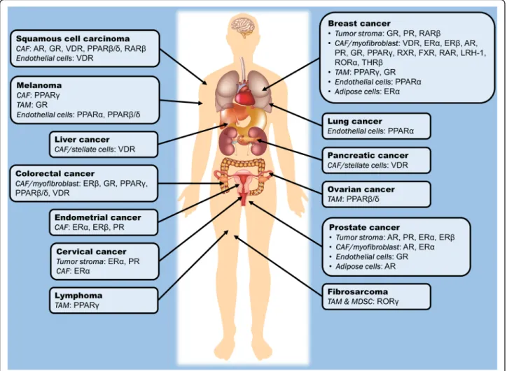
mutations that may lead to resistance to drug treatments are minimal compared with the large tumor mutation burden observed in cancer cells. By consolidating the NR profile of various stromal cells across different tumor types, we can highlight NRs that have been thus far identified to regulate the assistive properties of tumor stroma in carcinogenesis, as summarized in Table 1and Fig. 2. Certain NRs are clearly consistently observed across different tumor types; for instance, VDR, PPARs, ER, GR and AR in CAFs, as well as GR and PPARs in TAMs and endothelial cells. Modulating the activities of these NRs in stromal cells may potentially serve as a common adjunct therapy for the treatment of a wide range of cancers. In this context, by targeting NRs in stromal cells, the resultant physiological changes and drug responses could be more predictable, explaining why selected NRs, notably PPARs and GR, are consis-tently found to be crucial modulators of tumorigenesis in a cancer type-independent manner.
For stroma-directed therapy to be a viable strategy as part of a multimodality approach or as adjunctive treat-ment to conventional tumor treattreat-ment, we also need to address the relative population of different stromal cells in different tumor types. For example, CAFs are rela-tively rare in brain, renal and ovarian cancers. In such instances, the depletion of CAFs or the disruption of CAF functions is likely to provide only marginal benefits. Similarly, while next-generation cancer treatment using immunotherapies such as PD-1 checkpoint blockade and Chimeric Antigen Receptor T-cell (CART) therapy are swiftly gaining attention, the efficacy of CAR-T therapy is dependent on the immune cell interactions in the TME [138, 139]. A recent characterization of immune infiltrates has shown that tumor genotypes, such as the tumor mutation burden, determine immunophenotypes and tumor escape mechanisms [140]. In cases where immunotherapy is less successful, stroma-directed therapy targeting other stromal cells may rise to be the predomi-nant player. Moreover, if the efficacy and universality of stroma-directed therapy by targeting NRs are validated, the strategy can even be used to treat rare cancers simply because of the comparable physiological functionality of stromal cells in TME. These speculations and effectiveness of NR-based stroma-directed therapy can be further tested by extensive exploration of the NR signatures in TME across different types of cancers.
Limitations, challenges, and future perspectives
To a certain extent, manipulating NRs of key tumor-supporting cells can sensitize tumor cells to anti-cancer treatments by interfering with the stroma-tumor cross-talk. However, current knowledge is still too incomplete for reliable translation into favorable clinical outcomes for different cancer types because of several limitations.
First, the available data are derived primarily from hormone-dependent tumors, most notably breast and prostate cancers. Hence, our understanding of the roles of NRs in TME is fundamentally based on cancer-associated cells that are more actively involved in steroid hormone modulation and signaling. The effects of steroids differ from cancer to cancer [141], raising concerns about the generalizability of the results to cancers that are less hormone-dependent. Second, concerning the abovemen-tioned limitation, current findings mostly include studies of steroid hormone NRs such as GR, ER, AR, and PR, be-cause the development of hormone-dependent cancers is highly sensitive to steroids, facilitating detection of the biological roles of steroid receptors in tumorigenesis. As a result, our knowledge about NRs in TME is markedly skewed towards steroid receptors. In contrast, orphan NRs such as ERRs, RORs and LRH-1 have demonstrated a strong linkage with carcinogenesis [142]. However, exploiting them as a potential cancer therapy is underap-preciated due to the lack of well-characterized ligands. This situation is anticipated to change in the near future because the US Food and Drug Administration has re-cently approved the first use of RNA-based gene silencing drug (siRNA) to knock down the expression of defective transthyretin for the treatment of polyneuropathy in patients with hereditary transthyretin-mediated amyloi-dosis [143, 144]. Given that targeting orphan NRs with RNA interference technology could someday become a therapeutic option, the recent approval is believed to have sparked more intensive research on the impacts of orphan NR suppression in cancer development.
Furthermore, the roles of NRs in TME have been established mainly based on cell culture studies via coculturing methods or with the use of conditioned medium from tumor-supporting cells. Empirical data from in vivo animal studies of TME are limited because cell-specific activation or inhibition of an NR, especially with a pharmacological approach, is remarkably challen-ging in animal models. Although genetic engineering can be used to obtain targeted stimulation or knock-down in animals [36, 37], it is associated with tedious preparation, relatively high costs and arduous adminis-tration, rendering this approach less desirable in actual clinical settings compared with the use small molecules. However, cell-specific modulation of the specifically targeted NR is crucial because the same receptor can have opposing effects in different cancer-associated cells. This phenomenon is demonstrated by GR, the activation of which in cancer-associated myofibroblasts reduces tumor proliferation [57] but promotes the M2 pheno-type in macrophages, thus contributing to TAM diffe-rentiation and consequently tumor promotion [83]. Hence, given the heterogeneity of cellular populations in TME and their diverse physiological response to NR
Table 1 Summary of existing research studies that exploited NRs in different tumor stromal cells and investigated the impacts on carcinogenesis and tumor mi croenvironment Strom al cell type s Can cer type s Mod els Target NR(s) Agoni sts/ antagon ists K e y fin dings Referen ces CAF Cut aneous squam ous cell carcinoma (SCC ) Cell culture; Mice with SC C + CAF xenog raft 48 know n NRs in cell-bas ed studies; AR an d RAR β in mi ce mod els Tra nsfection of siRNA/e xpression vect ors of targeted NRs into CAFs; PPAR β /δ agonis t – GW07 42; VDR agonis t – EB 1089; GR agonis t – Flut icasone propi onate; RAR β antagonist – LE13 5; AR an tagonist – Bical utamide • PP AR β /δ , VDR, GR , RAR β and AR in CAF s are import ant mod ifiers of tumori gen ic activit ies. • Co ncurrent therapy o f cisplatin, LE135 and bical utami de atten uated chemor esistanc e in mice tumor xenog rafts. [ 15 ] CAF Pros tate canc er Cell culture; Mice with prostate cance r (PC3) + CAF xenog raft AR Tra nsfection of AR-exp ressing vect ors into CAFs •AR -ex press ing stromal cells sup pressed prostate cancer growt h and invas ivenes s in vitro an d in vivo. [ 20 ] CAF Pros tate canc er Cell culture; Mice with prostate cance r (PC3) + CAF xenog raft AR Tra nsfection of AR-exp ressing vect ors into CAFs • Lo w stro mal AR exp ression decreased castration-ind uced apoptosis. • Lo ss of AR sig naling in CAFs dis rupted extrace llular matrix integ rity, prom oting cancer cell s invas ion. [ 22 ] CAF Pros tate canc er Cell culture AR Tra nsfection of siRNA into CAF s • K nockdow n of AR in CAF s downre gulat ed the expre ssion of variou s growt h factors and im paired the growt h of tumor cells. [ 28 ] CAF Pros tate canc er Cell culture AR Agoni st – R188 1; Anta gonist – RD162 • Mi gration of prostate cance r cells was inhibit ed by condit ioned medi um of CAFs treated with R1 881, but was reversed by RD162. [ 29 ] CAF Pros tate canc er Cell culture AR Kno ckdown wit h AR antise nse oligo nucleo tides • Su ppression of AR expre ssion in CAFs inhibit ed cancer cell growt h, but promo ted stem ce ll phenoty pes. [ 30 ] CAF Bre ast cancer Cell culture AR Agoni st – Mibo lerone • Exp osure of cond itioned me dium from Mibolero ne-treate d CAFs reduced bre ast cancer cell mot ility. [ 31 ] CAF Pros tate canc er Cell culture; Mice with prostate cance r (22RV 1) + CAF xenog raft ER α Tra nsfection of ER α -ex pressing vect ors into CAFs • C onditi o n e d m ed ium fr o m ER α-exp re ssing CA Fs sti m ul ated pr o liferat ion o f various pro stat e cancer cell lines. • Co -implantation of ER α -ex pressing CAFs and prostate canc er cells increased tu mor size in mi ce. [ 36 ] CAF Pros tate canc er Cell culture; Mice with prostate cance r (22RV 1) + CAF xenog raft ER α Tra nsfection of ER α -ex pressing vect ors into CAFs • Stro mal ER α reduced cancer cell invas ion. • Mi ce co-i njected with ER α-expre ssing CAF s and prostate canc er cells had le ss tumor foci, less meta stases and reduced angiogenes is. [ 37 ] CAF Pros tate canc er Cell culture; Mice with prostate cance r (22RV 1) + CAF ER α Tra nsfection of ER α -ex pressing vect ors into CAFs •ER α-expre ssing CAF s suppre ssed cance r invasive ness via reduc ed macrophage infiltration . [ 38 ]
Table 1 Summary of existing research studies that exploited NRs in different tumor stromal cells and investigated the impacts on carcinogenesis and tumor mi croenvironment (Continued) Strom al cell type s Can cer type s Mod els Target NR(s) Agoni sts/ antagon ists K e y fin dings Referen ces xenog raft CAF Bre ast cancer Cell culture ER α , P R U sing CAFs isolated fr om ER α + /P R + or ER α −/P R −br ea st tum o rs • Can cer cell s co-cultured with ER α + /PR + tumor-derived CAFs had highe r tamoxi fen sensitivity. [ 39 ] CAF Cer vical canc er Cell culture ER α Agoni st – Estra diol; Anta gonist – ICI 18278 0, methyl piperidino py razole • ER α antagon ists downre gulated genes associ ated with cell cyc le, meta bolism and an giogeni c proces ses. [ 40 ] CAF Pros tate canc er Cell culture PR Tra nsfection of PR α -and PR β -ex pressing vecto rs into CAF s • Co nditione d medi um from PR -expre ssing CAFs inh ibited canc er cell migrat ion and invasive ness, but not cell proliferation. [ 51 ] CAF Pros tate canc er Cell culture PR Tra nsfection of PR α -and PR β -ex pressing vecto rs into CAF s • PR reg ulated prostate stromal cell different iation. [ 52 ] CAF Co lorectal canc er Cell culture GR Agoni st – Dex amet hasone • D exametha sone induced GR translocation into CAF nuc leus, negatively regul ating the expre ssion of pro-i nflamm atory genes an d paracrine fact ors that promo te canc er invas iveness. [ 56 ] CAF Co lorectal canc er Cell culture GR Agoni st – Dex amet hasone • Co nditione d medi um from dexam ethasone -treated CAFs de creased cancer cell proli feration and invasive ness. [ 57 ] CAF Co lorectal canc er Cell culture GR Agoni st – Dex amet hasone • Co nditione d medi um from dexam ethasone -treated CAFs im paired endothe lial cell migrat ion by alt ering CAF secret ome. [ 58 ] CAF Panc reatic cancer Cell culture; Mice with panc reatic ductal ad eno car cin o ma (PDA) xen ograf t VDR Agoni st – Calci pot riol • Calci pot riol main tained the quies cent state of pancreatic st ellate cell s. • Co -administration of calcipotriol and gemcitabine de creas ed tumor volume , enhanc ed intratumoral gemcitabine an d increased sur vival rates of mice with tumor xenograf t. [ 62 ] CAF Live r canc er Cell culture; p6 2KO mi ce VDR Agoni st – Calci pot riol • p6 2 is a medi ator of calc ipotriol -induc ed VDR activation which pre vented hep atic stellate cell activation. [ 64 ] CAF Panc reatic cancer Cell culture VDR Agoni st – Calci triol • Calci trio l mod ified miRNA compos ition in CAF exos omes. [ 65 ] CAF Bre ast cancer Cell culture PPAR γ, RXR PPAR γ agonist – Piogli tazone ; RXR agonis t – 6-OH -11-O-hyd rophen anthren e • PP AR γ/R XR agonists inh ibited NF-κB and me talloproteinas e activ ities in CAFs. [ 70 ] CAF Me lanom a Cell culture PPAR γ PPAR γ agonist – Ciglitazone , trog litazone, W Y 14643 , 15d-PG J2 • 15 d-PGJ2 inh ibited the growth of CAF s and tu be format ion of endothe lial cells. [ 71 ] CAF Co lorectal canc er Cell culture; PPAR β /δ PP AR β/ δ gen e knock out • Fi broblast-specific PP AR β/ δ knocko ut mice [ 72 ]
Table 1 Summary of existing research studies that exploited NRs in different tumor stromal cells and investigated the impacts on carcinogenesis and tumor mi croenvironment (Continued) Strom al cell type s Can cer type s Mod els Target NR(s) Agoni sts/ antagon ists K e y fin dings Referen ces Fibro blast-specific PP AR β /δ knock out mi ce had prolon ged survival and fewer intesti nal polyps . • CAF s with PP AR β/ δ dele tion red uced oxidati ve stress in canc er epith elium. CAF Bre ast cancer Cell culture; Mice with bre ast cance r (MCF-7 ) + CAF xenog raft FXR Agoni st – GW40 64 • Co nditione d medi um from GW406 4-treated CAFs inhibit ed leptin signali ng, growth , motil ity and invasive ness of cance r cell s. • GW 4064-t reated tumors ha d small er sizes in in vivo xenog raft studies. [ 77 ] CAF Bre ast cancer Cell culture FXR Agoni st – GW40 64 • GW 4064 reduced m igration and contrac tility of CAFs besides inh ibiting growth and motil ity of cance r cells. [ 78 ] CAF Bre ast cancer RAR β knock out mi ce RAR β RAR β ge ne knoc kout •RAR β knock out mice had reduced angiogenes is, inf lammatory cell infiltration an d myof ibroblas t coun t. [ 79 ] TAM – Cell culture GR Agoni st – Dex amet hasone • D exametha sone-depe ndent GR activation promo ted alternative differ entiation of monoc ytes to macropha ges with a M 2 phenoty pe. [ 83 ] TAM Bre ast cancer Mice with bre ast cance r (TS/A) + TAM xenog raft GR Agoni st – Gluc ocortico id • TAMs expose d to a mi xture cont aining condit ioned medi um from MS4A8A -ex press ing tumors , interl eukin -4 and glu cocort icoids enhanc ed tumor growth in mice with tumor xenog raft. [ 84 ] TAM – Inf lammatory cell -specific ER α an d ER β knock out mice ER α ,E Rβ ER α and ER β gene knocko ut; ER agonis t – 17 β -estradiol • ER α signaling prom oted alternative activation of macrophage s. [ 90 ] TAM Bre ast cancer Cell culture; Fema le MMT V-PyMT mi ce PPAR γ –• Cle avage of PPAR γ by cas pase-1 promo ted TAM differ entiation . • Inh ibition of caspase-1 atte nuated caspase-1/PPAR γ interact ion and suppressed tumor growth. [ 98 ] TAM O varian cance r Cell culture PPAR β /δ Agoni st – L165 041; Inve rse agonist – ST247 , PT-S 264 • Ac tivation of PPAR β /δ upr egula ted immu nity-and tumori genes is-related genes in TAMs . • Inve rse agonis ts of PPAR β /δ revers ed the abnorm al gen e expre ssion . [ 99 ] TAM – Cell culture PPAR γ Agoni st – Rosi glitazone , 15d-P GJ 2 • PP AR γ agonis ts reversed the suppressive effect of TAMs on anti tumor cytotoxi c T-cells. [ 100 ] TAM Bre ast cancer Cell culture; Macrop ha ge PPAR γ knoc kout mi ce PPAR γ Agoni st – Rosi glitazone • Macro phage PP AR γ abl ation prom oted breast tumor growth and nullifie d anti-tumor effect s o f rosiglitazone. [ 101 ]
Table 1 Summary of existing research studies that exploited NRs in different tumor stromal cells and investigated the impacts on carcinogenesis and tumor mi croenvironment (Continued) Strom al cell type s Can cer type s Mod els Target NR(s) Agoni sts/ antagon ists K e y fin dings Referen ces TAM & MDSC – Mice with fibro sarcoma (MN/MCA1) xenograft; MMTV-PyMT mice ROR γ RORC1 ge ne knoc kout • ROR γ protect ed MDSCs from apoptos is, promo ted TAM differ entiation an d prevented neu trop hil infiltrat ion into tumor, thu s leading to tumor growth and me tastasis . [ 107 ] End othelia l cell – Cell culture; Tie2 CrePPAR γ flox/flox mi ce PPAR γ PP AR γ ge ne knoc kout • D eletion of PP AR γ im paired an giogene sis and cell ular migrat ion in vitro an d in vivo. [ 114 ] End othelia l cell Me lanom a, lung cance r, gli oblastoma, fibro sarcoma Cell culture; Mice with me lanom a (B16-F 10), Lewis lung car ci nom a, g lio bl as to m a (U87) or fibrosarcoma (HT1080) xenog raft PPAR α PP AR α gene knocko ut; Agoni st – Fen ofibrate, gemfib rozil, be zafibrate, WY14 643 and 5, 8, 11 , 14-e icosatetray noic acid • Fe nofibrate strongly suppre ssed endothe lial cell prolif eration, angiogenes is and prim ary tumor growth in mice. • An ti-angi ogenic eff ect of feno fibrate was revers ed by PP AR α knoc kout. [ 115 ] End othelia l cell – Cell culture; C57B L6 mi ce PPAR β /δ Agoni st – GW50 1516 • GW 50151 6 induced end othelia l cell proliferation an d angioge nesis in vitro and in vivo. [ 116 ] End othelia l cell Lung cance r PP AR β /δ knock out mi ce PPAR β /δ PP AR β/ δ gen e knock out •PP AR β/ δ knoc kout im paired end othe lial cell maturation, causin g dimini shed blood flo w to the tumors and abnorm al microvascular structures. [ 117 ] End othelia l cell Sq uamous cell carcin oma Cell culture VDR Agoni st – Calci triol • Tu mor-derived endothe lial cells were sensitive to the anti-prol iferati ve effect s of calc itriol. [ 119 ] End othelia l cell Sq uamous cell carcin oma Cell culture VDR Agoni st – Calci triol • Calci trio l induced cell cycle arre st and apoptosis in tumor-derive d end othelia l cells, which we re attri butab le to CYP2 4 inhibit ion. [ 121 ] End othelia l cell Sq uamous cell carcin oma Cell culture VDR Agoni st – Calci triol • Me thylation silencing of CYP2 4 prom oter led to differ ential sen sitivit y to calcitriol-depe nde nt growth inh ibition in endothe lial cells. [ 123 ] End othelia l cell – Cell culture GR A g o n is t– Dexa metha son e, cort isol; Anta gonist – RU384 86 • GR agon ists block ed microvessel tubule formation , but did not affe ct viab ility. The anti-angi oge nic effect s were revers ed by RU3848 6. [ 128 ] End othelia l cell Me lanom a Cell culture; Mice with me lanom a (B16. F10) xenog raft GR Agoni st – Predn isolone, dexam ethasom e, budes onide, methy lprednisolon e • All GR agon ists inhib ited tumor size and growth of end othe lial cell s. [ 129 ] Extra- hemat opoie tic Tie2-pos itive cell s Me lanom a, lung cance r, bre ast cancer Ovariectom ized mice with me lanom a (B16K 1), Lewis lung car cinoma (LL2) or bre ast canc er (4 T1) ER α Agoni st – Estra diol • Extra-hem atopo ietic Ti e2-expre ssing cells were res ponsible for inc reased tu mor growth and intratumoral vessel density induced by estradiol treatmen t. [ 130 ]
Table 1 Summary of existing research studies that exploited NRs in different tumor stromal cells and investigated the impacts on carcinogenesis and tumor mi croenvironment (Continued) Strom al cell type s Can cer type s Mod els Target NR(s) Agoni sts/ antagon ists K e y fin dings Referen ces xenog raft Adipoc yte Pros tate canc er Cell culture; Mice with prostat e cancer (22RV1) xe nogr af t AR –• Re cruitment of adi pocyte s to prostate ca ncer cells e nha nced ca ncer inva siv e ness vi a suppression of AR ac tivity and indu ction of TG F-β 1/Sm ad /MMP9 signa ls. [ 135 ] Adipoc yte Bre ast cancer Cell culture ER α –• Adi pocyte s exp osed to hyp oxic cond ition triggere d ER α suppre ssion and prom oted endothe lial-t o-mesen chym al transit ion of breast cance r cell s. [ 136 ]
modulation, future research should also focus on the development of cell-specific drug delivery to achieve targeted manipulation of NR signaling in relevant cells.
The effects of NRs in TME on exosomes have scarcely been explored. Considering the vital roles of exosomes in cell-cell communication, which mediates various oncogenic processes, it is worthwhile to investigate how NR signaling in cancer-supporting cells calibrates the stroma-tumor interaction by regulating the exosomal content and liber-ation. Additionally, stroma-tumor communication is a dy-namic and reciprocal action. Therefore, understanding how neighboring cancer cells affect NR signaling in the cancer-associated cells and downstream functional alter-ations can further reveal the true nature of TME. Essen-tially, in-depth dissection of the interplay between tumor-supporting cells and malignant cells may reveal add-itional exploitable targets to improve cancer therapy.
Conclusions
NRs of tumor-supporting cells in TME play an essential role in various oncogenic processes. The NR signature of TME can serve as a crucial marker to pinpoint the fragility of the disease and guide the therapeutic strat-egy, with the ultimate goal of improving cancer progno-sis. In light of the striking druggability of NRs, the future clinical prospect of developing a TME-oriented cancer therapy by targeting these receptors is promis-ing. Among the 48 NRs in humans, the oncogenic functions of VDR, PPARs, AR, ER and GR in tumor-supporting cells are the best-characterized to date. Evidence of other receptors, such as RARβ, RORγ, and FXR, is limited yet promising. Given the
heterogen-eity of cellular populations within TME, more
intensive research in understanding the molecular mechanisms of cell-cell interactions and how to
Fig. 2 Summary of the so far identified nuclear receptors in the tumor microenvironment which play an active role in the modulation of oncogenic processes in different cancer types. AR, androgen receptor; CAF, cancer-associated fibroblast; ER, estrogen receptor; FXR, farnesoid X receptor; GR, glucocorticoid receptor; LHR-1, liver homolog receptor-1; MDSC, myeloid-derived suppressive cells; PPAR, peroxisome proliferator-activated receptor; PR, progesterone receptor; RAR, retinoic acid receptor; ROR, RAR-related orphan receptor; RXR, retinoic X receptor; TAM, tumor-associated macrophage; THR, thyroid hormone receptor; VDR, vitamin D receptor
master intercellular communication is of paramount importance. The ability to exploit NRs in TME in a highly specific and precise manner, in this case, can lay the foundation for precision medicine in cancer therapy and may even allow us to transform tumor-supporting cells into tumor foes.
Abbreviations
AR:Androgen receptor; CAF: Cancer-associated fibroblast; ECM: Extracellular matrix; ER: Estrogen receptor; FXR: Farnesoid X receptor; GR: Glucocorticoid receptor; IL: Interleukin; LRH-1: Liver receptor homolog-1; MDSC: Myeloid-derived suppressor cells; NR: Nuclear receptor; PGC-1β: PPARγ coactivator 1β; PPAR: Peroxisome proliferator-activated receptor; PR: Progesterone receptor; RAR: Retinoic acid receptor; ROR: RAR-related orphan receptor; RXR: Retinoic X receptor; TAM: Tumor-associated macrophage; TGFβ: Transforming growth factorβ; TME: Tumor microenvironment; TNFa: Tumor necrosis factor α; VDR: Vitamin D receptor; VEGF: Vascular endothelial growth factor Acknowledgments
Not applicable. Funding
This work was supported by Singapore Ministry of Education under Singapore Ministry of Education Academic Research Fund Tier 2 (2014-T2 –1-012; 2017-T1–002–103) and Academic Research Fund Tier 1 (2015-T1–001-034) to NST, and by the Lee Kong Chian School of Medicine, Nanyang Technological University Start-up Grant to WW.
Availability of data and materials Not applicable.
Authors’ contributions
NST and WW designed the framework of the review. HSC and JXTL collected related studies and drafted the manuscript. NST, WW, HSC and JXTL participated in the manuscript editing. All authors have read and approved the final manuscript, and consent to publish.
Ethics approval and consent to participate Not applicable.
Consent for publication Not applicable. Competing interests
The authors declare that they have no competing interests.
Publisher’s Note
Springer Nature remains neutral with regard to jurisdictional claims in published maps and institutional affiliations.
Author details
1School of Biological Sciences, Nanyang Technological University Singapore,
60 Nanyang Drive, Singapore 637551, Singapore.2Lee Kong Chian School of Medicine, Nanyang Technological University Singapore, 11 Mandalay Road, Singapore 308232, Singapore.3INRA ToxAlim, UMR1331, Chemin de Tournefeuille, Toulouse Cedex 3, France.4Center for Integrative Genomics,
University of Lausanne, Le Génopode, CH-1015 Lausanne, Switzerland.
Received: 12 December 2018 Accepted: 21 February 2019
References
1. Metivier R, Reid G, Gannon F. Transcription in four dimensions: nuclear receptor-directed initiation of gene expression. EMBO Rep. 2006;7:161–7. 2. Sever R, Glass CK. Signaling by nuclear receptors. Cold Spring Harb Perspect
Biol. 2013;5:a016709.
3. Stender JD, Kim K, Charn TH, Komm B, Chang KCN, Kraus WL, et al. Genome-wide analysis of estrogen receptorα DNA binding and tethering
mechanisms identifies Runx1 as a novel tethering factor in receptor-mediated transcriptional activation. Mol Cell Biol. 2010;30:3943–55. 4. Gadaleta RM, Magnani L. Nuclear receptors and chromatin: an inducible
couple. J Mol Endocrinol. 2014;52:R137.
5. Richer JK, Jacobsen BM, Manning NG, Abel MG, Wolf DM, Horwitz KB. Differential gene regulation by the two progesterone receptor isoforms in human breast cancer cells. J Biol Chem. 2002;277:5209–18.
6. Strand DW, Jiang M, Murphy TA, Yi Y, Konvinse KC, Franco OE, et al. PPARγ isoforms differentially regulate metabolic networks to mediate mouse prostatic epithelial differentiation. Cell Death Dis. 2012;3:e361. 7. Dhiman VK, Bolt MJ, White KP. Nuclear receptors in cancer—
uncovering new and evolving roles through genomic analysis. Nat Rev Genet. 2017;19:160.
8. Long MD, Campbell MJ. Pan-cancer analyses of the nuclear receptor superfamily. Nucl Receptor Res. 2015;2:101182.
9. Chen F, Zhuang X, Lin L, Yu P, Wang Y, Shi Y, et al. New horizons in tumor microenvironment biology: challenges and opportunities. BMC Med. 2015;13:45.
10. Eyden B. The myofibroblast: phenotypic characterization as a prerequisite to understanding its functions in translational medicine. J Cell Mol Med. 2008;12:22–37.
11. Gascard P, Tlsty TD. Carcinoma-associated fibroblasts: orchestrating the composition of malignancy. Genes Dev. 2016;30:1002–19.
12. Liao Z, Tan ZW, Zhu P, Tan NS. Cancer-associated fibroblasts in tumor microenvironment– Accomplices in tumor malignancy. Cell Immunol. 2018; (17)30222–8.https://doi.org/10.1016/j.cellimm.2017.12.003.
13. Knower KC, Chand AL, Eriksson N, Takagi K, Miki Y, Sasano H, et al. Distinct nuclear receptor expression in stroma adjacent to breast tumors. Breast Cancer Res Treat. 2013;142:211–23.
14. Siletz A, Kniazeva E, Jeruss JS, Shea LD. Transcription factor networks in invasion-promoting breast carcinoma-associated fibroblasts. Cancer Microenviron. 2013;6:91–107.
15. Chan JSK, Sng MK, Teo ZQ, Chong HC, Twang JS, Tan NS. Targeting nuclear receptors in cancer-associated fibroblasts as concurrent therapy to inhibit development of chemoresistant tumors. Oncogene. 2017;37:160. 16. Leach D, Buchanan G. Stromal androgen receptor in prostate cancer
development and progression. Cancers. 2017;9:10.
17. Olapade-Olaopa EO, MacKay EH, Taub NA, Sandhu DPS, Terry TR, Habib FK. Malignant transformation of human prostatic epithelium is associated with the loss of androgen receptor immunoreactivity in the surrounding stroma. Clin Cancer Res. 1999;5:569–76.
18. Henshall SM, Quinn DI, Lee CS, Head DR, Golovsky D, Brenner PC, et al. Altered expression of androgen receptor in the malignant epithelium and adjacent stroma is associated with early relapse in prostate cancer. Cancer Res. 2001;61:423–7.
19. Ricciardelli C, Choong CS, Buchanan G, Vivekanandan S, Neufing P, Stahl J, et al. Androgen receptor levels in prostate cancer epithelial and peritumoral stromal cells identify non-organ confined disease. Prostate. 2005;63:19–28.
20. Li Y, Li CX, Ye H, Chen F, Melamed J, Peng Y, et al. Decrease in stromal androgen receptor associates with androgen-independent disease and promotes prostate cancer cell proliferation and invasion. J Cell Mol Med. 2008;12:2790–8.
21. Wikström P, Marusic J, Stattin P, Bergh A. Low stroma androgen receptor level in normal and tumor prostate tissue is related to poor outcome in prostate cancer patients. Prostate. 2009;69:799–809.
22. Leach DA, Need EF, Toivanen R, Trotta AP, Palenthorpe HM, Tamblyn DJ, et al. Stromal androgen receptor regulates the composition of the microenvironment to influence prostate cancer outcome. Oncotarget. 2015;6:16135–50. 23. Grossmann M, Cheung AS, Zajac JD. Androgens and prostate cancer;
pathogenesis and deprivation therapy. Best Pract Res Clin Endocrinol Metab. 2013;27:603–16.
24. Nash C, Boufaied N, Mills IG, Franco OE, Hayward SW, Thomson AA. Genome-wide analysis of AR binding and comparison with transcript expression in primary human fetal prostate fibroblasts and cancer associated fibroblasts. Mol Cell Endocrinol. 2018;471:1–14.
25. Berry PA, Maitland NJ, Collins AT. Androgen receptor signalling in prostate: effects of stromal factors on normal and cancer stem cells. Mol Cell Endocrinol. 2008;288:30–7.
26. Shigemura K, Isotani S, Wang R, Fujisawa M, Gotoh A, Marshall FF, et al. Soluble factors derived from stroma activated androgen receptor