Converging Roles of Neurodevelopment and Wnt Signaling in
Neuropsychiatric Disorders
by
Omer Durak
B.S. Biology
California Institute of Technology, 2009
Submitted to the Department of Brain and Cognitive Sciences
in Partial Fulfillment of the Requirements for the Degree of
DOCTOR OF PHILOSOPHY IN NEUROSCIENCE
at the
MASSACHUSETTS INSTITUTE OF TECHNOLOGY
February 2017
2017 Massachusetts Institute of Technology. All right reserved
Signature of Author
Certified by
Accepted by
MASSACHUSETTS INSTITUTE OF TECHNOLOGYDEC 2
0
2016
LIBRARIES
ARCHIVES
Signature redacted
' Departmbnt of Brain and Cognitive Sciences
September 29, 2016
Signature redacted
Li-Huei Tsai, PhD
Picower Professor of Neuroscience,
Department of Brain and Cognitive Sciences
Direc or, the Picower Institute for Learning and Memory
(Signature redacted
I-th-ew A. Wilson
erma
airchild Professor of Neuroscience and Picower Scholar
ctor of Graduate Education of Brain and Cognitive Sciences
Converging Roles of Neurodevelopment and Wnt Signaling in Neuropsychiatric
Disorders
by
Omer Durak
Submitted to the Department of Brain and Cognitive Sciences on October 15, 2016, in
partial fulfillment of the requirements for the degree of Doctor of Philosophy in
Neuroscience
Abstract
Neuropsychiatric Disorders are the leading category contributing to disability-adjusted
life years (DALYs) in the U.S. according to the World Health Organization. These findings
underline the vast burden caused by neuropsychiatric disorders on patients. However, effective
treatments do not exist for many of the neuropsychiatric disorders mostly due to lack of
understanding of disease pathology. Evidence from whole genome sequencing of psychiatric
disorder patients increasingly suggest that Wnt signaling and cortical development - in addition
to other perturbations - may underlie the pathophysiology of multiple disorders. Furthermore,
besides autism spectrum disorder, contribution of neurodevelopmental dysregulations to
disease etiology in late-onset disorder such as schizophrenia are becoming widely accepted.
Therefore, a better understanding of cortical development and functions of Wnt signaling could
prove critical in determining the cellular and molecular mechanisms underlying the causes of
psychiatric disorders.
The work presented in this thesis aims to understand the functions of multiple
neuropsychiatric disorder risk genes in brain development, and the converging role of Wnt
signaling in neurodevelopment. First, we determined ASD risk gene Chd8 to be a positive
regulator neural progenitor proliferation in the developing mouse brain through its transcriptional
regulation of cell cycle and Wnt signaling genes. Surprisingly, Chd8 exhibits a cell type-specific
modulation of Wnt signaling. Furthermore, knockdown of Chd8 in the upper cortical layer
neurons caused ASD-related behavioral abnormalities in adult mice, which could be rescued via
induction of Wnt signaling. Secondly, we made the novel observation that bipolar disorder risk
gene Ank3 (ankyrin-G) plays a crucial role in cortical neurogenesis through regulation of
subcellular localization of
P-catenin,
which is an essential component of Wnt signaling. Finally,
the effects of brain-specific deletion of BcI9 on brain development and behavior were
characterized using a heterozygous BcI9 deletion transgenic mouse line. Behavioral and brain
development defects associated with Bc19 were shown to mimic some of the clinical symptoms
observed in patients. Collectively, our results demonstrate a central role for Wnt signaling and
cortical development in pathophysiology of neurodevelopmental and neuropsychiatric disorders.
Thesis Supervisor: Li-Huei Tsai
Title: Picower Professor of Neuroscience,
Department of Brain and Cognitive Sciences
Director, the Picower Institute for Learning and Memory
Dedication
This work is dedicated to my late father
who taught me the value of
hard work and compassion
Acknowledgments
For me, the past five years in the Tsai lab as a graduate student have been a journey of
growth both as a person and scientist. I would like to thank my Ph.D. advisor, Dr. Li-Huei Tsai
for her support starting when I joined the lab as a summer student. During my time in the lab,
Li-Huei's passion for science and determination to pursue new research areas with dedication
taught me the value of hard-work and never-give-up attitude when it comes to one's
experiments. Li-Huei always encouraged me to ask questions and make comments during
meetings, which I value greatly as scientists should never feel embarrassed to question. A good
mentor should not only be judged by their scientific contribution to one's work, but also be
appreciated for how they keep the morale high in the lab. Li-Huei never turned down an
opportunity to have a good time in the lab, be it mimosas at lab meetings, birthday celebrations
during the day or just to relaxing after a long week of work on Friday.
Besides Li-Huei, I had many graduate student and postdoc mentors in the lab. I would
like to especially thank Froylan Calderon de Anda, whose drive in pursue of scientific knowledge
has inspired me for years to come. I truly appreciated Froy's patience toward my hot-headed
attitude, and enjoyed listening to his quotes starting with "As we say in Mexico...". Furthermore,
I was lucky enough to overlap with Ram Madabushi for the majority of my time in the lab. Ram
is one of the best scientists I met, whose eagerness to discuss every aspect of his or my
experiments, although sometimes for too long, taught me to be a better listener. Also, without
his help, writing manuscripts would be almost impossible. I would like to thank other postdocs
who taught me great deal of technical skills and for just being great friends: Jemmie Cheng, for
tirelessly talking to me about histone marks, and introducing me to her dog "Peaches". Alexi
Nott for being one of the nicest people I met, and teaching me how to run behavioral assays.
Karun Singh, for teaching me in utero electroporation and for being a great friend both in the lab
and outside; Yingwei Mao for teaching me the workings of biochemical experiments; Johannes
Graff for introducing me to the great singer "Gunther" and always allowing me to speak my mind freely.
During graduate school, I have been lucky to mentor, and arguably be mentored by, many undergraduate, graduate students and research technicians. I am grateful to Scarlett Barker, Richard Rueda, Anthony Martorell, Marlian Montesinos and Carol Liu for letting me experience what a wonderful feeling of mentoring is.
Additionally, I am grateful that I have met many past and current members of the Tsai lab. I would like to thank Takahiro Soda and Paola Giusti for being very welcoming when I joined to the lab. They have been very valuable friends. Joshua Buchman for trusting me with
his ASPM project. Ryan Stott for being a great bay mate and for all those English lessons and cultural insight about American life. Yea Jin Kaeser-Woo for all the help with the Chd8 project.
Fatema Abdrrob, Nina Dedic, Matt Dobbin, Sara Elmsaouri, Tyler Gillingham, Hannah laccarino,
Nadine Joseph Martin Kahn, Oleg Kritskiy, Hiruy Meharena, Jay Penney, Damien Rei, Anna Rosario, Emma Quinn, Megumi Sasaki, Jinsoo Seo, Ashley Watson, and Alyssa Baccarella for
making life very enjoyable in the Tsai lab.
Although as a graduate student, I spent a lot of time in the lab with members of the Tsai lab, I also made friends outside of the lab whose support has been invaluable. My classmates at MIT, Shaiyan Keshvari, Julian Jara-Ettinger, Galen Lynch, Laura Stoppel, Alex Paunov, Emily Mackevicius, Idan Blank, Max Siegel, Wilma Bainbridge and Zenna Tavares for the amazing times we had over the years. Also, I could not thank enough to my friends from high school who
are still so supportive. Dilaver Velioglu, for being the best man at my wedding. Sezen Unluonen, Goker Arpag, Deniz Cetin, Mert Corbaci and Gozde Erdem for all the Turkish gatherings.
I would also like to thank my committee members, Professors Yingxi Lin and Mriganka
Sur for taking time and effort to contribute my thesis work. And Professor Karun Singh for agreeing to serve as my external committee member on such as short notice.
I would not be able to get to where I am without the love and support of my family. I would like to thank my mom and my late father for always being there and never once
questioning my decisions since I started school. They taught me the value of education,
friendship, respect and endurance. My siblings, Naime, Ali, Hanife and Cemile for being like my
second parents, but also keeping their childish attitudes.
Above all, I would like to thank my wife Demet "Demi", without her support this work would never be finished. Demi has been there for me since high school - the last 13 years. She
has seen all the good and the bad of graduate school, has always been there to support me. Although, she is not in the field of science, she was always eager to listen to what I got to say about my work. When I could not differentiate the colors on the images I have taken because of my color blindness, she took the seat to help me count those green cells. I would like to thank her for being an understanding partner and staying up late with me when I could not go any longer. Her cheerfulness and belief in me is what kept me going all these years.
Table of Contents
Abstract
Acknowledgements
Dedication
Table of Contents
Chapter 1: Introduction
Chapter 2: Chd8 mediates cortical neurogenesis through transcriptional regulation of
cell cycle and Wnt signaling genes
Chapter 3: Ankyrin-G regulates neurogenesis and Wnt signaling by altering the
subcellular localization of
P-catenin
Chapter 4: The role of Bc19 in brain development and cognition
Chapter 5: Conclusions and Future Directions
CHAPTER 1:
Introduction
Neuropsychiatric Disorder
Neuropsychiatric Disorders are the leading category contributing to disability-adjusted life years (DALYs) in the U.S. according to the World Health Organization. These findings underline the vast burden caused by neuropsychiatric disorders on patients. However, there is a lack of effective treatments for many of the neuropsychiatric disorders. Therefore, there is an urgent need for deeper understanding of the pathophysiology of these disorders in order to identify better drug targets. In the past decade, genome-wide association and whole-exome sequencing studies identified many genes that are associated with increased risk for mental disorders such as bipolar disorder, schizophrenia and autism spectrum disorder (ASD)1 3.
Interestingly, these studies indicated that some of these risk factors are common for seemingly different neuropsychiatric disorders4. Furthermore, many of these risk genes regulate signaling
pathways implicated in neuronal development both in embryonic and postnatal brain. Based on
this, one could hypothesize 1) converging roles of common signaling pathways in the
pathophysiology of multiple disorders, and 2) these pathways can be used to develop drugs that
target specific symptoms present in more than one disorder.
Bipolar Disorder: Etiology and Risk Factors
Bipolar disorder, which affects about 2% of the adult population, is characterized by episodes of mania or hypomania and depression56' . Concordance rate of bipolar disorder I about 45% among monozygotic twins suggesting high heritability7. Recent genome-wide association studies identified high risk genetic factors for bipolar disorder including ANK3
(ankyrin-G) and CACNA1C (voltage-dependent calcium channel, L-type, alpha 1C subunit).6'8
Ankyrin-G is a scaffolding protein which localizes to axon initial segment (AIS) and nodes of Ranvier"'1 0. Ankyrin-G is required for the assembly and maintenance of the AIS, which is established through its interaction with scaffolding and transmembrane proteins, and voltage-dependent sodium and potassium channels011' . Although the etiology of bipolar disorder is not well understood, it has been proposed that disruption in limbic, anterior paralimbic and
prefrontal structures may contribute to bipolar disorder symptoms1 2. Indeed, differences in
volume within anterior paralimbic and heteremodal cortices were observed between adolescent bipolar disorder patients and healthy subjects, indicating neurodevelopmental differences contributing to disease etiology13.
Schizophrenia: Etiology and Risk Factors
Schizophrenia is a complex brain disorder affecting approximately 1 % of the population14 Symptoms of schizophrenia include positive symptoms (i.e. delusions or hallucinations),
negative symptoms (i.e. diminished emotional expression or avolition) and cognitive symptoms (i.e. deficits in learning and memory or attention)15'16. Schizophrenia displays high genetic
inheritance as it is more common in some families and high concordance rate among identical twins17.Recent findings showed that copy number variations (CNV) in multiple chromosomal loci, including 1q21.1, 3q29, 15q11.2, 15q13.3, 16pl1.2, 16p12.1, 16p13.11, 17pl2 and
22q1 1.2, are associated with risk for schizophrenia. Interestingly, some of these CNVs were
also shown to be associated with other neurodevelopmental disorders and affect signaling pathways implicated in regulation of neurodevelopmental processes, suggesting disrupted brain development could contribute to schizophrenia disease pathology19
,20. Alterations in cortical morphology, especially regions concerning language processing, increased ventricle size and changes in hippocampal circuitry are central to pathophysiology of schizophrenia 2 1 22.
Autism Spectrum Disorder (ASD), which affects approximately 1 in 88 children, is an etiologically and clinically complex neurodevelopmental disorder that is typically characterized
by social deficits, communication difficulties, stereotyped behaviors and cognitive delays23,24. In
many cases, these clinical symptoms are preceded by altered brain and head growth apparent
as early as the first year of age, strongly implicating perturbed neural development in ASD
etiology24
-26. Prefrontal cortex, which is involved in higher cognitive functions such as social
interaction and communication, has been shown to be the most commonly affected brain region in ASD, exhibiting increased dendritic spine density, neuron number and soma size in
postmortem samples from ASD patients27
-31 It is well established that ASD has a strong genetic origin as supported by a number of genome-wide association and exome sequencing studies.
Many of the ASD risk genes identified are shown to often encode proteins involved in chromatin-remodeling, transcriptional regulation and synapse formation or function 1,32-3. O these genes, mutations in CHD8 (chromodomain helicase binding protein 8) were identified in
ASD subjects from exome sequencing of trio families1' 34
,37. CHD8 is an ATP-dependent
chromatin remodeler which was initially identified as a binding partner and negative regulator of
P-catenin
signaling and was shown to be enriched in the promoters of transcriptionally active genes38-41. Furthermore, CHD8 was also shown to be a binding partner of the transcription factor, E2F1, and necessary for E2F1-dependent cell cycle gene activation during the G1/S transition, suggesting a role proliferation39. CHD8 is a strong gene risk candidate for ASD,
however, its precise role brain development and ASD etiology is not yet well understood.
Shared Genetic Risk Factors Between Psychiatric Disorders
A recent insight from genome sequencing studies suggest that same gene risk factors
can be associated with multiple psychiatric disorders such as schizophrenia, bipolar disorder,
ASD and intellectual disability. In accordance with this idea, Fromer and colleagues showed that
with ASD and intellectual disability4. Copy number variations (CNVs) in the 1q21.1 region has
previously been identified to be associated with multiple disorders such as intellectual disability, autism spectrum disorder (ASD), mental retardation and schizophrenia19,42-44. Furthermore,
overlap of genetic risk factors between schizophrenia, ASD and bipolar disorder has been implicated by multiple studies6
,36' 45,46
.
These findings suggest that neurodevelopmental disorders, such as ASD, and late-onset psychiatric disorders schizophrenia and bipolardisorders are likely to share some aspects of pathogenesis. The overlap of risk factors between these disorder further implicate that same signaling pathways could contribute to the etiology of seemingly different psychiatric disorders. For example, canonical Wnt signaling has been
implicated in pathogenesis of ASD, schizophrenia and bipolar disorder4
7-49. Furthermore,
neurodevelopmental theory of schizophrenia, which proposes that mental illness has its origins in disturbed developmental nervous system, has been supported by animal models. DISCI (disrupted-in-schizophrenia 1) has been shown to regulate embryonic cortical development through regulation of Wnt signaling50. In sum, these findings advocate that better understanding of embryonic brain development could shed further light onto the pathogenesis of multiple psychiatric disorders.
Development of Mammalian Cerebral Cortex
As mentioned above, disruptions in cerebral cortical development have been implicated
in multiple neuropsychiatric disorder such as ASD, schizophrenia and bipolar disorder. The cerebral cortex is central brain region responsible for execution of higher-order brain functions such as cognition and sensory perception5 1. The fully developed mammalian cerebral cortex is a six-layered structure derived from the telencephalic hemispheres, which originates from the most anterior region of neural tube5 2. The telencephalon is divided into ventral subpallium and
subpallium develops into the basal ganglia
53. The mammalian neocortex has expanded
immensely during evolution distinguishing mammals from lower vertebrates54.
The process of cerebral cortical development is preceded by expansion of single sheet
of neuroepithelial cells (neural stem cells) through rapid symmetric division in telencephalon
55.
During mid-gestation at around embryonic day (E) 11 in mouse, these cells start dividing
asymmetrically to produce neuronal daughter cells of the cerebral cortex, which last
approximately until birth
5. With the initiation of neuronal production, the developing cortex is
divided into distinct zones: 1) ventricular and subventricular zones (VZ/SVZ) which is composed
of neural progenitors; 2) intermediate zone (IZ) where migrating bipolar and multipolar cells are
found; and 3) cortical plate (CP) consisting of differentiated neurons. The six-layered structure
of the mature cortex is produced through an inside-out process meaning earliest-born neurons
occupy the deeper layers of the cortical plate, and successively later-born neurons migrate pass
these layers and make up the more superficial layers (Figure 1-1)57. Following the termination of
neurogenesis, glial cells (astrocytes and oligodendrocytes) are produced are produced from the
same set of progenitors in the ventricular zone, which continues during postnatal
development
56. This stepwise production of different cell types allows the neuronal cell
population of the cortex to establish majority of their connections, such as axonal projections,
before glial cell populate the cortical structures. Finally, two major neuronal groups, inhibitory
interneurons and excitatory projections neurons, exist in the cerebral cortex. Whereas the
excitatory neurons are produced from the pallial (dorsal) proliferative zone of the telencephalon
and migrate radially, interneurons are produced in the subpallial (ventral) proliferative zone and
migrate tangentially to occupy cortical plate
57'
58. Although decades of research have been aimed
at understanding the underlying molecular and cellular mechanisms of cortical development, it is
still not well understood how cell diversity in the cortex is achieved, and dysregulation of this
process contribute to neuropsychiatric disorders.
1.0
ElO
4
E15
-~
p ~E-z
VZ
/
p4
I
Figure 1-1: Embryonic cortical development
Dueing early cortical development (left), the ventricular zone is expanded through rapid proliferation of neuroepithelial and radial glial cells. At E15, multiple cortical regions and cell types start to emerge. The ventricular and subventricular zones are contain radial glial cells (blue) and intermediate/basal progenitors.
Newborn neurons (green) migrate towards cortical plate, using radial glial processes as their guidance. However, some can migrate without attaching to radial glial processes. Mature neurons (yellow) occupy the cortical plate, which in adult brain is composed of six-layers.
15 000
CP
IZ
SVZ
VZ
Ie*T
Diversity of Neural Progenitor Cells
During evolution, the diversity and number cells in the mammalian cerebral cortex has undergone immense expansion, which probably in part due to existence of diverse neural
progenitor cells5 9. These progenitor cells can be classified into four main populations:
neuroepithelial cells, radial glial cells (apical progenitors), basal/intermediate progenitors and recently discovered outer radial glia cells60.
Neuroepithelial cells are the main progenitor cells forming the pseudostratified
neuroepithelium before the onset of neurogenesis. These cells are highly polarized along their apical-basal axis, which requires the integrity of the adherens junctions61. With the start of
neuronal generation, a new class of progenitors start to arise in the ventricular zone, the most apical region of the developing cortex which lines the ventricles, called radial glial cells. The switch to neurogenesis brings about changes in neuroepithelial cells such as loss of tight
junctions6 2. The new class of progenitors, radial glial cells, resemble neuroepithelial cells in their
expression of intermediate-filament protein nestin, the maintenance of apical-basal polarity, contact with both apical and basal lamina and interkinetic nuclear migration (which will be discussed later)62-65. Radial glial cells can be distinguished from neuroepithelial cells by their
expression of astrocyte-specific glutamate tranporter (GLAST), S100p, glial fibrillary acidic protein (GFAP), vimentin and brain-lipid -binding protein (BLBP)66
-68. Furthermore, radial glial
cells display more fate restriction compared to neuroepithelial cells, meaning whereas neuroepithelial cell can contribute to all cell types found in the central nervous system, radial
glial cells contribute to single cell types, such as astrocytes, oligodendrocytes or neurons69. Around the same time radial glial cells are produced, a second pool of proliferating cells start to
appear in the subventricular zone called basal/intermediate progenitors. These progenitors differ from neuroepithelial and radial glial cells in several aspects: 1) basal progenitors undergo
surface of ventricular
70; 2) apical process, which contact the ventricular surface, found in
neuroepithelial and radial glial cells are absent in basal progenitors
71, and finally 3) expression
of transcription factor Tbr2
72. Furthermore, whereas neuroepithelial and radial glial cells
generally undergo symmetric proliferative division increase or asymmetric division to maintain
progenitor pool, basal progenitors divide symmetrically to produce two daughter neurons after
73
one or two round of proliferative division
Finally, in the past few years a new class of neural progenitors have been identified in
primate brain called outer radial glia (oRG) cells localized to outer subventricular zone
74, which
later are shown to exist in rodent brain as well
7 5. Outer subventricular zone is drastically larger
in gyrencephalic brains (primates) compared to lissencephalic brain of rodents. This led
researchers to suggest that oRG undergoes multiple rounds of proliferative symmetric and
asymmetric division, similar to radial glial cells, to push the boundaries of oSVZ and increase
the basal progenitor pool before dividing symmetrically to produce daughter neurons
76. Unlike
apical radial glial cells that are bipolar, oRGs display a unipolar morphology lacking apical
process that descends toward the ventricle
74. Finally, oRG cells were shown to exhibit a mitotic
somal translocation preceding cell division, in contrast to interkinetic nuclear migration observed
in radial glial cells74.
Modes of Neural Progenitor Cell Division
Proper development of cortical development and cellular diversity depends on balanced
proliferation vs differentiation. In this section, modes of neural progenitor proliferation will be
discussed. Cortical neural progenitors exhibit symmetric and asymmetric division properties
which is defined by the cellular identity of the daughter cells. In principle, the modes of neural
progenitor division can be classified into four: 1) symmetric proliferative, 2) symmetric
neurogenic division, 3) asymmetric self-renewing, and 4) asymmetric neurogenic division. In
glial cell can divide symmetrically to produce to two radial glial cells, which increases the
progenitor pool, or two daughter neurons which causes depletion of progenitor pool. On the
other hand, asymmetric division of a radial glial cell can produce one daughter radial glial cell
and a daughter neuron to maintain progenitor pool while increasing the number of neurons, or
produce a self-renewing radial glial cell and a basal progenitor which transiently increases the
progenitor pool
78. Although substantial amount of research has identified valuable information
about the molecular and cellular mechanisms controlling mode of cell division, we still do not
have a complete understanding. A better insight into the process is crucial since changes in the
mode of neural progenitor division have been implicated in abnormal cortical development, such
as microcephaly and macrocephaly
7 9. Below, some of the mechanism regulating neural
progenitor cell division will be discussed.
One prominent theory behind symmetric versus asymmetric division is differential
distribution of cellular components to the daughter cells
8 0. In this model, it has been proposed
that cleavage planes of during mitosis would determine the distribution of cellular constituents to
daughter cells. Therefore, apical-basal polarity neural progenitor cells would play an important
role for determining the mode of cell division. For example, a vertical cleavage plane (vertical to
ventricular surface) would result in equal distribution of the crucial apical and basal constituents
to the daughter cells causing symmetric division. With similar logic, slight deviation from vertical
cleavage plane or horizontal plane would cause unequal distribution to daughter cells resulting
in asymmetric division
8 1. Therefore, cellular machinery controlling this process need to be
precise and tightly controlled. Some of these mechanism include transcription factors such as
EMX2 which promotes vertical cleavage plane and symmetric division, whereas transcription
factor Pax6 promotes asymmetric, neurogenic division in radial glial cells
8 2 8 3.
Given that equal versus unequal inheritance of apical plasma membrane can be a
determining factor in type of cell division, distribution or manipulation of adherens junctions,
which are located to apical process in neuroepithelial and radial glial cells, can also affect the mode of cell division. Adherens junctions are intercellular protein complexes whose extracellular side mediate cohesive recognition by neighboring cell, and cytoplasmic domain is composed of cadherin-catenin complexes interacting with actin cytoskeleton84. Deletion of aE-catenin, a component of the cadherin-catenin complex, in neuroepithelial cells caused loss of adherens
junctions, disruption of cell polarity and resulted in overproliferation of neural progenitors85. Interplay between adherens junction and canonical Wnt signaling through control of
P-catenin,
which is a critical component of both adherens junctions and Wnt signaling, has been implicated before86. Interestingly, deletion of N-cadherin, a component of cadherin-catenin complex,resulted in increased neural progenitor proliferation via redistribution of P-catenin and induction of Wnt signaling8 7. Finally, Par3, which localizes to the apical cortex of mammalian
neuroepithelial cells in the vicinity of adherens junctions, is equally and unequally inherited by daughter cell in symmetric and asymmetric division, respectively88.
Another process to consider in cleavage plane determination is the positioning of the poles of the mitotic spindle. The mitotic spindle is a microtubule apparatus that binds to and ensures equal segregation of chromosome during mitotis. It has been shown in polarized
MDCKII cells, a spindle-pole orientations perpendicular to apical-basal axis is necessary for equal distribution of apical plasma membrane and symmetric dvision9. Interestingly, abnormal spindle-like microcephaly-associated (ASPM) gene, which is implicated in expansion of human
primate brain and cerebral cortical size determination, is shown to be concentrated at mitotic spindle poles of neuroepithelial cells, knockdown of Aspm caused less frequent appearance of vertical cleavage plane and symmetric division9092. Mutations in human ASPM causes primary
microcephalym, which is defined by reduced brain size, and we previously showed that
knockdown of Aspm mouse cortical neural progenitor cells causes reduced proliferation due to
increased cell cycle exit93,94
Interkinetic Nuclear Migration
One of the hallmarks of the neural progenitors is the behavior called interkinetic nuclear
migration (INM), which is a process whereby the nucleus of the bipolar neural progenitors migrate along the apical-basal axis during cell cycle73,95. During G1 phase of the cell cycle, the nucleus of the neural progenitor migrates basally away from the ventricular surface and stays there during the span of S phase. Then the nucleus migrates back to apical side of the
ventricular zone for mitosis. This type of behavior may allow neural progenitor cells maintain their polarity without directly being adjacent to ventricular lumen, and also create two
dimensional space in the apical-basal orientation for more neural progenitor cells to occupy the ventricular zone' 97. It is proposed that microtubules may regulate INM, and consistent with this idea, knockdown of LIS1 protein, which can be found in a complex with dynein and dynactin, affects microtubule dynamics and causes dysregulation of INM in neuroepithelial cells9 8.
Furthermore, disruption of INM is shown to cause mitosis in anywhere of the ventricular zone rather than only in the apical region, which in turn can cause more neurogenic division probably due to slow cell cycle progression9 9'1 00
Cell Cycle in Neural Progenitors
Formation of properly developed cerebral cortex depends on intricate balance between neural progenitor proliferation, cell cycle progress and finally neuronal differentiation through cell cycle exit. The function of cell cycle regulation in determining neuronal number depends on the
rate of cell cycle progression and the balance between cell cycle re-entry or exit. For example, variation in cell cycle length can directly determine the number of neurons produced given the total corticogenesis time stays constant. Furthermore, altering the mode of division, proliferative vs neurogenic, can directly affect the outcome, and thus changes in frequency of cell cycle exit
is one of the most important determinants in neuron production. Therefore, better understanding of molecular mechanisms regulating cell cycle in neural progenitors is necessary to appreciate
cortical development. The basic function of cell cycle is to replicate DNA before division and distribute the genetic material equally between two daughter cells. There are four basic steps in
neural progenitor cell cycle: 1) G1-phase (gap-1 phase) during which cell growth occurs, 2) DNA is replicated during S-phase (synthesis), 3) G2-phase (gap-2 phase) which is necessary for proofreading of replicated DNA to ensure proper replication and repair of errors, and finally 4) M-phase (mitosis) during which DNA packaging, chromosome segregation and cell division occurs.
As corticogenesis progresses, neural progenitor cells switch from proliferative division to differentiative neurogenic division. A seminal work on cell cycle length during cortical
neurogenesis showed that progression of neurogenesis is associated with an increase the average cell cycle length of neural progenitor cells01. Interestingly, this increase in cell cycle length could mainly be attributed to lengthening of G1 phase. Further research has shown that simply lengthening of neuroepithelial cell cycle, without blocking it, using a cyclin-dependent kinase inhibitor, was sufficient to initiate premature neurogenesis/neurogenic division00. Supporting this finding, Tis21-expressing neural progenitors, which are only neurogenic, showed significantly longer cell cycle than proliferating neural progenitors0 2 These findings indicate that lengthening of G1 phase can promote neurogenesis. Although, it is not well
understood why this is the case, it is possible that short cell cycle length does not allow enough
time for differentiation cues, which are received by the neural progenitors, to induce
differentiation, and therefore, short cell cycle length is correlated with symmetric proliferative division9.
Progression through cell cycle is regulated by checkpoints which are safe nets against defect occurring during cell cycle, especially defects concerning genomic integrity 0 3. These
checkpoints are governed by the functions of cyclins and their cyclin-dependent kinases0 3. One of these checkpoints exist at the G1 phase to S phase transition, during which a decision is
made to either commit to a round of cell cycle, exit the cell cycle permanently , or transiently exit
to a GO phase'
04. Transition from G1 to S phase is controlled by CyclinD-Cdk4/6 complex,
whose activation in turn phosphorylates and inhibits tumor suppressor Retinoblastoma (Rb)
103Rb proteins inhibit another cyclin-cdk complex, CyclinE-Cdk2, to constrain the G1-S phase
transition
0 5. It has been shown that deletion of Rb in neural progenitors results in increased
proliferation due to aberrant S phase entry
06. The primary targets of Rb protein are the E2F
transcription factors which regulate the expression of genes essential for cell proliferation
107.
Furthermore, overexpression of CyclinDl/Cdk4 complex in embryonic cortical neural
progenitors via in utero electroporation resulted in expansion of neural progenitor pool through
inhibition of progenitors switching from proliferation to neurogenesis
108.
These results suggest
that direct manipulation of cdk-cyclin complexes responsible for G1-S phase transition can have
lasting effects on neural progenitor proliferation and neuronal production.
Signaling Pathways in Regulation of Cortical Development
Various signaling pathways such as Notch, ERK, Sonic Hedgehog,
FGF
and Wnt
signaling, have been show to regulate cortical
development109.
Wnt signaling pathways
(canonical and non-canonical) are essential molecular mechanisms regulating various
processes of embryonic development, such as cell proliferation, cell determination and cell
polarity, and tissue homeostasis'0.
111Because of the multilevel involvement in embryonic and
adult development, many psychiatric disorder risk factors have been identified to perturb Wnt
signaling pathway
12. The work presented in this thesis explores the role of Wnt signaling in
brain development in relation to neuropsychiatric disorder. Therefore, and in-depth background
to Wnt pathways, especially canonical Wnt/p-catenin signaling, will be provided in the next
section. However, this does not imply that other signaling events are less important than Wnt
signaling in regulation of corticogenesis.
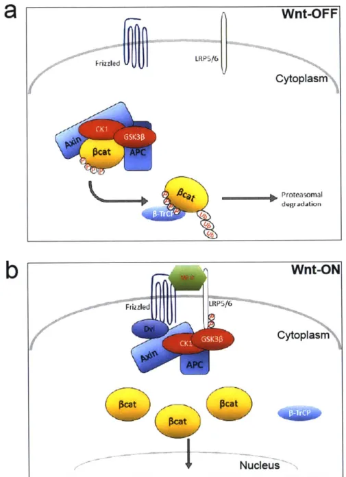
Canonical Wnt/p-catenin Signaling: Overview
The essence of canonical Wnt/p-catenin pathway is stabilization P-catenin in the cytoplasm and subsequent translocation into the nucleus to activate transcription of Wnt
signaling target genes'13. In the absence of Wnt ligand binding to its receptors, P-catenin protein
is targeted for proteolytic degradation through phosphorylation by the so-called destruction complex composed of scaffolding protein Axin, the Adenomatous Polyposis Coli (APC) gene product, casein kinase 1 (CK1) and glycogen synthase kinase 3P (Gsk3
P).
Phosphorylation ofP-catenin
is accomplished by phosphorylation first at Ser45 by CK1, followed by Ser33, Ser37,and Thr4l phosphorylation by GSK3P1 . Upon activation of canonical Wnt signaling via binding
of secreted Wnt ligands to cell surface receptors of the Frizzled family and LRP co-receptors,
P-catenin is stabilized in the cytoplasm through disassembly of the destruction complex. P-catenin
then enters the nucleus, where it binds to TCF/LEF family transcription factors to activate expression of Wnt target genes such as CyclinD1 1 5.
Wnt Signaling Components and Mechanism: Detailed Look into Canonical Wnt Signaling
On the cell membrane: Wnt proteins and receptors
Wnt proteins are conserved in all metazoan animals and there are 19 different Wnt ligands in human and mouse. The Wnt ligands are approximately 40 kDa in size cysteine rich glycosylated proteins which contain an N-terminal signal peptide for secretion1 6. Earlier efforts
117
to characterize Wnt proteins faced difficulties due to its highly hydrophobic nature' . Murine Wnt3a is the first purified and well characterized Wnt protein. In addition to its glycosylation, which is required for secretion, Wnt3a undergoes two lipid modifications which likely account for
its high hydrophobicity and insolubility1 . Whereas addition of palmitate to cysteine 77 was shown to be necessary for ability of Wnt3a to activate Wnt/p-catenin signaling1 1'71 191 20
attachment of palmitoleoyl to serine 209 is necessary for its secretion2 . Multiple proteins have been identified to regulate Wnt biogenesis and secretion. Porcupine (Porc) is a transmembrane
palmitoleoylation at serine 209, and deficiency of Porcupine results in Wnt3a accumulation in
ER
12 1Wntless (WIs) is a multipass transmembrane protein localized to Golgi apparatus,
endocytic compartments and the plasma membrane. Following glycosylation and lipid
modification by Porcupine in the ER, Wls is required to escort Wnt from golgi to the plasma
membrane for secretion'
18Secreted Wnts bind to two distinct receptor families, forming a heterodimeric receptor
complex, to trigger Wnt/p-catenin signaling
1 3. The major component of this complex is Frizzled
(Fzd) family of receptors which are seven-transmembrane receptors with a long amino-terminal
extension called cysteine-rich-domain (CRD) and topological homology to G-protein coupled
receptors122,2. There are 10 Frizzled receptors encoded in mammalian genome. The othercomponent is the single pass transmembrane molecule of the LDL receptor related protein 5
and 6
(LRP5/6)2212, 4.Wnt ligands bind to the
CRD
of Frizzled receptors which in turn recruits
the LRP5/6 to from a trimeric complex initiating the Wnt signaling
12 5In the cytoplasm: Wnt-off State
Degradation of cytoplasmic
P-catenin,
which acts as a transcriptional co-activator of the
signaling pathway, is the hallmark of Wnt/p-catenin signaling
3. In the absence of Wnt ligand
binding to Frizzled-LRP5/6 receptor complex,
P-catenin
levels are kept low by the destruction
complex mainly composed of tumor suppressor Axin, APC, and two serine/threonine kinase
families:
CK1
a, -5 and GSK3a, -P (Figure 1-2)126. Additional components of the destruction
complex include WTX (Wilms tumor gene on the X-chromosome) and serine/threonine protein
phosphatases PP1 and PP2A
127129. In this complex, Axin functions as a scaffolding protein
interacting with GSK3, CK1, APC and
P-catenin
1 26. This enables sequential phosphorylation of
P-catenin
first by
CK1
at Ser45, followed by Ser33, Ser37, and Thr4l phosphorylation by
GSK3P
1 14. Phosphorylated
P-catenin
is then recognized by the F box/WD repeat protein
vv nit-Or r
FrizzledULRP516
Cytoplasm kcAt A C ca Proteasomal degradationb
Wnt-ON
Friz zled LRP516 Cytoplas APC NucleusFigure 1-2: Canonical Wnt Signaling In the Cytoplasm
(a) In the absence of Wnt ligand binding to Frizzled-LRP5/6 receptor complex, P-catenin levels are kept low by the destruction complex mainly composed of tumor suppressor Axin, APC, and two serine/threonine kinase families: CK1 a, -6 and GSK3a, -0. (b) The major outcome of the canonical Wnt signaling activation is disassembly of the destruction complex and accumulation of stabilized cytosolic P-catenin, and
subsequent translation of P-catenin to nucleus.
leads to ubiquitination and proteasomal degradation of
P-catenin
130. Efficient degradation of
cytosolic
P-catenin
ensures blocking of
P-catenin
nuclear translocation and keep Wnt/
P-catenin
signaling inactive. Mutations effecting phosphorylation of
P-catenin,
as well as tumor
suppressors APC and Axin mutations are commonly observed in various cancers supporting the
notion that intact destruction complex is necessary to keep Wnt signaling at low levels
13 1.
In the cytoplasm: Wnt-on State
The major outcome of the canonical Wnt signaling activation is disassembly of the
destruction complex and accumulation of stabilized cytosolic
P-catenin
(Figure 1-2)110. Currently,
it is not very well understood how the binding of Wnt ligands to the receptors initiate destruction
complex disassembly, but clues from recent studies are starting to shed light on to molecular
mechanism underlying this process. Wnt-induced LRP5/6 phosphorylation by
CK1
or GSK3P
could be the key event in receptor activation
3 1 4. One possible mechanism is that Wnt binding
to the receptors would recruit Disheveled (Dsh or Dvl) protein to Frizzled receptor via its PDZ
domain
5, which in turn provides venue for Axin complex bound by GSK3P and
CK1
to relocate
to cell surface Wnt receptor complex
136. The relocation of Axin-GSK3P-CK1 complex would
promote the phosphorylation of LRP5/6 receptor, thereby initiating the downstream
signaling
1 34,1 36
,1 37
. Through disassembly of the destruction complex, Wnt signaling leads to
inhibition of
P-catenin
ubiquitination, and therefore accumulation in the cytoplasm.
In the Nucleus
Following accumulation in cytoplasm, stabilized
P-catenin
builds up in the nucleus
1 38However, the mechanisms underlying the nuclear transport of
P-catenin
are currently not well
understood. Earlier studies have suggested that
P-catenin
is imported to nucleus in a nuclear
139,140
a9|
b
Wnt-ON
Pcat
NuPcat
T CF/ LEF
Wnt Target GenesFigure 1-3: Canonical Wnt Signaling In the Cytoplasm
(a) In the absence of Wnt, TCF/LEF transcription factors bind to the Wnt responsive element (WRE) DNA consensus sequence to repress gene expression. In this context, TCF/LEF transcription factors recruit corepressors such as Groucho, and HDACs promoting histone deacetylation and chromatin compaction (b) In the presence of Wnt, P-catenin binds to TCF/LEF transcription factors and displaces Groucho and activates expression of Wnt target genes. This interaction further recruits additional transcriptional coactivators such Bc19 and Pygopus.
cleus
Wnt-OFF
Cytoplasm WH DAC Wnt target Genesindicated that Wnt-dependent activation of Racl-JNK pathway by phosphorylation of
P-catenin
at serines 191 and 605 promotes its nuclear translocation"'.
Once translocated to nucleus,
P-catenin
binds to the TCF/LEF family of DNA binding
factors to activate Wnt signaling target genes"
0. In the absence of Wnt, TCF/LEF transcription
factors bind to the Wnt responsive element (WRE) DNA consensus sequence to repress gene
expression
1 4 2.
In this context, TCF/LEF transcription factors recruit corepressors such as
Groucho, CtBP and HDACs promoting histone deacetylation and chromatin compaction (Figure
1-3)143-145. In the presence of Wnt,
P-catenin
binds to TCF/LEF transcription factors and
displaces Groucho
146. This interaction further recruits additional transcriptional coactivators such
Bc19 and Pygopus (Figure 1-3)147. Whereas the central Arm-repeats of
P-catenin
bind to
TCF/LEF,
the amino-terminal Arm-repeats bind to Bc19, which in turn bridges Pygopus to the
N-terminus of
P-catenin
147.
This complex has been implicated in retention of
P-catenin
in the
nucleus, though it is currently unclear if they also actively participate in shuttling
148. Other
coactivators of TCF/LEF-
P-catenin
complex includes p300/CBP and TRRAP histone
acetyltransferases, MLL1/2 histone methyltransferases, the SWI/SNF family of ATPases for
chromatin remodeling and Mediator complex all of which contributes to transcriptional activation
of Wnt target genes
Non-canonical Wnt signaling: Overview
In addition to canonical Wnt/p-catenin signaling, Wnt proteins initiate signaling in other
pathways called non-canonical Wnt signaling, which are independent of
P-catenin
transcriptional
activity. These pathways include the Wnt/PCP (planar cell polarity) and the Wnt/Ca2+
pathways". Activation of non-canonical Wnt pathways have been shown to be dependent on
Wnt binding to Frizzled receptors, but independent of LRP5/6. In the Wnt/PCP pathway,
Frizzled and Disheveled proteins function in concert with other proteins such as Celsr, Prickle,
vangl/Strabismus or PTK7 to establish cellular polarity by asymmetrical and polarized protein
localization1491 51
.
Dysregulation of Wnt/PCP pathway has been shown to disrupt the orientationof epithelial structures such as cuticle hairs and sensory bristles in Drosophila
1 52, and neural
tube closure in mammals
15 3. Activation of Wnt/Ca2+ pathway lead to release of intracellular Ca2+
through trimeric G proteins, which in turn can activate Ca
2+-sensitiveproteins such as PKC and
CamKI1
154'
155.Wnt/Ca2+ pathway has been shown to regulate dorsal axis formation, ventral cell
fate, convergent extension movements and heart development156.
Dual Role of
p-catenin
in Wnt Signaling and Cadherin Pathways
The cytoplasmic and nuclear pool of
P-catenin
participating in Wnt signaling is
dynamically regulated. However, there is another pool of
P-catenin
at the cell membrane, which
is relatively more stable. In addition to its central role in Wnt signaling,
@-catenin
also binds to
type I cadherins at the cell membrane, linking them to the actin cytoskeleton through binding to
a-catenin
157and thereby having a role in structural organization. Although it was previously
thought that the cadherin-bound pool of
P-catenin
cannot be made available for Wnt signaling,
recent studies have suggested instead the existence of an interplay between Wnt signaling and
this cell adhesion complex. Several lines of evidence suggest that altering the levels of
P-catenin localized to the P-catenin-cadherin complex can affect the availability of
P-catenin
for
participation in Wnt signaling. This includes the observation that the sequestration of
P-catenin
at the cell-cell adhesion sites via E-/N-cadherin overexpression downregulates Wnt signaling
and that the absence of E-cadherin in epithelial cells and embryonic stem cells results in the
accumulation of free
P-catenin
in the nucleus158~160.Wnt Signaling in Cortical Development
Mapping of canonical Wnt signaling in the developing mouse forebrain using TCF/LEF
binding sites-driven
#-galactosidase
expression showed that Wnt signaling is highly active in
cortical neural progenitors during early development, and as corticogenesis progresses, cortical
Wnt signaling decreases
6 11,
16 2. At the cellular level, active Wnt signaling has been identified in
proliferating radial glial cells and intermediate/basal progenitors, which then is downregulated in
differentiating cells
163,16 4. These findings implicate a dynamic regulation of Wnt signaling in
developing cortex, and temporal and spatial expression patterns are suggestive of an active role
Wnt signaling in neural progenitor proliferation and differentiation.
As described previously in the cortical development section, neural progenitors of the
developing cortex display multiple modes of cell division, and the intricate balance between
proliferation and differentiation regulates neural production. Transgenic mice expressing
stabilized
P-catenin,
which constitutively activates canonical Wnt signaling, displayed enlarged
forebrain due to horizontal expression of neural progenitor population
165. It was shown that
overexpression of
P-catenin
in neural progenitors caused reduced cell cycle exit, prolonging
proliferative division
165,166.Further supporting these findings, activation of Wnt signaling via
expression of
P-catenin/Lefl
fusion protein in E10 brain cased inhibition of neural differentiation,
whereas inactivation of
P-catenin
in Eli and E13 mouse brain promoted cell cycle exit and
thereby increased neuronal differentiation
163,
161.
Regulation of different progenitor pools - basal
versus intermediate progenitors - has also been implicated. Induced Wnt signaling in cortical
neural progenitors is correlated with reduced intermediate progenitor number, while
intermediate progenitor number increased when Wnt signaling was inhibited
1 67'
168. However,
regulation of intermediate progenitor population could be more of secondary effect of radial glial
cell regulation. Proliferative radial glial cells divide asymmetrically to produce one radial glia cell
and a transiently amplifying basal progenitor. Therefore, it is plausible that activation of Wnt
signaling increases proliferative symmetric division of radial glial cells initially, which in turn
increases radial glial cell pool while reducing intermediate progenitor pool. Such a proposal was
supported by findings showing that reduction in intermediate progenitor pool is recovered as
development progresses, suggesting a delay in intermediate progenitor production
166. Overall,
these studies suggest that Wnt signaling promotes symmetric division of radial glial cells while inhibiting cell cycle exit. This effect in part may be due to regulation of CyclinD1 expression by Wnt signaling, since overexpression of CyclinD1 has been shown to increases cell cycle
re-108
entry' .
Interestingly, Wnt signaling may differentially regulate cell cycle in radial glial cells and intermediate progenitor. In utero electroporation of Wnt3a at E13 cortex increases proliferation of radial glial cells consistent with symmetric division inducer role of Wnt signaling641 65' .
However, it was also discovered that Wnt3a overexpression promoted terminal differentiation of intermediate progenitors in developing mouse brain164. These findings point to context and temporal-dependent control of Wnt signaling in different cortical progenitors and their proliferation. In this model, during early corticogenesis, radial glial cells respond the Wnt stimulation by symmetric proliferative division increasing progenitor pool. Later in
corticogenesis, Wnt stimulation causes terminal differentiation of intermediate progenitors, while
it can still stimulate proliferative division in radial glial cells164'69'70.
The role of Wnt signaling and its components in dendrite development and
synaptogenesis has also been shown, though this role is relatively less well understood. Overexpression of Wnt7b or Wnt2 in cultured hippocampal neurons resulted in increase in
complexity of dendritic arborization171
,17 2. Furthermore, induced P-catenin expression in cultured hippocampal cells caused increased dendritic branching and number, however, this effect could be independent of canonical Wnt signaling173. Finally, Wnt signaling plays an important role in
174
synaptogenesis by promoting pre- and post-synaptic assembly in central nervous system
Conclusion
Evidence from whole genome sequencing of psychiatric disorder patients increasingly
underlie the pathophysiology of multiple disorders. Furthermore, besides autism spectrum
disorder, contribution of neurodevelopmental dysregulations to disease etiology in late-onset
disorder such as schizophrenia are becoming widely accepted. Therefore, a better
understanding of cortical development and functions of Wnt signaling could prove critical in
determining the cellular and molecular mechanisms underlying the pathophysiology of
psychiatric disorders. In my thesis research, I have attempted to characterize the functions of
three (ANK3, CHD8 and BCL9) neuropsychiatric risk factors in mouse cortical development and
Wnt signaling. First, ASD risk gene Chd8 was shown to be a positive regulator of Wnt signaling
and neural progenitor proliferation in a cell-type specific manner. Knockdown of Chd8 in upper
cortical layer neurons caused ASD-related behavioral abnormalities in adult mice (Chapter 2).
Secondly, we made the novel observation that bipolar disorder risk gene Ank3 (ankyrin-G) plays
a crucial role in cortical neurogenesis through regulation of subcellular localization of Wnt
signaling (Chapter 3). Finally, the effects of brain-specific deletion of BcI9 on brain development
and behavior were characterized using a heterozygous BcI9 deletion transgenic mouse line.
Behavioral and brain development defects associated with BcI9 were shown to mimic some of
the clinical symptoms observed in patients.
CHAPTER 2:
Chd8 mediates cortical neurogenesis through transcriptional
regulation of cell cycle and Wnt signaling genes
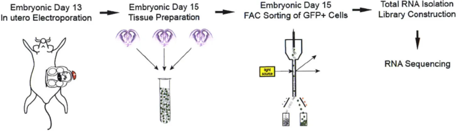
Omer Durak, Fan Gao, Yea Jin Kaeser-Woo, Richard Rueda, Anthony J. Martorell, Alexi Nott, Carol Y. Liu, L. Ashley Watson and Li-Huei Tsai
Abstract
De novo mutations in CHD8 are strongly associated with autism spectrum disorder
(ASD), however the underlying mechanisms remain elusive. Here we report that Chd8
knockdown during cortical development results in defective neural progenitor proliferation and differentiation that ultimately manifests in abnormal neuronal morphology and behaviors in adult
mice. Transcriptome analysis revealed that on the one hand Chd8 stimulates the transcription of cell cycle genes, while on the other it precludes the induction of neural specific genes by
regulating the expression of PRC2 complex components. Furthermore, knockdown of Chd8
disrupts the expression of key transducers of Wnt signaling, and enhancing Wnt signaling rescues the transcriptional and behavioral deficits caused by Chd8 knockdown. We propose that these roles of Chd8 and the dynamics of its expression during development help negotiate the fine balance between neural progenitor proliferation and differentiation. Together, these
observations provide new insights into the etiology of ASD.
O.D. and L.-H.T designed the study, and L.-H.T directed and coordinated the study. O.D. initiated, planned and performed the experiments. F.G conducted the bioinformatics analysis. Y.J.K cloned the human CHD8 construct and contributed to sample preparation for FAC-sorting. R.R prepared cultured various cell lines and helped with sample preparation for FAC-sorting. A.J.M. conducted some of the luciferase assays. A.J.M and A.N. contributed to behavioral experiments. C.Y.L conducted the neuronal
Introduction
ASD is a complex developmental disorder that manifests in social deficits,
communication difficulties, stereotyped behaviors, and cognitive delays23
,24. It is a highly heritable disorder with concordance rates of about 8% between siblings and 64% between monozygotic twins - much higher than the prevalence in the general population of about
1%23,175. Around 120 genes have been linked to ASD, often encoding proteins involved in chromatin remodeling, transcriptional regulation, and synapse formation or function1'3 23- . Of
them, at least 15 distinct mutations in the coding regions of chromodomain helicase DNA binding protein 8 (CHDB), an ATP-dependent chromatin remodeler, were identified in ASD subjects from exome sequencing of trio families 1
,34,37. Most of these mutations are predicted to be loss-of-function as they either result in frame shift or truncation of protein product as well as in frame single amino acid deletion37. Subjects with CHD8 mutations often display increased head circumference, cognitive deficits, as well as social interaction and communication difficulties34'37.
Little is known about the biological function of CHD8. It was initially identified as a binding partner and negative regulator of P-catenin signaling and was shown to be enriched in the promoters of transcriptionally active genes 38 4 1. Homozygous deletion of Chd8 in mice
results in early embryonic lethality resulting from massive apoptosis 4'17 6.However, in contrast
to expectations, there was no induction of Wnt/P-catenin signaling in Chd8 null mice76. CHD8
was also shown to be a binding partner of the transcription factor, E2F1, and necessary for E2F1-dependent cell cycle gene activation during the G1/S transition39. Down-regulation of
CHD8 in cultured cells resulted in impaired cell proliferation17 . In contrast, an increase in the number of mitotic cells and head size were observed following suppression of zebrafish chd8
178
ortholog'.
Whereas the genetic and molecular underpinnings of ASD are heterogeneous, an accumulating body of evidence indicates that disrupted embryonic cortical development could
be a common defect underlying the etiology of ASD2 4
,28
,179. As such, an understanding of how
CHD8 affects cortical development is likely to provide crucial insights into the pathophysiology
of ASD.
Cortical development is a spatially and temporally regulated process that occurs during mid- to late-embryogenesis"30'181. The process is defined by an early expansion of proliferative neural progenitor cells (NPCs) that reside in the ventricular zone (VZ) of cortical epithelium. At the onset of neurogenesis, NPCs undergo neurogenic divisions to produce pyramidal neurons that migrate radially to the cortical surface to generate the six layers of the neocortex182. The significant differences in cortical size between gyrecephalic (i.e. primates) and lissencephalic
(i.e. rodents) species are believed to be due to existence of gyrecephalic brain specific self-sustaining neural stem cells in the outer subventricular zone (SVZ), which later in corticogenesis divide to increase the number of upper layer cortical neurons18 1. Despite these differences,
mechanisms governing cortical development and the subsequent laminar organization of the neocortex are largely conserved across mammals. Diverse signaling pathways govern the intricate balance between continued proliferation and cell cycle exit/differentiation that is necessary to produce the full range and organization of cortical cell types. A large body of literature has established the role of canonical Wnt signaling in cortical neural progenitor
proliferation115,164,183,184. Induction of Wnt signaling via overexpression of stabilized
P-catenin
increased neural progenitor proliferation due to reduced cell cycle exit, suggesting that Wntsignaling negatively regulates cell cycle exit/differentiation185. The spatial and temporal control
of developmental genes by epigenetic mechanisms have also been increasingly appreciated in
recent year182,1s Studies have shown that polycomb group (PcG) proteins are important for temporal repression of genes necessary for differentiation86. Interestingly, these genes are
repressed under normal circumstances, but maintained in a "poised state", ready for activation when differentiation cues are present. Disruption of polycomb repressive complex 2 (PRC2),