HAL Id: hal-02268443
https://hal.archives-ouvertes.fr/hal-02268443
Submitted on 20 Aug 2019
HAL is a multi-disciplinary open access
archive for the deposit and dissemination of
sci-entific research documents, whether they are
pub-lished or not. The documents may come from
teaching and research institutions in France or
abroad, or from public or private research centers.
L’archive ouverte pluridisciplinaire HAL, est
destinée au dépôt et à la diffusion de documents
scientifiques de niveau recherche, publiés ou non,
émanant des établissements d’enseignement et de
recherche français ou étrangers, des laboratoires
publics ou privés.
Celine Marban-Doran, Faezeh Forouzanfar, Amina Ait Ammar, Faïza Bittich,
Hala El Mekdad, Fadoua Daouad, Olivier Rohr, Christian Schwartz
To cite this version:
Celine Marban-Doran, Faezeh Forouzanfar, Amina Ait Ammar, Faïza Bittich, Hala El Mekdad, et al..
Targeting the Brain Reservoirs: Toward an HIV Cure. Frontiers in Immunology, Frontiers, 2016, 7,
pp.397. �10.3389/fimmu.2016.00397�. �hal-02268443�
Edited by: Ihssane Zouikr, RIKEN Brain Science Institute, Japan Reviewed by:
Taisuke Izumi, Frederick National Laboratory for Cancer Research, USA Carine M. C. Van Lint, Université libre de Bruxelles, Belgium
*Correspondence: Olivier Rohr [email protected]; Christian Schwartz [email protected] Specialty section: This article was submitted to Multiple Sclerosis and Neuroimmunology, a section of the journal Frontiers in Immunology Received: 01 July 2016 Accepted: 20 September 2016 Published: 30 September 2016 Citation: Marban C, Forouzanfar F, Ait-Ammar A, Fahmi F, El Mekdad H, Daouad F, Rohr O and Schwartz C (2016) Targeting the Brain Reservoirs: Toward an HIV Cure. Front. Immunol. 7:397. doi: 10.3389/fimmu.2016.00397
Targeting the Brain Reservoirs:
Toward an Hiv Cure
Céline Marban
1, Faezeh Forouzanfar
2, Amina Ait-Ammar
2, Faiza Fahmi
2, Hala El Mekdad
2,3,
Fadoua Daouad
2, Olivier Rohr
2,3,4* and Christian Schwartz
2,3*
1 INSERM UMR 1121 Faculté de Chirurgie Dentaire, Université de Strasbourg, Strasbourg, France, 2 EA7292, DHPI,
Université de Strasbourg, Strasbourg, France, 3 IUT Louis Pasteur de Schiltigheim, Université de Strasbourg, Schiltigheim,
France, 4 Institut Universitaire de France, Paris, France
One of the top research priorities of the international AIDS society by the action
“Towards an HIV Cure” is the purge or the decrease of the pool of all latently infected
cells. This strategy is based on reactivation of latently reservoirs (the shock) followed
by an intensifying combination antiretroviral therapy (cART) to kill them (the kill). The
central nervous system (CNS) has potential latently infected cells, i.e., perivascular
macrophages, microglial cells, and astrocytes that will need to be eliminated. However,
the CNS has several characteristics that may preclude the achievement of a cure. In this
review, we discuss several limitations to the eradication of brain reservoirs and how we
could circumvent these limitations by making it efforts in four directions: (i) designing
efficient latency-reversal agents for CNS-cell types, (ii) improving cART by targeting HIV
transcription, (iii) improving delivery of HIV drugs in the CNS and in the CNS-cell types,
and (iv) developing therapeutic immunization. As a prerequisite to these efforts, we also
believe that a better comprehension of molecular mechanisms involved in establishment
and persistence of HIV latency in brain reservoirs are essential to design new molecules
for strategies aiming to achieve a cure for instance the “shock and kill” strategy.
Keywords: brain, reservoirs, latency, cure, cART, Hiv transcription
iNTRODUCTiON
Combination antiretroviral therapy (cART), introduced in 1996, has radically improved the
man-agement of HIV-1 infection and decreased both morbidity and mortality. However, despite initial
hopes to cure HIV, treatments were unable to fully eliminate the virus (
1
–
3
). Indeed, with very
sensitive methods (
4
–
6
), a remaining viremia is always noticed in patients on cART. Moreover,
HIV RNA returns to a measurable plasma level when cART is disrupted (
7
,
8
). The origin of this
persistent viremia is still a matter of debate (
9
–
11
). Latent persistence of HIV in long-lived cells,
such as the central memory CD4+ T-cells, hematopoietic stem cells, dendritic cells, and cells from
the monocyte–macrophages lineage in the form of proviruses have been described (
1
,
2
,
12
–
19
).
Moreover, these cells are located in a variety of anatomical sites, including tissues, such as blood,
brain, gut-associated lymphoid tissue, bone marrow, and genital tract (
20
), making it difficult to
purge the virus from all the reservoirs.
These latently infected cells are from time to time reactivated and produce HIV particles at low
levels, thus explaining the persistence of viremia. An alternative theory, the cryptic ongoing
replica-tion states that despite cART, HIV is continuously produced at low levels. The inefficiency of the
treatment in cells supporting ongoing replication could be due to poor drug penetration in
sanctuar-ies, such as the brain (
21
) or by cell-to-cell transfer of the virus (
22
). In theory, there are critical
therapeutic implications for cART as it is expected that during
ongoing replication, drug resistance might arise (
23
–
26
). The
potential mechanisms of HIV persistence have been discussed
recently in a review by Hong and Mellors (
27
).
One of the main debates in the field of HIV reservoir is whether
or not the central nervous system (CNS) constitutes a real viral
reservoir. Indeed, with its unique features, such as the existence
of a blood–brain barrier (BBB) with poor drug penetration, the
CNS might be considered as a sanctuary (
20
) made of specific
cell types (
28
) with reduced immune surveillance. Moreover,
the anatomy of the CNS is such that there is poor viral genetic
information exchange with the other sites and, thus, might be
referred as a compartment (
20
,
29
,
30
).
First, we will give our opinion on the existence of viral
reservoirs in the CNS referring to excellent recent reviews in
this topic. Next, we will discuss the importance to purge these
potential viral reservoirs. Indeed, in theory, it is possible to
acquire virus resistance to cART if there is an ongoing
replica-tion in the brain. Another major concern is the existence of
HIV-associated neurocognitive disorders (HAND). In up to 50% of
the HIV-infected patients on efficient cART and undetectable
virus load (≤50 copies/ml), HAND has been recorded. Several
mechanisms are evoked to explain the increase of less severe
forms of HAND in which production of some viral proteins
occurs during reactivation or cryptic ongoing HIV replication.
We will then review the state of art of what is known regarding
the molecular mechanisms underlying the establishment and
persistence of HIV in the potential reservoirs in the brain and,
finally, discuss the profound therapeutic implications of purging
reservoirs.
CAN THe CNS Be QUALiFieD
AS A Hiv ReSeRvOiR?
A viral reservoir is an infected cell population that allows
persis-tence of replication-competent virus in patients under cART (
20
).
According to this definition, the only true reservoirs are the
rest-ing CD4+ T-cells. Indeed, these cells fulfill all the criteria to be
considered as a real reservoir, i.e., presence of integrated virus in
long-lived cells, persistence of high levels of virus in a quiescent/
latent state in the reservoir and possible reactivation of the virus
with the formation of replication-competent particles (
31
).
There are several evidences that brain cells harbor
genome-integrated HIV (
28
). We know that the virus invades the brain
very soon following infection. Virus infection was shown
in astrocytes (
32
), in perivascular macrophages (
32
), and in
microglial cells (
33
). All three cell types are long-lived cells with
perivascular macrophages (
34
) and astrocytes (
35
) with a
half-life ranging from months and microglial cells with a half-half-life
of years (
36
). All these cells are infected at high frequency in
the brain. Astrocytes, the most abundant cell type in the brain,
are infected in up to 19% of the cell population (
37
). Similar
ratio of infected cells has been found among the perivascular
macrophages and the microglial cells (
33
,
38
). In addition,
several mechanisms, including epigenetic regulation, have been
evoked to induce latency in these cells notably in astrocytes and
microglial cells (
39
–
42
).
Due to ethical and technical problems, it is not possible to
evaluate the human brain-infected cells for their capacity to
produce replication-competent viruses. However, there are
several indirect evidences showing that CNS is a reservoir
for HIV. Indeed, HIV DNA has been detected in brain tissues
isolated from autopsies of HIV patients whose infection has
been controlled by cART (
33
,
39
). Moreover, there is a strong
correlation of the amount of HIV DNA found in astrocytes and
HIV-associated dementia (HAD) (
37
). Various animal models
have been used to show persistence of HIV infection in the CNS
as brain biopsy is not possible. Indeed, several animal models,
such as macaque, rats, and humanized BLT mouse, have been
used to mimic the condition of HIV-infected patients on cART,
which confirmed the presence of viral RNA or viral proteins
in the brain (
43
–
45
). Specifically, in the macaque model, a
mechanism of the establishment of transcriptional HIV latency
in the CNS has been suggested (
46
). They notably showed that
interferon beta repressed SIV LTR activity by inducing C/
EBPγ expression, a dominant negative isoform of C/EBPβ (
47
).
There are also several evidences supporting continuous CNS
perturbation despite an efficient cART (
48
) with an increase of
the prevalence of milder form of HAND. Moreover, in patients
under suppressive cART activation of the immune system is still
observed in the CNS with some biomarkers, such as neopterin
or NFL being detected in the cerebrospinal fluid (CSF) (
49
).
One explanation is the existence of an inflammatory process
that might be driven by low-level HIV replication in infected
cells (
50
,
51
). Interestingly, neuroimaging data are also in favor
of persistent CNS inflammation in patients on cART (
52
,
53
).
Finally, development of highly sensitive methods, such as
single-copy assay (SCA), has allowed the detection of HIV RNA in the
CSF from infected patients on cART or from elite controllers
whose HIV RNA level was initially undetectable in the plasma
and CSF (
54
–
56
). The recent discovery of a CSF viral escape in
patients on cART with undetectable plasma HIV RNA but with
neurological impairment argue also for the existence of a
per-sistent HIV reservoir in the brain (
55
–
59
). In conclusion, there
are now several evidences supporting that CNS is a reservoir for
HIV even if it is still controversial. Readers will be referred to the
following reviews that nourish the debate of whether or not CNS
serves as a HIV reservoir (
60
–
63
).
wHY iS iT iMPORTANT TO PURGe
THe CNS ReSeRvOiR OF Hiv?
The CNS is involved in the control of most functions of the
body and mind. The brain operates in a very well controlled
microenvironment separated from the other parts of the body
by two barriers: the choroid plexus and the BBB. The two
bar-riers, but predominantly the BBB, constitute physical barriers
and any perturbation of their integrity will be associated with
neurological diseases. There are several other features that
make the CNS unique. The CNS has specific immunological
features; principally an innate immune response through the
perivascular macrophages and the microglial cells. However, the
adaptive immune response has also been observed and, thus,
contributes to the immune surveillance in the CNS (
64
,
65
).
Indeed, leukocytes trafficking to the CSF either by traversing the
BBB to the perivascular space or the choroid plexus has been
detected (
66
). More interestingly, in patients having CSF/plasma
HIV discordance (patients having higher levels of HIV RNA in
CSF than in blood) even at very low levels it was demonstrated
that both innate (macrophages and microglial cells) and adaptive
(T CD4+ and CD8+ lymphocytes) are involved in CNS injury
(
67
–
70
). It has been shown that the percentage of a specific set
of T CD8+ lymphocytes that expresses interferon γ is higher in
the CSF than in blood. Moreover, this higher percentage of T
CD8+ cells in CSF versus blood contributes to the occurrence
of HAND (
67
) [reviewed in Ref. (
71
)]. Within 2 weeks following
acute infection by HIV, the virus enters the CNS. There are at
least two mechanisms to explain how HIV crosses the BBB,
including trafficking of cell free virus and infected cells (
72
). The
well-documented infection of the CNS is accomplished through
infected cells and, thus, has been named the “Trojan Horse”
mechanism (
73
). A recent study using natalizumab, an anti-α4
blocking antibody preventing both lymphocytes and monocytes
trafficking across the BBB, is in accordance with this mechanism.
Indeed, a drastic decrease of SIV DNA in the brain was observed
when natalizumab was given to rhesus macaque during acute
SIV infection (
74
). According to this theory, infected monocytes
cross the BBB and infect the perivascular macrophages, the
microglial cells, and the astrocytes that result in HIV-associated
neurological disorders (
75
). Since the introduction of cART, an
important decrease in the incidence of the severe form of HAND
has been noticed (
76
). However, there is an increase of milder
form of the infection (up to 50%), which might be largely under
diagnosed. Thus, better screening tools to detect HAND are
required in the future (
77
). The reasons for the increase of the
prevalence of milder forms of HAND are not fully understood.
One explanation might be related to the existence of quiescent/
latent viral reservoirs in the CNS that emphasizes the importance
of eradicating the reservoirs. Another major concern related to
the existence of such quiescent/latent reservoirs in the CNS is
that it might be a source of new particles that could replenish
the periphery blood. These notions will be discussed in the
later chapters.
Hiv-1 and Hiv-Associated Neurological
Disorders
HIV-associated neurocognitive disorders have been divided into
three subgroups according to the Frascati criteria, i.e.,
asymp-tomatic neurocognitive impairment (ANI), mild neurocognitive
disorder (MND), and HAD (
78
). These disorders are associated
with the entry of HIV into the CNS that occurs almost
imme-diately after systemic infection (
79
). The more severe form of
HAND, i.e., HAD has drastically decreased with the introduction
of cART. However, the less severe forms (MND and ANI) have
continued with a prevalence ranging from 20% to up to 50%,
while keeping in mind that these milder forms are often under
diagnosed (
80
,
81
). However, the details of persistence of these
less severe forms of HAND in patients on cART are not fully
understood. There are at least two hallmarks of HIV infection in
the brain, i.e., chronic immune activation and compromised BBB
integrity in which the central role for HIV neuropathogenesis
is played by the monocytes/macrophages (
82
–
85
). Importantly,
immune activation still occurs in patients on cART (
50
,
51
).
The exact mechanisms of such pathogenesis are not entirely
known and rely on two models: a direct and an indirect model
(
86
,
87
). In the direct model, infected cells will cause neuronal
death through the action of newly synthesized viral proteins,
such as Tat, gp120, Vpr, and Nef. The two major viral proteins
that lead to neuronal injury are Tat and gp120. Their effects
are mediated through their interaction with neuronal cell
recep-tors, such as the NMDA receptor and the chemokine receptors
(CCR5 and CXCR4). More details on the mechanisms involved
in the neuropathogenesis caused by viral proteins are found
in the review (
88
). In the indirect model, sustained chronic
inflammation is induced by secreting perivascular macrophages,
microglial cells, and to a lesser extent by astrocytes releasing
neurotoxic host factors. Among these secreted products, there
are proinflamatory cytokines (TNFα, IL-1β, IL-6, IL-8, and
INFα), chemokines (CCL2 and CCL5), and small molecules,
such as quinolinic acid and the platelet-activating factor.
Moreover, these viral proteins and cellular factors increase the
oxidative stress and alter the integrity of the BBB which in
turn results in the stimulation of even more infected cells in
the brain. Further investigations are needed to decipher the
exact mechanisms involved in CNS injury. Interestingly, Tat
might be involved in both direct and indirect processes that lead
ultimately to neuronal death. Potential roles and functions of Tat
in both direct and indirect neurotoxicities have been described
elsewhere (
89
,
90
). The importance of Tat is still a matter of
debate since there are controversies regarding the amount of Tat
present in the CNS cells environment and the amount of Tat
used in in vitro experiments. In favor of its importance is the
use of Tat transgenic animal model where CNS injury has been
observed (
91
,
92
). Therefore, it will be essential to detect Tat in
the brain from patients on cART. It is possible that this protein
might arise from quiescent/latent reservoirs and, therefore, be
responsible for the milder form of HAND. Improvement of
cART by targeting the production phase of HIV-1, including
transcription appears, therefore, crucial (
93
). Indeed, current
cART is not targeting this step and since the CNS infection
occurs almost immediately during acute infection, establishment
of infected reservoirs will not be prevented. Moreover, strategies
aiming to purge the reservoirs are based on HIV reactivation
with the risk that viral proteins, such as Tat will be produced
in the brain. HIV-1-mediated neuropathogenesis might also
involve a dynamic interaction between astrocytes and peripheral
blood mononuclear cells (PBMCs) (
94
). Indeed, a recent report
showed that astrocytes susceptibility to produce HIV infection
is enhanced by PBMCs producing interferon γ which in turn
inhibit HIV-1 production in PBMCs through the secretion of
small glycoprotein, i.e., the Wtns. These later proteins have been
shown to be involved in many CNS processes (
95
), such as
synaptic plasticity and neurotransmitter release, which might
explain partly HIV-1-mediated neuropathogenesis.
CNS Reservoirs as a Source of virus
The CNS has two special features making it difficult the
achievement of a cure. First of all, the CNS is considered as
a sanctuary for HIV by pharmacologic means as it is a site
with limited access to antiretroviral drugs (ARV) (
96
–
99
). As
an outcome, there is a risk to allow the occurrence of virus
resistant to the current drugs used in cART. Second, the CNS is
also considered as a compartment in which the virus is isolated
from other parts of the body (
29
,
100
). Because of poor genetic
information exchange with the other sites, neurotropic variants
of HIV might be selected, which most likely will not respond
to treatment in a similar way than the virus encountered in
the CD4+ T-cells, the main target in the body. There are now
numerous evidences supporting the fact that the CNS-resident
virus has evolved to become macrophage tropic (
101
). Indeed,
sequence analysis of the env gene and of the HIV-1 promoter
(LTR) argue for the compartmentalization of HIV variants in
the CNS (
102
–
105
). Variations in the promoter are important
since mechanisms involved in the establishment and
persis-tence of latency in the CNS might differ from the one described
in CD4+ T-cells. As mentioned above, this will impact the
effi-ciency of latency-reversing agents (LRA) in strategies aiming to
purge the latent/quiescent reservoirs (
106
,
107
). Another major
concern regarding the necessity to purge the CNS reservoirs
is related to the discovery of CNS viral escape in patients on
cART (
108
). Initial studies have shown occasional cases of virus
escapes in the CSF (
109
,
110
). Development of highly sensitive
assays has even allowed the detection of CSF HIV RNA, which
were not detectable with previous assays (
111
). Indeed in a
report, evaluation of CSF viral escape has been done in a cohort
of neurologically asymptomatic patients successfully treated
with cART. It was shown that around 10% of these patients
had detectable CSF HIV RNA, suggesting that viral escape may
be underestimated (
112
). The recent discovery of a CNS viral
escape in a cohort of 14 patients on cART with undetectable
plasma HIV RNA but who developed HIV-encephalitis argues
for the possibility that CNS is a real reservoir (
57
). Actually,
this study and others raise the question that CNS-specific viral
replication can occur in patients on cART from reactivated
reservoirs which in theory may have escaped therapy and
ultimately lead to drugs resistance (
58
,
59
,
113
). Very
interest-ingly a similar drug-privileged site, i.e., the lymphoid tissue
has been shown to have low access to drugs (
114
). The authors
notably showed that the virus is continuously produced and
might be a source of HIV from which replenishment of blood
occurs. However, and contrary to the brain, they do not show
that resistance to antiretroviral drugs arises. The authors
of this study suggest that this absence of resistance to ARV
might be explained by the too low level of drug concentration
in lymphoid tissue that is not sufficient to confer competitive
advantages to the development of drug-resistant viruses. This
study point out to the importance of developing new ways to
deliver drugs in all sanctuaries, including brain and lymphoid
tissues (
115
).
Overall, we suggest that it is crucial to eradicate brain
res-ervoirs since ARV-resistant viruses are capable to replenish the
systemic circulation from these reservoirs. It will also imply that
CSF analysis in patients on cART should be performed more
often since it will greatly help assessing the
compartmentaliza-tion of HIV in the brain and monitoring the efficiency of new
treatments (
116
). Notably, CSF might be used to evaluate HIV
drug resistance.
MOLeCULAR MeCHANiSMS OF Hiv-1
LATeNCY
Establishment and persistence of HIV latency occur in brain cells,
i.e., perivascular macrophages, microglial cells, and astrocytes.
Infection of these cells differs from the infection of blood cells
infected, mainly the CD4+ T-cells. Indeed, HIV infection in
macrophages is not lytic and these cells are far more resistant to
cytopathic effects. Moreover, infected monocyte–macrophage
cells are also more resistant to apoptosis, a major obstacle for the
eradication of the virus. These cells may harbor latent viruses
for months (perivascular macrophages) or for years (microglial
cells). Astrocytes are also thought to be infected by HIV-1 despite
the lack of the co-receptors CCR5 and CXCR4 probably through
the involvement of vesicles (
38
). However, the infection appears
to be non-productive with only early transcripts, such as tat and
nef, that are detectable at very low level (
117
).
Understanding the intimate mechanisms underlying HIV-1
latency in these CNS-specific cells is necessary to develop new
and original therapies for viral eradication. The molecular
mechanisms underlying these therapies are determined by the
cellular specificity of HIV gene transcription and the variability
of the LTR found in viruses having evolved in the brain (
61
,
118
). For example, it has been shown in microglial cells that
Sp3 and a truncated form of C/EBPβ (NF-IL6) inhibit the basal
transcriptional activity of HIV-1 (
47
). Such a reduced basal and
Tat-activated transcriptional activity has also been shown in
astrocytes. Transcriptional silencing has been associated with
low levels of TAR RNA binding proteins (TRBP) and with
muta-tions of the SP motifs found within the LTR of brain-derived
HIV-1. Mutations prevent the transcription factor Sp1 to bind
the promoter and, thus, inhibit transcriptional activation (
119
,
120
). However, the main mechanism involved in establishment
and persistence of latency involves epigenetic regulation (
41
,
121
,
122
). In our laboratory, we showed that the cellular factor
COUP-TF interacting protein (CTIP2) is a key factor in the
establishment and persistence of HIV latency in microglial cells
(
123
). We notably showed that this protein serves as a platform
to anchor several protein complexes having different functions.
Indeed, at least two different complexes containing CTIP2 are
involved in the establishment and the persistence of HIV-1
latency (Figure 1). Moreover, CTIP2 is also involved in the
con-trol of cellular genes of importance for the virus. Among these
factors, the cellular cyclin-dependent kinase inhibitor CDKN1A/
p21
wafhas been described to favor HIV-1 gene transcription in
the monocyte–macrophage lineage. This effect indirectly favors
HIV-1 latency since activation of the p21 gene stimulates viral
expression in macrophages (
124
). Moreover, CTIP2 counteracts
HIV-1 Vpr protein that is required for p21 expression (
125
). We,
therefore, suggested that CTIP2 generates a cellular environment
disfavoring viral reactivation and, thus, favoring HIV-1 latency.
The first CTIP2-associated complex described in our
labora-tory has been involved in the establishment of HIV-1 latency
through the induction of heterochromatin in the vicinity of the
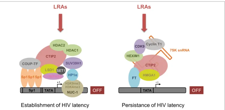
FiGURe 1 | CTiP2 promotes the establishment and persistence of Hiv-1 latency through the recruitment of two macromolecular complexes on the Hiv-1 promoter in microglial cells. CTIP2 participates in the establishment of HIV-1 latency by recruiting a chromatin-modifying complex at the HIV-1 promoter.
This complex consists of two histone deacetylases: HDAC1 and HDAC2 that are responsible for H3K9 deacetylation of Nuc-1, a nucleosome located immediately downstream of the HIV-1 transcriptional start site. The histone methyltransferase SUV39H1 takes also part of the complex and catalyzes the tri-methylation of H3K9 on Nuc-1. Finally, HP1α, a protein associated with heterochromatin, specifically recognizes H3K9me3 and spreads along the HIV-1 promoter, thus creating a domain of heterochromatin refractory to transcription. In parallel, CTIP2 also recruits the histone demethylase complex LSD1/COMPASS/SET1 that, in association with the histone marks H3K9me3 and H3K4me3, contributes to HIV-1 gene silencing and, therefore, the establishment of HIV-1 latency. Besides, by recruiting a transcriptional inhibitory complex at the HIV-1 promoter, CTIP2 is also involved in the prevention of HIV-1 reactivation. This complex is an inactive form of the elongation factor pTEFb and consists of pTEFb, HEXIM1, HMGA1, and the snRNA 7SK. Due to their involvement in HIV-1 establishment and persistence of HIV-1 latency, CTIP2-associated proteins from both complexes constitute new pharmacological targets to reverse HIV-1 latency. Accordingly, new latency-reversing agents (LRAs) are currently being developed or undergoing clinical trials with the aim of reversing HIV-1 latency and depleting HIV-1 reservoirs.
viral promoter (Figure 1, left complex). Indeed, we showed that
CTIP2 recruits a chromatin-modifying complex through the Sp1
sites of the proximal promoter (
42
). This complex contains the
histone deacetylases HDAC1, HDAC2, and histone
methyltrans-ferase SUV39H1 that specifically demethylates lysine 9 of histone
H3. This histone modification allows heterochromatin protein
1 (HP1) binding, heterochromatin formation, and hence HIV
silencing (
42
,
126
,
127
). In a consecutive study, we have shown
that CTIP2 interacts physically and functionally with the
lysine-specific demethylase (LSD1) to repress HIV-1 transcription and
viral expression in a synergistic manner (
128
). The recruitment
of LSD1 at the HIV-1 proximal promoter has been associated
with both H3K4me3 and H3K9me3 epigenetic marks, which is
linked to the recruitment of hSet1 and WDR5, two members
of the hCOMPASS complex, on the HIV-1 promoter (
128
).
Recruitment of CTIP2 on the p21 promoter also induces a
het-erochromatin environment. Moreover, CTIP2 has been shown
to silence p21 gene transcription by creating epigenetic marks
of repression, as described above for the HIV-1 promoter (
125
).
Interestingly epigenetic regulation of HIV-1 latency, which was
associated with the recruitment of HDACs and SUV39H1 has
also been described in astrocytes (
40
). Finally, in a recent report,
investigation of the neuropathology and the molecular alterations
associated with CNS latent HIV-1 infections provided evidence
that HIV-1 persistence in the brain is associated with high level
of CTIP2, HDACs, and HP1 (
39
).
We also showed that CTIP2 belongs to another complex
able to prevent HIV-1 reactivation (Figure 1, right complex)
(
129
). Indeed previous work has shown that CTIP2 represses
the late phase, Tat-dependent, of HIV-1 transcription (
127
).
In the absence of the trans-activator factor Tat, an inactive
form of the elongation factor pTEFb is found in a multiprotein
complex, including 7SK snRNA, CTIP2, and HEXIM1 anchored
to viral and cellular gene promoters (
129
). pTEFb is composed
of a regulatory subunit CyclinT1 and a catalytic subunit CDK9,
whose kinase activity is involved in the Ser2 phosphorylation of
the carboxyl terminal end of the RNA polymerase II and in the
phosphorylation of the negative transcriptional elongation
fac-tors NELF and DSIF. Following phosphorylation, the RNA pol
II processivity significantly increases, which leads to an efficient
transcription of genes (
130
). Interestingly, we have shown that
CTIP2 drastically repressed CDK9 kinase activity in this inactive
complex, thus, inhibited pTEFb function. Finally, we showed that
the cellular protein high mobility group AT-hook 1 (HMGA1),
which also belongs to the 7SK snRNA complex recruits the
inactive CTIP2/pTEFb complex to the HIV-1 and cellular target
promoters (
131
). As a consequence, protein complexes
contain-ing CTIP2 regulate viral and endogenous gene expression, thus
favoring HIV-1 persistence. Far more investigations are still
needed to decipher the precise molecular mechanisms involved
in these processes. We still do not fully understand how the
transition from transcription initiation into elongation (which
involves pTEFb) is controlled by cellular factors and/or the viral
transactivator Tat. We and others hypothesized that the inactive
form of the pTEFb complex is part of a 7SK complex that is
anchored to the promoter by either CTIP2 (
129
) or Kap1 (
132
),
thus available for RNApolII elongation through its activation.
The transition from the inactive to the active form of the pTEFb
complex through the action of Tat is not well understood but
may involve a phosphatase (PPM1G/PP2Cγ) that takes apart
pTEFb from the 7SK complex (
133
).
THeRAPeUTiC iMPLiCATiONS FOR THe
eRADiCATiON OF Hiv-1 FROM BRAiN
ReSeRvOiRS
Several considerations already mentioned [emergence of
mul-tidrug resistance (
24
,
113
), non-AIDS-related events (
134
–
136
)
etc.] urge the search of new ways to develop a sterilizing or a
functional cure for AIDS (
137
). The purge of viral reservoirs
by the “shock and kill” strategy (
138
) is a possible approach to
achieve such a cure. This strategy aims at purging or at least
reducing the size of cellular reservoirs by reactivating HIV
transcription (shock) followed by intensive cART therapy and
immune activation (kill) (
139
,
140
). As several reports suggested,
using LRA alone or in combination have proven the efficiency
of this strategy in the reactivation of quiescent/latent HIV
from CD4+ T-cells reservoirs (
138
,
141
–
145
). Several clinical
trials have been carried out and some others are in progress or
forthcoming (
146
). This strategy of reactivation needs to work
on all potential reservoirs, including brain reservoirs. However,
several limitations to the eradication of the brain reservoirs may
preclude a cure.
Limitations to the eradication of Brain
Reservoirs
It is essential to decipher the molecular mechanisms underlying
HIV persistence in all types of potential reservoirs, since some
important differences in those mechanisms have been noticed
in all latently infected cell types. For example, LSD1 has been
associated with activation of HIV transcription in CD4+ T-cells
(
147
). However, in microglial cells, LSD1 played a role in the
establishment of latency (
128
). LSD1 mediates HIV-1
transcrip-tion silencing in microglial by anchoring various factors at the
promoter rather than inducing HIV-1 transcription by its own
enzymatic activity in CD4+ T-cells. The dual role of LSD1
achieved by different mechanisms in the two main HIV-1 cellular
targets points to the complexity of the molecular mechanisms
of HIV latency (
148
). Hence, additional investigations of the
epigenetic regulation of HIV latency are needed in order to
develop efficient drugs targeting each potential viral reservoir.
Furthermore, as mentioned in the previous sections, there are
several characteristics of the CNS, which limits a cure by the
“shock and kill” strategy:
i. The CNS is a sanctuary with barriers (BBB and choroid
plexus) that reduce the access of some of the drugs currently
used to the brain (
97
).
ii. The main cellular targets are astrocytes and CNS-resident
macrophages. However, few drugs are able to target the
monocyte–macrophages lineage (
149
) and the effects of
cART on HIV replication in astrocytes are unknown or
neurotoxic (
150
).
iii. CNS has long been considered as an immunologically
privileged site (
151
). Therefore, achieving immune
activa-tion through cytotoxic T lymphocytes (CTL) activaactiva-tion to
eliminate the potential reservoirs may be difficult or even
deleterious in the brain.
iv. Another major concern is related to the fact that reactivation
of the virus with LRA will lead to the synthesis of neurotoxic
viral proteins, such as Tat and the gp120, as there are no
drugs currently available targeting HIV transcription.
Moreover, reactivation of the virus is often associated with
CNS inflammation through macrophage/microglial cell
activation (
152
,
153
).
How Can we Overcome These
Limitations?
With these limitations evoked in the previous section, it may be
difficult to achieve a purge in the CNS. The idea is to eliminate
or reduce the pool of latent/quiescent reservoirs with the aim
to mimic elite controllers able to control the HIV infection and
with very low amount of reservoirs. Introducing cART very early
following HIV infection has been proved to be efficient since it
limits the size of the latent/quiescent reservoirs (
154
–
156
).
In our opinion, achieving a sterilizing cure or a partial
functional cure in the brain needs efforts in four directions: (i)
designing efficient LRA for CNS-cell types, (ii) improving cART
by targeting HIV transcription, (iii) improving delivery of HIV
drugs in the CNS and in the CNS-cell types, and (iv) developing
therapeutic immunization.
Designing potent LRAs to reactivate HIV-1 transcription
from the CNS-cell types is crucial in a “shock and kill” strategy.
However, we and others have shown that the molecular
mecha-nisms involved in the establishment and persistence of latency
in these cells may differ from the mechanisms involved in the
CD4+ T-cells that are currently the main targets for LRAs (
106
,
107
,
137
). As a consequence, the outcome in the use of LRAs
may differ in CNS-cell types. Several HDAC inhibitors (HDACi)
have been tested in the U1 monocyte cell line and in primary
cells (astrocytes and macrophages) (
106
,
107
,
157
). Preliminary
data showed that some LRAs, including panobinostat (
158
) and
JQ1 (
159
), are relatively non-toxic and efficient to induce HIV
reactivation at a therapeutic concentration (
106
,
107
). On the
contrary, other LRAs, including disulfiram and vorinostat, which
were promising in CD4+ T-cells, were not working at therapeutic
concentration in the CNS-cell types (
106
,
107
). Among LRAs,
bryostatin-1 is very promising since it can cross the BBB to
activate brain Protein Kinase C especially in the two main targets
for HIV-1, i.e., microglial cells and astrocytes (
142
,
160
). This
PKC activator has already been used in both preclinical trials for
Alzheimer disease and in clinical trials to treat cancers [reviewed
in Ref. (
161
)]. Further investigations will be needed to
character-ize new targets, such as the hCompass complex, recruited on the
viral promoter by LSD1 in microglial cells. Preclinical studies
in animal models are also needed to test the efficacy of LRAs.
Combinations of LRAs have to be tested in vitro and in vivo as
well, since they may work in a synergistic manner as described
(
142
,
162
). Using combination of LRAs with lower dose may
also prevent some drug side effects when used alone at a higher
concentration [reviewed in Ref. (
163
)]. Finally, a recent pilot
study has suggested that administration of panobinostat, a potent
activator of HIV transcription in CNS-cell types, was not
associ-ated with side effect in the brain as assessed by CSF biomarkers,
such as neopterin, C reactive protein, and IP-10 (
164
).
Improving cART by targeting HIV transcription is also
cru-cial since there are currently no drugs targeting this step (
93
).
Moreover, reactivation of HIV leads to the synthesis of neurotoxic
viral proteins, such as Tat. We and others discussed in details the
importance of targeting this step and readers are referred to these
recent reviews (
93
,
165
). Particularly, inhibitors may be developed
against the two main targets that control HIV transcription, i.e.,
the cellular factor NF-KB and the viral transactivator Tat. Since
NF-KB also plays a central role in inflammation, new drugs
targeting this factor will also prevent or at least reduce chronic
inflammation in the brain (
166
,
167
). It is also important to target
the viral transactivator Tat since this factor is involved in the
regulation of HIV-1 and its secreted form induces neuronal death
by direct neurotoxicity. Several molecules, especially natural
compounds deserve attention (
168
,
169
). We believe that
charac-terization of new targets associated with the exploitation of new
technologies, such as bioengineering, high-throughput
screen-ing, computer-aided drug design, and combinatorial chemistry,
will considerably improve the discovery of new drugs. Among
the molecules that deserve attention, we can mention the dCA,
a chemical derivative of corticostatin, a natural steroidal alkaloid
isolated from a sponge (
170
–
172
). A promising Tat inhibitor has
been recently isolated from the plant Tripterygium wilfordii and
named triptolide (
173
). This molecule, which is currently in phase
III of a clinical trial, inhibits both HIV replication and
transcrip-tion by increasing the proteasomal degradatranscrip-tion of Tat. Another
family of protein that deserves attention is the DING proteins
(pDINGs), a family of potential therapeutic agents against HIV-1
(
174
–
178
). These molecules discovered in bacteria, plants, and
animals have been reported to inhibit HIV transcription. In
addi-tion, it has been shown that a phosphorylated form of pDING
is a neuroprotective factor and could be used to reduce neuro
inflammation due to HIV-1 (
88
,
179
).
Another major limitation to purge brain reservoirs is related
to the poor access of the drugs in the CNS due to the presence
of barriers, such as the BBB. Moreover, drugs have to target
macrophages and astrocytes. Indeed, it has been shown that all
drugs, except protease inhibitors, display reduced activity in
macrophages compared to CD4+ T-cells (
180
). We have already
mentioned that some LRAs have no effect in the CNS-cell types
at a therapeutic concentration. Different mechanisms have
been evoked to explain the lower EC50 values of these drugs
in macrophages/microglial cells. Drug penetration may be
reduced due to the differential expression of efflux transporter
and multidrug resistance proteins (
181
,
182
). Several ways are
explored to overcome these limitations and discussed in other
reviews (
183
,
184
). Improvement of both bioavailability and
bio-distribution of drugs used in cART will increase the access of
these drugs to the brain. Among the approaches used to improve
drug delivery in the brain, there is the development of carriers,
such as liposomes, dendrimers, and micelles. A particularly
promising approach is based on polymeric nanomedicines that
raise hope for eradication of HIV from all potential reservoirs
[reviewed in Ref. (
185
–
187
)]. Increase in treatment efficacy and
tolerance may be expected, hence favoring patient adherence.
These later strategies may also increase the distribution of drugs
in CNS-cell types, such as astrocytes and macrophages/ microglial
cells. Indeed, macrophages/microglial cells constitute an
impor-tant but neglected barrier for HIV eradication, which will need
efforts to circumvent (
149
,
188
). Several conventional and new
therapeutics against HIV-1 in macrophages, including PI3K/
Akt blocking agents, carbohydrate-binding agents, and small
interfering RNAs, have been discussed elsewhere and deserve
real attention (
184
,
189
).
Immune-based therapeutics should also be considered since
the size of the reservoir following treatment with LRAs is not
reduced and need immune activation to clear them (
190
). In
particular, CTL activation has been shown to clear HIV-1 from
infected CD4+ T-cells (
140
). Previous studies done in animal
models argued in favor of the importance of CTL in the clearance
of HIV-1 infected macrophages in the brain (
191
,
192
). A CD8+
T-cell response appears essential in the control of other brain
infections, such as toxoplasmosis (
193
). Dealing with immune
activation is not easy and constitutes a challenge for strategies
aiming to eradicate HIV-1 reservoirs. These approaches need
further investigations and development of adequate animal
models to ensure the feasibility of such treatments (
194
,
195
).
Another unexplored non-conventional way to clear reactivated
latently infected cells is based on the use of neutralizing
antibod-ies against HIV-1 with promising results obtained in humanized
mice (
143
,
196
).
CONCLUSiON
Reducing the size of reservoirs is fundamental for HIV+
patients to control their viral replication without any
treat-ment, a situation typical for elite controllers. The purge of HIV
reservoirs constitutes, therefore, one of the top research priority
of the International AIDS Society (IAS) through the action
“Toward an HIV Cure.” We may expect to get a sterilizing
cure by eradicating the virus from all the reservoirs but a more
realistic view would be a functional cure through the
reduc-tion of the pool of cellular reservoirs. A major problem is to
reduce/eradicate reservoirs located in the CNS. There are now
numerous direct and indirect arguments for the existence of a
pool of quiescent/latent reservoirs in the brain even if it has not
been demonstrated in human yet. The strategy called “shock
and kill” enables reactivation of quiescent/latent reservoirs
followed by an intensive cART to clear the reservoirs. Several
pilot clinical trials have been done and some are ongoing and
upcoming. The results of trials are encouraging but also point
to the need of additional interventions, such as immune
activa-tion, in order to clear the reservoirs. This immune activation
approach is needed to eliminate or reduce brain reservoirs
but might be difficult since the CNS has several unique
char-acteristics. Indeed, the CNS is a pharmacological sanctuary
and a compartment isolated from the other parts of the body.
In addition, latently infected cells in the brain, i.e., astrocytes
and macrophages/microglial cells are rather different from the
main memory T-cells reservoir. Altogether, intense efforts are
needed in several directions, including the design of efficient
LRAs for CNS-cell types, improving cART by targeting HIV
transcription, improving delivery of HIV drugs in the CNS and
in the CNS-cell types and developing therapeutic immunization
therapies in order to overcome the above discussed limitations.
We believe that we are at a crossroads to achieve a cure for
HIV. Indeed, there are several adequate animal models
(non-human primate, (non-humanized mice, etc.) to test the efficiency
of strategies aiming to purge reservoirs. Identification of new
targets and the availability of new technologies will also allow
the design of new original drugs. In particular, new natural
compounds and their derivatives could help in the design of
new class of molecules targeting HIV-1 transcription a step not
yet targeted by cART. This is especially crucial in a strategy
aiming to reactivate latent CNS-cell types. Finally, hope rises
also with the advent of nanotechnologies. Although still in the
early stage, nanotechnologies could be used in drug transport to
enable drugs to reach both the brain (by crossing barriers such as
the BBB) and the CNS-cell types (by crossing cell membranes).
Dosage is expected lower and in consequence less toxicity and
a better adherence to treatment is awaited.
AUTHOR CONTRiBUTiONS
CM revised the manuscript and made substantial contributions
to its final content and design. AA-A, FFo, FFa, FD, and HM
revised the manuscript. OR and CS drafted the manuscript. All
authors read and approved the final manuscript.
FUNDiNG
This work was supported by grants from the Agence Nationale de
Recherches sur le SIDA (ANRS) to OR, CS, and AA-A, Sidaction,
Ligue contre le cancer to OR and CS, from Institut Universitaire
de France to OR and from H2020-MSCA-RISE-691119 –
EU4HIVCURE to OR.
ReFeReNCeS
1. Chun TW, Carruth L, Finzi D, Shen X, DiGiuseppe JA, Taylor H, et al. Quantification of latent tissue reservoirs and total body viral load in HIV-1 infection. Nature (1997) 387(6629):183–8. doi:10.1038/387183a0
2. Chun TW, Stuyver L, Mizell SB, Ehler LA, Mican JA, Baseler M, et al. Presence of an inducible HIV-1 latent reservoir during highly active antiret-roviral therapy. Proc Natl Acad Sci U S A (1997) 94(24):13193–7. doi:10.1073/ pnas.94.24.13193
3. Zhang L, Ramratnam B, Tenner-Racz K, He Y, Vesanen M, Lewin S, et al. Quantifying residual HIV-1 replication in patients receiving combination antiretroviral therapy. N Engl J Med (1999) 340(21):1605–13. doi:10.1056/ NEJM199905273402101
4. Bouchat S, Delacourt N, Kula A, Darcis G, Van Driessche B, Corazza F, et al. Sequential treatment with 5-Aza-2’-deoxycytidine and deacetylase inhibi-tors reactivates HIV-1. EMBO Mol Med (2016) 8(2):117–38. doi:10.15252/ emmm.201505557
5. Di Mascio M, Dornadula G, Zhang H, Sullivan J, Xu Y, Kulkosky J, et al. In a subset of subjects on highly active antiretroviral therapy, human immunodeficiency virus type 1 RNA in plasma decays from 50 to <5 copies per milliliter, with a half-life of 6 months. J Virol (2003) 77(3):2271–5. doi:10.1128/JVI.77.3.2271-2275.2003
6. Dornadula G, Zhang H, VanUitert B, Stern J, Livornese L Jr, Ingerman MJ, et al. Residual HIV-1 RNA in blood plasma of patients taking suppressive highly active antiretroviral therapy. JAMA (1999) 282(17):1627–32. doi:10.1001/ jama.282.17.1627
7. Harrigan PR, Whaley M, Montaner JS. Rate of HIV-1 RNA rebound upon stopping antiretroviral therapy. AIDS (1999) 13(8):F59–62. doi:10.1097/ 00002030-199905280-00001
8. Zhang L, Chung C, Hu BS, He T, Guo Y, Kim AJ, et al. Genetic character-ization of rebounding HIV-1 after cessation of highly active antiretroviral therapy. J Clin Invest (2000) 106(7):839–45. doi:10.1172/JCI10565 9. Crowe S, Zhu T, Muller WA. The contribution of monocyte infection and
trafficking to viral persistence, and maintenance of the viral reservoir in HIV infection. J Leukoc Biol (2003) 74(5):635–41. doi:10.1189/jlb.0503204 10. Shen L, Siliciano RF. Viral reservoirs, residual viremia, and the potential of
highly active antiretroviral therapy to eradicate HIV infection. J Allergy Clin Immunol (2008) 122(1):22–8. doi:10.1016/j.jaci.2008.05.033
11. Maldarelli F. Targeting viral reservoirs: ability of antiretroviral therapy to stop viral replication. Curr Opin HIV AIDS (2010) 6(1):49–56. doi:10.1097/ COH.0b013e32834134ea
12. Chun TW, Finzi D, Margolick J, Chadwick K, Schwartz D, Siliciano RF. In vivo fate of HIV-1-infected T cells: quantitative analysis of the tran-sition to stable latency. Nat Med (1995) 1(12):1284–90. doi:10.1038/ nm1295-1284
13. Finzi D, Hermankova M, Pierson T, Carruth LM, Buck C, Chaisson RE, et al. Identification of a reservoir for HIV-1 in patients on highly active antiretroviral therapy. Science (1997) 278(5341):1295–300. doi:10.1126/ science.278.5341.1295
14. Perelson AS, Essunger P, Cao Y, Vesanen M, Hurley A, Saksela K, et al. Decay characteristics of HIV-1-infected compartments during combination therapy. Nature (1997) 387(6629):188–91. doi:10.1038/387188a0
15. Wong JK, Hezareh M, Gunthard HF, Havlir DV, Ignacio CC, Spina CA, et al. Recovery of replication-competent HIV despite prolonged suppres-sion of plasma viremia. Science (1997) 278(5341):1291–5. doi:10.1126/ science.278.5341.1291
16. Bailey JR, Sedaghat AR, Kieffer T, Brennan T, Lee PK, Wind-Rotolo M, et al. Residual human immunodeficiency virus type 1 viremia in some patients on antiretroviral therapy is dominated by a small number of invariant clones rarely found in circulating CD4+ T cells. J Virol (2006) 80(13):6441–57. doi:10.1128/JVI.00591-06
17. Alexaki A, Liu Y, Wigdahl B. Cellular reservoirs of HIV-1 and their role in viral persistence. Curr HIV Res (2008) 6(5):388–400. doi:10.2174/1570 16208785861195
18. Alexaki A, Wigdahl B. HIV-1 infection of bone marrow hematopoietic progenitor cells and their role in trafficking and viral dissemination. PLoS Pathog (2008) 4(12):e1000215. doi:10.1371/journal.ppat.1000215 19. Coleman CM, Wu L. HIV interactions with monocytes and dendritic cells: viral
latency and reservoirs. Retrovirology (2009) 6:51. doi:10.1186/1742-4690-6-51 20. Eisele E, Siliciano RF. Redefining the viral reservoirs that prevent HIV-1
eradication. Immunity (2012) 37(3):377–88. doi:10.1016/j.immuni.2012. 08.010
21. Varatharajan L, Thomas SA. The transport of anti-HIV drugs across blood-CNS interfaces: summary of current knowledge and recommendations for further research. Antiviral Res (2009) 82(2):A99–109. doi:10.1016/ j.antiviral.2008.12.013
22. Sigal A, Kim JT, Balazs AB, Dekel E, Mayo A, Milo R, et al. Cell-to-cell spread of HIV permits ongoing replication despite antiretroviral therapy. Nature (2011) 477(7362):95–8. doi:10.1038/nature10347
23. Griffiths PD. A perspective on antiviral resistance. J Clin Virol (2009) 46(1):3–8. doi:10.1016/j.jcv.2009.06.017
24. Kozal MJ. Drug-resistant human immunodefiency virus. Clin Microbiol Infect (2009) 15(Suppl 1):69–73. doi:10.1111/j.1469-0691.2008.02687.x 25. Nachega JB, Marconi VC, van Zyl GU, Gardner EM, Preiser W,
Hong SY, et al. HIV treatment adherence, drug resistance, virologic failure: evolving concepts. Infect Disord Drug Targets (2011) 11(2):167–74. doi:10.2174/187152611795589663
26. Seligman SJ. Possibility of HIV-1 resistance mutations in cerebrospinal fluid from persons receiving suppressive therapy. J Infect Dis (2011) 204(1):174. doi:10.1093/infdis/jir234 author reply 174–75
27. Hong FF, Mellors JW. Changes in HIV reservoirs during long-term antiretroviral therapy. Curr Opin HIV AIDS (2015) 10(1):43–8. doi:10.1097/ COH.0000000000000119
28. Kramer-Hämmerle S, Rothenaigner I, Wolff H, Bell JE, Brack-Werner R. Cells of the central nervous system as targets and reservoirs of the human immunodeficiency virus. Virus Res (2005) 111(2):194–213. doi:10.1016/ j.virusres.2005.04.009
29. Nickle DC, Jensen MA, Shriner D, Brodie SJ, Frenkel LM, Mittler JE, et al. Evolutionary indicators of human immunodeficiency virus type 1 reservoirs and compartments. J Virol (2003) 77(9):5540–6. doi:10.1128/ JVI.77.9.5540-5546.2003
30. Le Douce V, Herbein G, Rohr O, Schwartz C. Molecular mechanisms of HIV-1 persistence in the monocyte-macrophage lineage. Retrovirology (2010) 7(1):32. doi:10.1186/1742-4690-7-32
31. Blankson JN, Persaud D, Siliciano RF. The challenge of viral reservoirs in HIV-1 infection. Annu Rev Med (2002) 53:557–93. doi:10.1146/annurev. med.53.082901.104024
32. Churchill MJ, Gorry PR, Cowley D, Lal L, Sonza S, Purcell DFJ, et al. Use of laser capture microdissection to detect integrated HIV-1 DNA in mac-rophages and astrocytes from autopsy brain tissues. J Neurovirol (2006) 12(2):146–52. doi:10.1080/13550280600748946
33. Thompson KA, Cherry CL, Bell JE, McLean CA. Brain cell reservoirs of latent virus in presymptomatic HIV-infected individuals. Am J Pathol (2011) 179(4):1623–9. doi:10.1016/j.ajpath.2011.06.039
34. Koppensteiner H, Brack-Werner R, Schindler M. Macrophages and their relevance in human immunodeficiency virus type I infection. Retrovirology (2012) 9(January):82. doi:10.1186/1742-4690-9-82
35. Sofroniew MV, Vinters HV. Astrocytes: biology and pathology. Acta Neuropathol (2010) 119(1):7–35. doi:10.1007/s00401-009-0619-8 36. Soulet D, Rivest S. Bone-marrow-derived microglia: myth or reality? Curr
Opin Pharmacol (2008) 8(4):508–18. doi:10.1016/j.coph.2008.04.002 37. Churchill MJ, Wesselingh SL, Cowley D, Pardo CA, McArthur JC, Brew BJ,
et al. Extensive astrocyte infection is prominent in human immunodeficiency virus-associated dementia. Ann Neurol (2009) 66(2):253–8. doi:10.1002/ ana.21697
38. Gray LR, Turville SG, Hitchen TL, Wan-Jung C, Ellett AM, Salimi H, et al. HIV-1 entry and trans-infection of astrocytes involves CD81 vesicles. PLoS One (2014) 9(2):e90620. doi:10.1371/journal.pone.0090620
39. Desplats P, Dumaop W, Smith D, Adame A, Everall I, Letendre S, et al. Molecular and pathologic insights from latent HIV-1 infection in the human brain. Neurology (2013) 80(15):1415–23. doi:10.1212/WNL.0b013e 31828c2e9e
40. Narasipura SD, Kim S, Al-Harthi L. Epigenetic regulation of HIV-1 latency in astrocytes. J Virol (2014) 88(5):3031–8. doi:10.1128/JVI.03333-13 41. Redel L, Le Douce V, Cherrier T, Marban C, Janossy A, Aunis D, et al.
HIV-1 regulation of latency in the monocyte-macrophage lineage and in CD4+ T lymphocytes. J Leukoc Biol (2010) 87(4):575–88. doi:10.1189/jlb. 0409264
42. Marban C, Suzanne S, Dequiedt F, de Walque S, Redel L, Van Lint C, et al. Recruitment of chromatin-modifying enzymes by CTIP2 promotes HIV-1 transcriptional silencing. EMBO J (2007) 26(2):412–23. doi:10.1038/ sj.emboj.7601516
43. Petry H, Lüke W. Infection of Macaque monkeys with simian immuno-deficiency virus: an animal model for neuro-AIDS. Intervirology (1997) 40(2–3):112–21. doi:10.1159/000150538
44. Gorantla S, Gendelman HE, Poluektova LY. Can humanized mice reflect the complex pathobiology of HIV-associated neurocognitive disorders? J Neuroimmune Pharmacol (2012) 7(2):352–62. doi:10.1007/s11481-011- 9335-y
45. Vigorito M, Connaghan KP, Chang SL. The HIV-1 transgenic rat model of neuroHIV. Brain Behav Immun (2015) 48(August):336–49. doi:10.1016/ j.bbi.2015.02.020
46. Barber SA, Gama L, Dudaronek JM, Voelker T, Tarwater PM, Clements JE. Mechanism for the establishment of transcriptional HIV latency in the brain in a simian immunodeficiency virus-Macaque model. J Infect Dis (2006) 193(7):963–70. doi:10.1086/500983
47. Schwartz C, Catez P, Rohr O, Lecestre D, Aunis D, Schaeffer E. Functional interactions between C/EBP, Sp1, and COUP-TF regulate human immuno-deficiency virus type 1 gene transcription in human brain cells. J Virol (2000) 74(1):65–73. doi:10.1128/JVI.74.1.65-73.2000
48. Hellmuth J, Valcour V, Spudich S. CNS reservoirs for HIV: implications for eradication. J Virus Erad (2015) 1(2):67–71.
49. Jessen Krut J, Mellberg T, Price RW, Hagberg L, Fuchs D, Rosengren L, et al. Biomarker evidence of axonal injury in neuroasymptomatic HIV-1 patients. PLoS One (2014) 9(2):e88591. doi:10.1371/journal.pone.0088591
50. Yilmaz A, Price RW, Spudich S, Fuchs D, Hagberg L, Gisslén M. Persistent intrathecal immune activation in HIV-1-infected individuals on antiretrovi-ral therapy. J Acquir Immune Defic Syndr (2008) 47(2):168–73. doi:10.1097/ QAI.0b013e31815ace97
51. Edén A, Price RW, Spudich S, Fuchs D, Hagberg L, Gisslén M. Immune acti-vation of the central nervous system is still present after >4 years of effective highly active antiretroviral therapy. J Infect Dis (2007) 196(12):1779–83. doi:10.1086/523648
52. Harezlak J, Cohen R, Gongvatana A, Taylor M, Buchthal S, Schifitto G, et al. Predictors of CNS injury as measured by proton magnetic resonance spectroscopy in the setting of chronic HIV infection and CART. J Neurovirol (2014) 20(3):294–303. doi:10.1007/s13365-014-0246-6
53. Harezlak J, Buchthal S, Taylor M, Schifitto G, Zhong J, Daar E, et al. Persistence of HIV-associated cognitive impairment, inflammation, and neuronal injury in era of highly active antiretroviral treatment. AIDS (2011) 25(5):625–33. doi:10.1097/QAD.0b013e3283427da7
54. Dahl V, Peterson J, Spudich S, Lee E, Shacklett BL, Price RW, et al. Single-copy assay quantification of HIV-1 RNA in paired cerebrospinal fluid and plasma samples from elite controllers. AIDS (2013) 27(7):1145–9. doi:10.1097/ QAD.0b013e32835cf235
55. Dahl V, Gisslen M, Hagberg L, Peterson J, Shao W, Spudich S, et al. An example of genetically distinct HIV type 1 variants in cerebrospinal fluid and plasma during suppressive therapy. J Infect Dis (2014) 209(10):1618–22. doi:10.1093/infdis/jit805
56. Dahl V, Peterson J, Fuchs D, Gisslen M, Palmer S, Price RW. Low levels of HIV-1 RNA detected in the cerebrospinal fluid after up to 10 years of sup-pressive therapy are associated with local immune activation. AIDS (2014) 28(15):2251–8. doi:10.1097/QAD.0000000000000400
57. Lescure F-X, Moulignier A, Savatovsky J, Amiel C, Carcelain G, Molina J-M, et al. CD8 encephalitis in HIV-infected patients receiving cART: a treatable entity. Clin Infect Dis (2013) 57(1):101–8. doi:10.1093/ cid/cit175
58. Canestri A, Lescure F-X, Jaureguiberry S, Moulignier A, Amiel C, Marcelin AG, et al. Discordance between cerebral spinal fluid and plasma HIV replication in patients with neurological symptoms who are receiving suppres-sive antiretroviral therapy. Clin Infect Dis (2010) 50(5):773–8. doi:10.1086/ 650538
59. Peluso MJ, Ferretti F, Peterson J, Lee E, Fuchs D, Boschini A, et al. Cerebrospinal fluid HIV escape associated with progressive neurologic dysfunction in patients on antiretroviral therapy with well controlled plasma viral load. AIDS (2012) 26(14):1765–74. doi:10.1097/QAD.0b013e32 8355e6b2
60. Brew BJ, Robertson K, Wright EJ, Churchill M, Crowe SM, Cysique LA, et al. HIV eradication symposium: will the brain be left behind? J Neurovirol (2015) 21(3):322–34. doi:10.1007/s13365-015-0322-6
61. Churchill MJ, Cowley DJ, Wesselingh SL, Gorry PR, Gray LR. HIV-1 transcriptional regulation in the central nervous system and implications for HIV cure research. J Neurovirol (2015) 21(3):290–300. doi:10.1007/ s13365-014-0271-5