HAL Id: hal-01489263
https://hal.sorbonne-universite.fr/hal-01489263
Submitted on 14 Mar 2017HAL is a multi-disciplinary open access archive for the deposit and dissemination of sci-entific research documents, whether they are pub-lished or not. The documents may come from teaching and research institutions in France or abroad, or from public or private research centers.
L’archive ouverte pluridisciplinaire HAL, est destinée au dépôt et à la diffusion de documents scientifiques de niveau recherche, publiés ou non, émanant des établissements d’enseignement et de recherche français ou étrangers, des laboratoires publics ou privés.
Response Assessment in Myeloma: Practical Manual on
Consistent reporting in an era of dramatic therapeutic
advances
Laurent Garderet, Anita d’Souza, Paulette Jacobs, Anja van Biezen, Stefan
Schönland, Nicolaus Kroeger, Curly Morris, Parameswaran Hari
To cite this version:
Laurent Garderet, Anita d’Souza, Paulette Jacobs, Anja van Biezen, Stefan Schönland, et al.. Response Assessment in Myeloma: Practical Manual on Consistent reporting in an era of dra-matic therapeutic advances. Biology of Blood and Marrow Transplantation, Elsevier, 2017, �10.1016/j.bbmt.2017.03.009�. �hal-01489263�
Response Assessment in Myeloma: Practical Manual on Consistent reporting in an era of
dramatic therapeutic advances
Laurent Garderet1
, Anita D’Souza2, Paulette Jacobs3, Anja van Biezen4, Stefan Schönland5, Nicolaus
Kroeger6, Curly Morris7
, Parameswaran Hari2
Author affiliations:
1. INSERM, UMR_S 938, Proliferation and differentiation of stem cells, F-75012, Paris, France, AP-HP, Hôpital Saint Antoine, Département d'hématologie et thérapie cellulaire, F-75012, Paris, France, Université Pierre et Marie Curie-Paris 6, France
2. Center for International Blood and Marrow Transplant Research, Medical College of Wisconsin, Milwaukee, WI, USA
3. Clinical Trials Office, Medical College of Wisconsin, Milwaukee, USA
4. EBMT Data Office Leiden, Department of Medical Statistics & Bioinformatics, S-05-P, Leiden University Medical Center (Poortgebouw-Noord, Room 1.07), PO Box 9600, 2300 RC, Leiden, The Netherlands
5. Department of Internal Medicine V, Hematology, Oncology and Rheumatology, Amyloidosis Center, University Hospital Heidelberg, Germany
6. Department of Stem Cell Transplantation University Hospital Hamburg-Eppendorf Martinistrasse 52 D-20246 Hamburg/Germany
7. Queens University, Belfast, Northern Ireland, UK
Corresponding author:
Dr Laurent Garderet Address:
Hôpital Saint Antoine
Service d’hématologie et thérapie cellulaire 184, rue du Faubourg Saint Antoine
75012, Paris, France
Email: [email protected] Tel: + 33 1 49 28 26 24
Fax: + 33 1 49 28 32 00
Journal: Biology of Blood and Marrow Transplantation Article type: regular article
Running head title: Myeloma response and relapse assessment
Highlights:
How to evaluate response to myeloma treatments How to evaluate relapse/progression in myeloma Myeloma treatments
Abstract
The understanding and treatment of multiple myeloma have dramatically improved in the last years. However, accurate assessment of the response of myeloma to therapy and its subsequent relapse remains a difficult task. Criteria have changed over time and new parameters have recently been incorporated to evaluate the minimal residual disease status. We present a practical approach to assess response and relapse/progression in myeloma in the context of its treatment. A robust reporting schema is crucial to correctly evaluate any treatment protocol and do cross trial comparisons.
Multiple Myeloma (MM) is a highly heterogeneous disease with multifarious manifestations. In order to assess the tumor load decline after treatment and its increase during relapse/progression, numerous parameters need to be taken into account. As our ability and the tools to measure low levels of disease have improved over time, so have the accepted definitions of response, most recently in August 2016. The goal of this article is to define, describe and clarify the practical methodological aspects of disease evaluation in response to therapy and in progression or relapse. We expect this practical manual will help myeloma professionals and research workers in data collection for registries, databases and clinical trial reporting.
A/ Definitions
1. Plasma cell disorder
Plasma cell disorders are characterized by clonal proliferation of plasma cells and clinical
consequences secondary to the malignant plasma cell burden and/or the secretion of monoclonal intact immunoglobulins or free light chains (1). In normal individuals, the output of normal plasma cells results in polyclonal immunoglobulin production. Malignant plasma cells in multiple myeloma (MM) generally secrete specific clonally derived intact immunoglobulins or their component light chains. This abnormal immunoglobulin component is called the monoclonal immunoglobulin, monoclonal protein (M protein/M spike/M component) or paraprotein. A measurable M protein level is generally defined as an M protein band in serum measuring > 1 g/dL (10 g/L) and an M protein excretion in urine of > 200 mg/24 h. The minimum baseline threshold of 1 g/dL defining measurable M protein should be distinguished from the usual level of 0.5 g/dL (5 g/L) necessary to define progression of disease. In a subset of patients, the myeloma plasma cells secrete only monoclonal light chains which are detected in the urine, while an intact immunoglobulin paraprotein may not be detectable in the serum. Patients without measurable M protein in the serum, but with at least 200 mg/24 h excretion of M protein in the urine are said to have secretory light chain only disease. Up to 97% of patients have secretory multiple myeloma with a detectable paraprotein in the serum and/or urine. Those with no
measurable M protein in the urine/serum (who do not meet the criteria for measurable disease) may have free light chain (FLC) abnormalities in the serum, measurable using the “FreeliteTM assay”. A measurable FLC level in the blood is generally defined as 10 mg/dL (100 mg/L) of involved light chain and with an associated abnormal kappa:lambda ratio (normal range 0.26-1.65). It is important to note that there are at least currently two commercially available tests to measure circulating serum free light chains: the FreeliteTM assay developed by The Binding Site and the N latex FLC assay marketed by Siemens – these tests are not interchangeable and have different normal ranges. These tests are very useful to monitor response while tests measuring total light chain in serum and urines are not useful in clinical practice.
2. Types of plasma cell disorders
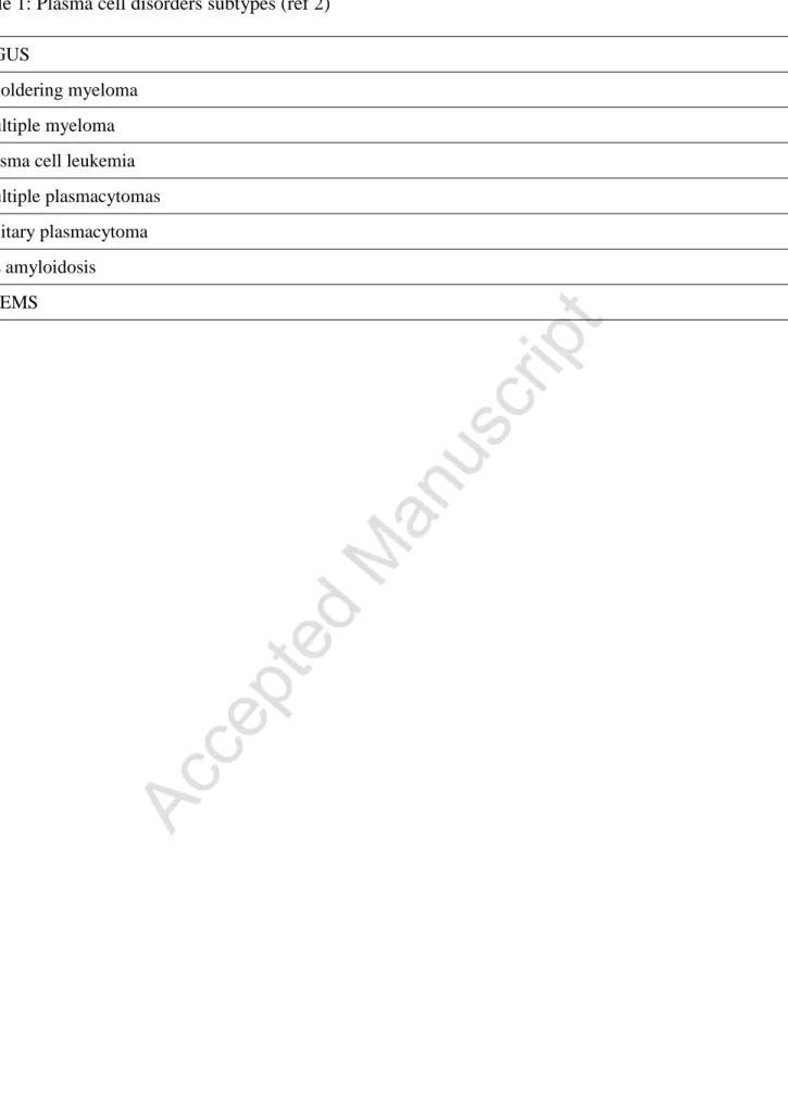
Plasma cell disorders are not limited to multiple myeloma. The spectrum of plasma cell disorders includes: MGUS (Monoclonal Gammopathy of Undetermined Significance), Smoldering Multiple Myeloma, Plasma Cell Leukemia, Solitary Plasmacytoma/Multiple Plasmacytomas, AL Amyloidosis and POEMS (Table 1 and ref 2). Moreover, paraprotein and free light chain abnormalities are not exclusive to plasma cell disorders and may be produced by lymphocytes in a variety of other malignant and non-malignant conditions.
2.1 MGUS
This is the most common plasma cell disorder and a minority of individuals with MGUS will develop multiple myeloma. In 2010, the International Myeloma Working Group (IMWG) defined MGUS as the presence of lower levels of serum M protein (< 3 g/dL), a small clonal plasma cell population in the bone marrow (< 10%) and the absence of the myeloma defining events described below in section 2.2.
2.2 Multiple myeloma
MM is defined using a combination of clinical and pathological criteria (3, 4).
Pathological criteria: Clonal bone marrow plasma cells ≥10% or biopsy-proven plasmacytoma and the
presence of one or more clinical myeloma defining events or biomarkers of MM.
Myeloma defining events:
Evidence of end organ damage which can be attributed to the underlying plasma cell proliferative disorder, specifically:
- Hypercalcemia: serum calcium > 0.25 mmol/L (>1 mg/dL) higher than the upper limit of normal, or > 2.75 mmol/L (>11 mg/dL)
- Renal insufficiency: creatinine clearance <40 mL per min or serum creatinine >177 μmol/L (>2 mg/dL)
- Anemia: hemoglobin value of >20 g/L (>2 g/dL) below the lower limit of normal, or a hemoglobin value of <100 g/L (<10 g/dL)
- Bone lesions: one or more osteolytic lesions on the skeletal radiography, CT or PET-CT scan. This combination of end organ damage defines the acronym CRAB (standing for hyper Calcemia, Renal impairment, Anemia and Bone lesions).
Myeloma defining biomarkers of malignancy (in the absence of CRAB) (5):
- Clonal bone marrow plasma cell percentage ≥60% - Serum free light chain ratio (involved:uninvolved) ≥100
- >1 focal lytic bone or bone marrow lesion (>5 mm) on a MRI scan.
These last three parameters are not only biomarkers of myeloma; if present (one of them is enough), they also indicate that treatment should be initiated. The term “symptomatic myeloma” is a clinical term which refers to the occurrence of end organ damage as described above and indicates the need for therapy (rather than the presence of actual symptoms). These new biomarkers add to the criteria for initiating treatment.
Defining the type of myeloma:
Intact immunoglobulin M protein (heavy chain) and/or light chain type:
The hallmark of MM is the production of monoclonal immunoglobulins and/or light chains by the clonal plasma cells. The specific isotype of the heavy or light chain is characterized by immunofixation electrophoresis (IFE). In light chain only myeloma, no heavy chain component is secreted while in non-secretory myeloma there is no monoclonal component at all. In most cases of MM, only one type of M component is produced throughout the course of the illness. However, in rare cases, two or more (IgG kappa and IgA kappa, for example) may be present or appear occasionally during the course of the disease in serum/urine (biclonal gammopathy).
For data collection and entry, the type and magnitude of the M protein needs to be specified as below. IgG-IgA-IgM-IgE-IgD indicates the type of heavy chain of the M component, while Kappa-Lambda indicates the type of light chain of the M component.
For biclonal gammopathy with two different M protein types, one should note both M protein types on the data collection form and indicate which has the highest value. The M protein with the highest value needs to be considered more significant and entered into the database. The data entry should still note the presence of the two chain types in the comments field. If a patient has more than one M protein spike in the serum (or urine) at the start of treatment, the sum of the M proteins should be followed to assess the response. If the sum of both M proteins is still not at a measurable level, one may use the free light chain test or the bone marrow plasma cell infiltration assessment. In the context of deep responses obtained during autologous stem cell transplantation, one or more new electrophoretic bands
(oligoclonal bands) may appear. These should not be considered pathological or classified as relapse; it is related to B cell immune reconstitution and these bands disappear within a few months post
-transplant.
Magnitude of the M protein: - Secretory myeloma:
The M protein serves as the major biochemical tumor marker for response evaluation and its magnitude at start of therapy and at each point of retreatment for progression serves as a baseline to evaluate the response. In most cases, the response criteria for MM are dependent on the magnitude of the M protein and/or its detection. In rare cases, tumor dedifferentiation during recurrent myeloma may cause plasma cells previously secreting intact paraproteins, e.g., IgG kappa, to produce only a light chain or even no myeloma related protein at all. In such patients, one should follow the light chain or non-secretory myeloma instructions described below.
The magnitude of the monoclonal protein MP in serum (g/L or mg/dL) is a key parameter to monitor patients with secretory intact immunoglobulin myeloma, where an intact monoclonal protein with both heavy and light chain components is the measurable M component, but not to monitor light chain only myeloma or non-secretory MM. The monoclonal protein level is obtained from the serum protein electrophoresis (SPEP)/IFE report; the total IgG (quantitative IgG) level is not an acceptable alternative.
For IgA in some cases where a discrete M protein quantitation cannot be done and IgD myelomas, quantitative immunoglobulin measurements are acceptable for disease assessment. As compared to IgG, almost all IgD M proteins are quantitatively small, while sometimes the IgA spike on the SPEP is difficult to measure because of co-migration with other proteins. When using quantitative
immunoglobulin measurements, the same percentage change applies as for the serum M spike to define the response status. For IgD myeloma (and also in IgM and IgE myeloma), if quantitative measurement is also not possible, the level of free light chain, if elevated, is acceptable to monitor the disease but the myeloma type should be reported as IgD (or IgM/IgE as appropriate). In clinical practice, whenever light chains alone are predominantly detected, an IgD myeloma should be ruled out.
Urinary M proteins or monoclonal immunoglobulin light chains in urine (g/24 h):
For most patients with secretory light chain MM, this is the most important tumor marker. Those with intact immunoglobulin MM may also have measurable urinary light chains. Measurable disease for urinary M protein is defined as >200 mg of monoclonal light chains excreted during 24 h urine
collection. The current criteria still indicate urinary light chain excretion as the biochemical marker to follow, although free light chain measurement could supplant this parameter in the future. Indeed, a recent paper demonstrated an improved sensitivity and prognostic value of serum free light chains over urine measurements (6).
Free light chain in serum:
For the majority of patients without measurable M protein in the serum or 24 h urine collection
(oligosecretory MM), the serum free light chains (FLC) are the best available measurable tumor marker (7). The FLC level determined by the “freelite” assay at baseline provides a trackable tumor marker in these patients with oligosecretory MM. FLC is considered to be measurable in patients whose involved light chain (either kappa or lambda) is > 100 mg/L (10 mg/dL) and who have an abnormal
kappa:lambda ratio (abnormal is outside the range 0.26-1.65 with normal renal function). The FLC test is also used to quantify light chain myeloma when the proteinuria is not assessable. It is also one of the mandatory parameters to define stringent complete response and should be obtained if a complete response is documented.
- Non-secretory MM and marrow involvement:
The bone marrow plasmacytosis indicates the percentage of plasma cells among the total nucleated cells in cytologic bone marrow studies. The marrow plasmacytosis is the only measurable marker in the minority of patients who are non-secretory by electrophoresis of serum/urine and by FLC analysis. The percentage of plasma cells may also be estimated by other techniques such as immunohistochemical
(IHC) staining of marrow trephine biopsies using plasma cell specific markers like CD 138. Flow cytometry of marrow samples typically underestimates the plasma cell content for technical reasons and should not be used to calculate/report the plasmacytosis burden.
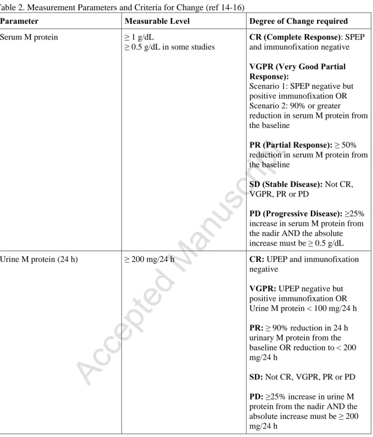
All these parameters are summarized in Table 2.
Staging markers:
Serum beta-2 microglobulin (mg/L): the beta-2 microglobulin level at diagnosis is an important
prognostic factor and is used to stage the disease. It reflects a mixture of tumor load and renal function. Serum albumin (g/dL): together with beta-2 microglobulin, this parameter serves to divide MM into 3 groups according to the International Staging System (ISS) (8).
Lactate dehydrogenase: if elevated at diagnosis this is an adverse prognostic factor and has been incorporated into the new Revised ISS system along with high risk genetic markers (9, 10)). The genetic markers are obtained after an immunomagnetic beads CD138 selection is performed and a fluorescent in situ hybridization (FISH) analysis is carried out on the nuclei from these purified plasma cells. Other prognostic staging systems exist, especially based on molecular risk stratification (11).
2.3 Smoldering MM
Asymptomatic patients who meet biochemical or histopathological criteria for MM but do not have myeloma defining events are considered to have “smoldering myeloma”. Treatment is not currently recommended for these patients except in clinical trials. Symptoms generally develop as a result of hypercalcemia, renal insufficiency, anemia and bone lesions as summarized in the acronym CRAB, although further symptoms such as those caused by hyperviscosity may also require active therapy. As noted previously, even in “asymptomatic” patients, three additional biomarkers or myeloma defining events have now been accepted by the IMWG as indicators for treatment (12). Using the most recent definition of myeloma requiring treatment, smoldering myeloma is an indolent form of myeloma with more than 10% bone marrow plasmacytosis but less than 60% and without myeloma defining
symptoms (CRAB) or biomarkers (free light chain ratio >100 or >1 focal lytic bone lesion on MRI scan).
2.4 Plasma Cell Leukemia (PCL)
PCL is an aggressive form of plasma cell myeloma characterized by an absolute plasma cell count of at
least 2.0 x 10⁹/L (2,000 cells/mm3) in peripheral blood or more than 20% plasma cells in the peripheral differential white blood cell count. When discovered de novo at diagnosis it is called primary PCL; when discovered at the time of disease relapse it is referred to as secondary PCL. Secondary PCL should be considered as progressive or relapsed myeloma and therefore should not be reported as primary PCL. The response criteria for PCL are summarized in Table 3 (13).
2.5 Solitary Plasmacytoma/ Multiple Plasmacytomas
Plasmacytomas are bone or soft tissue lesions composed of plasma cells with no other findings meeting the criteria for MM (no significant increase in the percentage of marrow plasma cells or CRAB criteria/ biomarkers of myeloma; bone destruction may be associated with a single plasmacytoma but this does not justify the diagnosis of myeloma). If numerous plasmacytomas are present, the diagnosis is multiple plasmacytomas and these cases fulfill the criteria for treatment of myeloma. If the pathological criteria for MM are met in the presence of abnormal plasmacytomas, the disease is defined as myeloma with plasmacytoma(s).
Waldenstrom macroglobulinemia is not classified as a plasma cell disorder because it is considered a lymphoma subtype.
B/ Assessing Myeloma Response
In order to assess the response to therapy or progression of disease in MM, one needs to consider the variations over time of the biochemical markers, i.e., serum or urine M protein or free light chain assays, bone marrow plasmacytosis and imaging studies for plasmacytomas and bone lesions. Response
evaluation is usually done (at least) every three months in routine clinical practice whereas in clinical trials the frequency is usually every four weeks.
1. M proteins and Free Light Chains – What is measurable disease?
For patients with intact immunoglobulin secreting MM, measurable disease is defined as a baseline level of >1 g/dL or 10 g/L of serum M protein (some studies allow >0.5 g/dL) or >200 mg/24 h of urine M protein. For those with less than this level of M protein production, a serum involved FLC level of ≥10 mg/dL (100 mg/L) is considered to be a measurable marker and if this marker is not informative then it is necessary to assess the bone marrow plasmacytosis. Truly non-secretory disease with no measurable M protein or FLC secretion can only be assessed by marrow plasma cell estimation. Response levels in MM are defined on the basis of changes in these biochemical markers. The magnitudes of change in these parameters and the corresponding response/progression levels are summarized in Table 4 (14-16). Patients should be categorized as having stable disease (SD) until they meet the criteria for a response category or display progressive disease (PD).
2. IMWG response criteria and levels of response/progression
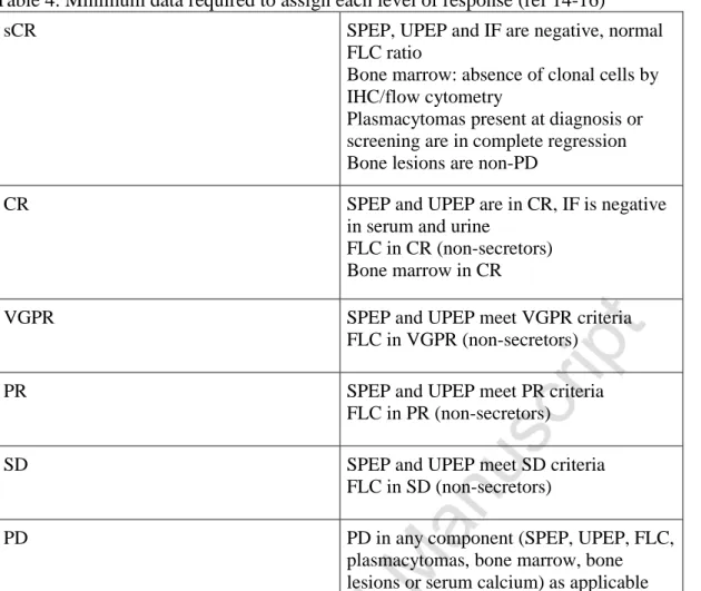
The first set of criteria defining response to treatment and progressive/relapsing disease were published in 1998 by the EBMT and CIBMTR (14). Since then, they have been updated in 2006 with additional clarifications in 2011, 2014 and 2016, by the IMWG (12, 14-16). The parameters needing to be measured and the criteria for change are reported in Table 2 and the minimum data required to assign each response level are given in Table 4.
By definition, relapse occurs when the patient, who was in complete remission, experiences a
reappearance of myeloma, while progression refers to patients with an increasing disease burden from a baseline of persistent residual disease. The minimum level of increase for progression or relapse is an increase in the monoclonal peak of at least 25% from the baseline and at least 0.5 g/dL in magnitude.
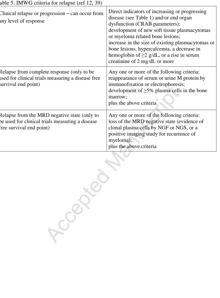
There are three subcategories of relapse: clinical relapse, relapse from complete response and relapse from minimal residual disease (MRD) negativity (Table 5) (12).
The date of relapse/progression is the date when it was first detected (although confirmation with an additional assessment is mandatory). Relapse or progression is not necessarily an indication to start treatment again right away. When treatment is resumed, sometimes weeks, months or even years later, this defines the starting date of the subsequent therapy and a new baseline for response to this line of therapy. By definition, the time to the next treatment is the time interval between the initiation date of a specific line of therapy and the date of beginning a subsequent line of therapy (which may comprise the same drugs as the initial one).
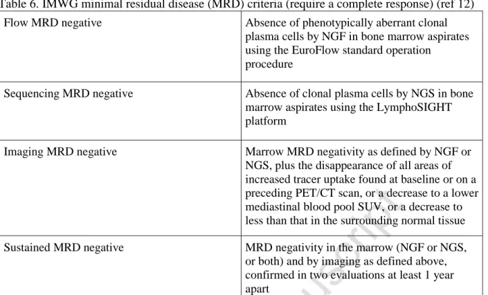
Recently, criteria for responses deeper than CR and assessment of minimal residual disease (MRD) were defined by consensus (12). There are currently three approaches to evaluate MRD (17): cell-based, molecular-based or imaging-based. Bone marrow minimal residual disease can be assessed by multicolor flow cytometry (MFC) which is a cell-based technique (18), while the molecular techniques include ASO-PCR and Next Generation Sequencing. The current modern flow cytometric method for MRD includes 8-color flow cytometry plus kappa and lambda determination for clonality and is known as Next Generation Flow; the sensitivity is 1 in 105 cells and could reach 1 in 106 if enough cells are counted (19). Next Generation Sequencing such as the LymphoSIGHT platform is sensitive to the level of one remaining malignant cell in 106. On the basis of these methods, the IMWG has defined a bone marrow MRD-negative response category. At present, this committee recommends the use of next generation sequencing (NGS) or next generation flow (NGF) for the detection of minimal residual disease in the bone marrow, depending on the availability of the techniques and the feasibility of individual clinical trials.
Accordingly, when minimal residual disease results are reported, the assessment should be qualified by the method(s) used (flow minimal residual negative or sequencing minimal residual disease-negative) and the level of sensitivity (e.g., one in 105 or one in 106 cells). Imaging-based MRD
assessment includes evaluation of the extramedullary disease. Two techniques are available: PET/CT and MRI (20). Table 6 summarizes the consensus on MRD criteria.
C/ Myeloma treatment in the context of response assessment
Treatment options are determined mainly by the ability or not to proceed to autologous transplantation, which is closely related to frailty and fitness (lack of comorbidities) (21). For example, among patients older than 70 years, autologous stem cell transplantation (ASCT) is recommended less often since the procedure may be considered to be too toxic for the elderly population because of the potential concomitant comorbidities. In contrast, for younger patients, ASCT is generally considered at some point in the course of the disease, although significant comorbidities may be a contraindication even in younger individuals.
1. Autologous Stem Cell Transplantation
1.1 Transplant eligible patients
In the initial (induction) phase, the treatment usually combines a proteasome inhibitor (PI) with an immunomodulatory drug (IMiD) and steroids. This triplet therapy has many variants considering that there are currently at least three approved PIs, bortezomib (Velcade®), ixazomib (Ninlaro®) and carfilzomib (Kyprolis®), and three IMiDs, thalidomide, lenalidomide (Revlimid®) and pomalidomide (Pomalyst® in the US and Imnovid® in Europe). The two most common combinations are Velcade, Thalidomide and Dexamethasone (VTD, mostly used in Europe) and Velcade, Revlimid and
Dexamethasone (VRD or RVD, mostly used in the US). A treatment cycle lasts typically 3 weeks and the patients receive 3 to 4 cycles before ASCT. Occasionally, the IMiD component may be replaced by doxorubicin or cyclophosphamide (CyBorD: Cyclophosphamide, Bortezomib and Dexamethasone). One study has shown superiority of VTD over CyBorD with a higher response (22). An ongoing study,
comparing carfilzomib plus lenalidomide plus dexamethasone (KRD) vs bortezomib plus lenalidomide plus dexamethasone (VRD), will help define which one is the better proteasome inhibitor in the
induction phase (23). After induction, the patients usually achieve remission and non-progressive patients proceed to high dose chemotherapy followed by autologous stem cell transplantation.
Approximately three months after ASCT, disease evaluation is performed and further consolidation or maintenance therapy may be initiated thereafter. Post-transplant consolidation generally comprises a short period of treatment of about 2-3 months using full doses of anti-myeloma drugs. Maintenance consists of much longer therapy involving lower doses of drugs which are given until the next relapse or for a fixed duration.
1.2 Non-transplant eligible patients
Historically, the treatment for non-transplant eligible patients was alkylator-based with a doublet combining melphalan and the steroid prednisone (MP) (24). Combinations of MP with newer drugs such as thalidomide (T), lenalidomide (Revlimid®, R) or bortezomib (Velcade®,V) for a defined period
have been shown to be superior to MP in clinical trials (25) so that MPT or sometimes C
(cyclophosphamide) D (dexamethasone)T has become the standard of care at induction, with VMP (26) and more recently MPR assuming this role, the choice of novel agent often being determined by national funding policies or insurers willingness to pay. More recently, the combination of
lenalidomide plus dexamethasone given on an extended basis was found to be superior to MPT and represents a new standard of care for this patient population (27). The triplet combination of Velcade, Revlimid and Dexamethasone (VRD or RVD) has been shown to be associated with superior overall survival (28). Additional second generation agents such as carfilzomib, pomalidomide, and ixazomib are now being incorporated into first line clinical trials, while a further range of effective agents including panobinostat (HDAC inhibitor) and monoclonal antibodies such as daratumumab are becoming available.
2. Allogeneic Stem Cell Transplantation
This procedure is performed less frequently due to its relatively high mortality and morbidity. The two main indications are: 1/ for young patients in first line with a high risk of disease progression (e.g., poor prognosis cytogenetics) and 2/ for patients in first relapse who respond to the relapse treatment
(chemosensitive disease) (29). Various conditioning regimens have been employed but myeloablative conditioning is now rarely used because of the higher transplant related mortality. The current
conditioning regimens are mostly of reduced intensity (RIC: Reduced Intensity Conditioning) or NMA (Non Myeloablative) regimens.
Therefore currently in myeloma, allogeneic stem cell transplantation is mostly performed in the relapse setting (30). The EBMT data registry shows that the numbers are still increasing over the years. Most reports on allografting as a rescue strategy after a previous autograft have been single-center or retrospective registry analyses. Some retrospective studies have compared allogeneic to autologous stem cell transplantation. Overall, these studies showed the feasibility of allografting in relapsed
multiple myeloma even though they included heterogeneous patient groups, differences in conditioning regimens and supportive care (31).
Since myeloma patients invariably relapse despite the improved initial therapies and survival, the case may be made that relapsed patients after an autograft may most benefit from the potentially curative effect of graft versus myeloma, especially if high-risk features are present at diagnosis (32).
3. Tandem and multiple transplantations
3.1 Tandem autologous transplantation
High dose melphalan followed by autologous transplantation remains one of the best forms of treatment for myeloma as it induces deep remission, which often translates into a long period without
progression/relapse. Two planned autologous stem cell transplants (tandem ASCT) performed three to six months apart, usually in the context of first line therapy, have been shown to be superior to single ASCT for some patients and this approach is still commonly employed, especially in Europe. It is important to note that there should be no progression of MM between the two transplants.
Recently two studies addressing the role of tandem autologous transplantation have shown opposite results. The European study demonstrated superiority of tandem versus single transplantation (33), especially for high risks patients, while the US study by the Blood and Marrow Transplant Clinical Trials Network (BMT CTN 0702) did not show any benefit (34).
3.2 Tandem autologous allogeneic transplantation
In this type of tandem transplantation, ASCT is often performed before RIC allogeneic transplantation as part of a planned therapeutic strategy, which is therefore termed tandem auto-allo transplantation. The two procedures are usually carried out within 3-6 months in the absence of any intervening progression of MM (35, 36).
3.3 Multiple transplants (more than two)
Salvage transplantation (transplantation as second line therapy or later) has now been shown to be an effective treatment strategy (37). With the patients living longer, some of them may receive more than two transplants. This can happen if: 1/ they initially have tandem ASCT and a third ASCT after relapse, 2/ they undergo one ASCT and tandem ASCT at relapse, or 3/ they receive single ASCT, a second transplant at relapse and a third transplant after a subsequent relapse.
D/ Practical issues in response evaluation
Multiple levels of confirmation are necessary for data accuracy. The research coordinator or data manager makes the initial evaluation. This should be confirmed by the principal investigator
(physician) and if the patient is participating in a clinical trial, it will be checked again by the medical monitor of the trial where applicable. If the medical monitor disagrees with the physician’s evaluation, a query is sent to the physician. Nowadays, in international trials, a panel of myeloma experts review independently all the response/relapse/progression evaluations. They constitute an Independent Review Committee (IRC).
There are currently two commonly used sets of international criteria: the EBMT/CIBMTR criteria (14) and the IMWG criteria (15, 16) incorporating the Free Light Chain test. A specific trial may arbitrarily use either of them or a modified version. Recently, the minimal residual disease (MRD) assessment has been added to the IMWG criteria (12).
1. How to evaluate response
In order to evaluate response, the following considerations are important:
1.1 What is the baseline?
At diagnosis or at screening?
If the patient is included in a clinical trial, the M protein is often determined at three time points: at diagnosis, at screening (when checking to verify that the patient can be included in the trial) and at baseline, which is day one of cycle 1 of treatment according to the protocol. The baseline is the reference for the measurement of M protein. If the patient is not participating in a clinical trial, the baseline is defined by the first measurement at diagnosis or at the start of therapy. These two time points are generally the same unless there is a significant interval between diagnosis and starting the treatment, as may happen in the case of smoldering myeloma progressing to myeloma. In this case, the baseline to use would be the result at the time of progression to active myeloma (usually the last value before starting therapy).
In order to assess response/progression, defining the baseline for assessment is critical. The baseline, for newly diagnosed patients beginning therapy, is clearly the peak values of M protein (serum or
urine), FLC and marrow plasmacytosis immediately prior to the start of therapy. If a baseline value is not available, an induction response cannot be determined (unless the patient achieves CR to
induction).
In patients proceeding to planned upfront transplantation after induction without any evidence of disease progression between induction and transplant, the baseline for assessing the response to transplantation remains the initial time point prior to induction therapy. The response to transplant should be part of the continuum of response to initial therapy. For example, a patient who has a VGPR going into ASCT and has a continued VGPR remains in VGPR. If there is any evidence of disease progression, the baseline for the subsequent line of therapy after induction is redefined as the peak M protein at the time of progression (or at the time of starting the new line of therapy). Once again, if the transplant follows the new line of therapy as a planned intervention without any additional subsequent progression, the baseline for assessing the response to transplantation is the peak M protein at the time of progression (not at the time point immediately prior to the transplant).
1.2 What is a line of treatment?
A line of therapy is defined as one or more cycles of a planned treatment program. When patients have undergone sequential phases of treatment without intervening progression, such as induction, collection of peripheral blood stem cells (PBSCs), transplantation and consolidation/maintenance, this is
considered as one line of treatment. A new line of therapy is initiated as a result of disease progression or relapse. Such a new line of therapy for progression or relapse is associated with a new baseline for disease evaluation.
1.3 What are the necessary criteria to define the disease status?
All the criteria must be met in order to define a response according to the guidelines.
There are three common time points when the response to a line of treatment is assessed: at specific time points in the course of the disease (e.g., after 2 or 4 cycles of therapy), immediately before a
transplant or on day 100 after the transplant. Alternatively, one may choose the time of the best response, which is the time when the M protein is at its lowest level or CR (or sCR) is reached. For the most common type of MM (secreting an intact monoclonal immunoglobulin M protein, e.g., IgG kappa), the M protein value measured at baseline (the highest) is compared with the lowest M protein level after the beginning of therapy. For light chain myeloma, kappa or lambda, values of the light chain proteinuria (g/24 h) may be compared or if no measurable M protein can be detected, one may use serum free light chain measurements. The percentage of plasma cells in bone marrow can be determined by aspiration or biopsy (preferred method). In either case, the origin should be noted. When the bone marrow plasma cell infiltration is assessed by both BM aspirate and biopsy, the highest value of the plasma cell infiltration should be reported.
If a critical data point to establish a level of response is missing, the evaluation is downgraded to the next lower level, e.g., VGPR instead of CR if a marrow assessment is unavailable/not done.
The near CR (nCR) status is generally not used anymore, except by some investigators, although it is still a level of response in the EBMT criteria. Therefore, if a patient has no further serum monoclonal peak and a normal bone marrow but a positive or unknown serum immunofixation status, the
appropriate level would be nCR according to the EBMT criteria but VGPR according to the IMWG criteria. However, nCR is not generally in common use and it is usually merged together with the VGPR status.
For stringent CR, all the following criteria must be met: all CR criteria plus the absence of clonal marrow plasmacytosis by IHC or flow cytometry and a normal free light chain ratio. If even a single criterion is not met or the data are missing, the response cannot be reported as sCR and must be downgraded to CR.
Each status should be confirmed by second tests giving consistent results.
Confirmation should be obtained for biochemical markers but is not necessary for bone marrow or imaging studies. There is no specific time interval required between the two evaluations (it used to be 6
weeks apart but this interval is no longer necessary). The response date is not the date of confirmation but the initial date when the assessment met the end point. In other words, the second test is
confirmatory. If the result is not confirmed by a second evaluation, the status is either Non Evaluable (NE) and the prior disease status remains valid e.g. for a patient who has achieved a confirmed partial response, a single evaluation meeting the CR end point but not confirmed would mean that the status is still a PR..
After achievement of a best response and with ongoing monthly evaluations, it is not recommended to downgrade the response, e.g., from VGPR to PR, unless there is clear evidence for disease progression. Thus, if the monoclonal protein slightly increases (without meeting the progression criteria) and then decreases at the next evaluation, the response level should remain VGPR. We do not recommend reporting VGPR followed by PR and then back to VGPR in this situation. The status should remain VGPR for all the three time points above until the criteria for progressive disease are satisfied.
When the M spike is reported as “too small to quantitate” in responding patients, one should assign the M protein a value of 0 to allow the subsequent calculation of an absolute increase to determine disease progression. Nevertheless, this situation does not correspond to CR since the paraprotein is detectable. The immunoelectropheresis should be reported as positive for the M protein. A negative IEP means that the SPEP proteins would likely be oligoclonal bands.
2. How to evaluate progression/relapse
The standard criteria for PD are:
- Relapse occurs whenever the patient moves from CR to any status with signs of myeloma disease, either biological and/or clinical.
- Disease progression can be either biochemical (increase in an existing monoclonal peak) or clinical. The former patients may sometimes have no symptoms and may not need therapy for a long period of
time. In other circumstances, the M protein increases and symptoms or organ damage occur concomitantly (clinical relapse).
- Patients are considered to have progressive disease (PD) if they meet the criteria for progression for a parameter which was not considered to be measurable at baseline. For example, a patient initially having a measurable serum M protein but no measurable proteinuria, who subsequently reaches complete remission but during follow-up presents a significant monoclonal proteinuria, without any serum monoclonal peak, should be classified as having PD. However, for patients with a measurable serum or urine M spike at baseline, progression cannot be defined on the basis of increases in serum FLC alone.
- When defining relapse/progression, the result should be confirmed by a second set of tests. We recommend the test to be repeated within 6 weeks of original measurement. If it is confirmed, the date of relapse/progression is the initial date (not that of the confirmatory second test). Second bone marrow biopsies are not required to confirm relapse/progression, even in the assessment of non-secretory patients. Similarly, when a new lytic lesion in bone or an increase in the size of a plasmacytoma or bone lesion is detected, it is not necessary to repeat the imaging procedure.
It is important to realize that in many circumstances, especially in the case of biochemical relapse or progression, treatment may not be reinitiated immediately. The patient may experience a reappearance of the M protein with slow progression but be treated months or years later.
Conversely, if a new line of anti-myeloma therapy was initiated before confirming PD, one should use the starting date of the treatment as the date of progression.
3. How to deal with missing and/or contradictory data
- If baseline information on the values of the involved protein is missing, the response cannot be evaluated (Non Evaluable=NE), except when CR or sCR was achieved or PD was reported.
- If the IEP result for serum or urine is missing, the test is considered to be positive and CR cannot be reported (one should downgrade to VGPR if the other criteria for CR are met).
- If there is no bone marrow evaluation, complete response cannot be reported even if all other parameters including serum and urine IEP and FLC are normal.
- If no 24 h urine monoclonal protein measurement is available, the serum free light chain test can be used in clinical situations, but by strict criteria the urine result is needed for response assessment.
- When data are missing for two or more consecutive cycles, one should report NE for the specific missing cycle assessments.
4. Evaluation of response in Plasma Cell Leukemia
PCL is the most aggressive variant among plasma cell dyscrasias and is defined by the presence of >20% plasma cells in peripheral blood and an absolute peripheral blood plasma cell count exceeding 2×10⁹/L. The clinical features and natural history and its poorer prognosis have led to the development of modified consensus criteria for response in primary plasma cell leukemia (13). In addition to the MM criteria described above, evaluation of the response in PCL includes criteria similar to those for leukemia. A careful assessment of the extramedullary disease at diagnosis and response evaluation is required for all PCL patients, since there is a higher propensity for extramedullary disease in PCL. Measurement of the residual disease in marrow by flow cytometry is necessary when there is no evidence of plasma cell infiltration using routine morphologic examinations. The additional tests needed to determine the response in PCL, which are complementary to the biochemical criteria for MM, are summarized in Table 2.
Monoclonal antibodies (daratumumab, elotuzumab) are increasingly used in the treatment of myeloma with great efficacy. However these therapeutic antibodies may be detected by serum electrophoresis or immunofixation and confound the measurement of myeloma associated M protein in patients who have recently been treated. The current limit of detection of most serum IFE assays is approximately 150 μg/ml M-protein, which is below the serum concentration of most monoclonal antibodies dosed in the therapeutic range. Therapeutic monoclonal antibodies so far approved in myeloma interfere with the detection IgG kappa M-protein (38). Several mitigation strategies are being developed to validly detect low levels of myeloma protein in the presence of therapeutic monoclonal antibody levels in order to confirm VGPR and CR responses. At this time, these strategies are not widely available but are an issue for patients with IgG Kappa myeloma achieving deep responses with monoclonal antibodies. However, there is an assay for daratumumab that will account for the antibody in the IEP but not for the Bristol Myers Squibb compounds (Nivolumab and Elotuzumab).
Assessing myeloma is an expert discipline and the guidelines have to be revised on a regular basis as the field is moving very fast. New forms of therapy have been developed, such as the monoclonal antibodies, providing a greater depth of response. As of now, we will therefore need to evaluate the minimal residual disease (MRD) status using new tools such as molecular study of the bone marrow by PCR or flow cytometry and recent skeletal imaging techniques like PET-CT. These new types of assessment will soon be incorporated into the armament for disease evaluation and new
References
1/ Röllig C, Knop S, Bornhäuser M. Multiple myeloma. Lancet. 2015; 385:2197-2208. 2/ Palumbo A, Anderson K. Multiple myeloma. N Engl J Med. 2011; 364:1046-1060.
3/ Rajkumar SV, Dimopoulos MA, Palumbo A, et al. International myeloma working group updated criteria for the diagnosis of multiple myeloma. Lancet Oncol. 2014;15:e538-548.
4/ Rajkumar SV. Updated diagnostic criteria and staging system for multiple myeloma. Am Soc Clin Oncol Educ Book. 2016;35:e418-423.
5/ Rajkumar SV. Evolving diagnostic criteria for multiple myeloma. Hematology Am Soc Hematol Educ Program. 2015;2015:272-278.
6/ Dejoie T, Corre J, Caillon H, et al. Serum free light chains, not urine specimens, should be used to evaluate response in light-chain multiple myeloma. Blood. 2016;128 :2941-2948.
7/ Dispenzieri A, Kyle R, Merlini G, et al. International Myeloma Working Group guidelines for serum-free light chain analysis in multiple myeloma and related disorders. Leukemia 2009;23:215-224. 8/ Greipp PR, San Miguel J, Durie BG, et al. International staging system for multiple myeloma. J Clin Oncol. 2005;23:3412–3420.
9/ Moreau P, Cavo M, Sonneveld P, et al. Combination of international scoring system 3, high lactate dehydrogenase, and t(4;14) and/or del(17p) identifies patients with multiple myeloma (MM) treated with front-line autologous stem-cell transplantation at high risk of early MM progression-related death. J Clin Oncol. 2014;32:2173–2180.
10/ Palumbo A, Avet-Loiseau H, Oliva S, et al. Revised International Staging System for Multiple Myeloma: A Report From International Myeloma Working Group. J Clin Oncol. 2015;33:2863-2869. 11/ Decaux O, Lode L, Magrangeas F, et al. Prediction of survival in multiple myeloma based on gene expression profiles reveals cell cycle and chromosomal instability signatures in high-risk patients and hyperdiploid signatures in low-risk patients: a study of the Intergroupe Francophone du Myelome. J Clin Oncol. 2008;26:4798–4805.
12/ Kumar S, Paiva B, Anderson KC, et al. International Myeloma Working Group consensus criteria for response and minimal residual disease assessment in multiple myeloma. Lancet Oncol. 2016; 17: e328–346.
13/ Fernandez de Larrea C, Kyle RA, Durie BG, et al. Plasma cell leukemia : consensus statement on diagnostic requirements, response criteria and treatment recommendations by the International Myeloma Working Group. Leukemia. 2013;27:780-791.
14/ Bladé J, Samson D, Reece D, et al. Criteria for evaluating disease response and progression in patients with multiple myeloma treated by high-dose therapy and haematopoietic stem cell
transplantation. Br J Hematol. 1998; 102: 1115-1123.
15/ Durie BG, Harousseau JL, Miguel JS , et al. International uniform response criteria for multiple myeloma. Leukemia. 2006;20:1467-1473.
16/ Rajkumar SV, Harousseau JL, Durie B, et al. Consensus recommendations for the uniform reporting of clinical trials: report of the International Myeloma Workshop Consensus Panel 1. Blood. 2011;117:4691-4695.
17/ Mailankody S, Korde N, Lesokhin AM, et al. Minimal residual disease in multiple myeloma: bringing the bench to the bedside. Nat Rev Clin Oncol. 2015 ;12:286-295.
18/ Paiva B, Merino J, San Miguel JF, et al. Utility of flow cytometry studies in the management of patients with multiple myeloma. Curr Opinion Oncol. 2016 ;28:511-517.
19/ Royston DJ, Gao Q, Nguyen N,et al. Single-Tube 10-Fluorochrome Analysis for Efficient Flow Evaluation of Minimal Residual Disease in Plasma Cell Myeloma. Am J Clin Pathol. 2016 ;146:41-49. 20/ Dimopoulos M, Terpos E, Comenzo RL, et al. International myeloma working group consensus statement and guidelines regarding the current role of imaging techniques in the diagnosis and monitoring of multiple myeloma. Leukemia. 2009;23:1545-1556.
21/ Harousseau JL, Moreau P. Autologous hematopoietic stem-cell transplantation for multiple myeloma. N Engl J Med. 2009;360:2645-2654.
22/ Moreau P, Hulin C, Macro M, et al. VTD is superior to VCD prior to intensive therapy in multiple myeloma: results of the prospective IFM2013-04 trial. Blood. 2016;127:2569-2574.
23/ Randomized Phase III Trial of Bortezomib, Lenalidomide and Dexamethasone (VRd) Versus Carfilzomib, Lenalidomide, Dexamethasone (CRd) Followed by Limited or Indefinite Lenalidomide Maintenance in Patients With newly Diagnosed Symptomatic Multiple Myeloma. Clinicaltrials.gov. NCT01863550.
24/ Palumbo A, Sezer O, Kyle R, et al. International Myeloma Working Group guidelines for the management of multiple myeloma patients ineligible for standard high-dose chemotherapy with autologous stem cell transplantation. Leukemia. 2009; 23:1716-1730.
25/ Facon T, Mary JY, Hulin C, et al. Melphalan and prednisone plus thalidomide versus melphalan and prednisone alone or reduced-intensity autologous stem cell transplantation in elderly patients with multiple myeloma (IFM 99-06): a randomised trial. Lancet. 2007 ;370:1209-1218.
26/ San Miguel JF, Schlag R, Khuageva NK, et al. Bortezomib plus melphalan and prednisone for
initial treatment of multiple myeloma. N Engl J Med. 2008 ;359:906-917.
27/ Benboubker L, Dimopoulos MA, Dispenzieri A, et al. Lenalidomide and dexamethasone in transplant-ineligible patients with myeloma. N Engl J Med. 2014;371:906-917.
28/ Durie BG, Hoering A, Abidi MH, et al. Bortezomib with lenalidomide and dexamethasone versus lenalidomide and dexamethasone alone in patients with newly diagnosed myeloma without intent for immediate autologous stem-cell transplant (SWOG S0777): a randomised, open-label, phase 3 trial. Lancet. 2016. pii: S0140-6736(16)31594-X.
29/ Lokhorst H, Einsele H, Vesole D, et al. International Myeloma Working Group consensus
statement regarding the current status of allogeneic stem-cell transplantation for multiple myeloma. J Clin Oncol. 2010;28:4521–4530.
30/ Giralt S, Garderet L, Durie B, et al. American Society of Blood and Marrow Transplant, European Society of Blood and Marrow Transplantation, Blood and Marrow Transplant Clinical Trials Network and International Myeloma Working Group Consensus conference on salvage hematopoietic cell transplantation in patients with relapsed multiple myeloma. Biol Blood Marrow Transplant. 2015;21:2039–2051.
31/ Festuccia M, Martino M, Ferrando F, et al. Allogeneic stem cell transplantation in multiple myeloma: immunotherapy and new drugs. Expert Opin Biol Ther. 2015;15:857–872.
32/ Kroger N, Shimoni A, Schilling G, et al. Unrelated stem cell transplantation after reduced intensity conditioning for patients with multiple myeloma relapsing after autologous transplantation. Br J Haematol. 2010;148:323–331.
33/ Cavo M, Petrucci MT, Di Raimondo F,et al. Upfront single versus double autologous stem cell transplantation for newly diagnosed multiple myeloma: an intergroup, multicenter, phase III study of the European Myeloma Network (EMN02/HO95 MM Trial). ASH Annual Meeting Abstracts 2016, abst 991.
34/ Stadtmauer EA, Pasquini MC, Blackwell B, et al. Comparison of Autologous Hematopoietic Cell Transplant (autoHCT), Bortezomib, Lenalidomide (Len) and Dexamethasone (RVD) Consolidation
with Len Maintenance (ACM), Tandem AutoHCT with Len Maintenance (TAM) and AutoHCT with Len Maintenance (AM) for up-Front Treatment of Patients with Multiple Myeloma (MM): Primary Results from the Randomized Phase III Trial of the Blood and Marrow Transplant Clinical Trials Network (BMT CTN 0702 – StaMINA Trial). ASH Annual Meeting Abstracts 2016, abst LBA-1.
35/ Bruno B, Rotta M, Patriarca F, et al. A comparison of allografting with autografting for newly diagnosed myeloma. N Engl J Med. 2007 ;356:1110-1120.
36/ Gahrton G, Iacobelli S, Björkstrand B, et al. Autologous/reduced-intensity allogeneic stem cell transplantation vs autologous transplantation in multiple myeloma: long-term results of the EBMT-NMAM2000 study. Blood. 2013 ;121:5055-5063.
37/ Cook G, Williams C, Brown JM, et al. High-dose chemotherapy plus autologous stem-cell
transplantation as consolidation therapy in patients with relapsed multiple myeloma after previous autologous stem-cell transplantation (NCRI Myeloma X Relapse [Intensive trial]): a randomised, open-label, phase 3 trial. Lancet Oncol. 2014;15:874-885.
38/ McCudden CR, Voorhees PM, Hainsworth SA, et al. Interference of monoclonal antibody therapies with serum protein electrophoresis tests. Clin Chem. 2010 ;56:1897-1899.
39/ Lonial S and Gertz MA. Eliminating the complete response penalty from myeloma response assessment. Blood. 2008; 111:3297-3298.
Table 1: Plasma cell disorders subtypes (ref 2)
MGUS
Smoldering myeloma Multiple myeloma Plasma cell leukemia Multiple plasmacytomas Solitary plasmacytoma AL amyloidosis POEMS
Table 2. Measurement Parameters and Criteria for Change (ref 14-16)
Parameter Measurable Level Degree of Change required
Serum M protein ≥ 1 g/dL
≥ 0.5 g/dL in some studies
CR (Complete Response): SPEP
and immunofixation negative
VGPR (Very Good Partial Response):
Scenario 1: SPEP negative but positive immunofixation OR Scenario 2: 90% or greater
reduction in serum M protein from the baseline
PR (Partial Response): ≥ 50%
reduction in serum M protein from the baseline
SD (Stable Disease): Not CR,
VGPR, PR or PD
PD (Progressive Disease): ≥25%
increase in serum M protein from the nadir AND the absolute increase must be ≥ 0.5 g/dL Urine M protein (24 h) ≥ 200 mg/24 h CR: UPEP and immunofixation
negative
VGPR: UPEP negative but
positive immunofixation OR Urine M protein < 100 mg/24 h
PR: ≥ 90% reduction in 24 h
urinary M protein from the baseline OR reduction to < 200 mg/24 h
SD: Not CR, VGPR, PR or PD
PD: ≥25% increase in urine M
protein from the nadir AND the absolute increase must be ≥ 200 mg/24 h
Free Light Chain (only if serum and urine M protein are not measurable)
≥10 mg/dL for the involved chain CR: Normal FLC ratio of 0.26 to
1.65
VGPR: > 90% decrease from the
baseline in the difference between the levels of involved and
uninvolved FLC
PR: ≥ 50% decrease from the
baseline in the difference between the levels of involved and
uninvolved FLC
SD: Not CR, VGPR, PR or PD
PD: ≥ 25% increase from the
nadir in the difference between the levels of involved and uninvolved FLC. The absolute increase must be ≥ 10 mg/dL from the nadir (only if serum and urine M protein are not measurable, otherwise no impact)
Marrow plasma cells ≥ 30% sCR (stringent CR): Absence of marrow clonal cells by IHC / flow cytometry
CR: < 5% plasma cells in the
bone marrow
If serum and urine M protein and FLC are not measurable (true non-secretor):
PR: ≥ 50% reduction in plasma
cells from the baseline instead of a reduction in M protein, provided the baseline bone marrow plasma cell content was ≥ 30%
PD: Absolute increase in marrow
Plasmacytoma Diameter > 2 cm After radiation, the plasmacytoma is not evaluable to assess the response but must be monitored to assess PD. Determine the size from the sum of the perpendicular diameters or the longest diameter (if only one is reported)
CR: Complete regression of the
plasmacytoma
VGPR: > 90% decrease in the
size of the plasmacytoma
PR: ≥ 50% decrease in the size of
the plasmacytoma
PD: > 50% increase in the size of
the plasmacytoma AND at least 1 cm absolute increase
Bone lesions NA Only for PD – if there are new or increasing bone lesions
Table 3. Criteria for response in plasma cell leukemia (ref 13) - Stringent Complete Response (sCR)
In addition to the sCR criteria for MM, the following need to be met:
• Absence of malignant plasma cells in the peripheral blood by flow cytometry • Absence of extramedullary disease (this evaluation is necessary)
- Complete Response (CR)
In addition to the CR criteria for MM, the following need to be met: • Absence of plasma cells in the peripheral blood (by morphology) • Absence of extramedullary disease
- Very Good Partial Response (VGPR)
In addition to the VGPR criteria for MM, the following need to be met: • Less than 5% plasma cells in a bone marrow aspirate
• Absence of plasma cells in the peripheral blood • Absence of extramedullary disease
- Partial Response (PR)
In addition to the PR criteria for MM, the following need to be met: • Between 5% and 25% plasma cells in a bone marrow aspirate • Between 1% and 5% plasma cells in the peripheral blood
• A 50% or greater reduction in the extent of extramedullary disease - Stable Disease (SD)
• Patients who do not meet the criteria for sCR, CR, VGPR, PR or PD (defined below) are considered to have stable disease
- Progressive Disease (PD)
Any of the PD criteria for MM, OR the following criterion:
Table 4. Minimum data required to assign each level of response (ref 14-16)
sCR SPEP, UPEP and IF are negative, normal FLC ratio
Bone marrow: absence of clonal cells by IHC/flow cytometry
Plasmacytomas present at diagnosis or screening are in complete regression Bone lesions are non-PD
CR SPEP and UPEP are in CR, IF is negative in serum and urine
FLC in CR (non-secretors) Bone marrow in CR
VGPR SPEP and UPEP meet VGPR criteria FLC in VGPR (non-secretors)
PR SPEP and UPEP meet PR criteria FLC in PR (non-secretors)
SD SPEP and UPEP meet SD criteria FLC in SD (non-secretors)
PD PD in any component (SPEP, UPEP, FLC, plasmacytomas, bone marrow, bone lesions or serum calcium) as applicable
Foot notes: The baseline values of all measurable disease parameters should be checked serially to assess response. To confirm CR or sCR, marrow and FLC assessments should also be performed in all cases. A subsequent biochemical study should be carried out to confirm all biochemical responses, whereas marrow studies do not need to be reconfirmed. Missing urine electrophoresis results represent a frequent problem. At this time point, FLC results are not an adequate substitute and the level of response must be reported as non-evaluable (NE) if there was measurable urine M protein at baseline. Similarly, CR cannot be confirmed without urine immunofixation. Exceptions to these rules may be conceded only on the decision of a
response monitoring committee and by consensus. By definition, to assign CR, all parameters in the bone marrow, serum and urine should be in CR. Any plasmacytoma present at baseline should undergo
monitoring. All measurable plasmacytomas should have disappeared to be able to confirm CR, or have regressed to appropriate levels for the other categories of response.
Table 5. IMWG criteria for relapse (ref 12, 39)
Clinical relapse or progression – can occur from any level of response
Direct indicators of increasing or progressing disease (see Table 1) and/or end organ dysfunction (CRAB parameters);
development of new soft tissue plasmacytomas or myeloma related bone lesions;
increase in the size of existing plasmacytomas or bone lesions, hypercalcemia, a decrease in hemoglobin of ≥2 g/dL, or a rise in serum creatinine of 2 mg/dL or more
Relapse from complete response (only to be used for clinical trials measuring a disease free survival end point)
Any one or more of the following criteria: reappearance of serum or urine M protein by immunofixation or electrophoresis;
development of ≥5% plasma cells in the bone marrow;
plus the above criteria Relapse from the MRD negative state (only to
be used for clinical trials measuring a disease free survival end point)
Any one or more of the following criteria: loss of the MRD negative state (evidence of clonal plasma cells by NGF or NGS, or a positive imaging study for recurrence of myeloma);
Table 6. IMWG minimal residual disease (MRD) criteria (require a complete response) (ref 12)
Flow MRD negative Absence of phenotypically aberrant clonal plasma cells by NGF in bone marrow aspirates using the EuroFlow standard operation
procedure
Sequencing MRD negative Absence of clonal plasma cells by NGS in bone marrow aspirates using the LymphoSIGHT platform
Imaging MRD negative Marrow MRD negativity as defined by NGF or NGS, plus the disappearance of all areas of increased tracer uptake found at baseline or on a preceding PET/CT scan, or a decrease to a lower mediastinal blood pool SUV, or a decrease to less than that in the surrounding normal tissue Sustained MRD negative MRD negativity in the marrow (NGF or NGS,
or both) and by imaging as defined above, confirmed in two evaluations at least 1 year apart