HAL Id: hal-01913959
https://hal.archives-ouvertes.fr/hal-01913959
Submitted on 8 Jun 2021
HAL is a multi-disciplinary open access
archive for the deposit and dissemination of
sci-entific research documents, whether they are
pub-lished or not. The documents may come from
teaching and research institutions in France or
abroad, or from public or private research centers.
L’archive ouverte pluridisciplinaire HAL, est
destinée au dépôt et à la diffusion de documents
scientifiques de niveau recherche, publiés ou non,
émanant des établissements d’enseignement et de
recherche français ou étrangers, des laboratoires
publics ou privés.
Distributed under a Creative Commons Attribution| 4.0 International License
Leydig cell tumor in a patient with 49,XXXXY
karyotype: a review of literature
Salwan Maqdasy, Laura Bogenmann, Marie Batisse-Lignier, Béatrice Roche,
Frédéric Franck, Françoise Desbiez, Igor Tauveron
To cite this version:
Salwan Maqdasy, Laura Bogenmann, Marie Batisse-Lignier, Béatrice Roche, Frédéric Franck, et al..
Leydig cell tumor in a patient with 49,XXXXY karyotype: a review of literature. Reproductive Biology
and Endocrinology, BioMed Central, 2015, 13, pp.72. �10.1186/s12958-015-0071-7�. �hal-01913959�
R E V I E W
Open Access
Leydig cell tumor in a patient with
49,XXXXY karyotype: a review of literature
Salwan Maqdasy
1,2,3*†, Laura Bogenmann
1†, Marie Batisse-Lignier
1,2, Béatrice Roche
1, Fréderic Franck
4,
Françoise Desbiez
1and Igor Tauveron
1,2Abstract
49,XXXXY pentasomy or Fraccaro
’s syndrome is the most severe variant of Klinefelter’s syndrome (KS) affecting
about 1/85000 male births. The classical presentation is the triad: mental retardation, hypergonadotropic
hypogonadism and radio ulnar synostosis. Indeed, the reproductive function of Fraccaro
’s syndrome is
distinguished from KS. Besides, Leydig cell tumors are described in cases of KS, but never documented in the
Klinefelter variants.
We describe a young adult of 22 years old who presented with hyper gonadotropic hypogonadism, delayed puberty
and bilateral micro-cryptorchidism. Chromosomal pentasomy was confirmed since infancy. Bilateral orchidectomy
revealed a unilateral well-circumscribed Leydig cell tumor associated with bilateral Leydig cell hyperplasia.
Inspired from reporting the first case of Leydig cell tumor in a 49,XXXXY patient, we summarize the particularities of
testicular function in 49,XXXXY from one side, and the risk and mechanisms of Leydig cell tumorigenesis in Klinefelter
variants on the other side. The histological destructions in 49,XXXXY testes and hypogonadism are more profound than
in Klinefelter patients, with early Sertoli, Leydig and germ cell destruction. Furthermore, the risk of Leydigioma
development in KS and its variants remains a dilemma. We believe that the risk of Leydigioma is much higher in KS
than the general population. By contrast, the risk could be lower in the Klinefelter variants with more than 3
supplementary X chromosomes, owing to an earlier and more profound destruction of Leydig cells rendering them
irresponsive to chronic Luteinizing hormone (LH) stimulation.
Keywords: Klinefelter, 49,XXXXY, Fraccaro
’s syndrome, Leydig cell tumor, Leydigioma, Endocrine function
Background
Klinefelter syndrome (47,XXY or KS) is the commonest
aneuploidy. It affects 1/650 male births (0.2 % of general
population). Besides, more severe, fortunately rare,
aneu-ploidies are also described; these include: 48,XXXY,
48,XXYY and 49,XXXXY. 48,XXYY affects 1/8000-1/
40,000 male deliveries, while 48,XXXY affects 1/50,000.
Pentasomy 49,XXXXY incidence is around 1/85000 male
births [1, 2]. A 49,XXXXY karyotype is thought to arise
from maternal non-disjunction during both stages of
meiosis, retaining all the X chromosomes within the
oo-cyte. The major endocrine issues of aneuploidies are
hyper gonadotropic hypogonadism, testicular
degenera-tive changes and the risk of testicular tumorigenesis.
Indeed, Leydig cell tumors or Leydigioma are
occasion-ally described in cases of KS, but never in the Klinefelter
variants.
The objectives of this article are: we document herein,
the first case of Leydig cell tumor associated with
bilat-eral Leydig cell hyperplasia in a 49,XXXXY patient.
Fur-thermore, we will review the particularities of testicular
function in 49,XXXXY from one side and the
mecha-nisms and Leydig cell tumorigenesis in Klinefelter
variants on the other side.
Case presentation
A young patient born in 1990 is followed up in our
depart-ment for hypogonadism. At birth, facial dysmorphism
characterized by microcephaly, hypertelorism, inclination
of the palpebral fissures, small broad-based nose, micro
* Correspondence:smaqdasy@chu-clermontferrand.fr
†Equal contributors 1
Service d’endocrinologie, diabétologie et maladies métaboliques, CHU Clermont-Ferrand, F-63003 Clermont-Ferrand, France
2
UMR CNRS 6293, INSERM U1103, Université Clermont-Auvergne, Génétique Reproduction et Développement, BP 10448, 63177 Aubiere, France Full list of author information is available at the end of the article
© 2015 Maqdasy et al. This is an Open Access article distributed under the terms of the Creative Commons Attribution License (http://creativecommons.org/licenses/by/4.0), which permits unrestricted use, distribution, and reproduction in any medium, provided the original work is properly credited. The Creative Commons Public Domain Dedication waiver (http:// creativecommons.org/publicdomain/zero/1.0/) applies to the data made available in this article, unless otherwise stated.
retrognathia and a clinodactyly of the 5th finger were
no-ticed immediately. A parasternal systolic murmur of
left-to-right shunt was audible. The karyotype of the patient
revealed 49,XXXXY aneuploidy. During his childhood,
mental retardation and delayed milestones were
docu-mented with walking at age of 3.5 years, pronouncing
words of two syllables at age of 21 months. At age of
15 years, he was described as a joyful teenager, who showed
a substantial anxiety for unusual situations. His language
was limited to about ten words. He was able to write his
name correctly, to copy a short text and to count up to ten.
Clinical examination revealed a bilateral testicular
ecto-pia and hypoplasia of the external genitalia.
Musculoskel-etal examination revealed scoliosis and bilateral radio
ulnar synostosis. Moderate hypotonia was also noticed
since his childhood, along with a significant fatigability.
At age of 13 years, puberty was absent (Tanner stage P1)
and hyper gonadotropic hypogonadism was confirmed.
The hormonal profile was in favour of early severe
testicu-lar failure (FSH 27 UI/L, LH 17 UI/L with undetectable
testosterone levels). Testosterone therapy (50 mg/3 weeks,
increased progressively to 125 mg/3 weeks) was initiated
at the age of 15 years. Skeletal age was 12.5 years.
Testos-terone levels were consequently normalized to 5.4 ng/ml
under the substitutive treatment, with FSH reduced to 9.2
UI/L and LH levels to 5.1 UI/L (normal levels: FSH
2.2-9.8 UI/L; LH 1.8-7 UI/L).
Young adult, he measures 1.83 m and weighs 55 kg
with a slender silhouette. His testicles were impalpable.
An abdmino-pelvic tomography revealed both testicles
in the inguinal position (Fig. 1a).
Bilateral orchidectomy was realized in 2012. The
macroscopic examination reported right and left small
testicles (1.5x1 cm). Excised tissues were fixed using 4 %
paraformaldehyde (Sigma-Aldrich) and embedded in
paraffin. Microscopic examination after Hematoxylin/
Eosin staining (Sigma-Aldrich) on 5
μm tissue sections
revealed that fibrous involution replaced the
seminifer-ous tubules along with disappearance of Sertoli cells,
as-sociated with Leydig cell hyperplasia. No germ cells
were detected. A well-circumscribed right-sided Leydig
cell tumor was detected. The tumor was composed of
hexagonal eosinophilic cells. Lipofusceine grains were
ob-served without Reinke crystalloids. The nuclei were
rounded with small nucleoli (Fig. 1b). Calretinin immuno
staining (790-4467/Ventana Clone SP65) confirmed the
nature of the sex cord stromal tumor (Fig. 1c). The
ap-pearance was monomorphic, without any significant
pro-liferative activity (less than 1 %) (Fig. 1d). The propro-liferative
activity was evaluated by immuno staining with human
anti-Ki67 antibody (Ki67: 790-4286/Ventana Clone 30–9).
Immunohistochemistry for calretinin and Ki-67 was
con-ducted according to the manufacturer’s recommendations;
the slides were then counterstained with haematoxylin.
Two years post-orchidectomy computed scanner
re-vealed no metastases. Testosterone replacement
ther-apy permitted to virilise the external genitalia (Tanner
stage P3).
Discussion
1. General
Harry Klinefelter published a report on nine men with
testicular dysgenesis, elevated gonadotropins, micro
orchidism, azoospermia, and gynecomastia in 1942 [3].
The hallmark of KS is hyper gonadotropic
hypogonad-ism with testicular atrophy [4]. The diagnosis is usually
evoked in infants with delayed milestones [5], and in
male adults with sterility [6, 7].
49,XXXXY is the most severe form, accounting for
1.4-1.7 % of aneuploidies [2, 8, 9]. Described firstly in
1960 by
Fraccaro, since then, we detected 176 cases in
the literature. Some authors distinguish specific
charac-teristics of this syndrome [1, 10, 11]. The classical
pres-entation is the triad: mental retardation, hypogonadism
and radio ulnar synostosis [2, 10–12]. Hypogonadism is
severe and frequently associated with genital anomalies,
which are characterized by micro penis, small testicles
and scrotum, cryptorchidism and genital ambiguity.
Gynecomastia is rare [10].
Some paradoxical features seem to be specific to
49,XXXXY; 49,XXXXY are usually shorter than the
general population [13, 14]. Microcephaly is found in
some children with 20 % reduction in cerebral volume
[15], which is more severe than Klinefelter patients
[16]. Focal neurological signs are possible, which are
related to demyelination [15]. Language acquisition is
late, and usually other alternatives of communication
are used in adult age [17]. We analysed 176 published
cases of 49,XXXXY in the literature. We noticed two
principal malformations in the published cases: cardiac
(22 patients, mostly patent ductus arteriosus) and
uro-genital (7 patients). The global mortality in aneuploidies
in general and in 49,XXXXY in particular, studied in
de-tails in 3518 patients of a British cohort included
be-tween 1958 and 2003 (48 cases of 49,XXXXY), was
elevated (SMR: 1.5-2) with congenital heart diseases,
epilepsy and pulmonary embolism as major causes of
mortality [8].
Indeed, our patient presented the classical triad of
49,XXXXY syndrome, which was associated with facial
dysmorphism, psychological troubles, mental retardation
and cardiac malformation. But, he had eunuchoid
morph-ology as in Klinefelter patients, which is rare for
49,XXXXY.
2. Testicular function in 49,XXXXY
Indeed, the testicular anomalies are the hallmark of
aneuploidies, starting from birth and on-going to
adult-hood [18]. Histological destructions in 49,XXXXY testes
are more profound than Klinefelter patients. In contrast
to KS, micro orchidism occurs in almost all cases of
48,XXYY, 48,XXXY and 49,XXXXY, with adult testicular
volumes typically around 1–4 mL [14, 17].
In early adulthood, Klinefelter patients seem to enter
normally in the puberty, with incomplete development of
secondary sexual characters, but this is rare in the variants
which are characterized by absence of puberty [19]. The
endocrine profile of KS and the variants is characterized by
normal levels of gonadotropins, AMH and Inhibin B with
variable levels of testosterone in infancy [20–22]. In infants,
Leydig cells are normally sensitive to the proliferative effect
of LH but Sertoli cell sensitivity is questionable, post
pu-bertal Sertoli cell resistance to FSH is definite [23].
Hypo-gonadism is more profound in the variants than the
classical 47,XXY syndrome. This is due to a more profound
testicular damage, associated with lower testosterone
levels, even in infancy leading to ambiguous genitalia. The
endocrine profile of aneuploidies is summarized in Table 1.
In our case, the hypogonadism was profound with no
initiation of puberty. Gynecomastia was absent
indicat-ing no any testosterone secretion to be aromatized.
Tes-ticular ectopia was moderate but associated with a
complete testicular fibrosis, with no germ or Sertoli cells
identified. Therefore, we suggest that the chromosomal
anomaly is responsible for early Sertoli and germ cell
de-terioration. Indeed, we suppose that the degree of
damage of Sertoli cells, their sensitivity to follicle
stimulat-ing hormone (FSH) and germ cell destruction in patients
with more than 3 X chromosomes is more profound. This
is supported by the evidence that the chance of fertility or
successful retrieval of gametes is limited to Klinefelter
pa-tients especially in mosaic forms than other aneuploidies.
On the other side, Leydig cell deterioration is tardive. This
is supported by the testicular decent even if incomplete
(dependent on Insulin like 3 and testosterone secreted
from foetal Leydig cells), Leydig cell hyperplasia
(re-sponsiveness to chronic Luteinizing hormone
stimula-tion from puberty) and the presence of steroidogenesis
in early puberty. Nevertheless, In comparison to 47,
XXY, Leydig cell deterioration seems to be more
pro-found and earlier in 49,XXXXY patients, as steroid
synthesis is negligible manifested by higher risk of
am-biguous genitalia and absent puberty.
3. Risk of Leydig cell tumors or Leydigioma
Testicular tumors are relatively rare, corresponding to
1 % of all human cancers [24]. Germ cell tumors and
sex cord stromal tumors constitute the two
predomin-ant types. Since 1960s, the incidence of testicular cancer
doubled in most of western countries [25]. Fortunately,
only 20 % of these tumors appear during infancy (5–10
years old) [26]. Leydig cell tumors or Leydigioma
repre-sent 0.8–3 % of all testicular tumors in adulthood and
4-9 % of childhood testicular tumors [27–29]. Most of
these tumors are benign, especially in infancy [30].
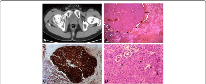
Fig. 1 Identification of a Leydigioma in a patient with 49,XXXXY karyotype with bilateral testicular ectopia. a Identification of 1 and 1.5 cm diameter testes within the inguinal groin by computed tomography in adolescence. b Identification of a well circumscribed 2 mm diameter tumor in the testis. Tumor cells are hexagonal, with round uniform prominent nuclei. The cytoplasm is eosinophilic, or slightly pale due to lipid accumulation. Lipofuschine pigment is identified in steroid producing tumors. Some calcification and hyalinization of the stroma could be identified. Reinke crystals, pathognomonic for Leydigioma are present in only 40 % of the cases. They were absent in this case. c Immuno staining of the tumor by Calretinin, a specific marker of stroma cell tumors. d Ki67 immuno staining. the proliferative index is low in the benign tumors
Testicular cancers are clearly linked to undescended
testis, testicular dysgenesis syndrome (TDS) and
infertil-ity [31–38]. Malignancy occurs in 3.5–14.5 % of
undes-cended testis. Thus, a higher risk of testicular cancers in
Klinefelter patients and the other variants has been
sup-posed. Leydig cell tumors and/or hyperplasia are not
un-common findings during the histological examination of
human testicular biopsies, especially in patients with
tes-ticular atrophy, cryptorchidism, KS and androgen
in-sensitivity syndrome [39, 40]. Indeed, a Danish Study
published in 1995 (696 patients) and a British cohort of
3518 patients with Klinefelter variants including 48
pa-tients with 49,XXXX, seem to be the most significant
publications to explore the tumor risk. They showed a
higher relative risk of mortality (RR = 1.5), but cancer
re-lated mortality was not different from the general
popu-lation [9, 41]. More specifically, the risk of testicular
cancers seemed to be equivocal to the general
popula-tion in these studies. Nevertheless, extra gonadal (mostly
mediastinal) germ cell tumors were more commonly
re-ported in patients with KS in comparison to the general
population, with a prevalence of 1 % [41–44].
Further-more, in another study, Ahmad
et al. measured the
vol-ume of Leydig cells histometrically on biopsies of 50
cases of KS; the mean volume was within normal limits
[45]. Meanwhile, it is important to mention that the first
two studies interested more specifically in cancer risk
and did not take in consideration the benign tumors or
hyperplasia and they were based on the data registries.
On the other hand, Ahmad
et al. analysis was based on
retrospective data. By contrast, a recent Italian study
screened the testes of 40 Klinefelter patients by
ultrasound, magnetic resonance imaging and tumor
markers. Over 3 years of follow up, 30 % of patients
pre-sented with either cysts or nodules, which were below
1 cm. But, no clinical, biological and radiological
argu-ments for testicular cancer were identified. Moreover, no
biological/morphological differences between those with
or without a history of cryptorchidism were noticed [46].
More specifically, a French cohort was based on
ultra-sound screening the testes of 141 KS patients. 158
tes-ticular nodules in 56 (40 %) patients were identified.
20 % of them had bilateral nodules. Indeed, only 12
pa-tients (7.6 %) were operated and all of them suffered
from Leydig cell hyperplasia and/or Leydig cell tumors.
On the other side, the karyotype anomalies in testicular
tumor context are not well recognized; unfortunately,
most studies interested in the testicular tumors neglected
the karyotype of these patients [37, 47]. A French study of
45 tumors in infertile patients with 11 Leydig cell
hyper-plasia and 17 benign Leydig cell tumors, revealed that KS
was found in 10 patients. Indeed, 12 Leydig cell tumors
were identified in these ten patients and fortunately all
were benign [48].
We analysed the literature for the published cases of
tes-ticular tumors in aneuploidies, over nearly 4000 patients
with KS and its variants, we identified only 34 patients
with gonadal and extra gonadal tumors. 20 patients had
Leydig cell tumors, fortunately only two were malignant;
teratomas were the second most common tumors
de-scribed (Table 2).
Returning to our patient, this is the first case of benign
Leydigioma associated with bilateral Leydig cell
hyper-plasia in a Klinefelter variant. It is important to say that
Table 1 Chronological evolution of clinical, histological and hormonal parameters of Klinefelter variants. Eunuchoid morphology and
gynecomastia are absent in 49,XXXXY karyotype
Parameter Infancy Early puberty (12 years) MidPuberty Tanner Puberty Tanner
II-III III-IV FSH N N ++ ++++ LH N N + +++ T - N or - + -E2 N ++ ++ ++ Inhibin B N N - - -AMH N N - - -INSL3 N N - -
-Germ cells Degeneration begun Progressive degeneration Accelerated degeneration in early puberty -Presence of spermatogonia only
Clinical Cryptorchidism Subnormal Testis weight Eunuchoid (+/−) Gynecomastia (+/−) Hypogonadism Testis atrophy
+, ++, +++, ++++: Mild, moderate, high, very high increase;−, − −, − − −: mild, moderate, severe decrease; − − − −: undetectable; N: Normal; FSH: Follicle stimulating hormone; LH: Luteinizing Hormone; T: Testosterone; E2: 17β oestradiol, AMH: Anti-Müllarian hormone; INSL3: Insulin Like 3
the literature today shows an equivocal or even a lower
risk of testicular tumors in KS in comparison to the
gen-eral population. Nevertheless, Leydigioma incidence is
possibly underestimated, as early preventive
orchidec-tomy is usually practiced; furthermore, no systemic
screening is consensual to detect such tumors. As we
reviewed above, 30 to 40 % of KS patients have testicular
masses on imaging. To-date the real incidence of Leydig
cell tumor in KS and its variants is difficult to be
esti-mated; we believe that the risk is much higher than the
general population. By contrast, the risk could be lower
in the Klinefelter variants with more than 3 supplementary
X chromosomes, owing to an earlier and more profound
destruction of Leydig cells rendering it irresponsive to
chronic Luteinizing hormone (LH) stimulation. That’s
why, no Leydig cell tumor is yet reported in the literature
(176 cases of 49,XXXXY).
4. Physiopathology of Leydig cell tumorigenesis
Undescended or ectopic testes are common in
aneu-ploidies (about 14-28 % of the cases, 6 times higher than
the male general population) [49, 50]. The peak age and
histological distribution of tumor in undescended testis
are similar to the scrotal testes. Most of the studies
re-ported germ cell tumors (more than 90 % of the tumors)
and rarely, Leydig cell tumors were described. Indeed,
the mechanisms are multiple and remain unclear. One
hypothesis is that cryptorchidism is not merely an
in-complete descent of the testis, but it reflects a
general-ized defect in embryogenesis and results in bilateral
dysgenetic gonads. The most compelling is that the risk
of testicular carcinoma is not limited to the undescended
testis, but it extends to the contralateral testis, even if it
is normally descended. Thus, the increased risk of
car-cinoma cannot be attributed only to the local
environ-mental factors, such as increased temperature in the
abdomen versus the scrotum [51, 52].
Epidemiological studies identified common risk factors
between infertility and testicular cancer. Hyper
estro-genic and hypo androestro-genic status are the most
com-monly accepted risk factors for testicular cancer and
infertility. Although cryptorchidism is a known risk of
testicular cancer and infertility, the risk of testicular
cancer among infertile men exceeds the frequency of
cryptorchidism in the same population [37].
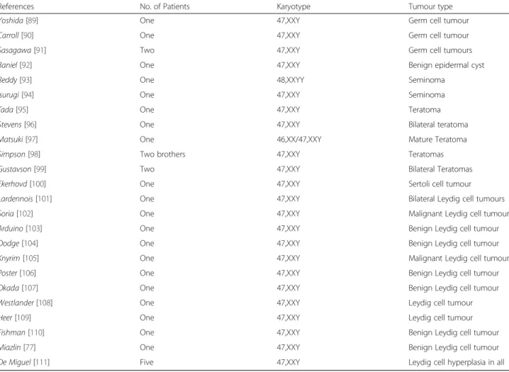
Table 2 Reported cases of testis-related tumours in aneuploidies
References No. of Patients Karyotype Tumour type
Yoshida [89] One 47,XXY Germ cell tumour
Carroll [90] One 47,XXY Germ cell tumour
Sasagawa [91] Two 47,XXY Germ cell tumours
Baniel [92] One 47,XXY Benign epidermal cyst
Reddy [93] One 48,XXYY Seminoma
Isurugi [94] One 47,XXY Seminoma
Tada [95] One 47,XXY Teratoma
Stevens [96] One 47,XXY Bilateral teratoma
Matsuki [97] One 46,XX/47,XXY Mature Teratoma Simpson [98] Two brothers 47,XXY Teratomas Gustavson [99] Two 47,XXY Bilateral Teratomas Ekerhovd [100] One 47,XXY Sertoli cell tumour Lardennois [101] One 47,XXY Bilateral Leydig cell tumours Soria [102] One 47,XXY Malignant Leydig cell tumour Arduino [103] One 47,XXY Benign Leydig cell tumour Dodge [104] One 47,XXY Benign Leydig cell tumour Knyrim [105] One 47,XXY Malignant Leydig cell tumour Poster [106] One 47,XXY Benign Leydig cell tumour Okada [107] One 47,XXY Benign Leydig cell tumour Westlander [108] One 47,XXY Leydig cell tumour
Heer [109] One 47,XXY Leydig cell tumour
Fishman [110] One 47,XXY Benign Leydig cell tumour Miazlin [77] One 47,XXY Benign Leydig cell tumour De Miguel [111] Five 47,XXY Leydig cell hyperplasia in all
Many mechanisms and animal models explain how a
Leydigioma could develop, nevertheless, the causes
re-main heterogeneous. Although LH plays an important
role in Leydig cell proliferation, Leydig cell maturation
and proliferation is affected by many other paracrine
and endocrine signals, including anti müllerian
hor-mone, inhibin, and other growth factors. The classical
pathway was suggested since 1980 with chronic LH
stimulation which induced Leydig cell
hyperplasia/aden-oma in rats [53]. Furthermore, anti-androgen therapy or
androgen insensitivity syndrome inducing LH secretion
re-sulted in the same phenotype [40, 54]. This hypothesis was
confirmed with the description of activating mutations of
LH receptors. Indeed, LH receptor is a trans membrane G
protein coupled receptor, expressed on Leydig cells.
Acti-vating mutations of this receptor are reported and induce
precocious puberty [55]. The chronic and permanent hyper
activation of this receptor leads to inappropriate
stimula-tion of cAMP pathway and Leydig cell hyperplasia. This
mutation is usually found in children who present with
Leydig cell tumor, and frequently responsible for
preco-cious puberty [56–65]. Actually, Asp578His mutation was
identified in more than 50 % of children with Leydig cell
tumors (13/24 patients) (reviewed in [57]).
Besides, mice models of KS with 41,XXY karyotype
[66], are characterized by small testes with Leydig cell
hyperplasia [67]. This supports the role of chronic and
permanent LH stimulation in the physiopathology of
Leydig cell tumor [67, 68].
Leydig cell tumor/hyperplasia were also linked to
many other situations than aneuploidies; these include:
McCune-Albright syndrome, Carney complex, fumarate
hydratase and cyclin dependent kinase (CDK) mutations.
The first two syndromes are multi tumor syndromes
af-fecting the endocrine system, including Leydig cells.
Their physiopathology is near to that of Klinefelter
variants with the activation of LH pathway (Activating
mutations of
α subunit of protein G (GNAS) for
McCune-Albright syndrome [69–72] and hyperactivity
of protein kinase A in Carney complex [73, 74]).
5. Management of Leydig cell tumors
Orchidectomy is indicated in cases of benign Leydig cell
tumors (single, unilateral, well circumscribed tumor
with-out hyper vascularization, necrosis, lithiasis or calcification
on ultrasound) [75–77]. Owing to the favourable course of
such usually small-sized tumors, some published studies
advocated conservative or testis-sparing surgery [78–82].
Such option does not seem to be associated with an
in-creased risk of recurrence. Conservative surgery seems to
be important for young patients with single testicle with
pa-ternity desire [83, 84]. Frozen sections largely help to
de-cide a more radical surgery or not [79, 82, 85]. The first 2
years of the diagnosis of malignant Leydig cell tumor are
the most crucial for the prognosis of the patients, however,
metastases up to 17 years post operation were also
ob-served [86]. More radical surgery with retroperitoneal
gan-glion removal, chemotherapy, and/or radiotherapy could
be used [30, 87, 88].
The dilemma of surgical choice is less advocated in
adult patients with KS associated with undescended
testis, advanced testicular failure or non-existence of
fer-tility challenge. These concerns are usually present in
Klinefelter variants. Thus, no benefits are awaited from
testis-sparing surgical procedures.
Conclusion
We present a case of 49,XXXXY sex polysomy, who shares
a number of characteristics of the other 176 patients cases
described in the literature, namely, hypogonadism with
cryptorchidism, facial dysmorphism, musculoskeletal and
cardiac malformations, and mental retardation seriously
affecting language skills. He presented with a benign
Leydig cell tumor. To our best knowledge, this is the first
case of testicular Leydig cell tumor described in a patient
affected by
Fraccaro syndrome.
Even if it shares some characteristics with KS,
49,XXXXY syndrome has to be distinguished and be
considered as the most severe form. Hypogonadism is
severe, together with genital ambiguity and absent
pu-berty evoking an earlier and more profound Sertoli,
Leydig and germ cell destruction.
The risk of Leydig cell tumors in aneuploidies remains a
dogma; it seems to be similar to the general population.
Nevertheless, this incidence is possibly underestimated, as
early preventive orchidectomy is usually practiced;
further-more, no systemic screening is consensual to detect such
tumors. On the other side, karyotype study in patients with
Leydig cell tumor/hyperplasia is seldom realized and needs
to be more systematic. Finally, the most agreed
physiopa-thology is related to chronic LH stimulation of Leydig cells.
Competing interests
The authors declare that they have no competing interests. Authors’ contributions
SM and LB wrote the paper; FF did the immuno histological study; IT manages and follows up the patient; MB, BR, FD and IT critically reviewed the manuscript. All the authors read and approved the final manuscript. Acknowledgements
No funding was dedicated to any of the authors concerning this work. Author details
1Service d’endocrinologie, diabétologie et maladies métaboliques, CHU
Clermont-Ferrand, F-63003 Clermont-Ferrand, France.2UMR CNRS 6293, INSERM U1103, Université Clermont-Auvergne, Génétique Reproduction et Développement, BP 10448, 63177 Aubiere, France.3Service de Médecine Nucléaire, Centre Jean Perrin, 58 rue Montalembert, F-63011
Clermont-Ferrand, France.4SIPATH Clermont-Ferrand, F-63003 Clermont-Ferrand, France.
Received: 5 May 2015 Accepted: 30 June 2015
References
1. Tartaglia N, Ayari N, Howell S, D’Epagnier C, Zeitler P. 48,XXYY, 48,XXXY and 49,XXXXY syndromes: not just variants of Klinefelter syndrome. Acta Paediatr Oslo Nor 1992. 2011;100:851–60.
2. Linden MG, Bender BG, Robinson A. Sex chromosome tetrasomy and pentasomy. Pediatrics. 1995;96(4 Pt 1):672–82.
3. Klinefelter HF, Reifenstein EC, Albright F. Syndrome characterized by gynecomastia, aspermatogenesis without A-Leydigism, and increased excretion of follicle-stimulating hormone1. J Clin Endocrinol Metab. 1942;2:615–27. 4. Fromantin M, Pesquies P, Serrurier B, Gautier D, Canivet B, Grenier J, et al.
Klinefelter’s syndrome in 19 year old adolescents. (100 cases detected during selection for National Service). Ann Méd Interne. 1977;128:239–44. 5. Visootsak J, Ayari N, Howell S, Lazarus J, Tartaglia N. Timing of diagnosis of
47, XXY and 48, XXYY: A survey of parent experiences. Am J Med Genet A. 2013;161:268–72.
6. Van Assche E, Bonduelle M, Tournaye H, Joris H, Verheyen G, Devroey P, et al. Cytogenetics of infertile men. Hum Reprod Oxf Engl.
1996;11 Suppl 4:1–24. discussion 25–26.
7. Groth KA, Skakkebæk A, Høst C, Gravholt CH, Bojesen A. Klinefelter syndrome—a clinical update. J Clin Endocrinol Metab. 2013;98:20–30. 8. Swerdlow AJ, Higgins CD, Schoemaker MJ, Wright AF, Jacobs PA, United
Kingdom Clinical Cytogenetics Group. Mortality in patients with Klinefelter syndrome in Britain: a cohort study. J Clin Endocrinol Metab. 2005;90:6516–22. 9. Hasle H, Mellemgaard A, Nielsen J, Hansen J. Cancer incidence in men with
Klinefelter syndrome. Br J Cancer. 1995;71:416–20.
10. Peet J, Weaver DD, Vance GH. 49, XXXXY: a distinct phenotype. Three new cases and review. J Med Genet. 1998;35:420–4.
11. Robinson A, Bender BG, Linden MG, Salbenblatt JA. Sex chromosome aneuploidy: the Denver prospective study. Birth Defects Orig Artic Ser. 1990;26:59–115.
12. Sprouse C, Tosi L, Stapleton E, Gropman AL, Mitchell FL, Peret R, et al. Musculoskeletal anomalies in a large cohort of boys with 49, XXXXY. Am J Med Genet C Semin Med Genet. 2013;163C:44–9.
13. Ottesen AM, Aksglaede L, Garn I, Tartaglia N, Tassone F, Gravholt CH, et al. Increased number of sex chromosomes affects height in a nonlinear fashion: a study of 305 patients with sex chromosome aneuploidy. Am J Med Genet A. 2010;152A:1206–12.
14. Tartaglia N, Davis S, Hench A, Nimishakavi S, Beauregard R, Reynolds A, et al. A new look at XXYY syndrome: medical and psychological features. Am J Med Genet A. 2008;146A:1509–22.
15. Blumenthal JD, Baker EH, Lee NR, Wade B, Clasen LS, Lenroot RK, et al. Brain morphological abnormalities in 49, XXXXY syndrome: A pediatric magnetic resonance imaging study. NeuroImage Clin. 2013;2:197–203.
16. Giedd JN, Clasen LS, Wallace GL, Lenroot RK, Lerch JP, Wells EM, et al. XXY (Klinefelter Syndrome): a pediatric quantitative brain magnetic resonance imaging case–control study. Pediatrics. 2007;119:e232–40.
17. Gropman AL, Rogol A, Fennoy I, Sadeghin T, Sinn S, Jameson R, et al. Clinical variability and novel neurodevelopmental findings in 49, XXXXY syndrome. Am J Med Genet A. 2010;152A:1523–30.
18. Boisen KA, Kaleva M, Main KM, Virtanen HE, Haavisto A-M, Schmidt IM, et al. Difference in prevalence of congenital cryptorchidism in infants between two Nordic countries. Lancet. 2004;363:1264–9.
19. Foresta C, Zuccarello D, Garolla A, Ferlin A. Role of hormones, genes, and environment in human cryptorchidism. Endocr Rev. 2008;29:560–80. 20. Christiansen P, Andersson A-M, Skakkebæk NE. Longitudinal studies of
inhibin B levels in boys and young adults with Klinefelter syndrome. J Clin Endocrinol Metab. 2003;88:888–91.
21. Salbenblatt JA, Bender BG, Puck MH, Robinson A, Faiman C, Winter JS. Pituitary-gonadal function in Klinefelter syndrome before and during puberty. Pediatr Res. 1985;19:82–6.
22. Ross JL, Samango-Sprouse C, Lahlou N, Kowal K, Elder FF, Zinn A. Early androgen deficiency in infants and young boys with 47, XXY Klinefelter syndrome. Horm Res. 2005;64:39–45.
23. Cabrol S, Ross JL, Fennoy I, Bouvattier C, Roger M, Lahlou N. Assessment of Leydig and Sertoli cell functions in infants with nonmosaic Klinefelter syndrome: insulin-like peptide 3 levels are normal and positively correlated with LH levels. J Clin Endocrinol Metab. 2011;96:E746–753.
24. Purdue MP, Devesa SS, Sigurdson AJ, McGlynn KA. International patterns and trends in testis cancer incidence. Int J Cancer J Int Cancer. 2005;115:822–7. 25. Chia VM, Quraishi SM, Devesa SS, Purdue MP, Cook MB, McGlynn KA.
International trends in the incidence of testicular cancer, 1973–2002. Cancer Epidemiol Biomark Prev. 2010;19:1151–9.
26. Ferlay J, Shin H-R, Bray F, Forman D, Mathers C, Parkin DM. Estimates of worldwide burden of cancer in 2008: GLOBOCAN 2008. Int J Cancer. 2010;127:2893–917.
27. Hekimgil M, Altay B, Yakut BD, Soydan S, Ozyurt C, Killi R. Leydig cell tumor of the testis: comparison of histopathological and immunohistochemical features of three azoospermic cases and one malignant case. Pathol Int. 2001;51:792–6.
28. Henderson CG, Ahmed AA, Sesterhenn I, Belman AB, Rushton HG. Enucleation for prepubertal leydig cell tumor. J Urol. 2006;176:703–5. 29. Agarwal PK, Palmer JS. Testicular and paratesticular neoplasms in
prepubertal males. J Urol. 2006;176:875–81.
30. Farkas LM, Székely JG, Pusztai C, Baki M. High frequency of metastatic Leydig cell testicular tumours. Oncology 2000, 59:118–121 31. Skakkebaek NE, Holm M, Hoei-Hansen C, Jørgensen N, Rajpert-De Meyts E.
Association between testicular dysgenesis syndrome (TDS) and testicular neopla-sia: evidence from 20 adult patients with signs of maldevelopment of the testis. APMIS Acta Pathol Microbiol Immunol Scand. 2003;111:1–9. discussion 9–11. 32. Hoei-Hansen CE, Holm M, Rajpert-De Meyts E, Skakkebaek NE. Histological
evidence of testicular dysgenesis in contralateral biopsies from 218 patients with testicular germ cell cancer. J Pathol. 2003;200:370–4.
33. Giwercman A, Grindsted J, Hansen B, Jensen OM, Skakkebaek NE. Testicular cancer risk in boys with maldescended testis: a cohort study. J Urol. 1987;138:1214–6.
34. Giwercman A, Bruun E, Frimodt-Møller C, Skakkebaek NE. Prevalence of carcinoma in situ and other histopathological abnormalities in testes of men with a history of cryptorchidism. J Urol. 1989;142:998–1001. discussion 1001–1002. 35. Prener A, Engholm G, Jensen OM. Genital anomalies and risk for testicular
cancer in Danish men. Epidemiol Camb Mass. 1996;7:14–9. 36. Møller H, Skakkebaek NE. Risk of testicular cancer in subfertile men:
case–control study. BMJ. 1999;318:559–62.
37. Paduch DA. Testicular cancer and male infertility. Curr Opin Urol. 2006;16:419–27. 38. Jacobsen R, Bostofte E, Engholm G, Hansen J, Skakkebaek NE, Møller H.
Fertility and offspring sex ratio of men who develop testicular cancer: a record linkage study. Hum Reprod Oxf Engl. 2000;15:1958–61. 39. Ferguson L, Agoulnik AI: Testicular Cancer and Cryptorchidism. Front
Endocrinol(Lausanne) 2013;432. doi: 10.3389/fendo.2013.00032. eCollection 2013.10.3389/fendo.2013.00032
40. Singh R, Shastry PK, Rasalkar AA, Singh L, Thangaraj K. A novel androgen receptor mutation resulting in complete androgen insensitivity syndrome and bilateral Leydig cell hyperplasia. J Androl. 2006;27:510–6.
41. Swerdlow AJ, Schoemaker MJ, Higgins CD, Wright AF, Jacobs PA. Cancer incidence and mortality in men with Klinefelter syndrome: a cohort study. J Natl Cancer Inst. 2005;97:1204–10.
42. Bojesen A, Juul S, Birkebaek N, Gravholt CH. Increased mortality in Klinefelter syndrome. J Clin Endocrinol Metab. 2004;89:3830–4.
43. Hasle H, Jacobsen BB, Asschenfeldt P, Andersen K. Mediastinal germ cell tumour associated with Klinefelter syndrome. A report of case and review of the literature. Eur J Pediatr. 1992;151:735–9.
44. Chetaille B, Massard G, Falcoz P-E. Les tumeurs germinales du médiastin : anatomopathologie, classification, tératomes et tumeurs malignes. Rev Pneumol Clin. 2010;66:63–70.
45. Ahmad KN, Dykes JRW, Ferguson-Smith MA, Lennox B, Mack WS. Leydig cell volume in chromatin-positive Klinefelter’s syndrome. J Clin Endocrinol Metab. 1971;33:517–20.
46. Accardo G, Vallone G, Esposito D, Barbato F, Renzullo A, Conzo G, et al. Testicular parenchymal abnormalities in Klinefelter syndrome: a question of cancer? Examination of 40 consecutive patients. Asian J Androl. 2015;17:154–8. 47. Carmignani L, Bozzini G. Re: Increased incidence of testicular cancer in men presenting with infertility and abnormal semen analysis: J. D. Raman, C. F. Nobert and M. Goldstein. J Urol. 2006;175:1574.
48. Butruille C, Marcelli F, Ghoneim T, Lemaitre L, Puech P, Leroy X, et al. Prise en charge des nodules testiculaires dans une population de patients infertiles. Prog En Uro. 2012;22:45–52.
49. Bojesen A, Juul S, Gravholt CH. Prenatal and postnatal prevalence of Klinefelter syndrome: a national registry study. J Clin Endocrinol Metab. 2003;88:622–6.
50. Bojesen A, Juul S, Birkebæk NH, Gravholt CH. Morbidity in Klinefelter syndrome: A Danish register study based on hospital discharge diagnoses. J Clin Endocrinol Metab. 2006;91:1254–60.
51. Hutson JM, Balic A, Nation T, Southwell B. Cryptorchidism. Semin Pediatr Surg. 2010;19:215–24.
52. Hutson JM. Undescended testis: the underlying mechanisms and the effects on germ cells that cause infertility and cancer. J Pediatr Surg. 2013;48:903–8. 53. Christensen AK, Peacock KC. Increase in Leydig cell number in testes of
adult rats treated chronically with an excess of human chorionic gonadotropin. Biol Reprod. 1980;22:383–91.
54. Prahalada S, Majka JA, Soper KA, Nett TM, Bagdon WJ, Peter CP, et al. Leydig cell hyperplasia and adenomas in mice treated with finasteride, a 5 alpha-reductase inhibitor: a possible mechanism. Fundam Appl Toxicol Off J Soc Toxicol. 1994;22:211–9.
55. Shenker A, Laue L, Kosugi S, Merendino Jr JJ, Minegishi T, Cutler Jr GB. A constitutively activating mutation of the luteinizing hormone receptor in familial male precocious puberty. Nature. 1993;365:652–4.
56. Liu G, Duranteau L, Carel JC, Monroe J, Doyle DA, Shenker A. Leydig-cell tumors caused by an activating mutation of the gene encoding the luteinizing hormone receptor. N Engl J Med. 1999;341:1731–6.
57. Olivier P, Simoneau-Roy J, Francoeur D, Sartelet H, Parma J, Vassart G, et al. Leydig cell tumors in children: contrasting clinical, hormonal, anatomical, and molecular characteristics in boys and girls. J Pediatr. 2012;161:1147–52. 58. Goji K, Teraoka Y, Hosokawa Y, Okuno M, Ozaki K, Yoshida M, et al.
Gonadotropin-independent precocious puberty associated with a somatic activating mutation of the LH receptor gene: detection of a mutation present in only a small fraction of cells from testicular tissue using wild-type blocking polymerase chain reaction and laser-capture microdissection. Endocrine. 2009;35:397–401.
59. Boot AM, Lumbroso S, Verhoef-Post M, Richter-Unruh A, Looijenga LHJ, Funaro A, et al. Mutation analysis of the LH receptor gene in Leydig cell adenoma and hyperplasia and functional and biochemical studies of activating mutations of the LH receptor gene. J Clin Endocrinol Metab. 2011;96:E1197–1205.
60. Sangkhathat S, Kanngurn S, Jaruratanasirikul S, Tubtawee T, Chaiyapan W, Patrapinyokul S, et al. Peripheral precocious puberty in a male caused by Leydig cell adenoma harboring a somatic mutation of the LHR gene: report of a case. J Med Assoc Thai Chotmaihet Thangphaet. 2010;93:1093–7. 61. D’ Alva CB, Brito VN, Palhares HMC, Carvalho FM, Arnhold IJP, Mendonca BB,
et al. A single somatic activating Asp578His mutation of the luteinizing hormone receptor causes Leydig cell tumour in boys with gonadotropin-independent precocious puberty. Clin Endocrinol (Oxf). 2006;65:408–10. 62. Canto P, Söderlund D, Ramón G, Nishimura E, Méndez JP. Mutational
analysis of the luteinizing hormone receptor gene in two individuals with Leydig cell tumors. Am J Med Genet. 2002;108:148–52.
63. Richter-Unruh A, Wessels HT, Menken U, Bergmann M, Schmittmann-Ohters K, Schaper J, et al. Male LH-independent sexual precocity in a 3.5-year-old boy caused by a somatic activating mutation of the LH receptor in a Leydig cell tumor. J Clin Endocrinol Metab. 2002;87:1052–6.
64. Kiepe D, Richter-Unruh A, Autschbach F, Kessler M, Schenk JP, Bettendorf M. Sexual pseudo-precocity caused by a somatic activating mutation of the LH receptor preceding true sexual precocity. Horm Res. 2008;70:249–53. 65. Kremer H, Martens JW, van Reen M, Verhoef-Post M, Wit JM, Otten BJ, et al.
A limited repertoire of mutations of the luteinizing hormone (LH) receptor gene in familial and sporadic patients with male LH-independent preco-cious puberty. J Clin Endocrinol Metab. 1999;84:1136–40.
66. Eicher EM, Hale DW, Hunt PA, Lee BK, Tucker PK, King TR, et al. The mouse Y* chromosome involves a complex rearrangement, including interstitial positioning of the pseudoautosomal region. Cytogenet Cell Genet. 1991;57:221–30. 67. Wistuba J, Luetjens CM, Stukenborg J-B, Poplinski A, Werler S, Dittmann M,
et al. Male 41, XXY* mice as a model for klinefelter syndrome: hyperactivation of leydig cells. Endocrinology. 2010;151:2898–910.
68. Wistuba J. Animal models for Klinefelter’s syndrome and their relevance for the clinic. Mol Hum Reprod. 2010;16:375–85.
69. Schwindinger WF, Francomano CA, Levine MA. Identification of a mutation in the gene encoding the alpha subunit of the stimulatory G protein of adenylyl cyclase in McCune-Albright syndrome. Proc Natl Acad Sci U S A. 1992;89:5152–6.
70. Weinstein LS, Shenker A, Gejman PV, Merino MJ, Friedman E, Spiegel AM. Activating mutations of the stimulatory G protein in the McCune-Albright syndrome. N Engl J Med. 1991;325:1688–95.
71. Fragoso MC, Latronico AC, Carvalho FM, Zerbini MC, Marcondes JA, Araujo LM, et al. Activating mutation of the stimulatory G protein (gsp) as a putative cause of ovarian and testicular human stromal Leydig cell tumors.
J Clin Endocrinol Metab. 1998;83:2074–8.
72. Boyce AM, Chong WH, Shawker TH, Pinto PA, Linehan WM, Bhattacharryya N, et al. Characterization and management of testicular pathology in McCune-Albright syndrome. J Clin Endocrinol Metab. 2012;97:E1782–1790. 73. Carney JA, Gordon H, Carpenter PC, Shenoy BV, Go VL. The complex of
myxomas, spotty pigmentation, and endocrine overactivity. Medicine (Baltimore). 1985;64:270–83.
74. Stratakis CA, Kirschner LS, Carney JA. Clinical and molecular features of the Carney complex: diagnostic criteria and recommendations for patient evaluation. J Clin Endocrinol Metab. 2001;86:4041–6.
75. Tazi MF, Ahallal Y, Khallouk A, Elfatemi H, Bendahou M, Tazi E, et al. Concomitant sertoli and leydig cell tumor of the testis: a case report. Rev Urol. 2011;13:173–5.
76. Carmignani L, Gadda F, Mancini M, Gazzano G, Nerva F, Rocco F, et al. Detection of testicular ultrasonographic lesions in severe male infertility. J Urol. 2004;172:1045–7.
77. Maizlin ZV, Belenky A, Kunichezky M, Sandbank J, Strauss S. Leydig cell tumors of the testis: gray scale and color Doppler sonographic appearance. J Ultrasound Med. 2004;23:959–64.
78. Loeser A, Vergho DC, Katzenberger T, Brix D, Kocot A, Spahn M, et al. Testis-sparing surgery versus radical orchiectomy in patients with Leydig cell tumors. Urology. 2009;74:370–2.
79. Carmignani L, Colombo R, Gadda F, Galasso G, Lania A, Palou J, et al. Conservative surgical therapy for leydig cell tumor. J Urol. 2007;178:507–11. discussion 511.
80. Nicolai N, Necchi A, Raggi D, Biasoni D, Catanzaro M, Piva L, et al. Clinical outcome in testicular sex cord stromal tumors: testis sparing vs. radical orchiectomy and management of advanced disease. Urology. 2015;85:402–6. 81. Giannarini G, Mogorovich A, Menchini Fabris F, Morelli G, De Maria M,
Manassero F, et al. Long-term followup after elective testis sparing surgery for Leydig cell tumors: a single center experience. J Urol.
2007;178(3 Pt 1):872–6. quiz 1129.
82. Bozzini G, Picozzi S, Gadda F, Colombo R, Decobelli O, Palou J, et al. Long-term follow-up using testicle-sparing surgery for leydig cell tumor. Clin Genitourin Cancer. 2013;11(3):321–4.
83. Masoudi JF, Van Arsdalen K, Rovner ES. Organ-sparing surgery for bilateral leydig cell tumor of the testis. Urology. 1999;54:744.
84. Giannarini G, Dieckmann K-P, Albers P, Heidenreich A, Pizzocaro G. Organ-sparing surgery for adult testicular tumours: a systematic review of the literature. Eur Urol. 2010;57:780–90.
85. Steiner H, Höltl L, Maneschg C, Berger AP, Rogatsch H, Bartsch G, et al. Frozen section analysis-guided organ-sparing approach in testicular tumors: technique, feasibility, and long-term results. Urology. 2003;62:508–13. 86. Grem JL, Robins HI, Wilson KS, Gilchrist K, Trump DL. Metastatic Leydig cell
tumor of the testis. Report of three cases and review of the literature. Cancer. 1986;58:2116–9.
87. Bertram KA, Bratloff B, Hodges GF, Davidson H. Treatment of malignant Leydig cell tumor. Cancer. 1991;68:2324–9.
88. Bokemeyer C, Harstrick A, Gonnermann O, Schober C, Kuczyk M, Poliwoda H, et al. Metastatic Leydig-cell tumors of the testis - report of 4 cases and review of the literature. Int J Oncol. 1993;2:241–4.
89. Yoshida T, Takao T, Tsujimura A, Tomita H, Aozasa K, Okuyama A. Testicular epidermoid cyst in Klinefelter’s syndrome. Int J Urol Off J Jpn Urol Assoc. 2006;13:478–80.
90. Carroll PR, Morse MJ, Koduru PP, Chaganti RS. Testicular germ cell tumor in patient with Klinefelter syndrome. Urology. 1988;31:72–4.
91. Sasagawa I, Nakada T, Kazama T, Sakamoto M, Satomi S, Katayama T. Epidermoid cyst of the testis in Klinefelter’s syndrome. Urol Int. 1987;42:398–400.
92. Baniel J, Perez JM, Foster RS. Benign testicular tumor associated with Klinefelter’s syndrome. J Urol. 1994;151:157–8.
93. Reddy SR, Svec F, Richardson P. Seminoma of the testis in a patient with 48, XXYY variant of Klinefelter’s syndrome. South Med J. 1991;84:773–5. 94. Isurugi K, Imao S, Hirose K, Aoki H. Seminoma in Klinefelter’s syndrome with
47, XXY, 15 s + karyotype. Cancer. 1977;39:2041–7.
95. Tada M, Takimoto Y, Kishimoto T. Immature teratoma of the testis associated with Klinefelter’s syndrome: a case report. Hinyokika Kiyo. 1990;36:1471–4.
96. Stevens MJ, Jameson CF, Hendry WF. Bilateral testicular teratoma in Klinefelter’s syndrome. Br J Urol. 1993;72:384–5.
97. Matsuki S, Sasagawa I, Kakizaki H, Suzuki Y, Nakada T. Testicular teratoma in a man with XX/XXY mosaic Klinefelter’s syndrome. J Urol. 1999;161:1573–4. 98. Simpson JL, Photopulos G. Letter: Bilateral teratoma of testis in 2 brothers
with 47, XXY Klinefelter’s syndrome. Clin Genet. 1976;9:380–1. 99. Gustavson KH, Gamstorp I, Meurling S. Bilateral teratoma of testis in two
brothers with 47, XXY Klinefelter’s syndrome. Clin Genet. 1975;8:5–10. 100. Ekerhovd E, Westlander G. Testicular sonography in men with Klinefelter
syndrome shows irregular echogenicity and blood flow of high resistance. J Assist Reprod Genet. 2002;19:517–22.
101. Lardennois B, El Hansa A, Bernier F, Birembaut P, Caron J, Lemaire P. Klinefelter’s disease and leydigiomas. A report of one case (author’s transl). J Urol (Paris). 1981;87:631–4.
102. Soria JC, Durdux C, Chrétien Y, Sibony M, Damotte D, Housset M. Malignant Leydig cell tumor of the testis associated with Klinefelter’s syndrome. Anticancer Res. 1999;19:4491–4.
103. ARDUINO LJ, GLUCKSMAN MA. Interstitial cell tumor of the testis associated with Klinefelter’s syndrome: a case report. J Urol. 1963;89:246–8.
104. Dodge OG, Jackson AW, Muldal S. Breast cancer and interstitial-cell tumor in a patient with Klinefelter’s syndrome. Cancer. 1969;24:1027–32.
105. Knyrim K, Higi M, Hossfeld DK, Seeber S, Schmidt CG. Autonomous cortisol secretion by a metastatic Leydig cell carcinoma associated with Klinefelter’s syndrome. J Cancer Res Clin Oncol. 1981;100:85–93.
106. Poster RB, Katz DS. Leydig cell tumor of the testis in Klinefelter syndrome: MR detection. J Comput Assist Tomogr. 1993;17:480–1.
107. Okada H, Gotoh A, Takechi Y, Kamidono S. Leydig cell tumour of the testis associated with Klinefelter’s syndrome and Osgood-Schlatter disease. Br J Urol. 1994;73:457.
108. Westlander G, Ekerhovd E, Granberg S, Hanson L, Hanson C, Bergh C. Testicular ultrasonography and extended chromosome analysis in men with nonmosaic Klinefelter syndrome: a prospective study of possible predictive factors for successful sperm recovery. Fertil Steril. 2001;75:1102–5. 109. Heer R, Jackson MJ, El-Sherif A, Thomas DJ. Twenty-nine Leydig cell tumors:
histological features, outcomes and implications for management. Int J Urol Off J Jpn Urol Assoc. 2010;17:886–9.
110. Fishman MDC, Eisenberg DA, Horrow MM. Klinefelter syndrome with leydig cell tumor/hyperplasia. Ultrasound Q. 2010;26:101–2.
111. De Miguel MP, Regadera J, Martinez-Garcia F, Nistal M, Paniagua R. Oncostatin M in the normal human testis and several testicular disorders. J Clin Endocrinol Metab. 1999;84:768–74.
Submit your next manuscript to BioMed Central
and take full advantage of:
• Convenient online submission
• Thorough peer review
• No space constraints or color figure charges
• Immediate publication on acceptance
• Inclusion in PubMed, CAS, Scopus and Google Scholar
• Research which is freely available for redistribution
Submit your manuscript at www.biomedcentral.com/submit