HAL Id: inserm-00919173
https://www.hal.inserm.fr/inserm-00919173
Submitted on 16 Dec 2013
HAL is a multi-disciplinary open access archive for the deposit and dissemination of sci-entific research documents, whether they are pub-lished or not. The documents may come from teaching and research institutions in France or abroad, or from public or private research centers.
L’archive ouverte pluridisciplinaire HAL, est destinée au dépôt et à la diffusion de documents scientifiques de niveau recherche, publiés ou non, émanant des établissements d’enseignement et de recherche français ou étrangers, des laboratoires publics ou privés.
glomerulopathy.
Olivia Boyer, Fabien Nevo, Emmanuelle Plaisier, Benoît Funalot, Olivier
Gribouval, Geneviève Benoit, Evelyne Huynh Cong, Christelle Arrondel,
Marie-Josèphe Tête, Rodrick Montjean, et al.
To cite this version:
Olivia Boyer, Fabien Nevo, Emmanuelle Plaisier, Benoît Funalot, Olivier Gribouval, et al.. INF2 mutations in Charcot-Marie-Tooth disease with glomerulopathy.. New England Journal of Medicine, Massachusetts Medical Society, 2011, 365 (25), pp.2377-88. �10.1056/NEJMoa1109122�. �inserm-00919173�
T h e n e w e n g l a n d j o u r n a l o f m e d i c i n e
n engl j med 365;25 nejm.org december 22, 2011 2377
original article
INF2
Mutations in Charcot–Marie–Tooth
Disease with Glomerulopathy
Olivia Boyer, M.D., Ph.D., Fabien Nevo, M.Sc., Emmanuelle Plaisier, M.D., Ph.D., Benoit Funalot, M.D., Ph.D., Olivier Gribouval, M.Sc., Geneviève Benoit, M.D., Evelyne Huynh Cong, M.Sc., Christelle Arrondel, M.Sc., Marie-Josèphe Tête, M.D.,
Rodrick Montjean, Ph.D., Laurence Richard, M.Sc., Alexandre Karras, M.D., Claire Pouteil-Noble, M.D., Ph.D., Leila Balafrej, M.D., Alain Bonnardeaux, M.D., Ph.D., Guillaume Canaud, M.D., Christophe Charasse, M.D., Jacques Dantal, M.D., Ph.D., Georges Deschenes, M.D., Ph.D., Patrice Deteix, M.D., Odile Dubourg, M.D., Ph.D.,
Philippe Petiot, M.D., Dominique Pouthier, M.D., Eric Leguern, M.D., Ph.D., Anne Guiochon-Mantel, M.D., Ph.D., Isabelle Broutin, Ph.D., Marie-Claire Gubler, M.D., Sophie Saunier, Ph.D., Pierre Ronco, M.D., Ph.D.,
Jean-Michel Vallat, M.D., Miguel Angel Alonso, Ph.D., Corinne Antignac, M.D., Ph.D., and Géraldine Mollet, Ph.D.
The authors’ affiliations are listed in the appendix. Address reprint requests to Dr. Antignac at INSERM U983, 6e étage, Tour Lavoisier, Hôpital Necker–Enfants Malades, 149 Rue de Sèvres, 75015 Paris, France, or at [email protected]. N Engl J Med 2011;365:2377-88.
Copyright © 2011 Massachusetts Medical Society.
A B S T R A C T
BACKGROUND
Charcot–Marie–Tooth neuropathy has been reported to be associated with renal diseases, mostly focal segmental glomerulosclerosis (FSGS). However, the common mechanisms underlying the neuropathy and FSGS remain unknown. Mutations in INF2 were recently identified in patients with autosomal dominant FSGS. INF2 en-codes a formin protein that interacts with the Rho-GTPase CDC42 and myelin and lymphocyte protein (MAL) that are implicated in essential steps of myelination and myelin maintenance. We therefore hypothesized that INF2 may be responsible for cases of Charcot–Marie–Tooth neuropathy associated with FSGS.
METHODS
We performed direct genotyping of INF2 in 16 index patients with Charcot–Marie– Tooth neuropathy and FSGS who did not have a mutation in PMP22 or MPZ, encoding peripheral myelin protein 22 and myelin protein zero, respectively. Histologic and functional studies were also conducted.
RESULTS
We identified nine new heterozygous mutations in 12 of the 16 index patients (75%), all located in exons 2 and 3, encoding the diaphanous-inhibitory domain of INF2. Patients presented with an intermediate form of Charcot–Marie–Tooth neuropathy as well as a glomerulopathy with FSGS on kidney biopsy. Immunohistochemical analy-sis revealed strong INF2 expression in Schwann-cell cytoplasm and podocytes. Moreover, we demonstrated that INF2 colocalizes and interacts with MAL in Schwann cells. The INF2 mutants perturbed the INF2–MAL–CDC42 pathway, resulting in cy-toskeleton disorganization, enhanced INF2 binding to CDC42 and mislocalization of INF2, MAL, and CDC42.
CONCLUSIONS
INF2 mutations appear to cause many cases of FSGS-associated Charcot–Marie–Tooth neuropathy, showing that INF2 is involved in a disease affecting both the kidney glom-erulus and the peripheral nervous system. These findings provide new insights into the pathophysiological mechanisms linking formin proteins to podocyte and Schwann-cell function. (Funded by the Agence Nationale de la Recherche and others.)
The New England Journal of Medicine
Downloaded from nejm.org on December 16, 2013. For personal use only. No other uses without permission. Copyright © 2011 Massachusetts Medical Society. All rights reserved.
C
harcot–Marie–Tooth disease refers to a heterogeneous group of inherited chronic peripheral motor and sensoryneu-ropathies.1 Affected persons typically present with
progressive distal-muscle weakness and atrophy, reduced tendon reflexes, and foot and hand defor-mities. Three Charcot–Marie–Tooth disease sub-types have been distinguished by means of elec-trophysiological and neuropathological studies — a glial myelinopathy (type 1) characterized by slow motor-nerve conduction velocities and demy-elinating neuropathy, an axonal form (type 2) as-sociated with normal or subnormal nerve conduc-tion velocities and axonal degeneraconduc-tion, and an intermediate form with demyelinating and axonal features in which patients from the same family may have either subnormal or reduced nerve
con-duction velocities.2 At least 40 different genes or
loci have been associated with this disease (as has
been reviewed by Lupski and colleagues).3
Auto-somal dominant Charcot–Marie–Tooth type 1 is the most prevalent form, with mutations in the periph-eral myelin protein 22 gene (PMP22) and the myelin
protein zero gene (MPZ) underlying most cases.4,5
An increased prevalence of nephropathies, par-ticularly focal segmental glomerulosclerosis (FSGS), has been documented in patients with Charcot–
Marie–Tooth neuropathy,6 but the
pathophysiologi-cal mechanism linking these two clinipathophysiologi-cal entities is unknown. FSGS is a histologic pattern of renal damage that is associated with a spectrum of pri-mary and secondary glomerular diseases, includ-ing isolated proteinuria and
glucocorticoid-resis-tant nephrotic syndrome.7 In the past few years,
the identification of genes involved in hereditary glomerulopathies has expanded knowledge about the crucial role of the podocyte, a glomerular epi-thelial cell with interdigitating foot processes, as well as its actin cytoskeleton, in the function of the
glomerular filtration barrier.8
INF2 mutations account for 12 to 17% of
auto-somal dominant cases of FSGS.9,10 The gene
en-codes a member of the diaphanous-related formin family, which is involved in remodeling the actin
and microtubule cytoskeletons.11 INF2 possesses
functional domains characteristic of other diaph-anous-related formins: an N-terminal diaphanous-inhibitory domain (DID), the formin homology domains FH1 and FH2, and a C-terminal
diaph-anous-autoregulatory domain (DAD).11 However,
INF2 has a unique ability to promote not only actin polymerization but also filament severing
and depolymerization.12
INF2 interacts with other diaphanous-related
formins, such as mDia1–DIAPH113 and the
Rho-GTPase CDC42,14,15 through its DID. In addition,
it has been shown to bind (through its C-terminal) the myelin and lymphocyte protein (MAL) in Jurkat
T cells15 and MAL2 in Madin–Darby canine kidney
cells and HepG2 hepatocytes14 to regulate
intracel-lular protein transport.14-16 Although very little
is known about the role of diaphanous-related formins in the peripheral nervous system, the
im-plication of CDC4217 and MAL18 in essential steps
of myelination and myelin maintenance led us to hypothesize that INF2 mutations may be responsi-ble for the association between FSGS and Charcot– Marie–Tooth neuropathy.
M e t h od s
Study Participants
The study was conducted from March 2010 through September 2011. Sixteen index patients (seven with apparent autosomal dominant inheritance and nine with sporadic disease) from 16 unrelated families were included in the study. Twelve were from our French FSGS DNA cohort, including 2 families
described previously.19,20 We contacted authors
of published cases of FSGS and Charcot–Marie–
Tooth neuropathy 6,21 and thereby obtained DNA
samples from 4 additional families. Twenty-five members of the patients’ families were also tested. All index patients presented with clinical mani-festations of Charcot–Marie–Tooth disease asso-ciated with FSGS. The index patients had all been diagnosed with Charcot–Marie–Tooth disease by a neurologist in their primary care centers on the basis of their clinical history, physical examina-tion, and electrophysiological or histologic test-ing. Mutations in PMP22 or MPZ were ruled out in all patients. Written informed consent was ob-tained from all study participants or their par-ents, and the study was approved by the Comité de Protection des Personnes Ile-De-France II.
Genetic, Histologic, and Functional Studies
INF2 exons 2, 3, and 4 were sequenced for all par-ticipants, and in the absence of mutations, the re-maining exons were sequenced (as previously
de-scribed10). Localization of INF2, MAL, and MAL2
in normal human kidney and peripheral-nerve specimens and in cultured Schwann cells was as-sessed by means of immunoperoxidase and immu-nofluorescence staining. The interaction of INF2 with MAL in Schwann cells was demonstrated by
IN F2 Mu tations in Ch a rco t –M a r ie –T oo th Dise a se
n engl j med 365;25 nejm.org december 22, 2011 2379
pull-down assay. The effects of INF2 mutant ex-pression on interaction with active CDC42 and IQGAP1 were evaluated by coimmunoprecipitation, and the effects of INF2 mutant expression on in-tracellular localization of MAL and CDC42 and on the actin cytoskeleton were evaluated by immuno-fluorescence. Two INF2 mutants associated with FSGS and Charcot–Marie–Tooth neuropathy and three INF2 mutants associated with FSGS were studied. A detailed description of the methods used is provided in the Supplementary Appendix, avail-able with the full text of the article at NEJM.org.
R e s u l t s
INF2 Mutations
Heterozygous INF2 mutations were detected in 12 of the 16 index patients (75%). Nine different mutations were identified: eight missense muta-tions and one in-frame deletion of three amino ac-ids. All were new mutations located in exons 2 and 3, which encode the DID domain (Fig. 1A, and Table 1S in the Supplementary Appendix), and all caused nonconservative changes in highly con-served amino acids. Scores from PolyPhen-2 soft-ware analysis (http://genetics.bwh.harvard.edu/
pph2) to predict the functional effects of mis-sense INF2 variants ranged from 0.993 to 1, pre-dicting that INF2 variants were probably damag-ing. No INF2 variants were present in any of the 670 control chromosomes assayed or referenced in the National Heart, Lung, and Blood Institute’s
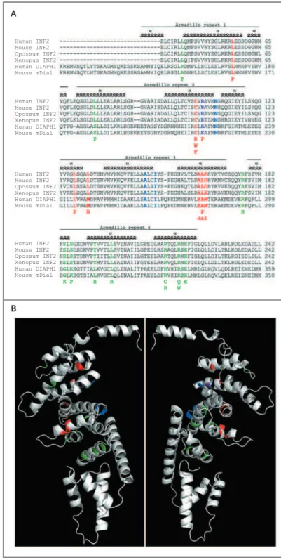
Figure 1. Summary of INF2 Substitutions in the 12 Study Patients.
Panel A shows substitutions identified in the study pa-tients (red, in Panels A and B) as well as those found in previous studies of focal segmental glomeruloscle-rosis (FSGS) alone (green, in Panels A and B), aligned with the C-terminal sequence (indicated with the string of single-letter amino acid symbols) of human, mouse, and xenopus INF2 proteins, opossum INF2-like pro-tein, and human and mouse DIAPH1 proteins. Resi-dues important for the DID–DAD interaction22
are shown in blue or, if the residues were also altered in a patient presenting with FSGS and Charcot–Marie– Tooth neuropathy, in purple (in Panels A and B). The numbering of amino acids from the translation initia-tion site is shown to the right of each sequence, and armadillo repeats are shown to highlight the distinct localization of mutants associated with FSGS and Charcot–Marie–Tooth neuropathy and of mutants as-sociated with FSGS alone. Panel B is a three-dimen-sional representation of the N-terminal portion of human INF2 viewed from op-posing directions, con-structed on the basis of the structure of mDia1. The residues associated with FSGS and Charcot–Marie– Tooth neuropathy (red and purple) are located in the inner face of the central core of the DID, whereas the mutant residues responsible for FSGS only (green) are more externally located.
B A Human INF2 Mouse INF2 Opossum INF2 Xenopus INF2 Human DIAPH1 Mouse mDial Human INF2 Mouse INF2 Opossum INF2 Xenopus INF2 Human DIAPH1 Mouse mDial Human INF2 Mouse INF2 Opossum INF2 Xenopus INF2 Human DIAPH1 Mouse mDial Human INF2 Mouse INF2 Opossum INF2 Xenopus INF2 Human DIAPH1 Mouse mDial
The New England Journal of Medicine
Downloaded from nejm.org on December 16, 2013. For personal use only. No other uses without permission. Copyright © 2011 Massachusetts Medical Society. All rights reserved.
Exome Sequencing Project server. Mutations seg-regated with the disease in each familial case, although intrafamilial variability was noted (Fig. 1S in the Supplementary Appendix). A de novo mu-tation was confirmed in all three sporadic cases for which DNA was available from both parents of the patient. Most mutations identified in pa-tients exhibiting FSGS and Charcot–Marie–Tooth neuropathy were localized in the 3′ end of exon 2 and in exon 3, in which no mutation has been iden-tified to date (to our knowledge). The INF2 vari-ants were clustered between nucleotides 300 and 500, whereas most isolated FSGS mutants were located downstream of nucleotide 500. To make functional predictions, we mapped mutants
asso-ciated with FSGS alone9,10 and those associated
with FSGS and Charcot–Marie–Tooth neuropathy onto a human INF2 DID in silico model (Fig. 1B); although all involved DID residues, mutations in the two groups of patients were distinctly lo-calized, the latter being located mostly in the second and third DID armadillo repeats and the former mostly in the fourth armadillo repeat (Fig. 1B).
To evaluate the potential role of INF2 in isolated Charcot–Marie–Tooth disease, we performed mu-tational analysis of INF2 exons 2, 3, and 4 in an additional group of 50 patients who presented with Charcot–Marie–Tooth disease without a known renal phenotype, nerve conduction velocities in the
intermediate range (25 to 45 m per second),2 and
without any PMP22 duplication or deletion or MPZ mutation. No pathogenic mutation was identified in this group.
phenotype of patients with INF2 Mutations
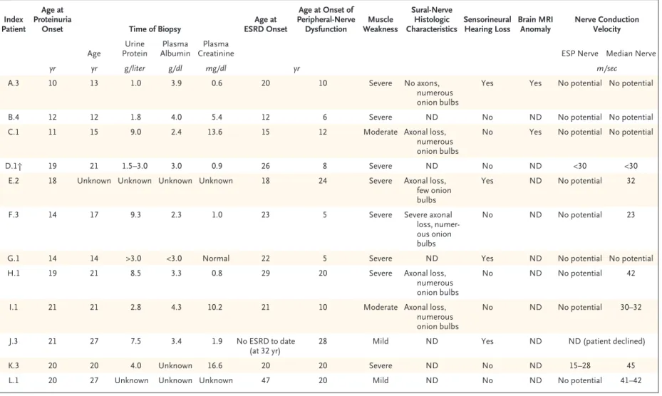
The 12 index patients with INF2 mutations present-ed with proteinuria at a mpresent-edian age of 18 years (range, 10 to 21), and end-stage renal disease (ESRD) developed in 11 patients at a median age of 21 years (range, 12 to 47) (Table 1). All patients exhibited lesions of FSGS (not otherwise
speci-fied)7 (Fig. 2A), but full-blown nephrotic syndrome
was noted in only 5.
The neurologic phenotype of index cases is de-tailed in Table 1 and in the Supplementary Appen-dix. The median age at onset of peripheral-nerve dysfunction was 13 years (range, 5 to 28). Ten of the 12 index patients had moderate to severe symp-toms at diagnosis, including walking difficulties and limited hand function, muscle wasting, and abolition of deep tendon reflexes in the lower and
upper limbs. Four patients also had mild or mod-erate sensorineural hearing loss. In addition, mag-netic resonance imaging of the brain, performed in 2 patients (15 and 48 years old), showed central nervous system anomalies characterized by white-matter hyperintensity and ventricular dilation, which were more severe in the older patient (Fig. 2B). Median-nerve conduction velocities were in the range of intermediate Charcot–Marie–Tooth neu-ropathy (23 to 45 m per second). Patient K.3 had almost normal median-nerve conduction velocities (45 m per second), whereas her maternal aunt had reduced velocities (30 m per second; data not shown). The six available sural-nerve biopsy spec-imens all showed a pattern of lesions with a com-bination of axonal and demyelinating changes, characterized by a marked decrease in myelinated fibers (Fig. 2C), as compared with that in age-matched controls, and numerous multilayered “on-ion bulbs” (Fig. 2D). Together, these data suggest an intermediate Charcot–Marie–Tooth phenotype in patients with INF2 mutations.
INF2 Expression in Podocytes and Schwann Cells
In peripheral nerves, we detected robust INF2 stain-ing in Schwann cells and lighter stainstain-ing in some axons (Fig. 3A, and Fig. 2S in the Supplementary Appendix). In the kidney, we confirmed that INF2 expression occurs predominantly in podocytes. We detected weak staining in the proximal and distal tubules and found no INF2 in vessels. In kidney-tissue and sural-nerve sections from the patients, INF2 staining persisted, but the severity of the le-sions precluded our drawing any conclusion with respect to putative overexpression.
INF2 and MAL in Schwann Cells
MAL interacts with INF215 and is a major
compo-nent of myelin.18 We therefore hypothesized that
mutant INF2 proteins could alter the INF2–MAL pathway in Schwann cells. We demonstrated the presence of INF2, together with MAL, in normal human peripheral-nerve serial sections and the endogenous colocalization of INF2 and MAL in mouse Schwann cells (Fig. 3A and 3B). Moreover, glutathione S-transferase–pull-down experiments revealed an interaction between the INF2 C-termi-nal and endogenous MAL in Schwann cells (Fig. 3C). We also confirmed INF2 and MAL2 localiza-tion in human podocytes, and the absence of MAL in the glomeruli (Fig. 3A).
I N F 2 M u t a t i o n s i n C h a r c o t – M a r i e – T o o t h D i s e a s e n e n g l j m e d 3 6 5; 2 5 n e jm .o r g d e c e m b e r 2 2 , 2 0 11 2381
Table 1. Neurologic and Renal Phenotype of the 12 Index Patients with INF2 Mutations. Index
Patient
Age at Proteinuria
Onset Time of Biopsy
Age at ESRD Onset Age at Onset of Peripheral-Nerve Dysfunction Muscle Weakness Sural-Nerve Histologic Characteristics Sensorineural Hearing Loss Brain MRI Anomaly Nerve Conduction Velocity Age Urine Protein Plasma Albumin Plasma
Creatinine ESP Nerve Median Nerve
yr yr g/liter g/dl mg/dl yr m/sec
A.3 10 13 1.0 3.9 0.6 20 10 Severe No axons,
numerous onion bulbs
Yes Yes No potential No potential
B.4 12 12 1.8 4.0 5.4 12 6 Severe ND No ND No potential No potential
C.1 11 15 9.0 2.4 13.6 15 12 Moderate Axonal loss,
numerous onion bulbs
No Yes No potential No potential
D.1† 19 21 1.5–3.0 3.0 0.9 26 8 Severe ND No ND <30 <30
E.2 18 Unknown Unknown Unknown Unknown 18 24 Severe Axonal loss,
few onion bulbs
Yes ND No potential 32
F.3 14 17 9.3 2.3 1.0 23 5 Severe Severe axonal
loss, numer-ous onion bulbs
No ND No potential 23
G.1 14 14 >3.0 <3.0 Normal 22 5 Severe ND Yes ND No potential No potential
H.1 19 21 8.5 3.3 0.8 29 20 Severe Axonal loss,
numerous onion bulbs
No ND No potential 42
I.1 21 21 2.8 4.3 10.2 21 10 Moderate Axonal loss,
numerous onion bulbs
No ND No potential 30–32
J.3 21 27 7.5 3.4 1.9 No ESRD to date
(at 32 yr)
28 Mild ND Yes ND ND (patient declined)
K.3 20 20 4.0 Unknown 16.6 20 20 Severe ND No ND 15–28 45
L.1 20 27 Unknown Unknown Unknown 47 20 Mild ND No ND No potential 41–42
* To convert the values for creatinine to micromoles per liter, multiply by 88.4. ESP denotes external sciatic popliteal, ESRD end-stage renal disease, MRI magnetic resonance imaging, and ND not done.
† This family has been previously described by Lemieux and Neemeh.21
The New England Journal of Medicine
Downloaded from nejm.org on December 16, 2013. For personal use only. No other uses without permission.
We then investigated the effects of INF2 mu-tants on MAL localization (Fig. 3D). In contrast to the perinuclear localization of the wild-type
form of INF2,9,25 INF2 mutants in patients with
FSGS and Charcot–Marie–Tooth disease were dif-fusely localized throughout the cytoplasm, similar
to the FSGS mutants studied here and by others.9
We also observed that MAL had a perinuclear localization when transfected alone or with the wild-type form of INF2, whereas its distribution
was diffuse throughout the cell with INF2 mu-tants, and that the two proteins were colocalized (Fig. 3C). Costaining of endogenous protein disul-fide isomerase (PDI) confirmed the predominant localization of wild-type INF2 to the endoplasmic
reticulum.25 In cells expressing INF2 mutants, the
mislocalization of INF2 coincided with a diffuse pattern of PDI staining, which could reflect cy-toskeleton disorganization (Fig. 3S in the Sup-plementary Appendix).
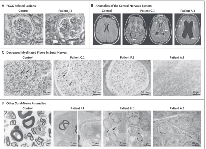
C Decreased Myelinated Fibers in Sural Nerves
A FSGS-Related Lesions B Anomalies of the Central Nervous System
Control Patient J.3 Control Patient C.1 Patient A.3
Control Patient C.1 Patient F.3 Patient A.3
D Other Sural-Nerve Anomalies
Control Patient I.1 Patient H.1 Patient A.3
2 µm 2 µm 2 µm 2 µm 2 µm 1 µm 10 µm 50 µm 50 µm 50 µm 50 µm 50 µm 2 µm 50 µm
Figure 2. Features on Pathological Analysis and Neuroimaging in Selected Patients with Focal Segmental Glomerulosclerosis (FSGS) and Charcot–Marie–Tooth Neuropathy.
Panel A shows the results of trichrome staining of kidney sections from a normal control and Patient J.3, who has typical lesions of FSGS not otherwise specified. Panel B shows the results of magnetic resonance imaging (MRI) of the brain (fluid-attenuated inversion recovery [FLAIR] sequences) of a normal control and two patients: one 15 years of age (Patient C.1) and the other 48 years of age (Patient A.3). Arrows point to bilateral symmetric hyperintensities visible in the internal capsule and the periventricular white matter. Panel C shows sural-nerve semithin sections revealing a marked loss of myelinated fibers in the patients (arrows). The severity of the lesions appears to increase with the patient’s age (from left to right, 15, 30, and 48 years); all myelinated fibers were absent in the oldest patient (Patient A.3). Panel D shows electron micrographs of sural-nerve sections revealing multilayered “onion bulbs” (in Patient I.1) and supernumerary elongated extensions from the cytoplasm of many nonmyelinating Schwann cells (in Patient H.1, arrows), occasionally including collagen fibers (in Patient H.1, arrowheads). In the oldest patient (Patient A.3), the endoneurium was completely replaced by numerous whorls of nonmyelinating Schwann-cell cytoplasm.
IN F2 Mu tations in Ch a rco t –M a r ie –T oo th Dise a se
n engl j med 365;25 nejm.org december 22, 2011 2383
INF2 Mutants and Actin cytoskeleton regulation
Cells expressing mutant INF2 exhibited less corti-cal actin and a reduced number of long actin stress fibers than those expressing wild-type INF2, and a disorganized microtubule network (Fig. 4S, 5S, and 6S in the Supplementary Appendix). Similar features were observed with the K/A–3L/A–INF2 mutant that lacks both the polymerization and
de-polymerization activities of wild-type INF2,14
al-though with this mutant the decrease in the con-tent of long actin filaments was less severe.
We therefore investigated whether the muta-tions in INF2 proteins affect their binding to CDC42, an actin-regulating Rho-GTPase known to interact, in its GTP-loaded active state, with the
INF2 DID.14 An enhanced interaction was observed
between the INF2 mutants and a constitutively active form of CDC42 (CDC42-Q61L) as compared with the wild-type INF2 protein (Fig. 4A and 4B). Moreover, INF2 mutants affected the subcellular localization of CDC42-Q61L, with the fraction of active CDC42 at the plasma membrane being lost in a large proportion of mutant cells as compared with cells expressing wild-type INF2 (Fig. 4C), but did not significantly perturb CDC42 activity (Fig. 7S in the Supplementary Appendix), a result con-sistent with INF2 being a downstream target of
CDC42.14,15 We also demonstrated the interaction
of INF2 with IQGAP1, a CDC42 effector known to
interact with mDia1.26,27 INF2 mutants still
in-teracted with IQGAP1 and altered the endogenous IQGAP1 subcellular distribution (Fig. 8S in the Supplementary Appendix), as they did for MAL and CDC42.
Di s c u s s ion
We have demonstrated that, in addition to leading to isolated FSGS, INF2 mutations are a major cause of Charcot–Marie–Tooth disease associated with FSGS, accounting for approximately 75% of all cas-es. These results shed new light on the genetic basis of the dual neurologic and renal phenotype first
described by Lemieux and Neemeh in 1967.21
Since the initial description, several cases of renal involvement (mostly glomerular disorders with FSGS lesions) have been reported in
associa-tion with Charcot–Marie–Tooth disease.6 Plaisier
and colleagues28 demonstrated the presence of
myelin protein zero (MPZ) in podocytes and an increased urinary albumin excretion in Mpz
knock-out mice, indicating a potential role of myelin components in glomerular permselectivity. No PMP22 or MPZ mutation has been reported in pa-tients with FSGS and Charcot–Marie–Tooth neu-ropathy. In contrast, three quarters of the patients in the present study had INF2 mutations. None of these mutations were present in patients with Charcot–Marie–Tooth disease only, suggesting that INF2 is not involved in cases of the disease with-out an apparent renal phenotype. Nevertheless, because of the individual and intrafamilial pheno-typic variability we observed, physicians should be alert for proteinuria in all patients who have Charcot–Marie–Tooth disease. Similarly, pes cavus was the only clinical sign of Charcot–Marie–Tooth disease in some members of patients’ families; therefore, a careful clinical neurologic evaluation should be considered for patients with FSGS.
Although INF2 mutations have been shown to be the major cause of autosomal dominant isolated
FSGS, accounting for 12 to 17% of all cases,9,10
the prevalence of INF2 mutations in association with FSGS and Charcot–Marie–Tooth disease is much higher (75%). Several lines of genetic and functional evidence indicate that these variants are pathogenic mutations. Given the high preva-lence of INF2 mutations, the detection of various distinct INF2 mutations, and the absence of tions in PMP22 and MPZ, the occurrence of a muta-tion in another gene underlying Charcot–Marie– Tooth disease in patients with INF2-related FSGS is unlikely. Moreover, it is improbable that the fre-quent de novo mutations we detected would occur in two distinct genes in the same patient. Further-more, all of the nine INF2 mutations we identified are new. Although the nine mutations encode DID residues, as do mutations associated with isolated FSGS, they had a distinct localization, correspond-ing mostly to the 3′ end of exon 2 as well as in exon 3, in which no isolated FSGS mutation has been identified.
We further explored the functional effects of some INF2 mutations. We postulated that the mechanisms linking INF2 to the development of Charcot–Marie–Tooth disease involved perturba-tion of cytoskeletal networks and thus intracellular transport of myelin components. Indeed, INF2 has been shown to regulate specialized routes of pro-tein targeting to the plasma membrane in various types of cells in association with CDC42 and MAL
or MAL2.14,15 This targeting involves vesicular
car-riers that associate with actin filaments and
re-The New England Journal of Medicine
Downloaded from nejm.org on December 16, 2013. For personal use only. No other uses without permission. Copyright © 2011 Massachusetts Medical Society. All rights reserved.
quires both the actin polymerization and
depo-lymerization properties of INF2.14,15 In addition,
CDC42 and MAL are fundamental players in
pe-ripheral myelination.17,18,29 Here we show that
INF2 is also expressed in Schwann cells and to a lesser extent in neurons. We also demonstrate the endogenous colocalization of INF2 and MAL and their in vivo interaction in Schwann cells, thereby providing a clear rationale for the role of INF2 mu-tations in Charcot–Marie–Tooth disease. In addi-tion, we show that INF2 mutations disrupt the INF2–MAL–CDC42 pathway. The reduction in cor-tical actin and stress fibers in cells expressing INF2 DID mutants was even more severe than in cells expressing the K/A–3L/A–INF2 mutant lack-ing both the polymerization and depolymerization properties of INF2. This suggests that INF2 DID mutants might not only alter these two functions but also have additional effects on INF2 partners. Indeed, we demonstrated an enhanced interaction between the INF2 DID mutants and CDC42 and a reduced fraction of active CDC42 at the plasma membrane. Together, our results suggest that the mislocalization of the INF2–MAL–CDC42 complex in the cytoplasm, as well as the defects in the po-lymerization and depopo-lymerization activities of INF2 required for actin dynamics, could disrupt protein targeting to the plasma membrane and therefore also disrupt proper myelin formation and maintenance.
The implication of the Rho-GTPase CDC42 pathway in the effect of the INF2 mutants is remi-niscent of mutations in two genes involved in dominant intermediate Charcot–Marie–Tooth dis-ease: DNM2, which encodes the GTPase protein
dynamin 2,30 and ARHGEF10, which encodes a
gua-nine exchange factor that activates Rho-GTPases.31
Finally, sensorineural hearing loss was present in 4 of the 12 families (33%) with an INF2 muta-tion, which is a prevalence significantly higher than the approximately 5% prevalence reported among patients with Charcot–Marie–Tooth
dis-ease.32 Mutations in DIAPH1, which encodes
mDia1, have been associated with autosomal
dominant sensorineural progressive hearing loss.33
The biologic role of mDia1 in hearing is likely to include regulation of actin polymerization in
hair cells of the inner ear. Sun and colleagues13
recently demonstrated that the INF2 DID interacts with the mDia1 DAD. Thus, similar mechanisms
are likely to be involved in deafness related to ei-ther DIAPH1 or INF2 mutations.
The reasons why INF2 mutations do not always lead to a neurologic phenotype still need to be clarified. One clue, however, is that the mutations underlying FSGS alone or in combination with Charcot–Marie–Tooth neuropathy are clustered in different parts of the DID. The latter mutations are located between two putative DID-binding pock-ets, suggesting that they could affect DID function more severely than mutations related to FSGS alone, by simultaneously disrupting the interaction of INF2 with multiple proteins, some of which could be specific myelin proteins. This is consis-tent with the renal phenotype that is more severe in patients with FSGS and Charcot–Marie–Tooth disease than in patients with FSGS (median age at
Figure 3 (facing page). Colocalization and Interaction of Wild-Type and Mutant INF2 with Myelin and Lym-phocyte Protein (MAL).
Panel A (immunoperoxidase stain) shows MAL, INF2, and MAL2 proteins in normal human kidney specimens and peripheral-nerve biopsy specimens. The serial sec-tions shown suggest that INF2 colocalizes with MAL2 in podocytes and MAL in Schwann cells. Panel B shows immunostaining of endogenous INF2 (green) and MAL (red) in MSC-80 mouse Schwann cells (with the merged image also shown).23
The scatter plot represents red and green pixel fluorescence intensities of the corre-sponding immunostaining. Both proteins were colo-calized (for eight samples analyzed: median Pearson’s coefficient, 0.909; range, 0.625 to 0.978).24
Panel C shows results of an INF2–MAL pull-down assay per-formed on the detergent-insoluble (I) membrane fraction of S16 rat Schwann cells separated from the soluble fraction (S) and the Ponceau red staining of the gluta-thione S-transferase proteins used at the bottom. In Schwann cells, endogenous MAL bound the C-terminal of INF2. Panel D shows the colabeling of wild-type or mutant FLAG-tagged INF2 (red) with myelocytomato-sis virus–associated sequence (MYC)–tagged MAL (green) transiently expressed in HeLa cells, with the corresponding scatter plot of red and green pixel fluo-rescence intensities on the right. In contrast to the peri-nuclear localization of wild-type INF2, the two INF2 mutants (with amino acid change L165P or R106P) in patients with focal segmental glomerulosclerosis (FSGS) and Charcot–Marie–Tooth (CMT) neuropathy were spread throughout the cytoplasm. As observed with endogenous proteins, wild-type INF2 and MAL re-combinant proteins were colocalized (see merged im-ages). For INF2 mutants, the colocalization persisted, leading to a broader cytoplasmic distribution of MAL.
IN F2 Mu tations in Ch a rco t –M a r ie –T oo th Dise a se
n engl j med 365;25 nejm.org december 22, 2011 2385
Green Pixel Intensity
60,000 40,000 20,000 0
0 20,000 40,000 60,000
Red Pixel Intensity
r=0.97
MSC-80 Cells
MAL Merge
INF2
HeLa Cells
Green Pixel Intensity
60,000 40,000 20,000 0 0 20,000 40,000 r=0.89
Green Pixel Intensity
30,000 20,000 10,000 0 0 20,000 40,000 60,000 r=0.79
Green Pixel Intensity
60,000 40,000 20,000 0
0 20,000 40,000 60,000
Red Pixel Intensity
r=0.84
MAL Merge
INF2
INF2 Wild Type
FSGS-CMT INF2 L165P INF2 R106P Kidney MAL MAL INF2 MAL2 Peripheral Nerve B A C D Input S I GST GST-INF2 C-terminal GST-INF2 C-terminal — GST —
The New England Journal of Medicine
Downloaded from nejm.org on December 16, 2013. For personal use only. No other uses without permission. Copyright © 2011 Massachusetts Medical Society. All rights reserved.
Relative Interaction of FLAG-INF2 and HA-CDC42–Q61L 60 40 20 0
Wild Type R106P L165P R177H R218Q Y193H B A FLAG-INF2 Wild type FSGS-CMT FSGS FLAG-INF2 Wild type FSGS-CMT FSGS P=0.04P=0.04 P=0.04
Cells with Membrane
Localization of CDC42-Q61L (%) 100 40 60 80 20 0 Wild Type L165P R106P R177H P=0.04 P=0.003 P=0.002 P=0.002
Not TransfectedFLAG-INF2–Wild TypeHA-CDC42–Q61LWild typeR106P L165P R177H R218Q Y193H Total Lysate IP HA FLAG-INF2 HA-CDC42–Q61L HA-CDC42–Q61L FLAG-INF2 FSGS-CMT FSGS HA-CDC42–Q61L+FLAG-INF2 HA-CDC42–Q61L Merge INF2 INF2 Wild Type INF2 R177H FSGS INF2 L165P INF2 R106P FSGS-CMT
IN F2 Mu tations in Ch a rco t –M a r ie –T oo th Dise a se
n engl j med 365;25 nejm.org december 22, 2011 2387
onset of proteinuria, 18 vs. 27 years, and of
end-stage renal disease, 21 vs. 36 years),10 although
this needs to be verified in a larger cohort. In conclusion, the identification of the
for-min INF2 as a crucial molecular entity in the occurrence of FSGS and Charcot–Marie–Tooth neuropathy provides additional insight into the role of similar cellular machinery in podocytes and Schwann cells, even though these two highly specialized cell types have distinct functions.
Supported by grants from the Association pour l’Utilisation du Rein Artificiel (to Dr. Antignac), Association Française con-tre les Myopathies (ANR-08-GENOPAT-017-01, to Dr. Antignac), Agence Nationale de la Recherche (PodoNet project number ANR-07-E-RARE-011-01 in the ERA-Net Consortium [JTC2007], to Dr. Antignac, and ANR-06-MRAR-024-01, to Dr. Leguern), Fondation pour la Recherche Médicale (project number DMP 2010-11-20-386, to Dr. Antignac, and doctoral funding, to Dr. Boyer), Association des Malades du Syndrome Néphrotique (to Dr. Mollet), Fonds de la Recherche en Santé du Québec (Fellow-ship Training Award to Dr. Benoit), and Ministerio de Ciencia e Innovación (BFU2009-07886 and CONSOLIDER COAT CSD2009-00016, to Dr. Alonso).
Disclosure forms provided by the authors are available with the full text of this article at NEJM.org.
We thank Scott J. Harvey for helpful discussions; Martin R. Pollak, Philippe Chavrier, and Christopher Carpenter for provid-ing FLAG-INF2-WT, GFP-IQGAP1-WT, and HA-CDC42–Q61L plasmids, respectively; Bernard Zalc for providing MSC-80 cells; Olivier Dorseuil for providing the GST-PAK construct; Catherine Lacroix, Laure-Hélène Noël, and Dominique Nochy for provid-ing histologic sections; Nicolas Goudin (of Plateau d’Imagerie Cellulaire, Institut Fédératif de Recherche Necker–Enfants Malades, Paris) for providing expert knowledge on confocal mi-croscopy; Drs. Bommelaer, Clavelou, Campone, Quérin, Rieu, Rouhart, Sarret, and Squalli Houssaini for providing medical information about their patients and the families who partici-pated in this study; and the NHLBI Exome Sequencing Project (http://snp.gs.washington.edu/EVS/) for providing exome variant cells for comparison.
Figure 4 (facing page). Effects of Disease-Causing INF2 Mutations on the INF2–CDC42 Interaction and CDC42 Subcellular Localization.
Panel A shows the in vivo interaction of the constitu-tively active form of CDC42 (human influenza hemag-glutinin [HA]-CDC42–Q61L) and FLAG-tagged wild-type and mutant INF2 constructs in HEK-293T cells. The histogram shows the amount of co-immunopre-cipitated INF2 protein normalized to the amount of immunoprecipitated CDC42 protein. The ratio of wild-type FLAG-INF2 to HA-CDC42–Q61L was set to 1, and all other values were calculated relative to it. Panel B shows HeLa cells transfected with plasmids encoding either a wild-type or mutant FLAG-tagged INF2 (red) and a constitutively active HA-tagged form of CDC42 (green). All cells expressing CDC42-Q61L alone or with wild-type INF2 exhibited cytoplasmic staining of CDC42, and about 60% also showed membrane localization. Mutant forms of INF2 led to a mislocalization of both proteins, the INF2 staining being diffuse in the cyto-plasm and CDC42 being less targeted to the cyto-plasma membrane. The associated histogram represents the mean percentage of cells showing membrane staining of CDC42 (with >30 cells cotransfected per experiment). CMT denotes Charcot–Marie–Tooth disease, and FSGS focal segmental glomerulosclerosis. T bars indicate the standard errors for three independent experiments. IP denotes immunoprecipitation.
Appendix
The authors’ affiliations are as follows: INSERM Unité 983 (O.B., F.N., O.G., G.B., E.H.C., C. Arrondel, M.-J.T., R.M., M.-C.G., S.S., C. Antignac, G.M.), Unité 702 (E.P., P.R.), and Unité 975 (E.L.); Service de Néphrologie Pédiatrique (O.B.), Service de Transplantation et Soins Intensifs (G.C.), and Service de Génétique (C. Antignac), Hôpital Necker–Enfants Malades, Assistance Publique–Hôpitaux de Paris (AP-HP); Service de Néphrologie et Dialyses (E.P., P.R.), Hôpital Tenon, AP-HP; Service de Néphrologie (A.K.), Hôpital Européen Georges Pompidou, AP-HP; Service de Néphrologie Pédiatrique (G.D.), Hôpital Robert Debré, AP-HP; Institut de Myologie (O.D.), Hôpital Pitié–Salpêtrière, AP-HP; Université Paris Descartes, Sorbonne Paris Cité (O.B., F.N., O.G., E.H.C., C. Arrondel, M.-J.T., R.M., A.K., G.C., I.B., M.-C.G., S.S., C. Antignac, G.M.); Université Pierre et Marie Curie (E.P., E.L., P.R.); Université Paris Diderot (G.D.); Centre National de la Recherche Scientifique Unité 7225 (E.L.) and Laboratoire de Cristallographie et RMN Biologiques (I.B.) — all in Paris; Laboratoire et Service de Neurologie, Centre Hospitalier Universitaire (CHU) et Université de Limoges, Limoges (B.F., L.R., J.-M.V.); Université de Lyon (C.P.-N.), Service de Néphrologie, Centre Hospitalier Lyon-Sud (C.P.-N.), and Service de Neurologie, Hôpi-tal de la Croix-Rousse (P.P.), Hospices Civils de Lyon — all in Lyon; Centre HospiHôpi-talier de Saint-Brieuc, Saint-Brieuc (C.C.); Service de Néphrologie et Immunologie Clinique, CHU Hôtel Dieu, Nantes (J.D.); CHU Gabriel Montpied, Université d’Auvergne, Clermont-Fer-rand (P.D.); Génétique Moléculaire, Pharmacogénétique et Hormonologie, CHU Bicêtre, Université Paris-Sud, Le Kremlin-Bicêtre (A.G.-M.) — all in France; Service de Néphrologie Pédiatrique, CHU Sainte-Justine, Université de Montréal (G.B.); and Centre de Recherche Guy-Bernier, Hôpital Maisonneuve–Rosemont (A.B.) — both in Montreal; Centre de Néphrologie et d’Hémodialyse Riad, Rabat, Morocco (L.B.); Service de Néphrologie, Centre Hospitalier de Luxembourg, Luxemburg (D.P.); and Centro de Biología Molecular Severo Ochoa, Consejo Superior de Investigaciones Cientificas–Universidad Autónoma de Madrid, Madrid (M.A.A.).
References
1. Skre H. Genetic and clinical aspects of Charcot-Marie-Tooth’s disease. Clin Genet 1974;6:98-118.
2. Pareyson D, Scaioli V, Laurà M.
Clini-cal and electrophysiologiClini-cal aspects of Charcot-Marie-Tooth disease. Neuromo-lecular Med 2006;8:3-22.
3. Lupski JR, Reid JG,
Gonzaga-Jau-regui C, et al. Whole-genome sequencing in a patient with Charcot-Marie-Tooth neuropathy. N Engl J Med 2010;362:1181-91.
The New England Journal of Medicine
Downloaded from nejm.org on December 16, 2013. For personal use only. No other uses without permission. Copyright © 2011 Massachusetts Medical Society. All rights reserved.
4. Patel PI, Roa BB, Welcher AA, et al. The gene for the peripheral myelin pro-tein PMP-22 is a candidate for Charcot-Marie-Tooth disease type 1A. Nat Genet 1992;1:159-65.
5. Hayasaka K, Takada G, Ionasescu VV. Mutation of the myelin P0 gene in Char-cot-Marie-Tooth neuropathy type 1B. Hum Mol Genet 1993;2:1369-72.
6. Paul MD, Fernandez D, Pryse-Phillips W, Gault MH. Charcot-Marie-Tooth dis-ease and nephropathy in a mother and daughter with a review of the literature. Nephron 1990;54:80-5.
7. D’Agati V. Pathologic classification of focal segmental glomerulosclerosis. Semin Nephrol 2003;23:117-34.
8. Machuca E, Benoit G, Antignac C. Ge-netics of nephrotic syndrome: connecting molecular genetics to podocyte physiolo-gy. Hum Mol Genet 2009;18:R185-R194.
9. Brown EJ, Schlöndorff JS, Becker DJ, et al. Mutations in the formin gene INF2 cause focal segmental glomerulosclero-sis. Nat Genet 2010;42:72-6.
10. Boyer O, Benoit G, Gribouval O, et al. Mutations in INF2 are a major cause of autosomal dominant focal segmental glo-merulosclerosis. J Am Soc Nephrol 2011;22:239-45.
11. Chesarone MA, DuPage AG, Goode BL. Unleashing formins to remodel the actin and microtubule cytoskeletons. Nat Rev Mol Cell Biol 2010;11:62-74.
12. Chhabra ES, Higgs HN. INF2 is a WASP homology 2 motif-containing for-min that severs actin filaments and ac-celerates both polymerization and depo-lymerization. J Biol Chem 2006;281: 26754-67.
13. Sun H, Schlöndorff JS, Brown EJ, Higgs HN, Pollak MR. Rho activation of mDia formins is modulated by an interac-tion with inverted formin 2 (INF2). Proc Natl Acad Sci U S A 2011;108:2933-8.
14. Madrid R, Aranda JF, Rodríguez-Frat-icelli AE, et al. The formin INF2 regulates
basolateral-to-apical transcytosis and lu-men formation in association with Cdc42 and MAL2. Dev Cell 2010;18:814-27.
15. Andrés-Delgado L, Antón OM, Madrid R, Byrne JA, Alonso MA. Formin INF2 regulates MAL-mediated transport of Lck to the plasma membrane of human T lym-phocytes. Blood 2010;116:5919-29.
16. Marazuela M, Alonso MA. Expression of MAL and MAL2, two elements of the protein machinery for raft-mediated transport, in normal and neoplastic hu-man tissue. Histol Histopathol 2004;19: 925-33.
17. Benninger Y, Thurnherr T, Pereira JA, et al. Essential and distinct roles for cdc42 and rac1 in the regulation of Schwann cell biology during peripheral nervous system development. J Cell Biol 2007;177:1051-61.
18. Frank M. MAL, a proteolipid in glyco-sphingolipid enriched domains: function-al implications in myelin and beyond. Prog Neurobiol 2000;60:531-44.
19. Deniau F, Guillot M, Plus A, et al. Charcot-Marie-Tooth disease and glomer-ular nephropathy. Arch Fr Pediatr 1986; 43:791-3. (In French.)
20. Fillod I, Cochat P, Colon S, Wright C, David L. Nephropathy and Charcot-Marie-Tooth disease: a case report. Pediatrie 1990;45:319-22. (In French.)
21. Lemieux G, Neemeh JA. Charcot-Marie-Tooth disease and nephritis. Can Med Assoc J 1967;97:1193-8.
22. Rose R, Weyand M, Lammers M, Ishizaki T, Ahmadian MR, Wittinghofer A. Structural and mechanistic insights into the interaction between Rho and mammalian Dia. Nature 2005;435:513-8.
23. Boutry JM, Hauw JJ, Gansmüller A, Di-Bert N, Pouchelet M, Baron-Van Ever-cooren A. Establishment and character-ization of a mouse Schwann cell line which produces myelin in vivo. J Neurosci Res 1992;32:15-26.
24. Bolte S, Cordelières FP. A guided tour into subcellular colocalization analysis in
light microscopy. J Microsc 2006;224:213-32.
25. Chhabra ES, Ramabhadran V, Gerber SA, Higgs HN. INF2 is an endoplasmic reticulum-associated formin protein. J Cell Sci 2009;122:1430-40.
26. Bashour AM, Fullerton AT, Hart MJ, Bloom GS. IQGAP1, a Rac- and Cdc42-binding protein, directly binds and cross-links microfilaments. J Cell Biol 1997;137: 1555-66.
27. Brandt DT, Marion S, Griffiths G, Watanabe T, Kaibuchi K, Grosse R. Dia1 and IQGAP1 interact in cell migration and phagocytic cup formation. J Cell Biol 2007;178:193-200.
28. Plaisier E, Mougenot B, Verpont MC, et al. Glomerular permeability is altered by loss of P0, a myelin protein expressed in glomerular epithelial cells. J Am Soc Nephrol 2005;16:3350-6.
29. Frank M, van der Haar ME, Schaeren-Wiemers N, Schwab ME. rMAL is a glyco-sphingolipid-associated protein of myelin and apical membranes of epithelial cells in kidney and stomach. J Neurosci 1998; 18:4901-13.
30. Schafer DA, Weed SA, Binns D, Karginov AV, Parsons JT, Cooper JA. Dy-namin2 and cortactin regulate actin as-sembly and filament organization. Curr Biol 2002;12:1852-7.
31. Verhoeven K, De Jonghe P, Van de Putte T, et al. Slowed conduction and thin myelination of peripheral nerves associ-ated with mutant rho guanine-nucleotide exchange factor 10. Am J Hum Genet 2003;73:926-32.
32. Szigeti K, Lupski JR. Charcot-Marie-Tooth disease. Eur J Hum Genet 2009;17: 703-10.
33. Lynch ED, Lee MK, Morrow JE, Welcsh PL, León PE, King MC. Nonsyndromic deafness DFNA1 associated with mutation of a human homolog of the Drosophila gene diaphanous. Science 1997;278:1315-8.
Copyright © 2011 Massachusetts Medical Society.
CLINICAL TRIAL REGISTRATION
The Journal requires investigators to register their clinical trials in a public trials registry. The members of the International Committee of Medical Journal Editors (ICMJE) will consider most reports of clinical
trials for publication only if the trials have been registered. Current information on requirements and appropriate registries