HAL Id: hal-02935548
https://hal.inrae.fr/hal-02935548
Submitted on 11 Sep 2020
HAL is a multi-disciplinary open access
archive for the deposit and dissemination of
sci-entific research documents, whether they are
pub-lished or not. The documents may come from
teaching and research institutions in France or
abroad, or from public or private research centers.
L’archive ouverte pluridisciplinaire HAL, est
destinée au dépôt et à la diffusion de documents
scientifiques de niveau recherche, publiés ou non,
émanant des établissements d’enseignement et de
recherche français ou étrangers, des laboratoires
publics ou privés.
Distributed under a Creative Commons Attribution| 4.0 International License
the Bacterial Genotoxin Colibactin
Camille Chagneau, Christophe Garcie, Nadège Bossuet-Greif, Sophie Tronnet,
Alexander Brachmann, Jörn Piel, Jean-Philippe Nougayrède, Patricia Martin,
Eric Oswald
To cite this version:
Camille Chagneau, Christophe Garcie, Nadège Bossuet-Greif, Sophie Tronnet, Alexander Brachmann,
et al.. The Polyamine Spermidine Modulates the Production of the Bacterial Genotoxin Colibactin.
MSphere, American Society for Microbiology., 2019, 4 (5), pp.00414-19. �10.1128/mSphere.00414-19�.
�hal-02935548�
The Polyamine Spermidine Modulates the Production of the
Bacterial Genotoxin Colibactin
Camille V. Chagneau,
a,bChristophe Garcie,
a,b* Nadège Bossuet-Greif,
bSophie Tronnet,
b* Alexander O. Brachmann,
cJörn Piel,
cJean-Philippe Nougayrède,
bPatricia Martin,
bEric Oswald
a,baCHU Toulouse, Service de Bactériologie-Hygiène, Toulouse, France bIRSD, Université de Toulouse, INSERM, INRA, ENVT, UPS, Toulouse, France
cInstitute of Microbiology, Eidgenössische Technische Hochschule (ETH), Zurich, Switzerland
ABSTRACT
Colibactin is a polyketide/nonribosomal peptide produced by
Esche-richia coli strains that harbor the pks island. This toxin induces DNA double-strand
breaks and DNA interstrand cross-links in infected eukaryotic cells.
Colibactin-producing strains are found associated with colorectal cancer biopsy specimens and
promote intestinal tumor progression in various murine models. Polyamines are
small polycationic molecules produced by both microorganisms and eukaryotic cells.
Their levels are increased in malignancies, where they contribute to disease
progres-sion and metastasis. In this study, we demonstrated that the endogenous
spermi-dine synthase SpeE is required for full genotoxic activity of colibactin-producing E.
coli. Supplying spermidine in a ΔspeE pks
⫹E. coli strain restored genotoxic activity.
Spermidine is involved in the autotoxicity linked to colibactin and is required for
di-rect damaging activity on DNA. The production of the colibactin prodrug motif is
impaired in ΔspeE mutants. Therefore, we demonstrated that spermidine has a direct
impact on colibactin synthesis.
IMPORTANCE
Colibactin-producing Escherichia coli strains are associated with
can-cerous and precancan-cerous colorectal tissues and are suspected of promoting
colorec-tal carcinogenesis. In this study, we describe a new interplay between the synthesis
of the genotoxin colibactin and the polyamine spermidine. Polyamines are highly
abundant in cancer tissue and are associated with cell proliferation. The need for
spermidine in genotoxic activity provides a new perspective on the role of these
metabolites in the pathogenicity of colibactin-producing E. coli strains in colorectal
cancer.
KEYWORDS
Escherichia coli, biosynthesis, colorectal cancer, genotoxic colibactin,
polyamines
T
he genotoxin colibactin is a secondary microbial metabolite synthetized by
Esche-richia coli and other enterobacteria. The genetic determinant of colibactin is a 54-kb
gene cluster, the pks genomic island (1). This highly conserved pathogenicity island is
predominately found in E. coli strains of the phylogenetic group B2 and in some other
species of Enterobacteriaceae (1, 2). The pks island carries the genes clbA to clbS, which
encode modular nonribosomal peptide synthetases (NRPSs), polyketide synthases
(PKSs), and accessory enzymes (1). This complex biosynthetic assembly line is
respon-sible for the synthesis of colibactin, which belongs to the chemical family of hybrid
polyketide/nonribosomal peptide (PK-NRP) compounds. Mature colibactin has a
com-plex structure of highly unstable and reactive molecules forming DNA adducts (3–12).
However, the complete structure of genotoxic colibactin is not yet fully elucidated.
Colibactin is genotoxic for eukaryotic (1) as well as prokaryotic cells when the ClbS
resistance protein is not produced (13). Colibactin-induced DNA damages result from
Citation Chagneau CV, Garcie C, Bossuet-Greif
N, Tronnet S, Brachmann AO, Piel J, Nougayrède J-P, Martin P, Oswald E. 2019. The polyamine spermidine modulates the production of the bacterial genotoxin colibactin. mSphere 4:e00414-19.https://doi .org/10.1128/mSphere.00414-19.
Editor Sarah E. F. D’Orazio, University of
Kentucky
Copyright © 2019 Chagneau et al. This is an
open-access article distributed under the terms of theCreative Commons Attribution 4.0 International license.
Address correspondence to Eric Oswald, eric.oswald@inserm.fr.
* Present address: Christophe Garcie, Centre Hospitalier de Pau, Laboratoire, Pau, France; Sophie Tronnet, the Laboratory for Molecular Infection Medicine Sweden (MIMS), Umeå Centre for Microbial Research (UCMR) and Department of Molecular Biology, Umeå University, Umeå, Sweden.
C.V.C. and C.G. contributed equally to this article. Received 10 June 2019 Accepted 9 September 2019 Published
RESEARCH ARTICLE
Host-Microbe Biology
2 October 2019on September 11, 2020 by guest
http://msphere.asm.org/
Downloaded from
the formation of interstrand DNA cross-links leading to DNA double-strand breaks (12,
14). These DNA damages can lead to gene mutations, chromosomal instability, and
senescence (15, 16), and in various mouse models colibactin-producing E. coli strains
promote intestinal tumor progression (17–20).
Epidemiological studies show a high prevalence of pks-carrying E. coli strains in
biopsy specimens from colorectal cancer patients (17, 21, 22). However, the presence of
a pks
⫹strain in gut microbiota is not sufficient to induce colorectal cancer. Other
factors have been shown to promote cell transformation and/or to potentiate the
bacterial genotoxicity of these bacteria. For example, deoxynivalenol, a food
contam-inant, exacerbates the genotoxic effect linked to colibactin in animals colonized by
pks
⫹E. coli (23). Environmental factors such as iron concentration can also directly
regulate colibactin production (24). However, inflammation is a more significant factor,
which seems to be required in colibactin-associated carcinogenesis (17, 20).
Inflamma-tion itself is critical for tumor progression (by promoting cell proliferaInflamma-tion, survival, and
migration) but it also drives modifications in microbiota composition and the expansion
of E. coli (25). It has been shown that a proinflammatory cancer microenvironment
could increase the expression of clb genes, at least at a transcriptional level (26).
Recently, an association between colibactin-producing E. coli and enterotoxigenic
Bacteroides fragilis, another procarcinogenic bacterial species, was also noted on the
colonic mucosa of patients with familial adenomatous polyposis (FAP), who are highly
susceptible to colorectal cancer (20). A synergy between these two bacterial species
was observed in tumor formation in an FAP murine model (20).
Polyamines are essential for cell proliferation and have been shown to play a crucial
role in carcinogenesis (27, 28). They are small aliphatic molecules involved in protein
synthesis and regulation, DNA integrity, stress resistance. (29). Infections by
microor-ganisms and chronic inflammation can interfere with polyamine catabolism and
in-crease the formation of damaging oxidative compounds, contributing in fine to
carci-nogenesis (30, 31). Polyamines are found at high concentrations in colorectal cancer
tissues even at precancerous states (32) but are also produced by intestinal bacteria
such as E. coli (33).
In this study, we investigated the putative roles of polyamines in colibactin
geno-toxic activity. We demonstrate that spermidine is involved in colibactin synthesis and
the associated genotoxicity.
RESULTS
The spermidine biosynthetic pathway is required for full genotoxicity of
colibactin-producing E. coli. To test the impact of the endogenous
spermidine-putrescine pathway on colibactin-producing E. coli genotoxic activity, mutants
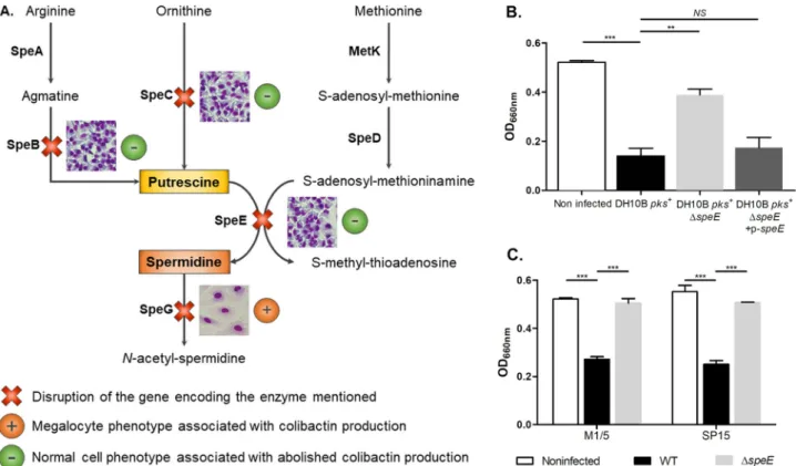
inacti-vated for the speB, speC, speE, and speG genes (Fig. 1A) were engineered in E. coli strain
DH10B, which harbors the pks island on a bacterial artificial chromosome (Table 1) (34).
The production of colibactin by each mutant was first monitored through
bacterium-host cell interactions and subsequent observation of the formation of large senescent
cells (megalocytosis) (Fig. 1). Inactivation of speB, speC, and speE genes but not of speG
resulted in a decrease in the megalocytosis effect (Fig. 1A), with no effect on bacterial
growth (data not shown). Therefore, spermidine biosynthesis, and especially the final
step catalyzed by the spermidine synthase SpeE, has more impact on colibactin
genotoxic activity than putrescine biosynthesis. We confirmed the loss of the
megalo-cytosis phenotype in the ΔspeE mutant and its restoration after plasmid
complemen-tation (Fig. 1B). The inactivation of speE in pks
⫹E. coli from other genetic backgrounds
(i.e., the E. coli M1/5 commensal strain and the E. coli SP15 pathogenic strain isolated
from neonatal meningitis) also resulted in a decrease in the megalocytosis effect
(Table 1 and Fig. 1C). We then measured the genotoxic activity of the bacteria by
quantifying histone H2AX phosphorylation in response to DNA damage in the infected
cells (1, 15, 35). We confirmed that the inactivation of the speE gene in the DH10B pks
⫹strain markedly decreased the genotoxic effect induced by colibactin (Fig. 2), whereas
the complemented mutant was fully genotoxic (Fig. 2).
on September 11, 2020 by guest
http://msphere.asm.org/
Spermidine is required for full genotoxicity of colibactin-producing E. coli. In
order to determine whether the decreased genotoxicity of the ΔspeE mutant is
asso-ciated with the spermidine synthase SpeE or to spermidine itself, we added increasing
concentrations of spermidine in the interaction medium during HeLa cell infection
FIG 1 Endogenous spermidine biosynthesis is involved in the E. coli colibactin-associated megalocytosis phenotype. (A) Presentation of the polyamine
biosynthetic pathways (34). DH10B pks⫹mutants with mutations of genes speB, speC, speE, and speG were tested for the megalocytosis phenotype in infected HeLa cells, as previously described (1). The phenotypes of HeLa cells resulting from infection with the different mutants are shown. (B and C) Cytotoxic effects of colibactin produced by E. coli strains DH10B pks⫹, M1/5, and SP15 and their derivatives were determined by quantification of megalocytosis. At the end of HeLa cell infection, the methylene blue protein staining was quantified by measurement of absorbance at the optical density at 660 nm. The multiplicity of infection (MOI) was 200. Data were pooled from three independent experiments.***, P⬍ 0.001, and **, P ⬍ 0.01, by 1-way analysis of variance (ANOVA). All bar graphs show mean values⫾ standard errors of the mean (SEM). NS, not significant.
TABLE 1 E. coli strains and plasmids used in this study
Strain or plasmid Genotype or phenotype Source
E. coli strains
DH10B pks⫹ K-12 laboratory strain carrying pBACpks, Cmr 1
DH10B pks⫹ΔspeB speB mutant of strain DH10B pBACpks, CmrKanr This study
DH10B pks⫹ΔspeC speC mutant of strain DH10B pBACpks, CmrKanr This study
DH10B pks⫹ΔspeE speE mutant of strain DH10B pBACpks, CmrKanr This study
DH10B pks⫹ΔspeG speG mutant of strain DH10B pBACpks, CmrKanr This study
DH10B pks⫹ΔspeE⫹ p-speE DH10B pks⫹ΔspeE carrying p-speE, CmrKanrAmpr This study
DH10B pks⫹ΔclbA clbA mutant of strain DH10B pBACpks, Cmr 1
DH10B pks⫹ΔclbS clbS mutant of strain DH10B pBACpks, Cmr 13
DH10B pks⫹ΔspeE ΔclbS speE clbS double mutant of strain DH10B pBACpks, Cmr This study
M1/5 Commensal E. coli strain isolated from feces of a healthy adult,
B2 phylogenetic group, colibactin genotoxin producer
35
M1/5 ΔspeE speE mutant of strain M1/5, Kanr This study
SP15 Extraintestinal pathogenic E. coli strain isolated from spinal fluid of neonate
with meningitis, O18:K1 serotype, colibactin genotoxin producer
50
SP15 ΔspeE speE mutant of strain SP15, Kanr This study
SP15 ΔspeG speG mutant of strain SP15, Kanr This study
Plasmid
p-speE pSC-A plasmid carrying wild-type speE gene, AmprKanr This study
Colibactin Synthesis Is Linked to Polyamine Metabolism
on September 11, 2020 by guest
http://msphere.asm.org/
(Fig. 3). The production of colibactin was again measured by quantifying histone H2AX
phosphorylation (Fig. 3) and megalocytosis assay (see Fig. S1 in the supplemental
material). Spermidine supplementation of the ΔspeE mutant restored its genotoxicity in
a dose-dependent manner (Fig. 3).
The spermidine acetyltransferase SpeG catalyzes spermidine acetylation into
phys-iologically inert N-acetylspermidine. While screening the impact of the
putrescine-spermidine pathway on colibactin-associated megalocytosis phenotype, we observed
no ΔspeG mutant defect (Fig. 1A). We hypothesized that by abolishing SpeG activity,
spermidine would accumulate in the bacteria and boost the genotoxic activity of pks
⫹E. coli. To test this hypothesis, we quantified H2AX phosphorylation in response to DNA
damages after HeLa cell infection by E. coli SP15 and deletion mutants of speE and speG
genes (Fig. 4). Compared to the wild-type strain, we observed a decrease in the
genotoxic activity of SP15 ΔspeE (Fig. 4). However, SP15 ΔspeG with impaired
catabo-lism of spermidine induced 20% more DNA damage than the wild-type SP15 strain
(Fig. 4). These results confirmed that spermidine is the key player in the interaction
between the putrescine-spermidine pathway and colibactin-associated genotoxic
ac-FIG 2 Deletion of the spermidine synthase SpeE impacts full genotoxicity of colibactin-producing E. coli.
The production of colibactin by E. coli strain DH10B pks⫹and derivatives was determined by quantifi-cation of H2AX phosphorylation, which correlates with DNA damage resulting from the genotoxic effect of colibactin. E. coli strain DH10B pks⫹, the ΔclbA mutant (negative control), and the ΔspeE mutant and its complemented derivative were cocultivated with HeLa cells in an In-Cell Western assay as previously described (35). The multiplicity of infection (MOI [i.e., the number of bacteria per cell) ranged from 100 to 400. Data shown in the graph are representative of three independent experiments. All bar graphs show mean values⫾ SEM.
FIG 3 Exogenous spermidine restores colibactin-associated genotoxicity in a ΔspeE mutant.
Colibactin-mediated genotoxicity was determined by infection of HeLa cells with E. coli strain DH10B pks⫹and derivatives and quantification of H2AX phosphorylation. When indicated, spermidine (g/ml) was added to the interaction medium during infection. MOI⫽ 400. NI, noninfected. Data were combined from three independent experiments.***, P⬍ 0.001, and *, P ⬍ 0.05, by 1-way ANOVA. All bar graphs show mean values⫾ SEM. NS, not significant.
on September 11, 2020 by guest
http://msphere.asm.org/
tivity. However, a ΔpotD spermidine import mutant of E. coli strain SP15 was not
impaired in its genotoxicity (see Fig. S2 in the supplemental material), suggesting that
spermidine endogenous production is sufficient to support colibactin production in this
in vitro assay. Furthermore, we tested other polyamines and showed that spermine and
norspermidine could restore the genotoxic activity in a ΔspeE mutant (see Fig. S3 in the
supplemental material).
Spermidine is directly involved in colibactin biosynthesis. To determine the level
at which spermidine acts, we quantified the autotoxicity linked to colibactin production
directly in bacteria. In fact, DNA damage can occur in toxigenic bacteria mutated for the
ClbS resistance protein, leading to activation of the SOS response and then decreased
growth (13). We constructed a ΔspeE ΔclbS double mutant in the E. coli strain DH10B
pks
⫹(Table 1) and compared its growth after 17 h in LB broth to ΔclbS and ΔspeE
mutants. As expected, there was a decrease in the number of ΔclbS mutant CFU
compared to the wild-type strain (Fig. 5). Reduced CFU counts were also observed for
the ΔspeE ΔclbS double mutant compared to the ΔspeE mutant, but to a lesser extent
than for the ΔclbS mutant (Fig. 5). Therefore, speE mutation significantly decreases
colibactin autotoxicity in a ΔclbS mutant, suggesting decreased production of
geno-toxin by the bacteria when the polyamine pathway is disrupted (Fig. 5).
To confirm the involvement of spermidine in colibactin production, we monitored
the DNA cross-linking activity of colibactin-producing E. coli in exogenous DNA, in an
acellular assay. Following incubation with bacteria, plasmid DNA was purified and
analyzed by electrophoresis on agarose gel under denaturing conditions to highlight
DNA interstrand cross-linking with delayed migration compared to linearized
single-stranded DNA (Fig. 6) (14). This experiment showed that speE mutation greatly
atten-FIG 4 Mutation of the spermidine acetyltransferase SpeG leads to an increase in colibactin-associated
genotoxicity. Colibactin-mediated genotoxicity was determined by infection of HeLa cells with E. coli strain SP15 and ΔspeE and ΔspeG mutants and quantification of H2AX phosphorylation. MOI⫽ 50 and 100. Data were pooled from two independent experiments in duplicate.***, P⬍ 0.001, **, P ⬍ 0.01, and *, P⬍ 0.05, by 1-way ANOVA. All bar graphs show mean values ⫾ SEM. NS, not significant.
Colibactin Synthesis Is Linked to Polyamine Metabolism
on September 11, 2020 by guest
http://msphere.asm.org/
uates DNA cross-linking activity, which was observed only for the highest bacterial dose
(Fig. 6). The ability of the ΔspeE mutant to induce DNA cross-links was restored by either
exogenous addition of spermidine or speE complementation (Fig. 6) or
transcomple-mentation by polyamine production of a wild-type E. coli strain without pks island in
coculture (see Fig. S4 in the supplemental material). These experiments performed
without eukaryotic host cells suggest that spermidine has a direct role in the
produc-tion of colibactin, which results in fully genotoxic bacteria.
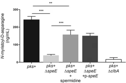
Considering that mature colibactin is not yet directly quantifiable, we took
advan-tage of the stability of the N-myristoyl-
D-asparagine moiety cleaved by the ClbP
peptidase in the late activation step of inactive precolibactin (3, 4) to indirectly access
the amount of colibactin produced, using LC-MS (36) (Fig. 7). Quantification of
N-myristoyl-
D-asparagine in culture supernatants revealed that the amount of colibactin
prodrug motif was drastically decreased in the ΔspeE mutant compared to the E. coli
DH10B pks
⫹strain, and partially restored by complementation with p-speE (Fig. 7).
FIG 5 Deletion of the spermidine synthase SpeE decreases colibactin autotoxicity in a ΔclbS mutant.
Enumeration of culturable bacterial cells in the stationary growth phase. The bacteria were pregrown in LB to reach an exponential growth (OD600⫽ 0.4). A total of 2 ⫻ 106bacteria/ml were then inoculated in LB and grown for 17 h before being plated on LB agar plates to determine CFU. The median and individual results of four independent experiments are shown.***, P⬍ 0.001, and **, P ⬍ 0.01, by 1-way ANOVA. NS, not significant.
FIG 6 Spermidine is involved in DNA cross-linking activity. DNA cross-linking was observed by
cultivat-ing the E. coli strains with linearized plasmid DNA. Two hundred nanograms of linearized pUC19 plasmid was added to 1⫻ 106, 2⫻ 106, or 4⫻ 106bacteria/well of wild-type (WT) E. coli strain DH10B pks⫹, the ΔclbA and ΔspeE mutants, and the complemented derivative. When indicated, 20g/ml of spermidine was added to the interaction medium during infection. DNA was then purified, loaded on agarose gel, and run under alkaline denaturing conditions. DNA with covalent interstrand cross-links is nondenatu-rable and displays delayed migration compared to denatured single-stranded DNA (lower band). This image is representative of three independent experiments.
on September 11, 2020 by guest
http://msphere.asm.org/
Supplementation of spermidine during growth increased the production of
N-myristoyl-
D-asparagine by the ΔspeE mutant (Fig. 7).
We tested whether spermidine altered clb gene expression by using clb reporter
strains previously designed in E. coli Nissle 1917 (37) (see Fig. S5 and Text S1 in the
supplemental material). We observed modifications of expression after both ΔspeE
mutation and spermidine supplementation, suggesting that spermidine can modify
expression of pks genes.
DISCUSSION
Our work demonstrates the role of spermidine in the production of the genotoxin
colibactin. We demonstrate that the endogenous spermidine biosynthesis pathway is
involved in colibactin-associated genotoxicity. As a ΔspeE mutant is impaired in its
genotoxic activity in acellular DNA cross-linking assay, a mechanism mediated by the
targeted eukaryotic cell (such as invasion) was ruled out. Spermidine is not necessary,
however, as traces of colibactin-related toxicity remain after speE mutation, as
demon-strated both by autotoxicity in the ΔspeE ΔclbS double mutant and DNA cross-link
formation at the highest bacterial concentration. Furthermore, N-myristoyl-
D-asparagine, a metabolite directly linked to colibactin synthesis, also undergoes
quan-titative alteration when the spermidine pathway is inactivated, showing a direct
involvement of spermidine in production of the toxin. Not only exogenous spermidine
but also other polyamines such as spermine and norspermidine can restore colibactin
biosynthesis in ΔspeE mutants. Noteworthy, the three polyamines that cause
comple-mentation share structural characteristics: i.e., 3 or more amine functions, 2 of which are
free at the ends. Proposed colibactin and precolibactin structures do not contain any
spermidine or spermidine-derived motif (3–12). Thus, polyamines may likely be
in-volved in colibactin synthesis with regard to monitoring its regulation. Precisely we
have observed changes in the expression of different clb genes related to speE mutation
or spermidine supplementation. Although the precise molecular mechanism remains
elusive, polyamines are known to be major regulators in E. coli gene expression,
especially at the translation step (38). Indeed, enhancement of mRNA and ribosome
interaction leads to increased translation of proteins that belong to the “polyamine
modulon” as it is called by the Igarashi and Kashiwagi team (38). Alternatively,
poly-amines could also act through facilitating transport or export of the genotoxin.
FIG 7 Spermidine is involved in colibactin biosynthesis. The colibactin prodrug motif N-myristoyl-D -asparagine, produced by the wild-type (WT) E. coli strain DH10B pks⫹, the ΔclbA and ΔspeE mutants, and the complemented derivative, was quantified by liquid chromatography-mass spectrometry (LC-MS). Bacteria were cultivated at 37°C for 18h in DMEM-HEPES, supplemented when indicated with 20g/ml of spermidine, and the levels of bacterial growth were similar under all conditions. The data presented in the graph were obtained from four biological replicates.***, P⬍ 0.001, and **, P ⬍ 0.01, by 1-way ANOVA. All bar graphs show mean values⫾ SEM.
Colibactin Synthesis Is Linked to Polyamine Metabolism
on September 11, 2020 by guest
http://msphere.asm.org/
Spermidine and polyamines have been described as important players in different
host-pathogen relationships. In the plague agent Yersinia pestis, for instance, an intact
putrescine-spermidine pathway is essential to achieve biofilm formation, which is in
turn important for transmission by fleas (39). A higher virulence linked to an increase
in the spermidine content exists in Shigella sp., a bacterium responsible for dysentery
and closely related to E. coli (40). During the pathoadaptive evolutionary changes in E.
coli that led to this pathogen, the speG gene became defective due to diverse
mutations. Spermidine accumulation in bacteria is a key factor for the invasion of
macrophages in the pathophysiology of Shigella sp. and entero-invasive E. coli (EIEC),
which share the same invasive process (40, 41). The production and response of colicin
E7, an E. coli bacteriocin, are also regulated by polyamines (42). Spermidine and
putrescine are indeed important for triggering toxin production after DNA damage
because polyamines are mediators of the SOS response that regulates colicin E7.
Exogenous spermidine decreases the susceptibility of E. coli to colicin E7, which can be
attributed to a downregulation of the transporters of the toxin by the polyamine (42).
Spermidine is highly linked to cancer, especially colorectal cancer. Polyamines are
associated with cell proliferation. Therefore, host cell polyamine content is upregulated
in colorectal cancer tissues. It relies on both an increase in polyamine production and
impaired catabolism (28). The host is not the only source of polyamine in the digestive
tract. Bacteria in gut microbiota also produce polyamines and are one of the main
sources of these compounds in the lower intestine (43). The diversity of the
microor-ganisms makes the metabolism and composition of the polyamine pool diverse (44,
45), and it can be even more complex as a result of collective pathways within
microorganisms as well as polyamines produced by eukaryotic cells (46). Food is
another source of polyamines in the gut, especially nutrients such as fruits, wheat
germs, cheeses, mushrooms, and nuts. (47). In our study, we demonstrated that
exogenous spermidine produced by another E. coli strain could support colibactin
biosynthesis by a spermidine mutant. This strongly suggests that in the gut, where
spermidine is abundant, colibactin-producing bacteria can take advantage of this
metabolite, whatever the origin of its production (microbiota, host, or food). Recently,
the external supply of polyamine spermidine was also suggested to extend life span
and to provide neuroprotective and cardioprotective properties, for example (48). To
date, the literature on these beneficial effects of spermidine has been limited to model
organisms and to epidemiological studies that link dietary uptake with longevity or
pathology. Clinical trials aimed at increasing the uptake of this polyamine or even the
administration of probiotics that enhance the microbial synthesis appear feasible (48).
However, whether spermidine supplementation in humans would increase tumor
growth or modify microbiota remains unknown and should be addressed in light of the
fact that a polyamine-enriched environment could promote and may even trigger the
production of the genotoxin colibactin. This reinforces the hypothesis of deleterious
synergy between microbiota and host cell metabolism during carcinogenesis.
MATERIALS AND METHODS
Bacterial strains, mutagenesis, and growth conditions. The bacterial strains used in this study are
listed in Table 1 and Table S1 in the supplemental material (Text S1). For genetic manipulations, E. coli strains were routinely grown at 37°C under shaking in 5 ml of lysogeny broth (LB Lennox; Invitrogen). Appropriate antibiotics were added to the medium when required (chloramphenicol, 25g/ml; kana-mycin, 50g/ml; carbenicillin, 50 g/ml). Inactivation of the genes speB, speC, speE, and speG was performed by using the lambda Red recombinase method (49) with pairs of primers presented in Table 2 and Table S2 in the supplemental material. Allelic exchanges were confirmed by PCR. For complemen-tation, the speE gene was PCR amplified using the primers speE_CompF and speE_CompR cloned into pSC-A-amp/kan using the StrataClone PCR kit (Agilent) (see p-speE in Table 1).
For the megalocytosis assay, genotoxicity quantification, DNA cross-linking assay, and
N-myristoyl-D-asparagine quantification, E. coli strains were pregrown overnight at 37°C with shaking in Dulbecco’s modified Eagle’s medium (DMEM)-HEPES (Gibco), a polyamine-free medium. Overnight cultures were then diluted 1:50 in DMEM-HEPES and grown until they reached an optical density at 600 nm (OD600) of 0.6 and then processed for experiments.
on September 11, 2020 by guest
http://msphere.asm.org/
For the autotoxicity assay, E. coli strains and derivatives were pregrown in LB to reach an exponential growth (OD600⫽ 0.4). A total of 2 ⫻ 106bacteria/ml were then inoculated in LB and grown for 17 h before being plated on LB agar plates to determine CFU counts.
Determining the megalocytosis and genotoxic effect induced by colibactin. The megalocytosis
and genotoxic effect induced by colibactin were determined as previously described (35). Briefly, HeLa cells were dispensed in a 96-well cell culture plate (5⫻ 103to 7.5⫻ 103cells/well) and incubated for 24 h. For bacterial infections, cell cultures were infected with a multiplicity of infection (number of bacteria per HeLa cell at the onset of infection) ranging from 100 to 400. Four hours postinoculation, cells were washed 3 times with Hanks balanced salt solution (HBSS) and incubated in cell culture medium with 200g/ml gentamicin until analysis.
For megalocytosis quantification, cells were incubated for 72h before protein staining with methyl-ene blue (1% wt/vol in 0.01 M Tris-HCl [pH 8.5]). The methylmethyl-ene blue was extracted with 01. N HCl. The quantification of staining was measured at OD660.
For H2AX phosphorylation quantification, cells were incubated for 3 h overnight before fixation with formaldehyde, permeabilization, and blocking, as previously described (35). Cells were then incubated for 2 h at room temperature with rabbit monoclonal anti-␥-H2AX antibody 9718 (1:200 [Cell Signaling Technology]). An infrared fluorescent IRDyeTM800CW-conjugated goat anti-rabbit secondary antibody (1:200 [Rockland]) was used to detect␥-H2AX. DNA was counterstained with RedDot2 (1:500 [Biotium]). DNA and␥-H2AX were visualized simultaneously using an Odyssey Infrared Imaging Scanner (Li-Cor Biosciences) with 680- and 800-nm channels. Relative fluorescence units for ␥-H2AX per cell (as determined by␥-H2AX divided by DNA content) were divided by untreated controls. Results were then divided by the mean ratio obtained for the wild-type strain after infection with the same infectious dose to determine percentage change in phosphorylation of H2AX levels relative to this reference strain.
DNA cross-linking assay. The assay was performed as previously described (14). Briefly, linearized
DNA was obtained by digesting pUC19 plasmid with BamHI (NEB). Purified linearized DNA was quantified and diluted to obtain a 200-ng/l stock solution. For bacterium-DNA interactions, 1 ⫻ 106to 6⫻ 106CFU were cultivated with linearized DNA for 4 h at 37°C without shaking. Following centrifugation for 5 min at 5,000⫻ g to pellet bacteria, the DNA present in the supernatants was purified using the PCR purification kit (Qiagen) according to the manufacturer’s recommendations.
Denaturing agarose gel was prepared by dissolving 1.0 g of agarose in 100 ml of a 100 mM NaCl and 2 mM EDTA solution (pH 8.0). The gel was then soaked (2 h) in an alkaline running buffer solution (40 mM NaOH and 1 mM EDTA [pH⬃12.0]). One hundred nanograms of each DNA sample was loaded on the agarose gel. The gel was run for 45 min at 1 V/cm and then 2 h at 2 V/cm. The gel was then neutralized for a total of 45 min in a 100 mM Tris (pH 7.4) buffer solution containing 150 mM NaCl, and the neutralizing solution was refreshed every 15 min. The gel was stained with GelRed for 20 min and revealed with UV exposure using the ChemiDoc imaging system (Bio-Rad).
N-Myristoyl-D-asparagine (colibactin prodrug motif) quantification by liquid chromatography-mass spectrometry. The colibactin prodrug motif was quantified as previously described (36). Briefly,
precultivated strains were grown in DMEM-HEPES at 37°C for 18 h under shaking (240 rpm). Supernatants of cultures were obtained by centrifugation of bacterial cells at 3,200⫻ g for 15 min and were filtered on 0.2-m-pore membranes. Each strain was cultured in triplicate (derived from three independent clones), and each supernatant was analyzed by liquid chromatography-tandem mass spectrometry (LC-MS/MS). Quantification experiments were performed with ultraperformance liquid chromatography high-resolution/heated electrospray ionization mass spectrometry (UPLC-HR/HESI-MS). The data were re-corded on a Thermo Scientific Q Exactive hybrid quadrupole-Orbitrap mass spectrometer coupled to a Dionex Ultimate 3000 UPLC. The following solvent gradient (A ⫽ H2O ⫹ 0.1% formic acid, B ⫽ acetonitrile⫹ 0.1% formic acid with B at 30% from0 to 1 min, 30 to 95% from 1 to 6 min, and 95% from 6 to 7 min at a flow rate of 0.5 ml/min) was used on a Phenomenex Kinetex 5-m EVO C18(50- by 2.1-mm) column at 30°C. The mass spectrometer was operated in positive-ionization mode at a scan range of 200 TABLE 2 Primers used in this study
Primer Sequence speB_P1 TACTGGCGTGCCGTTCGATATGGCCACTTCTGGTCGTGCGGTGTAGGCTGGAGCTGCTTC speB_P2 TAATAGCGCGATCGGAGGTCAGGCCGCCAATCACTGGCGTCATATGAATATCCTCCTTAG speB_F GCCTAACGACGCGGAAGG speB_R GTTTTACCCGTGCGCATCG speC_F CGCCATCTCCTTACATTCTCTC speC_R GTTGATTTTCGCTGGTTACTCC speE_P1 GGCATGAAACGCTACACGACCAGTTTGGGCAGTACTTTGGTGTAGGCTGGAGCTGCTT speE_P2 GATTGTAATAACGGCATTTCAGGCCAGAGGCGAGAAACATATGAATATCCTCCTTAG speE_F AGCTATTATGTTGCGCCCTT speE_R AGCCATGCAGTTTCAGTTTTT speE_CompF ATCTCGAGCCTTAAGCCTGCTTGCCTACG speE_CompR ATCTCGAGGTTTTGGCGTAGCAGATATCG speG_P1 GAAGCCTTTGTTGAACTCTCTGATCTGTATGATAAGCGTGTAGGCTGGAGCTGCTTC speG_P2 ATAGAGATTGAGAACGGTAAAGCCATAGTCCATTGCCATATGAATATCCTCCTTAG speG_F GCCGCGTTATTACCCCCTAA speG_R GGGTTTACACCATCAAAAATACGA Colibactin Synthesis Is Linked to Polyamine Metabolism
on September 11, 2020 by guest
http://msphere.asm.org/
to 500 m/z and a resolution of 35,000. The spray voltage was set to 3.5 kV, the S-lens to 35, the auxiliary gas heater temperature to 438°C, and the capillary temperature to 270°C. Absolute quantification was achieved by using a Schotten-Baumann reaction-derived N-myristoyl-L-asparagine (isomer of the
N-myristoyl-D-asparagine colibactin cleavage product) as a standard. Data were obtained from undiluted cell-free sample supernatants and analyzed for N-myristoyl-D-asparagine, and concentrations were calculated using Thermo Xcalibur 2.2 Quan Browser.
Statistical analysis. Statistical analyses were conducted using GraphPad Prism 6.01. The mean and
the standard error of the mean (SEM) are shown in the figures, unless otherwise stated. P values were calculated by a one-way analysis of variance (ANOVA) followed by a Bonferroni post hoc test. A P value of⬍0.05 was considered statistically significant.
SUPPLEMENTAL MATERIAL
Supplemental material for this article may be found at
https://doi.org/10.1128/
mSphere.00414-19
.
TEXT S1, DOCX file, 0.1 MB.
FIG S1, TIF file, 0.6 MB.
FIG S2, TIF file, 1.3 MB.
FIG S3, TIF file, 0.7 MB.
FIG S4, EPS file, 0.9 MB.
FIG S5, TIF file, 1.1 MB.
TABLE S1, DOCX file, 0.1 MB.
TABLE S2, DOCX file, 0.1 MB.
ACKNOWLEDGMENTS
We sincerely thank Michèle Boury and Jean Milhès for technical assistance and
Amplus (Lyon, France) for proofreading the American version of the manuscript.
This work was supported by the National French Cancer Institute
(INCA-PLBIO13-123) and the National French Institute of Health and Medical Research (INSERM). The
sponsors did not participate in the study design, data collection and interpretation, or
the decision to submit the work for publication.
REFERENCES
1. Nougayrède J-P, Homburg S, Taieb F, Boury M, Brzuszkiewicz E, Gottschalk G, Buchrieser C, Hacker J, Dobrindt U, Oswald E. 2006. Escherichia coli induces DNA double-strand breaks in eukaryotic cells. Science 313:848 – 851.https://doi.org/10.1126/science.1127059. 2. Putze J, Hennequin C, Nougayrède J-P, Zhang W, Homburg S, Karch H,
Bringer M-A, Fayolle C, Carniel E, Rabsch W, Oelschlaeger TA, Oswald E, Forestier C, Hacker J, Dobrindt U. 2009. Genetic structure and distribu-tion of the colibactin genomic island among members of the family Enterobacteriaceae. Infect Immun 77:4696 – 4703.https://doi.org/10 .1128/IAI.00522-09.
3. Brotherton CA, Balskus EP. 2013. A prodrug resistance mechanism is involved in colibactin biosynthesis and cytotoxicity. J Am Chem Soc 135:3359 –3362.https://doi.org/10.1021/ja312154m.
4. Bian X, Fu J, Plaza A, Herrmann J, Pistorius D, Stewart AF, Zhang Y, Müller R. 2013. In vivo evidence for a prodrug activation mechanism during colibactin maturation. Chembiochem Eur J Chem Biol 14:1194 –1197. https://doi.org/10.1002/cbic.201300208.
5. Brotherton CA, Wilson M, Byrd G, Balskus EP. 2015. Isolation of a metabolite from the pks island provides insights into colibactin biosynthesis and activity. Org Lett 17:1545–1548.https://doi.org/10 .1021/acs.orglett.5b00432.
6. Bian X, Plaza A, Zhang Y, Müller R. 2015. Two more pieces of the colibactin genotoxin puzzle from Escherichia coli show incorporation of an unusual 1-aminocyclopropanecarboxylic acid moiety. Chem Sci 6:3154 –3160.https://doi.org/10.1039/c5sc00101c.
7. Zha L, Wilson MR, Brotherton CA, Balskus EP. 2016. Characterization of polyketide synthase machinery from the pks island facilitates isolation of a candidate precolibactin. ACS Chem Biol 11:1287–1295.https://doi.org/ 10.1021/acschembio.6b00014.
8. Li Z-R, Li J, Gu J-P, Lai JYH, Duggan BM, Zhang W-P, Li Z-L, Li Y-X, Tong R-B, Xu Y, Lin D-H, Moore BS, Qian P-Y. 2016. Divergent biosynthesis yields a cytotoxic aminomalonate-containing precolibactin. Nat Chem Biol 12:773–775.https://doi.org/10.1038/nchembio.2157.
9. Zha L, Jiang Y, Henke MT, Wilson MR, Wang JX, Kelleher NL, Balskus EP. 2017. Colibactin assembly line enzymes use S-adenosylmethionine to build a cyclopropane ring. Nat Chem Biol 13:1063–1065.https://doi.org/ 10.1038/nchembio.2448.
10. Guntaka NS, Healy AR, Crawford JM, Herzon SB, Bruner SD. 2017. Struc-ture and functional analysis of ClbQ, an unusual intermediate-releasing thioesterase from the colibactin biosynthetic pathway. ACS Chem Biol 12:2598 –2608.https://doi.org/10.1021/acschembio.7b00479.
11. Xue M, Shine E, Wang W, Crawford JM, Herzon SB. 2018. Characterization of natural colibactin-nucleobase adducts by tandem mass spectrometry and isotopic labeling. Support for DNA alkylation by cyclopropane ring opening. Biochemistry 57:6391– 6394. https://doi.org/10.1021/acs.bio chem.8b01023.
12. Wilson MR, Jiang Y, Villalta PW, Stornetta A, Boudreau PD, Carrá A, Brennan CA, Chun E, Ngo L, Samson LD, Engelward BP, Garrett WS, Balbo S, Balskus EP. 2019. The human gut bacterial genotoxin colibactin alkylates DNA. Science 363:eaar7785. https://doi.org/10.1126/science .aar7785.
13. Bossuet-Greif N, Dubois D, Petit C, Tronnet S, Martin P, Bonnet R, Oswald E, Nougayrède J-P. 2016. Escherichia coli ClbS is a colibactin resistance protein. Mol Microbiol 99:897–908.https://doi.org/10.1111/mmi.13272. 14. Bossuet-Greif N, Vignard J, Taieb F, Mirey G, Dubois D, Petit C, Oswald E, Nougayrède J-P. 2018. The colibactin genotoxin generates DNA inter-strand cross-links in infected cells. mBio 9:e02393-17.https://doi.org/10 .1128/mBio.02393-17.
15. Cuevas-Ramos G, Petit CR, Marcq I, Boury M, Oswald E, Nougayrède J-P. 2010. Escherichia coli induces DNA damage in vivo and triggers genomic instability in mammalian cells. Proc Natl Acad Sci U S A 107: 11537–11542.https://doi.org/10.1073/pnas.1001261107.
16. Secher T, Samba-Louaka A, Oswald E, Nougayrède J-P. 2013. Escherichia coli producing colibactin triggers premature and transmissible senes-cence in mammalian cells. PLoS One 8:e77157.https://doi.org/10.1371/ journal.pone.0077157.
on September 11, 2020 by guest
http://msphere.asm.org/
17. Arthur JC, Perez-Chanona E, Mühlbauer M, Tomkovich S, Uronis JM, Fan T-J, Campbell BJ, Abujamel T, Dogan B, Rogers AB, Rhodes JM, Stintzi A, Simpson KW, Hansen JJ, Keku TO, Fodor AA, Jobin C. 2012. Intestinal inflammation targets cancer-inducing activity of the microbiota. Science 338:120 –123.https://doi.org/10.1126/science.1224820.
18. Cougnoux A, Dalmasso G, Martinez R, Buc E, Delmas J, Gibold L, Sauvanet P, Darcha C, Déchelotte P, Bonnet M, Pezet D, Wodrich H, Darfeuille-Michaud A, Bonnet R. 2014. Bacterial genotoxin colibactin promotes colon tumour growth by inducing a senescence-associated secretory phenotype. Gut 63:1932–1942.https://doi.org/10.1136/gutjnl-2013-305257.
19. Dalmasso G, Cougnoux A, Delmas J, Darfeuille-Michaud A, Bonnet R. 2014. The bacterial genotoxin colibactin promotes colon tumor growth by modifying the tumor microenvironment. Gut Microbes 5:675– 680. https://doi.org/10.4161/19490976.2014.969989.
20. Dejea CM, Fathi P, Craig JM, Boleij A, Taddese R, Geis AL, Wu X, DeStefano Shields CE, Hechenbleikner EM, Huso DL, Anders RA, Gi-ardiello FM, Wick EC, Wang H, Wu S, Pardoll DM, Housseau F, Sears CL. 2018. Patients with familial adenomatous polyposis harbor colonic bio-films containing tumorigenic bacteria. Science 359:592–597.https://doi .org/10.1126/science.aah3648.
21. Buc E, Dubois D, Sauvanet P, Raisch J, Delmas J, Darfeuille-Michaud A, Pezet D, Bonnet R. 2013. High prevalence of mucosa-associated E. coli producing cyclomodulin and genotoxin in colon cancer. PLoS One 8:e56964.https://doi.org/10.1371/journal.pone.0056964.
22. Raisch J, Buc E, Bonnet M, Sauvanet P, Vazeille E, de Vallée A, Déchelotte P, Darcha C, Pezet D, Bonnet R, Bringer M-A, Darfeuille-Michaud A. 2014. Colon cancer-associated B2 Escherichia coli colonize gut mucosa and promote cell proliferation. World J Gastroenterol 20:6560 – 6572.https:// doi.org/10.3748/wjg.v20.i21.6560.
23. Payros D, Dobrindt U, Martin P, Secher T, Bracarense A, Boury M, Laffitte J, Pinton P, Oswald E, Oswald IP. 2017. The food contaminant deoxyni-valenol exacerbates the genotoxicity of gut microbiota. mBio 8:e00007 -17.https://doi.org/10.1128/mBio.00007-17.
24. Tronnet S, Garcie C, Rehm N, Dobrindt U, Oswald E, Martin P. 2016. Iron homeostasis regulates the genotoxicity of Escherichia coli that produces colibactin. Infect Immun 84:3358 –3368.https://doi.org/10 .1128/IAI.00659-16.
25. Arthur JC, Jobin C. 2013. The complex interplay between inflammation, the microbiota and colorectal cancer. Gut Microbes 4:253–258.https:// doi.org/10.4161/gmic.24220.
26. Arthur JC, Gharaibeh RZ, Mühlbauer M, Perez-Chanona E, Uronis JM, McCafferty J, Fodor AA, Jobin C. 2014. Microbial genomic analysis reveals the essential role of inflammation in bacteria-induced colorectal cancer. Nat Commun 5:4724.https://doi.org/10.1038/ncomms5724.
27. Babbar N, Gerner EW. 2011. Targeting polyamines and inflammation for cancer prevention. Recent Results Cancer Res 188:49 – 64. https://doi .org/10.1007/978-3-642-10858-7_4.
28. Gerner EW, Meyskens FL. 2004. Polyamines and cancer: old molecules, new understanding. Nat Rev Cancer 4:781–792.https://doi.org/10.1038/ nrc1454.
29. Igarashi K, Kashiwagi K. 2010. Modulation of cellular function by poly-amines. Int J Biochem Cell Biol 42:39 –51.https://doi.org/10.1016/j.biocel .2009.07.009.
30. Murray Stewart T, Dunston TT, Woster PM, Casero RA. 2018. Polyamine catabolism and oxidative damage. J Biol Chem 293:18736 –18745. https://doi.org/10.1074/jbc.TM118.003337.
31. Goodwin AC, Shields CED, Wu S, Huso DL, Wu X, Murray-Stewart TR, Hacker-Prietz A, Rabizadeh S, Woster PM, Sears CL, Casero RA. 2011. Polyamine catabolism contributes to enterotoxigenic Bacteroides fragilis-induced colon tumorigenesis. Proc Natl Acad Sci U S A 108: 15354 –15359.https://doi.org/10.1073/pnas.1010203108.
32. Gerner EW, Bruckheimer E, Cohen A. 2018. Cancer pharmacoprevention: targeting polyamine metabolism to manage risk factors for colon cancer. J Biol Chem 293:18770 –18778. https://doi.org/10.1074/jbc .TM118.003343.
33. Tabor CW, Tabor H. 1985. Polyamines in microorganisms. Microbiol Rev 49:81–99.
34. Tabor CW, Tabor H, Xie QW. 1986. Spermidine synthase of Escherichia
coli: localization of the speE gene. Proc Natl Acad Sci U S A 83: 6040 – 6044.https://doi.org/10.1073/pnas.83.16.6040.
35. Martin P, Marcq I, Magistro G, Penary M, Garcie C, Payros D, Boury M, Olier M, Nougayrède J-P, Audebert M, Chalut C, Schubert S, Oswald E. 2013. Interplay between siderophores and colibactin genotoxin biosyn-thetic pathways in Escherichia coli. PLoS Pathog 9:e1003437.https://doi .org/10.1371/journal.ppat.1003437.
36. Garcie C, Tronnet S, Garénaux A, McCarthy AJ, Brachmann AO, Pénary M, Houle S, Nougayrède J-P, Piel J, Taylor PW, Dozois CM, Genevaux P, Oswald E, Martin P. 2016. The bacterial stress-responsive Hsp90 chap-erone (HtpG) is required for the production of the genotoxin colibactin and the siderophore yersiniabactin in Escherichia coli. J Infect Dis 214: 916 –924.https://doi.org/10.1093/infdis/jiw294.
37. Homburg S, Oswald E, Hacker J, Dobrindt U. 2007. Expression analysis of the colibactin gene cluster coding for a novel polyketide in Escherichia
coli. FEMS Microbiol Lett 275:255–262. https://doi.org/10.1111/j.1574 -6968.2007.00889.x.
38. Igarashi K, Kashiwagi K. 2018. Effects of polyamines on protein synthesis and growth of Escherichia coli. J Biol Chem 293:18702–18709.https:// doi.org/10.1074/jbc.TM118.003465.
39. Patel CN, Wortham BW, Lines JL, Fetherston JD, Perry RD, Oliveira MA. 2006. Polyamines are essential for the formation of plague biofilm. J Bacteriol 188:2355–2363.https://doi.org/10.1128/JB.188.7.2355-2363 .2006.
40. Barbagallo M, Di Martino ML, Marcocci L, Pietrangeli P, De Carolis E, Casalino M, Colonna B, Prosseda G. 2011. A new piece of the Shigella pathogenicity puzzle: spermidine accumulation by silencing of the speG gene. PLoS One 6:e27226.https://doi.org/10.1371/journal.pone .0027226.
41. Campilongo R, Martino MLD, Marcocci L, Pietrangeli P, Leuzzi A, Grossi M, Casalino M, Nicoletti M, Micheli G, Colonna B, Prosseda G. 2014. Molecular and functional profiling of the polyamine content in entero-invasive E. coli: looking into the gap between commensal E. coli and harmful Shigella. PLoS One 9:e106589.https://doi.org/10.1371/journal .pone.0106589.
42. Pan Y-H, Liao C-C, Kuo C-C, Duan K-J, Liang P-H, Yuan HS, Hu S-T, Chak K-F. 2006. The critical roles of polyamines in regulating ColE7 production and restricting ColE7 uptake of the colicin-producing Escherichia coli. J Biol Chem 281:13083–13091.https://doi.org/10.1074/jbc.M511365200. 43. Tofalo R, Cocchi S, Suzzi G. 2019. Polyamines and gut microbiota. Front
Nutr 6:16.https://doi.org/10.3389/fnut.2019.00016.
44. Hanfrey CC, Pearson BM, Hazeldine S, Lee J, Gaskin DJ, Woster PM, Phillips MA, Michael AJ. 2011. Alternative spermidine biosynthetic route is critical for growth of Campylobacter jejuni and is the dominant polyamine pathway in human gut microbiota. J Biol Chem 286: 43301– 43312.https://doi.org/10.1074/jbc.M111.307835.
45. Sugiyama Y, Nara M, Sakanaka M, Gotoh A, Kitakata A, Okuda S, Kurihara S. 2017. Comprehensive analysis of polyamine transport and biosynthe-sis in the dominant human gut bacteria: potential presence of novel polyamine metabolism and transport genes. Int J Biochem Cell Biol 93:52– 61.https://doi.org/10.1016/j.biocel.2017.10.015.
46. Johnson CH, Dejea CM, Edler D, Hoang LT, Santidrian AF, Felding BH, Ivanisevic J, Cho K, Wick EC, Hechenbleikner EM, Uritboonthai W, Goetz L, Casero RA, Pardoll DM, White JR, Patti GJ, Sears CL, Siuzdak G. 2015. Metabolism links bacterial biofilms and colon carcinogenesis. Cell Metab 21:891– 897.https://doi.org/10.1016/j.cmet.2015.04.011.
47. Ali MA, Poortvliet E, Strömberg R, Yngve A. 2011. Polyamines in foods: development of a food database. Food Nutr Res 55:5572.https://doi .org/10.3402/fnr.v55i0.5572.
48. Madeo F, Eisenberg T, Pietrocola F, Kroemer G. 2018. Spermidine in health and disease. Science 359:eaan2788.https://doi.org/10.1126/ science.aan2788.
49. Datsenko KA, Wanner BL. 2000. One-step inactivation of chromosomal genes in Escherichia coli K-12 using PCR products. Proc Natl Acad Sci U S A 97:6640 – 6645.https://doi.org/10.1073/pnas.120163297. 50. Johnson JR, Oswald E, O’Bryan TT, Kuskowski MA, Spanjaard L. 2002.
Phylogenetic distribution of virulence-associated genes among Esche-richia coli isolates associated with neonatal bacterial meningitis in the Netherlands. J Infect Dis 185:774 –784.https://doi.org/10.1086/339343. Colibactin Synthesis Is Linked to Polyamine Metabolism