ABSTRACT: A novel cyclodextrin derivative, i.e., permethy-lated-α-cyclodextrin (PM-α-CD), was used for removing glyc-erolipids from spinach thylakoid membranes and investigating their role in photosynthetic activities. A three-step selective re-moval of each lipid class was observed in treated membranes. Up to a concentration of 4 mM, PM-α-CD (in the presence of 75 µg chlorophyll a+b/mL), PM-α-CD displayed a marked se-lectivity for anionic lipids [sulfoquinovosyldiacylglycerol (SQDG) and phosphatidylglycerol (PG)] in comparison with galactolipids. At this concentration, half of PG and SQDG were removed. Within this range of concentration, the volume re-sponse of treated thylakoids to variation of osmolarity, an indi-rect mean of verifying the structural integrity of the membrane, was not altered. Similarly, neither photosystem II (PSII) nor pho-tosystem I (PSI) activity was affected. In contrast, the low-tem-perature fluorescence ratio F695/F740 drastically diminished from 1.45 to about 0.7, essentially due to the decrease of PSII fluorescence. The results derived from the fast fluorescence rise expressed in the form of a spider suggest that the fraction of in-activated (non-QAreducing) reaction centers (RC) increases while the active (QAreducing) RC remained intact. Raising the concentration of PM-α-CD from 4 to 7 mM resulted in a pro-gressive but greater diminution of the galactolipid level than that of SQDG and PG. Within this concentration range, the in-tegrity of the membrane was not altered, nor was either PSII or PSI activity, whereas the F695/F740 ratio decreased to about 0.45 as well as the fraction of inactivated RC. At concentrations above 7 mM of PM-α-CD, the integrity of the membrane was impaired, resulting in a decrease of both electron transport ac-tivities. At all concentrations, PM-α-CD did not show any se-lectivity toward either the acyl chains of the lipid molecules or the molecular species of PG. The results are discussed in terms of the role of glycerolipids in thylakoid membrane function and the relationship of the chemical structure of PM-α-CD and its lipid removal capacity.
Paper no. L8865 in Lipids 37, 201–208 (February 2002).
Besides proteins, acyl lipids are major components of thy-lakoid membranes (TM) in higher plants. They consist of four classes: monogalactosyldiacylglycerol (MGDG), digalacto-syldiacylglycerol (DGDG), sulfoquinovodigalacto-syldiacylglycerol (SQDG), and phosphatidylglycerol (PG). Galactolipids (MGDG and DGDG) are prominent and account for about 80% of total glycerolipids. Moreover, they are characterized by an exceptionally high content of trienoic acid, mainly α-linolenic acid (encountered in all lipid classes) and, in the so-called 16:3 plants, such as spinach, hexadecatrienoic acid (found in MGDG only). SQDG is enriched in palmitic acid (35 mol%), whereas PG contains a unique FA, trans-∆3 -hexa-decenoic acid [16:1 (3t)] (1,2). The FA composition of these acyl lipids gives rise to a great number of molecular species (3,4).
Glycerolipids display a marked transmembrane distribu-tion in the TM. The outer monolayer is highly enriched in MGDG (5,6) and PG (7,8), whereas the inner one contains a high level of DGDG (5,6); this confirms the general sided-ness of TM components. In contrast, recent analyses of glyc-erolipids in different domains of the membrane (e.g., intact thylakoids, grana lamellae vesicles, central core of the ap-pressed region, grana margin vesicles, and stroma lamellae vesicles) reveal that the level of the four lipid classes, the na-ture of their acyl chains, and the main molecular species of PG are identical in the intact TM as well as in the four mem-brane domains (9).
The role of membrane lipids in the photosynthetic func-tion have been particularly difficult to study. Although sev-eral approaches have been proposed in the literature (for a re-view, see Ref. 10), there is no consensus about the results ob-tained. Among them, the enzymatic digestion of lipids (e.g., 5,6,8,11,12), the use of specific antibodies against lipids (e.g., Refs. 13–15), the preparation of subthylakoid fractions (16,17), and the use of mutants deficient in certain lipids (18) have been extensively studied. All these techniques have ad-vantages and drawbacks. For instance, the use of enzymatic digestion with lipolytic enzymes results in the concomitant accumulation of FFA that are known to have detergentlike properties and to displace lipids in the membrane (19,20). The use of specific antibodies against lipids is often questionable due to the difficult access of the antibody to all the lipids at the surface of the membrane, especially if they are located in membrane hollows. Although the preparation of subthylakoid fractions seemed to have been for a long time the best tech-nique to associate structure and function, its greatest draw-back comes from utilization of high concentrations of deter-*To whom correspondence should be addressed at Laboratoire de
Physiolo-gie végétale, Université de Neuchâtel, Rue Emile-Argand 13, CH-2007 Neuchâtel, Switzerland.
E-mail: [email protected]
Abbreviations: ABS, light absorption; CD, cyclodextrin; α-CD, α-cyclodex-trin; β-CD, β-cyclodextrin; γ-CD, γ-cyclodextrin; Chl, chlorophyll; CSo, foliar cross section; DGDG, digalactosyldiacylglycerol; 2,6-DM-β-CD, 2,6-dimethyl-β-cyclodextrin; ETo, photosystem II electron transfer; F695, fluo-rescence at 695 nm; F740, fluorescence at 740 nm; LHCII, light harvesting chlorophyll a/b protein complex; MGDG, monogalactosyldiacylglycerol; PG, phosphatidylglycerol; PLA2, phospholipase A2; PM-α-CD, permethy-lated-α-cyclodextrin; PSI, photosystem I; PSII, photosystem II; RC, active reaction center; SQDG, sulfoquinovosyldiacylglycerol; TM, thylakoid mem-branes; TRo, photon trapping.
Cyclodextrins: A Potential Tool for Studying the Role
of Glycerolipids in Photosynthetic Membranes
Sylvie Duchêne and Paul-André Siegenthaler*
gents. This explains why a few authors have tried to obtain sub-thylakoid particles or domains without detergent by using the aqueous two-phase system (9,19,21,22). Although the mutant approach has permitted study of the effect of the degree of lipid polyunsaturation on the structure and function of the chloro-plast membrane (10,18), it is extremely difficult to evaluate the side effects of specific mutations on lipid metabolism.
Recently, cyclodextrins (CD) were reported to have the ca-pacity to remove lipids from the TM (23) and display he-molytic properties (24). CD molecules are cyclic oligosaccha-rides consisting of 6 (α-CD), 7 (β-CD), or 8 (γ-CD) glucopyra-nose units linked by α (1–4) bonds. These compounds are not perfectly cylindrical molecules but rather are somewhat cone-shaped. They adopt a torus shape and are able to bind lipids within their hydrophobic cavity to form water-soluble guest-CD inclusion complexes (25–27). So far, guest-CD have been used mainly in the biomedical and pharmaceutical fields.
In a previous contribution, we established the conditions under which TM glycerolipids can be extracted in the presence of α-CD, β-CD, and dimethyl-β-cyclodextrin (DM-β-CD) (23). We found that the extent of lipid, protein, pigment, and plastoquinone removal depends on both CD and chlorophyll (Chl) concentrations. Simultaneous with the removal of lipids, a few photosynthetic functions were found to be altered.
In this investigation, we used a novel CD derivative, the permethylated-α-cyclodextrin (PM-α-CD). This compound was found to display properties different from those of the CD used previously. At low concentrations, it had the capac-ity of removing anionic lipids (SQDG and PG) without con-comitant galactolipid extraction. These properties allowed us to study the relationship between the level of anionic lipids and various photosynthetic functions. Preliminary results of this investigation were presented elsewhere (28,29).
EXPERIMENTAL PROCEDURES
Materials. All the CD used in this study were obtained from Fluka (Buchs, Switzerland). The PM-α-CD was made by Dr. S. Claude (Department of Organic Chemistry, University of Neuchâtel, Switzerland). All chemicals were high-purity products from Fluka or Sigma, and Percoll was provided by Pharmacia (Uppsala, Sweden). Spinach leaves were pur-chased from the local market. They were washed, stored at 4°C and used within 2 d.
Preparation of TM. Spinach leaves (150 g) were ground for 10 s in 375 mL of a grinding medium containing 330 mM sor-bitol, 30 mM MOPS-KOH (pH 7.8), 2 mM EDTA-Na2, and 0.15% BSA by using a Waring Blender. The mixture was fil-tered through four layers of cheesecloth and two layers of mira-cloth, and the filtrate was subsequently centrifuged at 1500 × g for 2 min. The supernatant was discarded by aspiration, and the intact chloroplast pellet was resuspended in the grinding medium (20 mL) and layered on two centrifuge swinging-bucket tubes containing 20 mL of Percoll 40% supplemented with the grinding medium without EDTA-Na2and BSA. After centrifugation (2500 × g, 10 min), the chloroplast pellet was
resuspended in a low-osmotic medium (36 mL) containing 10 mM Tricine-KOH (pH 7.8) and 2 mM EDTA-Na2for 2 min. The osmolarity was reajusted by adding back 4 mL of 3 M sor-bitol. After centrifugation at 13,400 × g for 5 min, the thylakoid pellets were resuspended in a washing medium containing 300 mM sorbitol and 10 mM Tricine-KOH (pH 7.8). Fifteen milli-liters of Percoll (5% Percoll in 300 mM sorbitol and 10mM Tricine-KOH pH 7.8) were injected slowly below the suspen-sion (20 mL) of thylakoids. The tubes were centrifuged at 20,000 × g for 10 min, and the thylakoid pellets were washed in the same medium at 13,500 × g for 5 min and then re-suspended in the same medium at a concentration of 2 mg Chl/mL.
Incubation of thylakoids in the presence of CD. All CD were solubilized in 300 mM sorbitol and 10 mM Tricine-KOH (pH 7.8). The concentration of the stock solutions was 10 mM. CD at the desired concentrations were added to the thylakoid preparation (50–75 µg Chl/mL) and incubated at 0°C for 20–30 min. At the end of the reaction, thylakoids were centrifuged at 14,500 × g for 10 min and resuspended in 300 mM sorbitol and 10 mM Tricine-KOH (pH 7.8). The con-centration of Chl was readjusted to 1 or 2 mg Chl/mL depend-ing on the fate of the preparation.
Lipid analyses. Total lipids were extracted by adding 4 mL chloroform/methanol (53:37, vol/vol) and 2 mL 0.5 M KCl to the thylakoid suspension (150 µL of thylakoids containing 1 mg Chl/mL). This resulted in a two-phase system. The lipids of the lower phase were separated by TLC with acetone/ toluene/water (91:30:8) as solvents (30). Each lipid class was then hydrolyzed and esterified with 5% H2SO4in methanol at 85°C for 1 h. The resulting FAME were separated and identi-fied by GC and the molecular species of PG determined ac-cording to Xu and Siegenthaler (4). Chl concentrations were determined as described by Bruinsma (31).
Photosynthetic activities. Uncoupled photosystem II (PSII) electron flow was measured polarographically at 20°C in the presence of red actinic light (29 mW/cm2) in a medium containing 30 mM sodium phosphate buffer (pH 6.5), 3 mM NaCl, 60 mM sucrose, 0.2 mM phenyl-p-benzoquinone, 2 mM NH4Cl, and thylakoids corresponding to 20 µg Chl/mL, using a Clark-type oxygen electrode (evolution of O2). Un-coupled photosystem I (PSI) electron flow was determined in a medium containing 40 mM sodium phosphate buffer (pH 7.4), 1 mM NaCl, 10 µM 3-(3,4-dichlorophenyl)-1,1-di-methylurea; 32 mM ascorbate, 0.3 mM, 0.12 mM methylvio-logen, 0.6 mM NaN3, 2 mM NH4Cl and thylakoids corre-sponding to 20 µg Chl/mL, using a Clark-type electrode (ab-sorption of O2) (8). Fluoresence studies were carried out with freshly prepared thylakoids resuspended in 300 mM sorbitol and 10 mM Tricine-KOH (pH 7.8). The low-temperature flu-orescence spectra of thylakoids (diluted 1:9 in 80% glycerol) were carried out according to Rawyler and Siegenthaler (23). The fluorescence transients OJIP were measured as described by Srivastava and Strasser (32).
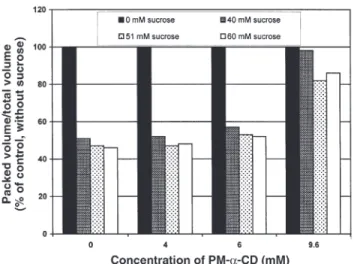
Response of thylakoids to osmolarity variation. The in-tegrity of thylakoid membranes during the removal of lipids
by CD was estimated by an osmolarity test (7,33). Thylakoids were treated with various concentrations of PM-α-CD (4,6, and 9.6 mM) as indicated previously. Then, each sample (2 mg Chl/mL), including the control without CD, was sus-pended in 40 mM Tricine-KOH (pH 8.0) and 3 mM MgCl2 and divided into four equal parts. Increasing concentrations of sucrose (40, 51, and 60 mM) were then added to each of the samples. Osmotic equilibrium was reached after 5 min, and then the thylakoid preparations were transferred to hemat-ocrit tubes and centrifuged at 23,000 × g (microcentrifuge; Christ, Zurich, Switzerland) for 5 min. The height of the thy-lakoid sediment in the tube was measured and compared to the total volume of the original suspension (ratio packed vol-ume/total volume, arbitrary units).
RESULTS
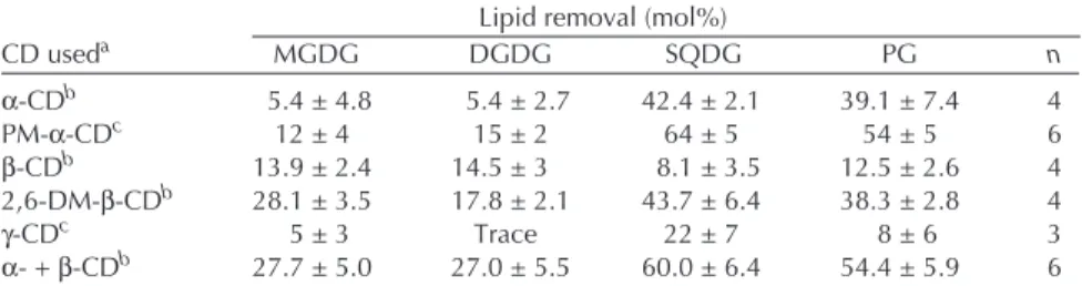
Several CD (α-, β-, γ-CD) and CD derivatives were compared regarding their lipid removal capacity and selectivity. The re-sults are presented in Table 1. α-CD [which contains six gly-copyranosidic units and presents an internal diameter of the cavity of 5.2 Å (27)] and its permethylated-α-derivative (PM-α-CD) displayed a much greater preference for SQDG and PG than for galactolipids. In contrast, β-CD [which contains seven glycopyranosidic units and presents an internal diame-ter of the cavity of 6.4 Å (27)] removed roughly 11 mol% of the four lipid classes. The derivative of β-CD (2,6-DM-β-CD) displayed a preference for the anionic lipids (SQDG and PG) over galactolipids but to a lesser extent than the α-CD and PM-α-CD. Other β-derivatives, such as permethylated-β-, carboxylated-β−, quaternary-β-, amphiphilic-β-, and sul-fated-β-CD, removed only lipids in trace amounts (data not shown). The selective capacity of γ-CD [which contains eight glycopyranosidic units and presents an internal diameter of the cavity of 8.3 Å (27)] was greater for SQDG than for all other lipid classes. But the amount of SQDG removed was rather low. A mixture of α- and β-CD removed about 30 mol% of galactolipids and 60 mol% of SQDG and PG but had no selective effect for one particular lipid class (Table 1). On
the basis of these results, we focused our attention on the ef-fect of PM-α-CD.
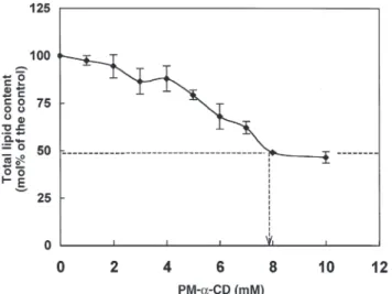
The results in Figure 1 show that the removability of total lipids in the thylakoid membrane increased as a function of the concentration of PM-α-CD up to 8 mM. At this concen-tration, 50 mol% of the total lipids were removed. A further addition of PM-α-CD had no effect.
Figure 2 illustrates the selective removal of each lipid class as a function of PM-α-CD concentration. Three steps can be distinguished: (i) Up to a concentration of 4 mM PM-α-CD, the content in MGDG remained constant, while that of DGDG decreased slightly. In contrast, the amount in anionic lipids decreased approximately by half of its original level. (ii) Raising the concentration of PM-α-CD from 4 to 8 mM resulted in a progressive but greater diminution of the galac-tolipid level than that of SQDG and PG. (iii) Higher concen-trations of PM-α-CD did not affect significantly the content of all four lipid classes. The concentration of PM-α-CD nec-essary to remove 50 mol% of each lipid was 8, 9.6, 3, and 3.5 mM for MGDG, DGDG, SQDG, and PG, respectively (see dotted lines in Fig. 2).
Figure 3 shows the relative content and composition of fatty acids in each lipid class remaining in the thylakoid mem-brane after a treatment with various concentrations of PM- α-CD. The PM-α-CD did not show any selectivity toward the acyl chains of the lipid molecules.
Spinach thylakoid membranes contain 10 PG molecular species (4). The three main molecular species [18:3/16:1 (3t); 18:3/16:0; 16:0/16:1 (3t)] represented 94.9 mol% of the total molecular species. The effect of PM-α-CD (0, 2, 5, and 9.6 mM) on their content in treated thylakoids was found to be 79.5 to 82.3 mol% for 18:3/16:1 (3t), 6.4 to 8.0 mol% for 18:3/16:0, and 3.2 to 5.6 mol% for 16:0/16:1 (3t). All these values presented no statistical difference between the control values and those of the different concentrations of PM-α-CD (individual data not shown). In conclusion, PM-α-CD did not show any selectivity toward the molecular species of PG, i.e., each molecular species was removed in the same proportion at each concentration of the CD.
TABLE 1
Comparison of the Removability of Lipid Classes in Thylakoids Treated by Various Cyclodextrins (CD)
Lipid removal (mol%)
CD useda MGDG DGDG SQDG PG n α-CDb 5.4 ± 4.8 5.4 ± 2.7 42.4 ± 2.1 39.1 ± 7.4 4 PM-α-CDc 12 ± 4 15 ± 2 64 ± 5 54 ± 5 6 β-CDb 13.9 ± 2.4 14.5 ± 3 8.1 ± 3.5 12.5 ± 2.6 4 2,6-DM-β-CDb 28.1 ± 3.5 17.8 ± 2.1 43.7 ± 6.4 38.3 ± 2.8 4 γ-CDc 5 ± 3 Trace 22 ± 7 8 ± 6 3 α- + β-CDb 27.7 ± 5.0 27.0 ± 5.5 60.0 ± 6.4 54.4 ± 5.9 6 aThylakoids were incubated with 5 mM of one of the cyclodextrins in a medium containing 300 mM
sorbitol and 10 mM Tricine-KOH (pH 7.8) at 0°C.
b50 µg chlorophyll/mL; 30 min incubation [results from Rawyler and Siegenthaler (23)].
c75 µg chlorophyll/mL; 20 min incubation. MGDG, monogalactosyldiacylglycerol; DGDG,
The effect of PM-α-CD on electron transport activity in spinach thylakoids is shown in Table 2. Up to a concentration of 7 mM PM-α-CD, neither PSII nor PSI activity was af-fected. However, a small stimulation of PSI activity at 5 mM PM-α-CD could be detected. At a concentration of 7 mM, 30 mol% of each galactolipid, 70 mol% of SQDG, and 60 mol% of PG were removed by this treatment (Fig. 2). A further in-crease of PM-α-CD (up to 9.6 mM) inhibited both activities,
although to different extents for PSII (35% inhibition) and PSI (56% inhibition). At this concentration, an additional treatment of the membrane with phospholipase A2(PLA2)
FIG. 1. Changes in the relative content of total lipids in thylakoid
mem-branes treated with various concentrations of permethylated α-cyclodextrin (PM-α-CD). The SD was calculated on the basis of six ex-periments (n = 6). The dotted line shows that 50 mol% of the total lipids was removed in the presence of 8 mM PM-α-CD. The 100% value cor-responded to 2600 ± 100 nmol/mg chlorophyll (Chl).
FIG. 2. Changes in the relative amount of individual diacyl lipid classes
(MGDG, DGDG, SQDG, PG) in thylakoid membranes treated with PM-α-CD. The standard deviation was calculated on the basis of nine ex-periments (n = 9). The dotted lines indicate the concentration at which PM-α-CD was necessary to remove 50 mol% of each lipid class. For each of the four lipid classes, the control amount, given in nmol lipid per mg Chl, was MGDG (1400), DGDG (707), SQDG (165), and PG (227). MGDG, monogalactosyldiacylglycerol; DGDG, digalactosyldia-cylglycerol; SQDQ, sulfoquinovosyldiadigalactosyldia-cylglycerol; PG, phosphatidyl-glycerol; for other abbreviation see Figure 1.
FIG. 3. Relative content and composition of FA in each lipid class
re-maining in the thylakoid membrane after treatment with PM-α-CD. Only the main FA of each lipid class are illustrated in this graph. This explains why some 100% values are not reached (e.g., DGDG and SQDG). The standard deviation was calculated on the basis of seven experiments (n = 7). The 100% values of the controls (0 mM PM-α-CD) corresponded in nmol per mg Chl to 1400 for MGDG, 707 for DGDG, 165 for SQDG, and 227 for PG. The 100% values for all the other his-tograms are different and can be calculated from the results of Figure 2. See Figures 1 and 2 for abbreviations.
further decreased PSII activity from 65 to 33% but had no ef-fect on PSI activity (Table 2).
It was important to verify the structural state of the mem-brane in PM-α-CD-treated thylakoids. This was achieved by testing the volume response of thylakoids to variations of the osmolarity (33). As shown in Figure 4, an increase in the os-molarity (from 49 to 109 Osmol) resulted in a decrease of about half of the packed volume of thylakoids. This occurred not only in the control samples but also in thylakoids treated with 4 and 6 mM PM-α-CD. However, in the presence of 9.6 mM PM-α-CD (Fig. 4) or 9.6 mM PM-α-CD + PLA2(data not shown), the osmotic response was nearly abolished.
Figure 5 shows the effect of various concentrations of PM-α-CD on low-temperature fluorescence. In the control thy-lakoid membrane, the ratio of fluorescence at 695 vs. 740 nm (F695/F740) was 1.45, indicating that the PSII fluorescence was greater than that of PSI. This ratio decreased progres-sively when the concentration of CD was raised. The extent
of this decrease was greater at low than at high concentrations of PM-α-CD. At a concentration of 4 mM, the ratio corre-sponded to half of the value of the original one. As shown in the inset of Figure 5, the decrease in the F695/F740 ratio was entirely due to the decrease of the fluorescence at 695 nm in the presence of PM-α-CD.
The results derived from the fast fluorescence rise (OJIP) (32,34) of CD-treated thylakoids are illustrated as a spider-plot presentation in Figure 6. This is a multiparametric de-scription of structure and function of each photosynthetic
TABLE 2
Effect of PM-α-cyclodextrin on Photosystems II and I Electron
Transport Activity in Spinach Thylakoids
PM-α-CD Photosystem II (PSII) Photosystem I (PSI) (mM) activity (%)a activity (%)b 0 100 100 2 97 ± 11 (n = 5) 98 ± 6 (n = 5) 5 95 ± 14 (n = 5) 132 ± 24 (n = 5) 7 92 ± 12 (n = 5) 104 ± 6 (n = 5) 9.6 65 ± 11 (n = 6) 44 ± 13 (n = 7) 9.6 + PLA2c 33 ± 6 (n = 2) 45 ± 3 (n = 2)
aPSII electron transport actitivy: 100% corresponded to 36 ± 12 micromol
O2/mg Chl × h (n = 7).
bPSI electron transport activity: 100% corresponded to 460 ± 90 micromol
O2/mg Chl × h (n = 7).
cPLA
2, phospholipase A2from porcine pancreas. After the permethylated-
α-cyclodextrin (PM-α-CD) treatment (20 min, 2°C), PLA2 (4 Boehringer
units/mg Chl) was added to the same reaction mixture and incubated at 20°C for 40 min.
FIG. 4. Osmotic response of thylakoids first pretreated by 4, 6, and 9.6
mM PM-α-CD, and then incubated in a medium containing 0, 40, 51, or 60 mM sucrose corresponding to 49, 89, 100, and 109 Osmol, re-spectively (other conditions as in the Experimental Procedure section). For abbreviation see Figure 1.
FIG. 5. Effect of lipid depletion by PM-α-CD on the low-temperature (77 K) fluorescence expressed as the ratio of the fluorescence at 695 nm (pho-tosystem II: PSII) to the fluoresence at 740 nm (pho(pho-tosystem I: PSI) in thy-lakoid membranes. Where indicated, the SD was calculated on the basis of six experiments (n = 6). For the other ones, n = 2. Inset: fluorescence spectra at 77 K of control sample (solid line) and PM-α-CD-treated (5 mM) thylakoids (dashed line). The shift of wavelength at 695 nm between the two samples is not significant. For abbreviation see Figure 1.
FIG. 6. Effect of lipid depletion by PM-α-CD on various parameters
characterizing the photochemistry of the thylakoid membranes; ABS, light absorption; TRo, photon trapping; ETo, PSII electron transfer; RC, active reaction center; CSo, foliar cross section; phi (Po) = TRo/ABS, yield of primary photochemistry. Symbols: ●●, 0 mM PM-α-CD; ■, 2 mM; ▲, 4 mM; x, 6 mM. The scale from 0 (center of the spider) to 1.5 corresponds to ratios of the various parameters of a treated sample and its control. For other abbreviation see Figure 1.
sample (control and PM-α-CD-treated thylakoids), presented by an octagonal line. This type of presentation facilitates the comparison of the effect of different treatments. The relative values (relative to the corresponding value of the control, which thus become equal to unity) of selected expressions, such as PSII electron transfer/cross-foliar section (ETo/CSo) and ETo/active reaction center (ETo/RC), etc., can be plotted using a spider-plot presentation. The fluorescence transients and the spider presentation are thoroughly described by Strasser et al. (34). On a sample basis (per CS), the light ab-sorption (ABS)/CSo (corresponding to the same amount of Chl) remained constant from 0 to 6 mM PM-α-CD, whereas the photon trapping (TRo)/CSo and electron transfers (ETo/CSo) decreased, in particular at 4 and 6 mM PM-α-CD. However, the specific trapping (TRo/RC) and, to a lesser ex-tent, electron transfers (ETo/RC) per reaction center were not affected up to about 6 mM. In contrast, the light absorption per active reaction center (ABS/RC) increased, especially from 0 to 4 mM PM-α-CD. In addition, the density of active reaction centers (RC/CSo) decreased steadily upon PM-α-CD addition, whereas the overall quantum yield phi (Po) de-creased only in the presence of the highest levels (4 to 6 mM) of CD.
DISCUSSION
The results of this investigation show that CD, in particular their permethylated derivative, PM-α-CD, offer interesting properties for removing glycerolipids from the TM. When comparing the lipid removal capacity of the different CD and derivatives (Table 1), we chose to investigate further the prop-erties of PM-α-CD for the following reasons: (i) This com-pound removed 50 to 70 mol% of anionic lipids (PG and SQDG) from the membrane (Fig. 2); (ii) at a concentration of 5 mM, it had almost no effect on galactolipids (Fig. 2); (iii) compared with α-CD, the permethylated derivative displays a greater cavity opening of the larger ring edge of the mole-cule, which might enhance its capacity for engulfing lipid molecules (35); (iv) compared with all β-CD and its deriva-tives, the α-CD and PM-α-CD have the advantage of display-ing a greater solubility in water (27); and (v) in contrast to de-tergent and enzymatic treatments, PM-α-CD, up to a concen-tration of 6 mM, had no deleterious effect on the TM (Fig. 4), thereby avoiding special protective precautions, e.g., using high concentrations of BSA to remove FFA resulting from lipid digestion (20). All these properties make PM-α-CD a suitable compound for studying lipid composition–function relationships in TM.
The first set of results shows that, up to a concentration of 4 to 5 mM, PM-α-CD displayed a marked selectivity for the re-moval of anionic lipids compared to galactolipids. Within this range of concentrations (Fig. 2), the structural integrity of the membrane was preserved (Fig. 4), and neither PSII nor PSI ac-tivity was affected (Table 2). For instance, 50% removal of both SQDG and PG did not impair PSII and PSI electron flow activities. This surprising finding can be explained in view of
earlier results concerning the effect of enzymatic digestion of membrane lipids by various phospholipase treatments (7,20). As is well established, the TM is constituted of two monolay-ers that display an asymmetric distribution of their lipids (2). For instance, the outer monolayer contains 70 mol% PG and the inner one, 30 mol%. On the other hand, the enzymatic digestion of all the PG located in the outer monolayer did not affect PSII and PSI activities, even after the removal of the prod-ucts (e.g., FA) resulting from the enzymatic digestion. In con-trast, the removal of a small amount of PG in the inner mono-layer drastically obliterates the activity of PSII (7).
Altogether, these experiments strongly suggest that the PG molecules that were removed by PM-α-CD originated from the outer monolayer only, since PSII and PSI activities were not altered. At a higher concentration of PM-α-CD (9.6 mM), half of both MGDG and DGDG molecules were removed from the TM by this treatment (Fig. 2). At this high concentration of PM-α-CD, the osmotic response was almost completely abol-ished, indicating that the structure of the TM was greatly im-paired. Interestingly, in vivo, similar results were found in an Arabidopsis mutant characterized by a defective MGDG syn-thase gene (MGD1). The amount of MGDG in this mutant is reduced by 42% compared with the wild type. The MGDG de-ficiency of the mgd1 mutant is correlated with striking defects in chloroplast ultrastructure (36). A similar observation was made in an Arabidopsis mutant dgd1 where DGDG lipid con-tent undergoes a 90% reduction compared to the wild type (37).
In contrast to electron flow activity, Chl fluorescence was the most sensitive photosynthetic parameter to be affected by a treatment of thylakoids with PM-α-CD. Indeed, the ratio F695/F740 decreased from 1.45 in the control to 0.4 in the pres-ence of 9.6 mM of PM-α-CD. Interestingly, the greatest de-crease in the ratio occurred only when anionic lipids were re-moved by the CD, i.e., at a concentration of 4 mM PM-α-CD, about 50% of SQDG and 50% of PG were removed, whereas the pools of MGDG and DGDG were essentially intact. The decrease in the ratio F695/F740 was mainly due to the diminu-tion of the fluorescence in PSII, thus suggesting that the re-moval of PG alters the organization of the light-harvesting Chl a/b protein complex (LHCII) (see Ref. 38 for a discussion of relations between the trimerization of LHCII, grana stacking, and light energy distribution in photosynthetic membranes).
These results also argue in favor of the existence of bulk and specific lipid molecules in the TM. The characteristics of these two types of molecules have recently been discussed (9,10). Thus, most of the PG and SQDG molecules that are removed from the outer monolayer at low concentrations of PM-α-CD (up to 4–5 mM) can be considered as being essen-tially bulk molecules since PSII and PSI electron flow activi-ties are not impaired. However, one cannot exclude that cer-tain specific anionic lipids, probably PG molecules, are also removed, thus altering the molecular organization of LHCII and its related fluorescence. At higher concentrations of PM-α-CD (>4 mM), the removal of only a few specific molecules of DGDG may change the conformation of LHCII proteins. Indeed, PG molecules have been reported to be essential in
the formation of LHCII trimers and its two- and three-dimen-sional crystallization. Furthermore, DGDG was found to bind to peripheral sites of LHCII (four molecules of DGDG per polypeptide) and to be crucial for the three-dimensional crys-tallization. Thus, specific PG and DGDG are both important molecules for the maintenance of the structure and function of LHCII (38–41). According to the spider-plot representa-tion, the electron transfer (beyond Q−A) decreased per cross section (ETo/CSo) and remained constant per active reaction center (ETo/RC) in PM-α-CD-treated TM. Thus, it is con-cluded that the fraction of reaction centers (RC) inactivated (inactive RC/active RC + inactive RC) by the removal of lipids increased while the active RC remained intact but de-creased in number. A few authors (14,15) have hypothesized how reaction centers can be inactivated. Certain bound SQDG and DGDG molecules are localized as prosthetic groups at the surface of the native D1/D2heterodimer, hold-ing the dimer together, whereas PG can be considered to be essential for the orientation and stabilization of the D1 -pro-tein. Ionic interactions and van der Waal’s forces in hy-drophobic pockets or clefts might be the principal binding system between D1and PG. For instance, after PLA2 treat-ment (14,15) or after removal of PG by PM-α-CD, the cleft could be closed and the function of the lipoprotein complex (D1(QB) and D2(QA) in the reaction center) obliterated. These changes might correspond to an increase of the frac-tion of inactivated reacfrac-tion centers.
The capacity of CD to engulf lipids is the result of several properties of not only the host (i.e., CD) but also the guest (i.e., lipids) molecules. Concerning the host molecule, the number of glucopyranose units, and the diameter of the cav-ity (26), the nature of the chemical groups (hydroxyl and methoxy groups) at the edges of the cavity (27,35), the polar-ity of each cyclic unit molecule, and the distribution of the charges, etc., are relevant factors in understanding the ability of CD to form water-soluble guest-CD inclusion complexes (27). On the other hand, the chemical properties of the guest molecules, such as the global charges of lipid molecules, the electrostatic properties of the lipid headgroups, and the na-ture of the acyl chains of the lipids (length and degree of un-saturation), are also determinative for generating this engulf-ing process (24). In our case, the proportion of removable lipids originating from a membrane should also depend on the specific characteristics of the membrane where lipids are in-serted, namely, the transmembrane distribution of lipids, the packing pressure of the membrane, and the accessibility of lipid molecules to CD. Although this investigation was not aimed at studying the host–guest interactions, our results may reveal information about this process.
First, let us consider the host characteristics: It has been reported that the permethylation of CD enlarges the whole cavity of the molecule (35). The methyl groups introduced into the O3position extend the O2–O3side of the cavity and make narrower the O6side (27), thus favoring the formation of inclusion complexes. Our results are in agreement with this observation (Table 1). The removal of all four classes of lipids was increased in the presence of PM-α-CD compared with
α-CD as well as in the presence of 2,6 DM-β-CD compared with the β-CD. When considering the guest molecule, the presence of negative charge in the molecule (SQDG and PG) allowed a better interaction with the host molecule (Table 1). Interestingly, Debouzy et al. (24) used liposomes (multi-lamellar vesicles) to study the overall sensitivity of different phospholipids for their interactions with α-CD. They found that among the most reactive phospholipids appeared PI and PS, which are both anionic lipids. However, PE, but not sphingomyelin and PC, which are characterized by a zero global charge, is also reactive toward α-CD. Obviously, the global charge of lipids appears not to be the only criterion in-fluencing the formation of the host-guest complex. On the other hand, the nature of the acyl chains of the anionic lipids is apparently not decisive for the recognition of the guest by the host (Fig. 3). An intriguing question concerns the high concentration of PM-α-CD (up to 5 mM) necessary to initiate the removal of galactolipids from the membrane. As dis-cussed by Bekers et al. (27), it may be possible that more than one molecule of the host is necessary to find complete accom-modation within the inclusion complex. In addition to that, the lack of global charges in galactolipids may require higher concentrations of PM-α-CD.
ACKNOWLEDGMENTS
This research was supported in part by the Swiss National Science Foundation (grants number 31,336,93,92 and 31,432,97,95). The au-thors thank very much Dr. Saturnin Claude for his generous gift of PM-α-CD and Dr. Reto Strasser (University of Geneva) for making his laboratory available for fluorescence transient analyses and for helping to interpret the fluorescence data. This work is part of a doc-toral program that was carried out by Sylvie Duchêne in the Labora-toire de Physiologie végétale, Université de Neuchâtel, Switzerland.
REFERENCES
1. Dubacq, J.P., and Trémolières, A. (1983) Occurrence and Func-tion of Phosphatidylglycerol Containing ∆3-trans-Hexadecenoic Acid in Photosynthetic Lamellae, Physiol. Vég. 21, 293–312. 2. Siegenthaler, P.A. (1998) Molecular Organization of Acyl
Lipids in Photosynthetic Membranes of Higher Plants, in Lipids in Photosynthesis: Structure, Function and Genetics (Siegen-thaler, P.A., and Murata, N., eds.), Vol. 6, pp. 119–144, Kluwer Academic Publishers, Dordrecht.
3. Nishihara, M., Yokota, K., and Kito, M. (1980) Lipid Molecular Species Composition of Thylakoid Membranes, Biochim. Bio-phys. Acta 617, 12–19.
4. Xu, Y.N., and Siegenthaler, P.A. (1996) Phosphatidylglycerol Molecular Species of Photosynthetic Membranes Analyzed by High-Performance Liquid Chromatography: Theoretical Con-siderations, Lipids 31, 223–229.
5. Rawyler, A., and Siegenthaler, P.A. (1985) Transversal Local-ization of Monogalactosyldiacylglycerol in Spinach Thylakoid Membrane, Biochim. Biophys. Acta 815, 287–298.
6. Rawyler, A., Unitt, M.D., Giroud, C., Davies, H., Mayor, J.P., Harwood, J.L., and Siegenthaler, P.A. (1987) The Transmem-brane Distribution of Galactolipids in Chloroplast Thylakoids Is Universal in a Wide Variety of Temperate Climate Plants, Pho-tosynth. Res. 11, 3–13.
7. Siegenthaler, P.A., Rawyler, A., and Smutny, J. (1989) The Phos-pholipid Population Which Sustains the Uncoupled Non-cyclic
Electron Flow Activity Is Localized in the Inner Monolayer of the Thylakoid Membrane, Biochim. Biophys. Acta 975, 104–111. 8. Duchêne, S., Smutny, J., and Siegenthaler, P.A. (2000) The
Topology of Phosphatidylglycerol Populations Is Essential for Sustaining Photosynthetic Electron Flow Activities in Thylakoid Membranes, Biochim. Biophys. Acta 1463, 115–120.
9. Duchêne, S., and Siegenthaler, P.A. (2000) Do Glycerolipids Display Lateral Heterogeneity in the Thylakoid Membrane? Lipids 35, 739–744.
10. Siegenthaler, P.A., and Trémolières, A. (1998) Role of Acyl Lipids in the Function of Higher Plants Photosynthetic Mem-branes, in Lipids in Photosynthesis: Structure, Function and Ge-netics (Siegenthaler, P.A., and Murata, N., eds.), Vol. 6, pp. 145–173, Kluwer Academic Publishers, Dordrecht.
11. Rawyler, A., and Siegenthaler, P.A. (1989) Change in the Mo-lecular Organization of Monogalactosyldiacylglycerol Between Resting and Functioning Thylakoid Membranes. Involvement of the CF0/CF1-ATP Synthetase, Biochim. Biophys. Acta 975, 283–292.
12. Siegenthaler, P.A., Sutter, J., and Rawyler, A. (1988) The Trans-membrane Distribution of Galactolipids in Spinach Thylakoid Inside-Out Vesicles Is Opposite to That Found in Intact Thy-lakoids, FEBS Lett. 228, 94–98.
13. Radunz, A. (1981) Application of Antibodies in the Analysis of Structural Configuration of Thylakoid Membranes, Ber. Deutsch. Bot. Ges. 94, 477–489.
14. Voss, R., Radunz, A., and Schmid, G.H. (1992) Binding of Lipids onto Polypeptides of the Thylakoid Membrane. I. Galac-tolipids and Sulpholipid as Prosthetic Groups of Core Peptides of the Photosystem II Complex, Z. Naturforsch. 47c, 406–415. 15. Kruse, O., and Schmid, G.H. (1995) The Role of
Phosphatidyl-glycerol as a Functional Effector and Membrane Anchor of the D1-Core Peptide from Photosystem-II Particles of the Cyanobac-terium Oscillatoria chalybea, Z. Naturforsch. 50c, 380–390. 16. Murata, N., Higashi, S.I., and Fujimura, K. (1990) Glycerolipids
in Various Preparations of Photosystem II from Spinach Chloro-plasts, Biochim. Biophys. Acta 1019, 261–268.
17. Trémolières, A., Dainese, P., and Bassi, R. (1994) Heterogenous Lipid Distribution among Chlorophyll-Binding Proteins of Pho-tosystem II in Maize Mesophyll Chloroplasts, Eur. J. Biochem. 221, 721–773.
18. Vijayan, P., Routaboul, J.M., and Browse, J. (1998) A Genetic Approach to Investigating Membrane Lipid Structure and Pho-tosynthetic Function, in Lipids in Photosynthesis: Structure, Function, and Genetics (Siegenthaler, P.A., and Murata, N., eds.), Vol. 6, pp. 263–285, Kluwer Academic Publishers, Dordrecht. 19. Henry, L.E.A., Mikkelsen, J.D., and Møller, B.L. (1983)
Pig-ment and Acyl Lipid Composition of Photosystem I and II Vesi-cles and of Photosynthetic Mutants in Barley, Carlsberg Res. Commun. 48, 131–148.
20. Siegenthaler, P.A., Smutny, J., and Rawyler, A. (1987) Involve-ment of Distinct Populations of Phosphatidylglycerol and Phos-phatidylcholine Molecules in Photosynthetic Electron-Flow Ac-tivities, Biochim. Biophys. Acta 891, 85–93.
21. Murphy, D.J., and Woodrow, I.E. (1983) Lateral Heterogeneity in the Distribution of Thylakoid Membrane Lipid and Protein Com-ponents and Its Implications for the Molecular Organization of Photosynthetic Membranes, Biochim. Biophys. Acta 725, 104–112. 22. Sundby, C., and Larsson, C. (1985) Transbilayer Organization of the Thylakoid Galactolipids, Biochim. Biophys. Acta 813, 61–67.
23. Rawyler, A., and Siegenthaler, P.A. (1996) Cyclodextrins: A New Tool for the Controlled Lipid Depletion of Thylakoid Membranes, Biochim. Biophys. Acta 1278, 89–97.
24. Debouzy, J.C., Fauvelle, F., Crouzy, S., Girault, L., Chapron, Y., Göschl, M., and Gadelle, A. (1998) Mechanism of α-Cy-clodextrin Induced Hemolysis. 2. A Study of the Factors Con-trolling the Association with Serine-, Ethanolamine-, and
Choline-Phospholipids, J. Pharm. Sci. 87, 59–66.
25. Szejtli, J. (1988) Topics in Inclusion Science, in Cyclodextrin Technology (Davies, J.E.D., ed.), pp. 1–306, Kluwer Academic Publishers, Dordrecht.
26. Szejtli, J. (1990) The Cyclodextrins and Their Applications in Biotechnology, Carbohydrate Polymers 12, 375–392.
27. Bekers, O., Uijendaal, E.V., Beijnen, J.H., Bult, A., and Under-berg, W.J.M. (1991) Cyclodextrins in the Pharmaceutical Field, Drug Dev. Indus. Pharm. 17, 1503–1549.
28. Duchêne, S., Strasser, R.J., Srivastava, A., and Siegenthaler, P.A. (1998) Cyclodextrins as a Tool for Studying the Role of Glycerolipids in Spinach Thylakoid Membranes, in Proceedings of the 10th International Congress on Photosynthesis, Budapest, Hungary (Garab, G., ed.), Kluwer Academic Publishers, Dordrecht.
29. Duchêne, S. (1998) Les Glycérolipides: Organisation Spatiale et Fonctions dans les Membranes Photosynthétiques de l’Epinard. Etude à l’Aide de Vésicules Inversées et de Cyclodextrines, Ph.D. thesis, University of Neuchâtel, pp. 1–118.
30. Xu, Y.N., and Siegenthaler, P.A. (1996) Effect of Non-Chilling Temperature and Light Intensity During Growth of Squash Cotyledons on the Composition of Thylakoid Membrane Lipids and Fatty Acids, Plant Cell Physiol. 37, 471–479.
31. Bruinsma, J. (1961) A Comment on the Spectrophotometric De-termination of Chlorophyll, Biochim. Biophys. Acta 52, 576–578.
32. Srivastava, A., and Strasser, R.J. (1996) Stress and Stress Man-agement of Land Plants During a Regular Day, J. Plant Physiol. 148, 445–455.
33. Nobel, P.S. (1970) Osmotic Responses of Chloroplasts, in Plant Cell Physiology, A Physicochemical Approach, pp. 56–57, W.H. Freeman and Company, San Francisco.
34. Strasser, R.J., Srivastava, A., and Tsimilli-Michael, M. (2000) The Fluorescence Transient As a Tool to Characterize and Screen Photosynthetic Samples, in Probing Photosynthesis: Mechanisms, Regulation, and Adaptation (Yunus, M., Pathre, U., and Mohanty, P., eds.), pp. 445–483, Taylor & Francis, Lon-don and New York.
35. Reinhardt, R., Richter, M., and Mager, P.P.(1996) Investigation of the Conformational Behaviour of Permethylated Cyclodex-trins by Molecular Modelling, Carb. Res. 291, 1–9.
36. Jarvis, P., Dörmann, P., Peto, C.A., Lutes, J., Benning, C., and Chory, J. (2000) Galactolipid Deficiency and Abnormal Chloro-plast Development in the Arabidopsis MGD Synthase 1 Mutant, Proc. Natl. Acad. Sci. USA 97, 8175–8179.
37. Dörmann, P., Balbo, I., and Benning, C. (1999) Arabidopsis Galactolipid Biosynthesis and Lipid Trafficking Mediated by DGD1, Science 284, 2181–2184.
38. Trémolières, A., and Siegenthaler, P.A. (1998) Reconstitution of Photosynthetic Structures and Activities with Lipids, in Lipids in Photosynthesis: Structure, Function, and Genetics (Siegenthaler, P.A., and Murata, N., eds.), Vol. 6, pp. 175–189, Kluwer Academic Publishers, Dordrecht.
39. Nussberger, S., Dörr, K., Wang, D.N., and Kühlbrandt, W. (1999) Lipid-Protein Interaction in Crystals of Plant Light-Harvesting Complex, J. Mol. Biol. 234, 347–356.
40. Kühlbrandt, W., Wang, D.N., and Fujiyoshi, Y. (1994) Atomic Model of Plant Light-Harvesting Complex by Electron Crystal-lography, Nature 367, 614–621.
41. Peterman, E.J.G., Hobe, S., Calkoen, F., van Grandelle, R., Paulsen, H., and van Ameronges, H. (1996) Low-Temperature Spectroscopy of Monomeric and Trimeric Forms of Reconsti-tuted Light-Harvesting Chlorophyll a/b Complex, Biochim. Bio-phys. Acta 1273, 171–174.
[Received July 2, 2001, and in revised form November 2, 2001; revision accepted November 9, 2001]