If You Don
’t Look, You Won’t See: Intravital Multiphoton
Imaging of Primary and Metastatic Breast Cancer
Laura Bonapace&Jeffrey Wyckoff&Thomas Oertner& Jacco Van Rheenen&Tobias Junt&
Mohamed Bentires-Alj
Received: 14 March 2012 / Accepted: 25 April 2012 / Published online: 12 May 2012 # Springer Science+Business Media, LLC 2012
Abstract A fundamental hallmark of cancer is progression to metastasis and the growth of breast cancer metastases in lung, bone, liver and/or brain causes fatal complications. Unfortunately, the cellular and biochemical mechanisms of the metastatic process remain ill-defined. Recent application of intravital multiphoton microscopy (MP-IVM) to image fluorescently labeled cells in mouse models of cancer has allowed dynamic observation of this multi-step pro-cess at the cellular and subcellular levels. In this article, we discuss the use of MP-IVM in studies of breast cancer metastasis, as well as surgical techniques for exposing tumors prior to imaging. We also describe a versatile multiphoton microscope for imaging tumor-stroma interactions.
Keywords Multiphoton microscopy . Intravital imaging . Breast cancer . Metastasis
Abbreviations
MP-IVM Multiphoton intravital microscopy MPM Multiphoton microscopy
SHG Second harmonic generation PMT Photomultiplier tube
FLIM Fluorescence-lifetime imaging microscopy FRET Fluorescence resonance energy transfer
Introduction
Metastases are the main cause of death in breast cancer patients[1]. In metastasis, cancer cells from the primary tumor migrate to and enter the blood stream directly, or indirectly through the lymphatic system. Surviving cancer cells transported to distant sites may then seed metastases. Metastatic spread and growth depend on properties of the cancer cell, its anatomical microenvironment at the primary site (the cancer stroma, i.e., fibroblasts, immune and vascu-lar cells and the extracelluvascu-lar matrix), and characteristics of the site of metastasis[2–4]. Primary tumor end-stage meas-urements (i.e., tumor volume) or classical biochemical assays provide a static“snapshot” of a large population of cancer cells, thereby obscuring the adaptive properties of the few neoplastic cells with the capacity to escape from the primary tumor, survive and adapt to changing environments at distant metastatic sites. In contrast, intravital imaging allows the study of metastatic cancer cells within the large population of non-metastasizing cells[5–7].
The application of intravital multiphoton microscopy (MP-IVM) allows real-time observation of many pathological pro-cesses in four dimensions (x, y, z and time) and at a single-cell level, including (a) tumor initiation and growth, (b) interaction of cancer cells and the tumor stroma at the primary site, (c) intra- and extravasation, (d) functional crosstalk between metastatic cells and the stroma at the metastatic site, (e) responses of cancer cells to chemotactic factors, and (f) responses of cancer cells to anti-cancer treatments[7,8]. Sev-eral tools developed for visualizing changes in cellular pro-cesses (e.g., cell death, proliferation, migration, and fate) or in
L. Bonapace
:
J. Wyckoff:
T. Oertner:
M. Bentires-Alj (*) Friedrich Miescher Institute for Biomedical Research (FMI), Maulbeerstr. 66,4058 Basel, Switzerland e-mail: bentires@fmi.ch L. Bonapace
:
T. JuntNovartis Institutes for Biomedical Research, Basel, Switzerland
J. Van Rheenen
Hubrecht Institute-KNAW and University Medical Center Utrecht, Utrecht, The Netherlands
signaling pathways (e.g., enzymatic activity) have been used successfully in MP-IVM experiments[7–9] and have led to new insights in cancer research[4,7,10,11]. For example, use of a chemotaxis-based in vivo invasion assay and multiphoton microscopy has provided the first direct evidence of a para-crine loop between macrophages and tumor cells, which is involved in the intravasation of breast tumor cells[7,12].
There are at least five requirements for MP-IVM exam-ination of mammary cancer: (a) a mouse model of mammary cancer[13] (e.g., immunocompetent transgenic mice, trans-planted tumor cells in immunodeficient mice); (b) techni-ques for surgical exposure and immobilization of the tumor such that perfusion of the tissue remains intact; (c) fluores-cent labels for the tumor and/or the stroma; (d) a multipho-ton microscope suitable for intravital imaging; and (e) an image-processing software for the analysis of dynamic recordings[8, 14,15]. In the present article, we describe a versatile, custom-built modular multiphoton microscope for imaging tumor-stroma interactions at both primary and metastatic sites. We also compare surgical techniques for exposing the tumor prior to imaging.
Multiphoton Microscope
Confocal and multiphoton microscopes have both been used to image mammary tumors in living mice[16–19]. In confocal microscopy, single photons of the visible spectrum excite fluorophores from the electronic ground state to an excited
state and light is emitted as these molecules return to the ground state (Fig.1a). This excitation takes place throughout a broad focal volume and to make the optical section appear crisp, out-of-focus light is shielded from the detector by a pinhole. In multiphoton microscopy (MPM), fluorophores are excited when they simultaneously absorb two or more low-energy photons of the infrared spectrum generated by a highly focused short laser pulses (Fig.1a). At the focal plane, the density of the photons is such that two or more photons interact concurrently with the same fluorophore with high probability. Since simultaneous absorption of two photons by fluorophores is restricted to the focal plane, this thwarts out-of-focus excitation and emission[20] and, thus, a crisp optical section is obtained without the need for a pinhole. Because of the focal excitation of fluorophores, emitted light does not travel back through the optical path of the scanner, with result-ing losses of emission light at each optical element. Instead, emitted fluorescence is detected close to the objective, which greatly increases sensitivity. The localization of excitation to the focal plane in MPM offers two additional advantages making MP-IVM superior to confocal microscopy for imaging of multi-cellular tissues (e.g., mammary tumors, Fig.1b). Pho-tobleaching and photodamage[18,21] are reduced and, since there is no out-of-focus absorption, more excitation light can penetrate the tissue to a greater depth. Furthermore, low-energy photons of the infrared light used in MP-IVM penetrate up to 1,000μm in tissue, as compared to only <100 μm for the high-energy photons of visible light used in confocal microscopy. A further advantage of excitation with pulsed, coherent infrared
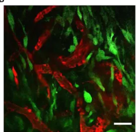
Figure 1 Jablonski energy diagram and a MP-IVM image of a mouse mammary tumor. a Energy diagram showing absorption of a single photon (1P, blue arrow) or the simultaneous absorption of 2 lower-energy photons (2P, red arrows), which trigger excitation of the fluo-rophore. After either of the excitation processes, part of the energy is dissipated as heat (grey arrow) and the rest is emitted as fluorescence (black arrow). b MP-IVM merged images of the murine breast cancer cell line 4 T1 expressing EGFP orthotopically injected into the fourth
mammary gland of Balb/c mice. A mammary imaging window was implanted surgically when the tumor was palpable (3 days after injec-tion). Two days after surgery, Texas Red dextran was injected i.v. to visualize the blood circulation (red signal), and an image of tumor cells (green signal) was acquired through the mammary imaging window. The image is a single optical section taken from a 100μm z-stack using a 20X (N.A. 0.95) objective at wave length 920 nm. Scan images of the tissue were acquired at a 5μm increment. Scale bar represents 50 μm
light is the formation of second harmonic generation (SHG) signals at fibrous structures such as collagen, which allows the visualization of extracellular matrix components in the tumor microenvironment.
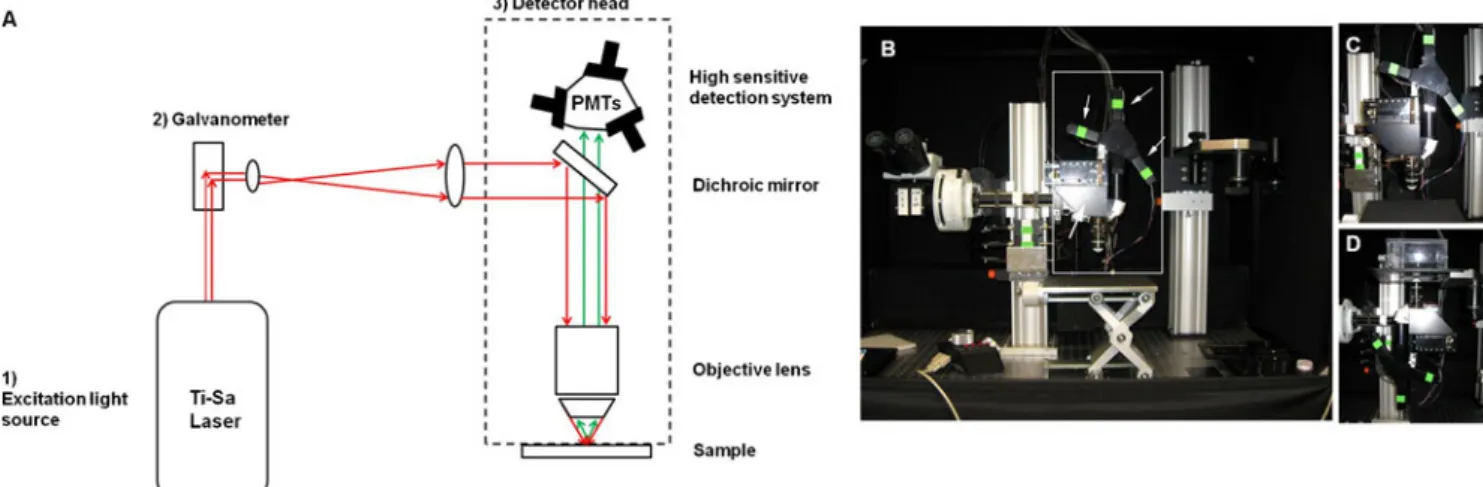
Typical MP-IVM consists of three basic components: (a) an excitation light source (i.e., a pulsed infrared laser); (b) a fluorescence microscope containing galvanometer-driven mir-rors for scanning of the tissue line by line; (c) a highly sensitive detection system of photomultiplier tubes (PMTs) (Fig.2a).
In our own research, we face the challenge of being able to image both orthotopic mammary tumors and metastatic sites such as lymph nodes or bone marrow. An upright microscope with an objective located above the specimen is more conve-nient for imaging the skull bone marrow[22] than an inverted microscope with an objective below the specimen. However, an upright microscope is not suitable for imaging orthotopic mammary tumors or the inguinal lymph node, because the mouse would have to rest on its back and movement caused by breathing would result in unacceptable z-drift of the images. To meet this challenge, we built a microscope ab initio based on a previously published design[23], using a commercially available kit from Sutter Inc (Fig.2b–d). Our MPM has a rotating detector head for optimal imaging of bone marrow (with an upright head, Fig.2c) or orthotopic mam-mary tumors and inguinal lymph nodes (with an inverted head, Fig.2d). In order to switch between upright and inverted configurations without laser realignment, the axis of rotation of the detector head is around the laser beam. The laser light (Mai Tai Millennia, Newport Spectra physics) passes first through a galvanometer-driven scanhead, which allows for line-scanning acquisition of images. The light then passes
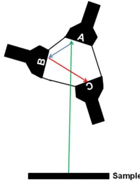
through a dichroic mirror posted in the detector head of the microscope at 45° relative to the light path and is reflected through the objective to the sample. This setup permits rota-tion of the detector head without changing the light path from the laser to the objective. For detection of the emitted fluores-cence, we use three separate PMTs (Hamamatsu photomulti-plier tubes R6357 multialkali) (Fig.3). Each PMT contains a bandpass filter that collects a specific wavelength and has an 18° angle of reflectance (Chroma, Bellows Falls, Vt., USA) to send remaining light to the next PMT. Signals from the microscope are acquired and converted to real-time images by a custom-made algorithm based on Matlab (MathWorks, Natick, Mass. USA), before being analyzed offline.
Surgical Preparation of Mice for Imaging of Primary Tumors and Metastases
As tumor growth and metastasis are slow processes, it is best to use surgical techniques that allow repeated imaging sessions with the same animal over several days. The skin-flap technique and a dorsal skinfold chamber were first used for exposing and repetitively imaging tumors[24–27]. The skin-flap technique involves a small incision to remove the skin and directly expose the tumor for imaging. While this method is suitable for single imaging sessions of ~6–24 h[7, 16, 17], some researchers have closed and then re-opened wounds for multiple imaging sessions, although this presents the risk of tissue inflammation[7,27]. In contrast, the dorsal skinfold chamber was invented as a set-up dedi-cated to repeated imaging sessions. The chamber is
Figure 2 Schematic of the three basic components of an MPM and an image of our versatile custom-built microscope. a Schematic of an MPM. As an excitation light source, we use [1] a femtosecond titanium-sapphire (Ti-Sa) laser with a tuning range of 700–1,040 nm. From the laser, light (red arrows) travels to [2] the galvanometer-driven scan mirrors. Next, the laser reaches [3] the detector head where it hits a dichroic mirror. This reflects the excitation light through the objective towards the sample. The detection system used for collecting
fluorescence emitted by the sample (green arrows) is composed of photodetectors, including photomultiplier tubes (PMTs). b Overview of our custom-built MPM. The box outlined in white represents the detector head, the thin arrows indicate the PMTs and the bold arrow shows the dichroic mirror. c Image of the detector head in the upright position (e.g., used for imaging bone marrow). d Image of the detector head in the inverted position for imaging mammary tumors and ingui-nal lymph nodes
essentially a metal frame with a glass window implanted on the back of the animal. A disadvantage of the dorsal skinfold chamber has been that mammary cells are grown in an ectopic site and, therefore, only cell line-based tumor models can be used. Moreover, the tumor volume is restricted by the size of the chamber and loss of heat is another disad-vantage of dorsal skinfold chamber[28]. However, the recent development of a mammary imaging window implanted ventrally, on top of orthotopic xenografts or genetically-induced tumors, extends the time and the frequency of imaging of the same animal and eliminates heat dispersion and the risk of an inflammatory reaction upon prolonged imaging[29].
As yet, metastatic sites such as lymph nodes and bone marrow can be imaged only for single imaging sessions of ~6–8 h and not longitudinally in the same mouse, as imag-ing windows for these organs do not exist. While imagimag-ing windows for the brain are available and imaging windows for the calvarial bone marrow seem feasible, other meta-static sites (e.g., liver) are not currently accessible. Imaging windows for the lung were recently developed but are not suitable for repetitive imaging[30,31].
Not only advanced surgery but also the development of new fluorophores should provide new insights into the metastatic process. To visualize orthotopically injected mammary cancer cells and track the same cells over several imaging sessions, photo-switchable fluorophores have been used[29]. For example, the fluorophore Dendra2 can be photoconverted irreversibly from green to red fluorescence upon blue-light radiation, and has been used successfully for quantifying and comparing the metastatic behavior of cells in different microenvironments[29].
What is Next?
Most studies using MP-IVM imaging have described early steps of the metastatic process within the primary site such as stroma-tumor interaction, cell migration and intravasation. In contrast, little is known about the behavior of breast cancer cells at metastatic sites in the lungs, lymph nodes, liver or bone marrow, which are not easy to expose surgically or to subject to long-term imaging. It is hoped that future development of two photon micro-endoscopy and of novel imaging windows will allow metastatic site observations in four dimensions.
Short-pulse infrared lasers allowing measurement of the fluorescent lifetime (FLIM) of fluorophores and, thus, the discrimination of multiple fluorophores with the same color can multiply the number of imaging channels by at least a factor of three. FLIM has been used successfully to discrimi-nate autofluorescence of healthy and tumorigenic tissues[5]. Furthermore, the technique can be used to measure changes in fluorescence resonance energy transfer (FRET) of FRET-biosensors, allowing the quantification of second messengers (e.g., calcium), the activation status of signaling pathways (e.g., PLC), as well as the extent of protein-protein interactions and further cellular processes (e.g., apoptosis) in a single tumor cell[5]. In addition, the probes and biosensors now in devel-opment for assessing signaling pathways in situ will allow fidelitous assessment of changes in signaling cascades within cancer cells following interaction with the tumor stroma and/or in response to therapies. This will ultimately increase our understanding of the metastatic process and hopefully lead to the identification of novel therapeutic targets in breast cancer metastasis.
Acknowledgments We thank J. Rietdorf and S. Bundschuh (FMI) for helping us set up the MPM and R. Friedrich (FMI) and J. Stein (Theodor Kocher Institute, Bern) for helpful discussions. We thank A. de Graaff and the Hubrecht Imaging Center for their support. Research in the lab of JvR is supported by VIDI fellowships (91710330), equipment grants (175.010.2007.00) and (834.11.002) from the Dutch Organization of Scientific Research (NWO), and a grant from the Dutch Cancer Society (KWF: HUBR 2009–4621). Research in the lab of M.B-A. is supported by the Novartis Research Foundation, the European Research Council (ERC starting grant 243211-PTPsBDC), the Swiss Cancer League, and the Krebsliga Beider Basel.
Figure 3 PMT design for the rotating detector head microscope. Schematic of the three PMTs arranged in a triangle. There is no beam splitter inside and the emission beam is reflected by a system of filters with specific bandpasses that are positioned in front of each PMT with an 18° angle of reflectance. The emitted light (green arrow) encounters the first PMT (A), which is optimized to allow transmittance of light in the range of fluorophores such as GFP (500-550 nm). The light is then reflected (blue arrow) to the second PMT (B), which is optimized for light in the blue spectrum (435-485 nm) and used for SHG detection and CFP. The rest of the emitted spectrum (red arrow) goes to the third PMT (C), which is optimized for light in the red spectrum (573-647 nm), exciting fluorophores such as mRFP and Texas red
References
1. Valastyan S, Weinberg RA. Tumor metastasis: molecular insights and evolving paradigms. Cell. 2011;147(2):275–92.
2. Fidler IJ. The pathogenesis of cancer metastasis: the‘seed and soil’ hypothesis revisited. Nat Rev Cancer. 2003;3(6):453–8.
3. Nguyen DX, Bos PD, Massague J. Metastasis: from dissemination to organ-specific colonization. Nat Rev Cancer. 2009;9(4):274–84. 4. Friedl P, Alexander S. Cancer invasion and the microenvironment:
plasticity and reciprocity. Cell. 2011;147(5):992–1009.
5. Beerling E, Ritsma L, Vrisekoop N, Derksen PW, van Rheenen J. Intravital microscopy: new insights into metastasis of tumors. J Cell Sci. 2011;124(Pt 3):299–310.
6. Zomer A, Beerling E, Vlug EJ, van Rheenen J. Real-time intravital imaging of cancer models. Clin Transl Oncol. 2011;13(12):848–54. 7. Condeelis J, Weissleder R. In vivo imaging in cancer. Cold Spring
Harb Perspect Biol. 2010 Dec;2(12).
8. Pittet MJ, Weissleder R. Intravital imaging. Cell. 2011;147 (5):983–91.
9. Mahmood U, Tung CH, Bogdanov Jr A, Weissleder R. Near-infrared optical imaging of protease activity for tumor detection. Radiology. 1999;213(3):866–70.
10. Sahai E. Illuminating the metastatic process. Nat Rev Cancer. 2007;7(10):737–49.
11. Giampieri S, Manning C, Hooper S, Jones L, Hill CS, Sahai E. Localized and reversible TGFbeta signalling switches breast cancer cells from cohesive to single cell motility. Nat Cell Biol. 2009;11 (11):1287–96.
12. Wyckoff J, Wang W, Lin EY, Wang Y, Pixley F, Stanley ER, et al. A paracrine loop between tumor cells and macrophages is required for tumor cell migration in mammary tumors. Cancer Res. 2004;64 (19):7022–9.
13. Borowsky AD. Choosing a mouse model: experimental biology in context—the utility and limitations of mouse models of breast cancer. Cold Spring Harb Perspect Biol. 2011;3(9):a009670. 14. Jain RK, Munn LL, Fukumura D. Dissecting tumour pathophysiology
using intravital microscopy. Nat Rev Cancer. 2002;2(4):266–76. 15. Kedrin D, Wyckoff J, Sahai E, Condeelis J, Segall JE. Imaging
tumor cell movement in vivo. Curr Protoc Cell Biol. 2007 Jun; Chapter 19:Unit 19 7.
16. Egeblad M, Ewald AJ, Askautrud HA, Truitt ML, Welm BE, Bainbridge E, et al. Visualizing stromal cell dynamics in different tumor microenvironments by spinning disk confocal microscopy. Dis Model Mechanobiol. 2008;1(2–3):155–67. discussion 65. 17. Wyckoff J, Gligorijevic B, Entenberg D, Segall J, Condeelis J.
High-resolution multiphoton imaging of tumors in vivo. Cold Spring Harb Protoc. 2011;2011(10):1167–84.
18. Ahmed F, Wyckoff J, Lin EY, Wang W, Wang Y, Hennighausen L, et al. GFP expression in the mammary gland for imaging of mammary tumor cells in transgenic mice. Cancer Res. 2002;62 (24):7166–9.
19. Lohela M, Werb Z. Intravital imaging of stromal cell dynamics in tumors. Curr Opin Genet Dev. 2010;20(1):72–8.
20. Eggeling C, Volkmer A, Seidel CA. Molecular photobleaching kinetics of Rhodamine 6G by one- and two-photon induced confocal fluorescence microscopy. ChemPhysChem. 2005;6 (5):791–804.
21. Helmchen F, Denk W. Deep tissue two-photon microscopy. Nat Methods. 2005;2(12):932–40.
22. Sipkins DA, Wei X, Wu JW, Runnels JM, Cote D, Means TK, et al. In vivo imaging of specialized bone marrow endothelial micro-domains for tumour engraftment. Nature. 2005;435(7044):969–73. 23. Euler T, Hausselt SE, Margolis DJ, Breuninger T, Castell X, Detwiler PB, et al. Eyecup scope—optical recordings of light stimulus-evoked fluorescence signals in the retina. Pflugers Arch. [Research Support, N.I.H., Extramural Research Support, Non-U.S. Gov’t]. 2009 Apr;457(6):1393–414.
24. Wyckoff JB, Jones JG, Condeelis JS, Segall JE. A critical step in metastasis: in vivo analysis of intravasation at the primary tumor. Cancer Res. 2000;60(9):2504–11.
25. Lehr HA, Leunig M, Menger MD, Nolte D, Messmer K. Dorsal skinfold chamber technique for intravital microscopy in nude mice. Am J Pathol. 1993;143(4):1055–62.
26. Alexander S, Koehl GE, Hirschberg M, Geissler EK, Friedl P. Dynamic imaging of cancer growth and invasion: a modified skin-fold chamber model. Histochem Cell Biol. 2008;130 (6):1147–54.
27. Yang M, Baranov E, Wang JW, Jiang P, Wang X, Sun FX, et al. Direct external imaging of nascent cancer, tumor progression, angiogenesis, and metastasis on internal organs in the fluorescent orthotopic model. Proc Natl Acad Sci U S A. 2002;99(6):3824–9. 28. Moy AJ, White SM, Indrawan ES, Lotfi J, Nudelman MJ, Costantini SJ, et al. Wide-field functional imaging of blood flow and hemoglobin oxygen saturation in the rodent dorsal window chamber. Microvasc Res. 2011;82(3):199–209. 29. Kedrin D, Gligorijevic B, Wyckoff J, Verkhusha VV, Condeelis J,
Segall JE, et al. Intravital imaging of metastatic behavior through a mammary imaging window. Nat Methods. 2008;5(12):1019–21. 30. Looney MR, Thornton EE, Sen D, Lamm WJ, Glenny RW, Krummel
MF. Stabilized imaging of immune surveillance in the mouse lung. Nat Methods. 2011;8(1):91–6.
31. Kreisel D, Nava RG, Li W, Zinselmeyer BH, Wang B, Lai J, et al. In vivo two-photon imaging reveals monocyte-dependent neutrophil extravasation during pulmonary inflammation. Proc Natl Acad Sci U S A. 2010;107(42):18073–8.