as a Cognitive Brain Network Disorder
The MIT Faculty has made this article openly available.
Please share
how this access benefits you. Your story matters.
Citation
Dauvermann, Maria R., Heather C. Whalley, André Schmidt,
Graham L. Lee, Liana Romaniuk, Neil Roberts, Eve C. Johnstone,
Stephen M. Lawrie, and Thomas W. J. Moorhead. “Computational
Neuropsychiatry - Schizophrenia as a Cognitive Brain Network
Disorder.” Frontiers in Psychiatry 5 (March 25, 2014).
As Published
http://dx.doi.org/10.3389/fpsyt.2014.00030
Publisher
Frontiers Research Foundation
Version
Final published version
Citable link
http://hdl.handle.net/1721.1/88101
Terms of Use
Article is made available in accordance with the publisher's
policy and may be subject to US copyright law. Please refer to the
publisher's site for terms of use.
Computational neuropsychiatry – schizophrenia as a
cognitive brain network disorder
Maria R. Dauvermann1*, Heather C. Whalley1, André Schmidt2,3, Graham L. Lee4, Liana Romaniuk1, Neil Roberts5, Eve C. Johnstone1, Stephen M. Lawrie1and Thomas W. J. Moorhead1
1Division of Psychiatry, Royal Edinburgh Hospital, University of Edinburgh, Edinburgh, UK 2Department of Psychiatry, University of Basel, Basel, Switzerland
3
Medical Image Analysis Center, University Hospital Basel, Basel, Switzerland
4
McGovern Institute for Brain Research, Massachusetts Institute of Technology, Cambridge, MA, USA
5
Clinical Research Imaging Centre, QMRI, University of Edinburgh, Edinburgh, UK
Edited by:
Stefan Borgwardt, University of Basel, Switzerland
Reviewed by:
Philip R. Corlett, Yale School of Medicine, USA
Lorenz Deserno, Max-Planck-Institute for Human Cognitive and Brain Sciences, Germany
*Correspondence:
Maria R. Dauvermann, Division of Psychiatry, Royal Edinburgh Hospital, University of Edinburgh, Kennedy Tower, Morningside Park, Edinburgh EH10 5HF, UK
e-mail: [email protected]. uk,maria.dauvermann@childrens. harvard.edu
Computational modeling of functional brain networks in fMRI data has advanced the under-standing of higher cognitive function. It is hypothesized that functional networks mediating higher cognitive processes are disrupted in people with schizophrenia. In this article, we review studies that applied measures of functional and effective connectivity to fMRI data during cognitive tasks, in particular working memory fMRI studies. We provide a conceptual summary of the main findings in fMRI data and their relationship with neurotransmitter systems, which are known to be altered in individuals with schizophrenia. We consider possible developments in computational neuropsychiatry, which are likely to further our understanding of how key functional networks are altered in schizophrenia.
Keywords: computational neuropsychiatry, schizophrenia, fMRI, dynamic causal modeling, cognition, neurotransmitter, dopamine, glutamate
INTRODUCTION
Schizophrenia is a severe psychiatric disorder, which is initially manifested through positive symptoms including delusions, hal-lucinations, and disorganized thoughts. As the illness progresses negative symptoms such as avolition, alogia, and apathy may occur. Prior to diagnosis of illness, cognitive deficits can occur and illness progression can also be associated with cognitive deficits (1,2). It is widely established that such cognitive deficits are considered a core symptom of schizophrenia (3) and are associated with reduc-tions in working memory performance. Working memory deficits are one of the main neurocognitive impairments found in subjects
Abbreviations: AC/ACC, anterior cingulate/anterior cingulate cortex; ARMS, at-risk mental state; BMS, Bayesian model selection; BOLD response, blood-oxygen-level dependent response; D1receptor, D1subtype of the dopamine receptor; D2 receptor, D2subtype of the dopamine receptor; D2/3receptors, D2/3subtype of the dopamine receptor; DCM, Dynamic Causal Modeling; DMN, default-mode network; DLPFC, dorsolateral prefrontal cortex; EC, effective connectivity; EST, sub-jects with EST; FC, functional connectivity; FEP, subsub-jects with first episode psychosis; FES, subjects with first episode schizophrenia; GABA,γ-aminobutyric acid; GBC, global-based connectivity; HR, subjects at high risk of schizophrenia; HR+, subjects at high familial risk of schizophrenia with transient psychotic symptoms; HR−, sub-jects at high familial risk of schizophrenia without transient psychotic symptoms; HRill, subjects at high familial risk of schizophrenia who subsequent to scanning developed schizophrenia; HSCT, Hayling sentence completion task IFG inferior frontal gyrus; MFG, middle frontal gyrus; MRS, magnetic resonance spectroscopy; NMDA, N -Methyl-d-aspartate acid; PC, parietal cortex; PET, positron emission tomography; rCBF, regional cerebral blood flow; SPECT, single-photon emission computed tomography; SPL, superior parietal lobe; STG, superior temporal gyrus.
with first episode schizophrenia (FES) (4,5) and in people with established schizophrenia (EST) (6). Similar deficits also occur in individuals at high risk of schizophrenia [HR; Ref. (2)]. Fur-thermore, recent evidence has been presented, which indicates a relationship between severity of working memory deficits and the severity of negative symptoms (7). The severity of working mem-ory deficits that is evident at the first episode of schizophrenia can predict the quality of life at the established stage of the illness (8,9). Two major neurotransmitter circuits have been implicated in clinical and cognitive symptoms in subjects with schizophrenia: these are the dopamine and glutamate neurotransmitter circuits. Evidence has been presented for separate alterations/disruptions of dopamine and glutamate as well as an interactive role between both neurotransmitters1. The two main neurobiological hypotheses in schizophrenia are based on the theories of altered dopamin-ergic transmission (“dopamine hypothesis of schizophrenia”) and altered glutamatergic transmission (“glutamate hypothesis of schizophrenia”). It is thought that both dopamine and glu-tamate modulate the dorsolateral prefrontal cortex (DLPFC) and in schizophrenia alter the performance in cognitive processes such as in working memory (10–13). Such work supports the notion of schizophrenia as a brain disorder. FMRI and positron emission tomography (PET) findings of altered functional activation and functional connectivity (FC) during working memory have been
1It is noted that other neurotransmitter circuits are interacting with dopaminergic and/or glutamatergic circuits such as serotonin and GABA (24,29,149).
reported in people with schizophrenia when they are compared to healthy controls (14,15). Furthermore, PET studies have presented evidence for indirect markers of altered dopamine transmission, which was correlated with working memory performance (2,16). Alterations of indirect measures of glutamate concentrations have been reported by proton magnetic resonance spectroscopy (MRS) studies (17).
One subfield within the emerging field of computational neu-ropsychiatry is based on modeling fMRI networks and the evi-dence of (i) altered dopaminergic and/or glutamatergic transmis-sion in (ii) cognitive function (i.e., working memory) in people with schizophrenia. Therefore, the objectives are the investigation of impaired cognitive function mediated by large-scale networks in combination with underlying neurobiological circuits such as dopamine and glutamate. Researchers in computational neuropsy-chiatry examine and model altered cognitive brain function in terms of functionally integrated regions [i.e., effective connectiv-ity (EC)] (18), which may be mediated by genetic factors and neurotransmitter circuits (19–21). Mechanistic responses can be inferred from the computational modeling of cognitive brain func-tion where the localized brain funcfunc-tion is monitored through the BOLD response (22). This modeling approach allows com-putational neuropsychiatry to further our understanding of the neurobiological processes, which underlie altered cognitive brain function in individuals with schizophrenia. Thus, advancing our knowledge of schizophrenia as a cognitive brain network disorder. In this review, we summarize fMRI findings in verbal/numeric working memory2in context of (i) the understanding of schizo-phrenia as a cognitive brain disorder (from clinical and cognitive neurosciences) and (ii) the understanding of schizophrenia as a cognitive brain network disorder (from computational neu-ropsychiatry). We discuss these sets of findings in context of the dopamine and the glutamate hypotheses of schizophrenia. We consider two key research questions for the discussion of each set of findings:
(i) To what extent do these sets of findings support the dopamine hypothesis and/or the glutamate hypothesis in subjects with schizophrenia?
(ii) Do the findings from computational neuropsychiatry lead to a better understanding of schizophrenia than that obtained from clinical and cognitive neurosciences?
The review is structured as followed: first, the dopamine and glutamate hypotheses of schizophrenia are summarized (Section Schizophrenia as a Brain Disorder). Second, exemplary findings of verbal/numeric working memory deficits from fMRI studies in subjects with schizophrenia are summarized. These findings are discussed in context of the dopamine hypothesis and the glu-tamate hypothesis of schizophrenia (Section Schizophrenia as a Cognitive Brain Disorder). Third, we present a brief introduc-tion to computaintroduc-tional neuropsychiatry. We provide examples from
2In this review, we focus on the “2-back” task [verbal “2-back”, (104); numeric “2-back”, (97)] to review/discuss brain function and PET findings of compara-ble experimental paradigms, psychological/ cognitive domains/components and activated brain regions.
computational neuropsychiatry and the application to the investi-gation of cognitive brain large-scale networks in people with schiz-ophrenia3. Finally, we consider current methodological limitations of the methods (Section From Computational Neuropsychiatry Towards Schizophrenia as a Cognitive Brain Network Disorder). We outline potential future influences of computational advances in schizophrenia that may shape our understanding of schizophre-nia with the aim of developing more effective treatments for the disorder (Section Understanding of Schizophrenia).
SCHIZOPHRENIA AS A BRAIN DISORDER
Neurobiological research into alterations of dopaminergic and/or glutamatergic neurotransmission has paved the way for the under-standing of schizophrenia as a disorder of the brain. The dopamine hypothesis posits that dopamine function is altered in schizo-phrenia and that this dysfunction may be the pathophysiological pathway leading to clinical and cognitive symptoms (23, 24). The glutamate hypothesis proposes that the altered dopaminergic dysfunction may be secondary to aberrant glutamatergic dysreg-ulation, which may contribute to clinical and cognitive symptoms in schizophrenia (25–27).
DOPAMINE HYPOTHESIS OF SCHIZOPHRENIA
The origin of the dopamine hypothesis of schizophrenia is based on the discovery of antipsychotic drugs by Delay et al. (28) in 1952. Carlsson and Lindqvit reported the first findings of an effect of antipsychotic drugs on the metabolism of dopamine (29). The dopamine hypothesis posits that alterations of dopaminergic receptors may underlie the clinical symptoms of schizophrenia (30). Over last three decades, the dopamine hypothesis of schiz-ophrenia has undergone reformulations in light of newly avail-able preclinical and clinical findings. Here, we consider the three main hypotheses: (i) the “dopamine receptor hypothesis,” (ii) the “modified dopamine hypothesis of schizophrenia,” and (iii) the “dopamine hypothesis: version III.”
The dopamine receptor hypothesis goes back to studies report-ing antipsychotics affectreport-ing the affinity of dopamine receptors (31–33). Further evidence for the hypothesis was presented with increased synaptic monoamine levels during the induction of psychotic symptoms (34). The focus of this hypothesis rests on the excess of dopamine receptors. Thus, the clinical treatment is aimed at blocking the dopamine D2 subtype of the dopamine
receptors (35).
The modified dopamine hypothesis of schizophrenia has been formulated to integrate new findings (36). Preclinical and clini-cal studies (i.e., post-mortem, metabolite, and dopamine receptor neuroimaging studies) have advanced the understanding of rela-tionships between affinity and occupancy of D2and D1subtypes of
the dopamine receptors and regional specificity (37). Furthermore, it was assumed that findings of altered regional dopaminergic receptor function from preclinical and indirect clinical studies could be linked to clinical symptomatology in schizophrenia (36). The hypothesis suggests that “hypofrontality,” as measured with reduced regional cerebral blood flow (rCBF) in the PFC may indi-cate low dopamine levels in the PFC (36). Findings from preclinical
lesion studies proposed that prefrontal “hypodopaminergia” lead to striatal “hyperdopaminergia” (38,39). In addition, it is hypoth-esized that prefrontal “hypodopaminergia” could cause negative symptoms, whereas striatal “hyperdopaminergia” could lead to positive symptoms (36).
The dopamine hypothesis: version III synthesizes published findings on dopamine and its potential role in schizophrenia from the main fields into one unifying hypothesis. The hypothesis aims to provide a framework for findings from developments in clini-cal research into genetic (risk) factors, environmental risk factors, neurochemical and neuroimaging studies, and preclinical studies, which may be related to increased presynaptic striatal dopamin-ergic function in schizophrenia (23). The hypothesis is comprised of four components: (i) The interaction of “hits” such as fronto-temporal dysfunction, genes, stress, and drugs may lead to striatal dopamine dysregulation (i.e., increased presynaptic dopamine synthesis capacity) and therefore to psychosis. (ii) It is hypoth-esized that the primary dopaminergic dysfunction is located at the presynaptic dopaminergic level instead of the D2 receptor
level. (iii) The hypothesis assumes that the dopamine dysregu-lation combined with cultural and societal factors could lead to future clinical diagnosis of “psychosis” rather than schizophrenia. (iv) It is proposed that the dopamine dysfunction could change the perception and judgment of stimuli (possibly through aberrant salience), which could result in cognitive deficits (40,41).
Recent meta-analyses, which examined markers of striatal dopamine alterations in schizophrenia, reported evidence of dif-ferent types of elevated dopamine dysfunction. Supporting evi-dence for the dopamine hypothesis has been shown by increased striatal presynaptic dopaminergic function in medication-free or medication-naïve patients with schizophrenia contrasted to healthy controls (42) and increased striatal dopamine synthesis capacity (43). Contradictory findings have however been reported by Fusar-Poli and Meyer-Lindenberg (44), who found no dif-ference in striatal dopamine active transporter density between patients with schizophrenia and healthy controls.
In summary, while both the dopamine receptor hypothesis and the modified dopamine hypothesis of schizophrenia have their origins in the neurobiological investigation of the mode of action of antipsychotics, the dopamine hypothesis: version III aims at integrating advances in research of schizophrenia into one unifying dopamine hypothesis. The scope of understanding of dopaminergic dysregulation has become more defined, rang-ing from the whole brain perspective, through the perspective of regional specificity between (DL)PFC and striatum, to the current perspective of elevated presynaptic striatal dopaminergic function. The development of the dopamine hypothesis over the three ver-sions has helped shape the understanding of schizophrenia as a brain disorder.
GLUTAMATE HYPOTHESIS OF SCHIZOPHRENIA
The origin of the glutamate hypothesis of schizophrenia was based on the discovery of psychotomimetic effects of ketamine and phen-cyclidine, which elicited psychotic symptoms in healthy people. Symptoms such as delusions and hallucinations experienced by healthy individuals were compared to positive symptoms seen in FES (45,46). The glutamate hypothesis postulates a mechanistic
process of altered interacting glutamatergic and/or dopaminergic neurotransmitter circuitries implicated in the pathophysiology of clinical and cognitive symptoms in schizophrenia (47–50). In this review, we consider three models of the glutamate hypothesis with relevance to the investigation of altered working memory function in people with schizophrenia: (i) the “N -Methyl-d-aspartate acid (NMDA) receptor hypofunction model” of schizophrenia, (ii) the “acute ketamine model,” and (iii) the “dysconnection hypothesis” of schizophrenia.
The NMDA receptor hypofunction model of schizophrenia posits that the subtype of the glutamate receptor is implicated in multiple pathological brain mechanisms of schizophrenia rang-ing across cellular, chemical, and neuronal levels (51–54). It has been proposed that NMDA receptor hypofunction could under-lie the pathophysiology of negative and cognitive symptoms in schizophrenia (29,51,55,56). Clinical trials with agents modulat-ing NMDA receptor in addition to treatment with first-generation antipsychotics (FGA; such as chlorpromazine, haloperidol, per-phenazine) and second-generation antipsychotics (SGA; such as clozapine and olanzapine) presented supporting evidence for amelioration of negative and cognitive symptoms (51,57,58). Evidence for the involvement of NMDA receptor hypofunction through interactions among different neurotransmitters such as γ-aminobutyric acid (GABAergic) interneurons (51) and dopamine (59,60) has also been reported.
Evidence for the glutamate hypothesis in humans is based on clinical studies with ketamine in healthy subjects. Results suggest that glutamatergic alterations could explain the pathophysiolog-ical mechanisms resulting in positive symptoms predominantly experienced by FES and those with first episode psychosis (FEP) (45,61). While findings from ketamine injection studies have aided the understanding of glutamatergic signaling in the development of delusions and hallucinations, evidence for altered glutamatergic transmission in negative and cognitive symptoms is scarce. FMRI findings from ketamine studies in healthy subjects propose that altered glutamatergic signaling could be implicated in working memory (12, 45, 62). These findings are in keeping with evi-dence from glutamatergic animal models, which report aberrant working memory function after the inhibition of glutamatergic receptors (63–66).
The dysconnection hypothesis of schizophrenia posits that altered NMDA receptor-mediated synaptic plasticity may be the underlying pathophysiological mechanism in individuals with schizophrenia (20,21,67). The authors propose that altered synap-tic plassynap-ticity may explain both clinical symptoms and cognitive deficits in people with schizophrenia neurobiologically by altered NMDA receptor neuromodulation. Therefore, the dysconnection hypothesis synthesizes neurobiological findings (i.e., dopamine as one of the main neuromodulators leading to aberrant NMDA receptor function) with clinical and cognitive neuroscientific find-ings (i.e., cognitive impairment) in individuals with schizophrenia. One of the main objectives of the dysconnection hypothesis is to offer a new approach and therefore new interpretation of neuro-physiological and neuroimaging data. This may be used to assist in the understanding of altered cognitive function in people with schizophrenia. For functional neuroimaging data, the biophysi-cal modeling approach of dynamic causal modeling [DCM; Ref.
(18)] has been proposed to infer biophysical processes (namely, NMDA receptor-dependent synaptic plasticity) underlying the blood-oxygen-level-dependent (BOLD) responses. In addition, the authors provide arguments that the development of positive symptoms such as delusions can be explained by a “failure of self-monitoring mechanism” or “corollary discharge” (20). Abnormal EC findings from EEG and fMRI studies across a range of cognitive tasks in subjects with schizophrenia in contrast to healthy con-trols have been reported (68–70). These lead to a new insight into altered connectivity above those provided by FC studies, which are formulated under different theoretical frameworks, specifi-cally DCM findings enable the inference of biophysical processes underlying neural responses (18,19,71).
In summary, the three hypotheses, the NMDA receptor hypo-function model, the acute ketamine model, and the dysconnection hypothesis, have motivated researchers to investigate biophysical circuit processes implicated in glutamatergic and dopaminer-gic interaction in negative symptoms and cognitive function in schizophrenia. These circuit mechanisms are thought to under-lie altered working memory function in schizophrenia. Research on the NMDA receptor hypofunction model has its roots in the pharmacological examination of antipsychotics, the development of new agents, and its effects on clinical and cognitive symptoms in preclinical and clinical research in schizophrenia. The focus of researchers examining the acute ketamine model and the dyscon-nection hypothesis lies on elucidating proposed neurobiological processes of blockade of NMDA receptor underlying altered cog-nitive brain function in schizophrenia. The study designs of both versions differ in the investigation of (i) the pharmacological effect of ketamine on altered cognitive brain function and clinical symp-tomatology in healthy controls (the acute ketamine model) and (ii) altered synaptic plasticity during altered cognitive brain function in subjects with schizophrenia. Despite the different approaches, researchers of both versions of the glutamate hypothesis share the common aim of increasing our insight into schizophrenia by the translation of neurobiological knowledge from basic research to clinical research in schizophrenia. Furthermore, researchers share the common methodological approach of large-scale net-work analysis of fMRI data. Taken together, development over the three versions of the glutamate hypothesis of schizophrenia have presented promising evidence for shaping the understanding of schizophrenia as a cognitive brain network disorder.
SCHIZOPHRENIA AS A COGNITIVE BRAIN DISORDER
Clinical and cognitive neuroscience studies have applied in vivo neuroimaging techniques of fMRI, PET, and single-photon emis-sion computed tomography (SPECT) to assess neurobiological processes that underlie working memory function in people with schizophrenia. Techniques such as PET and SPECT use injec-tions of positron-emitting radionuclide as tracer (for PET) or gamma-emitting radionuclide as tracer (for SPECT) in the liv-ing brain. Although these nuclear medical imagliv-ing techniques are non-invasive they require the administration of tracers. FMRI pro-vides non-invasive in vivo imaging, which measures brain function by means of the BOLD response (72).
In the last two decades, the fields of clinical and cognitive neurosciences merged to provide a multidisciplinary approach to
research into schizophrenia. This approach has created the notion of schizophrenia as a cognitive brain disorder (15,73,74).
EXAMPLES OF fMRI AND PET STUDIES INVESTIGATING ALTERED WORKING MEMORY FUNCTION IN SUBJECTS WITH SCHIZOPHRENIA
Working memory tasks were initially investigated with fMRI in healthy subjects (75–78). These initial findings led to the use of fMRI as a tool for examining neurobiological markers that could be related to working memory performance. The examination of working memory function was extended to individuals with schizophrenia.
Reported findings of brain function during working ory (among several domains and components of working mem-ory tasks) in healthy controls have led to the understanding that dopamine modulates working memory in healthy controls (79–81). This evidence of dopaminergic involvement in working memory was extended by the findings of altered dopaminergic modulation in schizophrenia (74,82). Subsequently, converging findings were reported that regions such as DLPFC, anterior cin-gulate cortex (ACC), and parietal cortex (PC) are activated in working memory in both healthy controls and in subjects with schizophrenia (83–86). However, in those with schizophrenia, these regions exhibit increased or reduced functional activations and FC between prefrontal and parietal regions as well as between prefrontal and temporal regions in contrast to healthy controls. Alterations in FC occur at all stages of the illness (87,88): (i) in HR subjects (89); (ii) in FES and FEP (90), and (iii) in subjects with EST (91).
Systematic reviews and meta-analyses of working memory fMRI studies in people with schizophrenia do not report con-sistent findings (92–95). Some studies report increased activation of the DLPFC, commonly referred to as “hyperfrontality,” how-ever, others report decreased activation or “hypofrontality.” This picture of differing functional activation in terms of the direc-tion, extent, and/or pattern of BOLD responses was attributed to the variation of domains and components of working memory tasks (92–95). Also it was considered that methodological factors in the applied analyses would contribute to these variations in functional activation (93,95,96). In addition, differences in med-ication could contribute to variation in the reported functional activation between studies.
Here, we review exemplary fMRI studies using the numeric or verbal “N-back” task in subjects with EST and healthy controls, which reported functional activation and FC findings (Table 1). The reviewed studies present group differences between subjects with schizophrenia and healthy controls. In functional activa-tion studies, evidence was reported for increased activaactiva-tion in DLPFC, PFC, ventral PFC, medial frontal gyrus, and AC during high working memory load in subjects with EST (89,97–101). However, reduced activation in prefrontal regions, such as ven-tral PFC, DLPFC, AC, and parietal regions was found during high working memory load in subjects with EST (97,98,102). One study in FES found a reduction of activation in inferior frontal gyrus (IFG), superior frontal gyrus, and AC during high working memory load (103). We note three factors, which contributed to difficulties in comparing the findings across the reviewed stud-ies: (i) missing information of phase of schizophrenia (100), (ii)
T able 1 | Understanding of sc hiz ophr enia as a cognitiv e br ain disor der – I summary of main findings in v erbal/numer ic w or king memory . St udy Subjects – phase of sc hiz ophr enia: HC – HR, FES , ES T Medication Exper imental par adigm F unctional connectivity method; seed regions/R OIs/V OIs; seed regions/ R OIs/V OIs; definition; spher e siz e Main finding(s) fMRI S TUDIES – F A ( 97 ) 1 8 HC; 1 3 ES T a Not reported Numeric “2-bac k” N/A ↑ W ith increasing WM load in right DLPFC, lef t PFC, lef t A C; ↓ with increasing WM load includ. right A C, right PC, lef t vPFC ( 1 02 ) 1 6 HC; 1 7 ES T 1 7 ES T, st able inject able FG A for 2 months V erbal “2-bac k” N/A Main ef fect of group: ↑ subgenual A C gyr us; group × WM load interaction for high WM load: ↓ in right DLPFC ( 98 ) 1 4 HC; 1 4 P atients b subdivided into HP: 8 HC, 7 patients; LP: 6 HC, 7 patients 1 4 P atients, 47 6.3 (291 .7) c; 7 P atients, 556.0 (1 57 .0) c; 6 P atients, 237 .7 (96.4) c Numeric “2-bac k” N/A ↑ and ↓ for high WM load in dif ferent subdivisions of the right and lef t DLPFC in 1 4 patients; bilateral prefront al areas of ↑ and ↓ for high WM load in HP patients; bilateral prefront al areas of ↓ for high WM load in LP patients ( 99 ) 22 HC; 1 4 ES T Not reported V erbal “2-bac k” N/A ↑ F or high WM load in right medial FG; ↑ for hits during for high WM load in right medial FG fMRI S TUDIES – FC ( 1 0 0 ) 26 HC; 1 5 P atients b, subdivided into HP: 1 4 HC, 8 patients; LP: 1 2 HC, 7 patients 1 5 P atients, 50 1 (337 .0) c Numeric “2-bac k” Seed-based cross-cor relation; seed regions: right dPFC and lef t vPFC; functional R OIs 1 0 mm sphere siz e ↑ F A with increasing WM load in bilateral vPFC in 1 5 patients; ↑ FC bet w een lef t vPFC and lef t SPL in 1 5 patients; ↓ FC bet w een right dPFC and bilateral IPL in 1 5 patients ( 89 ) 1 53 HC; 78 ES T b 75 ES T, FG A, and SG A; 3 ES T, dat a missing Numeric “2-bac k” Seed-based cross-cor relation d; Seed regions: right DLPFC; functional R OIs 6 mm sphere siz e ↑ F A for high WM load in right DLPFC; ↓ FC bet w een right DLPFC and bilateral HF; ↓ FC bet w een right DLPFC and right IPL ( 1 03 ) 28 HC; 30 FES a Not reported Numeric “2-bac k” Seed-based cross-cor relation; seed regions: lef t gyr us rect us, lef t IFG, lef t SFG, lef t A C, right PHG, right am y gdala; functional R OIs sphere siz e not reported ↑ F A for high WM load in lef t gyr us rect us, lef t IFG, lef t SFG, lef t A C, right PHG, right am y gdala; ↓ FC bet w een medial FG and right precuneus; bet w een medial FG and lef t OFG; bet w een medial front al gyr us and right precentral gyr us ( 1 0 1 ) 28 HC; 28 ES T a,b 24 ES T, 294.45 (31 6.36) c Numeric “2-bac k” R OI-to-R OI FC; R OIs: bilateral DLPFC, vlPFC, put amen, caudate nuclei, IPL; functional R OIs sphere siz e not reported ↑ F A for high WM load in bilateral put amen, lef t DLPFC, OFC, cuneus, and PC; ↓ FC bet w een lef t put amen and right vlPFC; ↓ FC bet w een lef t put amen and lef t vlPFC; bet w een right IPL and right vlPFC ↑ Increased in subjects with sc hiz ophrenia in contrast to HC. ↓ Decreased in subjects with sc hiz ophrenia in contrast to HC. A C, anterior cingulate; DLPFC, dorsolateral prefront al corte x; dPFC, dorsal prefront al corte x; ES T, subjects with est ablis hed sc hiz ophrenia; F A, functional activ ation; FC, F unctional connectivit y; FES , subjects with first episode sc hiz ophrenia; FG, front al gyr us; FG A, First-generation antipsy chotics; HC, health y controls; HF , hippocampal formation; HP , high-perf ormers; HR, subjects at high risk of sc hiz ophrenia; IFG, inf erior front al gyr us; IPL, inf erior pariet al lobe; LP , lo w -perf ormers; OFG, orbitofront al gyr us; PC, posterior cingulate; PHG, parahippocampal gyr us; R OI, region of interest; SG A, second-generation antipsy chotics; SFG, superior front al gyr us; SPL, superior pariet al lobule; vPFC, v entral prefront al corte x; vlPFC, v entrolateral PFC; WM, w orking memor y. aP atients with dif ferent sc hiz ophrenia subt ypes, suc h as paranoid subt ype, sc hiz oaf fectiv e subt ype, undif ferentiated subt ype. bPhase of illness, illness onset, and illness duration not reported. Phase of illness based on symptoms scores. cChlorpromazine equiv alents in milligrams per da y. dSeed-based connectivit y only reported here.
heterogeneous groups of subjects with EST (97,101, 103), and (iii) limited information on antipsychotic treatment (89,97–101,
103). Fundamentally, none of the functional activation findings was interpreted in context of the dopamine or glutamate hypothe-sis. The lack of a clear understanding in terms of neural activation and pathophysiological mechanism suggests there is a need for studies examining wider prefrontal circuitry underlying working memory deficits in schizophrenia (93,95).
Functional connectivity studies applied voxel-based seed approaches to the BOLD response (89,100,103), with the excep-tion of one study, which applied an ROI-to-ROI approach (101). Despite equivalent methodological approaches, the FC findings are not entirely comparable due to the use of different seed loca-tions. Findings of reduced connectivity involving subregions of the PFC were found in FES and EST. Reduced FC findings in subjects with schizophrenia and EST were reported in the major-ity of studies: (i) Reduced prefrontal–parietal4 FC in subjects with schizophrenia (100); (ii) Reduced prefrontal–hippocampal, prefrontal–striatal, and within-PFC FC in EST (89); and (iii) Reduced parieto-prefrontal FC and between putamen and ventro-lateral PFC in EST (101). Further evidence for reduced FC between medial frontal gyrus and putamen was found in FES (103). In con-trast to most studies that report reduced FC in the early and late phases of the illness, increased FC between the ventral PFC and posterior PC was shown in subjects with schizophrenia (100). The findings of both reduced and increased FC between subregions of the PFC and the posterior PC may be related to variations in behavioral response to task load for subjects with schizophrenia (100). Similar difficulties in comparing the FC findings among the studies are present as in the comparison of the functional acti-vation studies due to unclear and missing information regarding the illness phase, diagnosis, and medication treatment. Similarly, no reference is made to the dopamine or glutamate hypothesis in interpreting the FC findings.
In summary, findings presented by FC studies during the “N-back” task have paved the way for the understanding of large-scale functional networks in working memory. Furthermore, the insight of brain alterations in subjects with schizophrenia has advanced with FC from individually activated regions to con-nectivity between brain regions. The perspective of circuit-based neurobiology and cognitive brain function opens the doors for translational research from preclinical and clinical research in schizophrenia. However, FC is limited as the connection assess-ments are based upon regional correlations and this approach does not allow inferences of directions or causality between connected regions (18).
Positron emission tomography and SPECT imaging in schiz-ophrenia research are used to investigate indirect markers of dopamine measures such as D2/3 receptors, presynaptic dopamin-ergic function, dopamine synthesis capacity, dopamine release, and dopamine transporters. Three [H215O] PET studies consis-tently reported reduced rCBF in DLPFC and PC in verbal/numeric “2-back” in subjects with EST in contrast to healthy controls (104–
106). Reduced prefrontal–hippocampal FC findings in subjects
4Reduced FC between the dorsal PFC and posterior PC.
with schizophrenia in contrast to healthy controls (105, 106) confirmed the hypothesis of reduced functional connections in working memory. Correlational PET studies provided support for dopaminergic alterations and measures of the “2-back” task in subjects with schizophrenia (2,16).
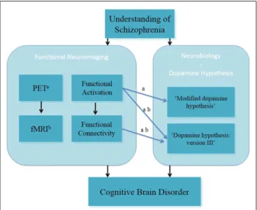
In summary, fMRI and PET studies in the field of clinical and cognitive neurosciences have been used to investigate brain function during working memory in people with schizophrenia (Figure 1). Both fMRI and PET findings have advanced the under-standing of altered working memory performance and brain func-tion in subjects with schizophrenia. This has led to better insight into the interaction between altered working memory function and experimental/clinical factors (such as cognitive domains of working memory function, performance level, phases of illness, clinical symptomatology, and effects of antipsychotic medication) in individuals with schizophrenia.
EXAMPLES OF fMRI STUDIES INVESTIGATING ALTERED SPATIAL WORKING MEMORY FUNCTION – GLUTAMATE HYPOTHESIS OF SCHIZOPHRENIA
The role of the DLPFC in working memory deficits has been associated with glutamatergic alterations and more specifically in dopamine–glutamate interactions (10,50,51). Furthermore, it has been reported that ketamine, a NMDA receptor antagonist, can induce psychosis-like symptoms in healthy subjects (45). Here, we briefly summarize the main functional activation and FC findings of fMRI studies on the spatial “N-back” task in the context of the glutamate hypothesis of schizophrenia (Table 2).
Anticevic et al. (12) presented ketamine-induced reduced func-tional activation in task-activated regions (such as the DLPFC and the precuneus) and task-deactivated regions of the default-mode network (DMN). In addition, the combination of a spiking local-circuit model of performance during the spatial “N-back” task and the functional activation findings revealed that the modulation of
FIGURE 1 | Understanding of schizophrenia as a cognitive brain disorder – verbal/numeric “N-back” task.a
fMRI;b
PET, positron emission tomography.
T able 2 | Sc hiz ophr enia as a cognitiv e br ain disor der II – summary of main findings in spatial w or king memory . St udy Subjects HC Medication/k etamine injection Exper imental par adigm F unctional connectivity method Computational modeling Main finding(s) Seed regions/R OIs Seed regions/R OIs definition Spher e siz e fMRI + KET AMINE S TUDIES ( 1 2 ) 1 9 HC One saline injection; initial k et amine bolus 0.23 mg/k g for I min; subsequent k et amine bolus 0.58 mg/k g for 1 h Spatial “2-bac k” and “4-bac k” Seed-based cross-cor relation Modeling of the acute k et amine ef fect of local and long-range E–I connections K et amine at tenuated task-activ ated regions (i.e., DLPFC and precuneus) Seeds: FP –DMN pair; CO–DMN pair; anatomical R OIs 1 5 mm sphere siz e Spiking E and I cell local-circuit models: task-activ ated module and task-deactiv ated module K et amine at tenuated task-deactiv ated regions o v erlapping the DMN; ↓ E–I conduct ance led to at tenuation in task-activ ated regions; modulation of task-activ ated FC bet w een FP –DMN net w orks during dela y of WM ( 62 ) 22 HC One saline injection; one initial k et amine bolus 0.23 mg/k g for 1 min; one subsequent k et amine bolus 0.58 mg/k g for 1 h Spatial “2-bac k” and “4-bac k” (1) Seed-based cross-cor relation; seed regions: bilateral MFG, IFG, SFG, and Hesc hl’ s gyr us; anatomical R OIs 1 0 mm sphere siz e N/A K et amine ef fect using (1): ↓ FC bet w een right DLPFC and MFG, IFG, front al OC, insula, medial FG; angular gyr us; k et amine ef fect using (2): ↓ FC within lef t DLPFC (2) Global-based connectivit y; see det ails as in (1) ↓ R educed. CO , cingulo-occipit al; DLPFC, dorsolateral prefront al corte x; DMN, def ault-mode net w ork; E cells, e x cit ator y cells; FC, functional connectivit y; FG, front al gyr us; FP , fronto-pariet al; HC, health y controls; I cells, inhibitor y cells; IFG, inf erior front al gyr us; MFG, middle front al gyr us; NMD A, N-Meth yl-d -aspart ate acid; OC, orbit al corte x; R OI, region of interest; SFG, superior front al gyr us; WM, w orking memor y.
ketamine alters the association between the task-activated and the task-deactivated networks. Finally, it was shown that ketamine modulates FC between the fronto-parietal and DMN networks. In a recent study, Driesen et al. (62) provided further support for ketamine-induced reduced prefrontal FC during the spatial “N-back” task. Two FC approaches with the same seed regions were employed, seed-based FC and global-based connectivity (GBC), which revealed both decreased FC within the DLPFC. The seed-based analysis resulted in reduced FC between DLPFC and mid-dle frontal gyrus [MFG, IFG, and insula (among other regions) under ketamine in contrast to saline]. The GBC analysis showed decreased FC of the DLPFC under ketamine.
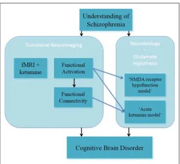
In summary, these studies on altered spatial working memory function inform on the glutamate hypothesis, through the acute ketamine model (Figure 2). In this, they have advanced the under-standing of NMDA receptor-modulated brain function in healthy subjects.
FROM COMPUTATIONAL NEUROPSYCHIATRY TOWARD SCHIZOPHRENIA AS A COGNITIVE BRAIN NETWORK DISORDER
Clinical and cognitive neurosciences have advanced the under-standing of altered working memory function in subjects with schizophrenia. FMRI studies in working memory among other neuroimaging and electrophysiological techniques, have reported on functional activation and FC findings in subjects with schizo-phrenia. Both findings of functional activation and FC revealed methodological, cognitive, and clinical factors related to our understanding of altered working memory function in patients with schizophrenia. In particular, FC findings mark the begin-ning of the notion of “disconnection” and “dysconnection” (20,
21,67,107) in working with people with schizophrenia. FC is defined as the statistical association or dependency among two or more anatomically distinct time-series (107). FC findings cannot be interpreted in terms of causal effects between connected regions and thus, does not allow for a mechanistic inference of the BOLD responses.
The modeling of functional large-scale networks5during work-ing memory function in schizophrenia could provide mechanistic explanations for altered brain function in individuals with schiz-ophrenia. The advantage of modeling functional large-scale net-works in terms of EC over FC is that inferences can be drawn on mechanistic processes, which are not directly observable in the BOLD response.
COMPUTATIONAL NEUROSCIENCE AND COMPUTATIONAL NEUROPSYCHIATRY
Marr proposes a theoretical framework for computational research on the brain on three levels (1976). At the first level, researchers should aim to gain knowledge of the high-level computations of the brain such as working memory (“computational level”). At the next level, the testing of the brain’s methods and algorithms for the high-level working memory function is led by hypotheses derived from the acquired knowledge and testing how appropriate an algo-rithm such as Bayesian inference is for modeling the working
5As one subfield within computational psychiatry.
FIGURE 2 | Understanding of schizophrenia as a cognitive brain disorder – spatial “N-back” task.
memory brain function (“algorithmic level”). Finally, when an algorithm is found, which is valid and more likely than alterna-tive algorithms to predict known brain function/behavior, then the investigation of the biological implementation can be pursued (“physical level”).
Computational neuropsychiatry is an emerging field within computational neuroscience. Computational neuropsychiatry aims to provide an explanatory bridge between altered cogni-tive function and neurobiological mechanisms associated with the development of mental illness (108, 109, Huys, unreferred preprint). Computational neuropsychiatry in humans has been defined by outlining a set of components, which include bio-physical modeling and computational modeling (109). Differ-ent types of computational models at differDiffer-ent neural levels are used dependent on the study hypothesis (108, Huys, unreferred preprint).
COMPUTATIONAL NEUROPSYCHIATRY AND MODELING OF FUNCTIONAL LARGE-SCALE NETWORKS IN SUBJECTS WITH SCHIZOPHRENIA
Connectionist and neural network models in working mem-ory/cognitive control in subjects with schizophrenia have added to our understanding of both the brain function and the neuro-biological mechanism underlying working memory (74,76). The strength of these models is based on the translational link between human brain function (i.e., functional activation) and preclini-cal neurobiologipreclini-cal evidence (namely, dopaminergic modulation) during working memory.
Following on from the work of Cohen and Braver, evidence for the understanding of schizophrenia as a cognitive network disor-der has been presented by both preclinical studies (8,10,110–113) and human FC studies in working memory (89,100,101,103). Recent studies examining biophysical mechanisms underlying
altered functional large-scale networks aim to bridge the gap between the human functional network used in working mem-ory and the preclinical neurobiological processes. Examples of such computational neuropsychiatric studies, including EC dur-ing workdur-ing memory in subjects with schizophrenia, are reviewed. In this, we focus on DCM studies investigating the numeric/verbal “N-back” task in subjects with schizophrenia and healthy controls. This is considered in the context of the dopamine and glutamate hypotheses of schizophrenia. Both neurobiological hypotheses have contributed to the formulation of research objectives in computational neuropsychiatry (114) and the development of computational modeling techniques of fMRI data in subjects with schizophrenia (20).
Dynamic causal modeling for fMRI – examples of modeling functional large-scale networks
Dynamic causal modeling for fMRI has been introduced as a method to provide insight into the notion of “functional inte-gration” during cognitive performance. “Functional inteinte-gration” has been advanced from the historic notion of “functional special-ization” (115), which is defined by context-dependent interactions among different brain regions (18,116–118).
Dynamic causal modeling has been described as a biophysical modeling of neuronal dynamic processes (18,19)6, which can be used as a method for the computation of synaptic plasticity from fMRI task-based studies (20,21). Together biophysical modeling and Bayesian inference analysis form the framework for DCM (71,
117,118). Thus, DCM is a modeling approach, which combines defined network models (i.e., hypotheses) with Bayesian inver-sion methods (19,117). Specifically, DCM assesses inter-regional EC through assessment of experimentally induced changes (18) and therefore allows for mechanistic inferences from neuronal function.
Bilinear DCM infers dynamics at the neuronal level by translat-ing modeled neuronal responses into predicted BOLD measure-ments (18). Non-linear DCM for fMRI (71,119) is an advanced approach for increasing the biological plausibility of DCMs by the means of modeling “gain modulation” (i.e., non-linear modula-tion of neuronal connecmodula-tions) (19,117,118). In non-linear DCM, the modulation of connection strengths by experimental inputs is supplemented by direct modulation of neural activity in one or more network regions (18,119). The computations for gating in neural networks use the multiplicative computation of non-linear modulation (120,121). Accordingly, non-linear DCM can be used for inferring that the strength of a connection is modulated by activity of other neuronal populations (119,122).
Findings of altered effective connectivity during working mem-ory in subjects with schizophrenia. The first DCM studies in
healthy controls described large-scale networks in working mem-ory and a similar task [continuous performance test; (123–125)]. A recent study in healthy controls built the linkage between EC results and underlying dopaminergic modulation of large-scale
6We consider DCM as the generative model approach as introduced in the seminal article by Friston et al. (18).
networks comprising of the DLPFC and PC during verbal memory performance (126).
To date four DCM studies have examined the verbal/numeric “N-back” task in subjects with schizophrenia using bilinear DCM (127–130) (Table 3). These provide novel insights into reduced task-dependent EC and increased task-independent EC measures through modeling large-scale networks in schizophrenia.
In the first study, increased fronto-temporal intrinsic connec-tivity was found to be associated with increased functional acti-vation of the superior temporal gyrus (STG) during the numeric “N-back” task in the subjects at the prodromal and at the acutely psychotic stage of schizophrenia in contrast to the healthy controls. This suggests a potential marker for vulnerability to the disorder (127). Furthermore, progressively decreased intrinsic connectiv-ity between the STG and the MFG in subjects at-risk mental state (ARMS) and FES subjects in contrast to the healthy controls was reported. This finding suggested that functional activation may resemble increased task-independent EC between the PFC and the STG. However, the results of the study are not comparable to other DCM studies because (i) only one model was examined and (ii) the biological plausibility of the EC measures is not clearly accessible. No reference to the dopamine or glutamate hypotheses was made.
The second study investigated the working memory-dependent modulatory effect for the prefrontal–parietal connectivity in sub-jects with EST and healthy subsub-jects during the numeric “N-back” task (128). The large-scale networks included the right DLPFC, the PC, and the visual cortex with bidirectional connection between all regions. The main finding was decreased task-dependent EC from the DLPFC to the PC in the subjects with EST. Thus, this finding could resemble evidence for the glutamate hypothesis of schizo-phrenia, specifically the NMDA receptor hypofunction model and the dysconnection hypothesis.
The third study examined possible vulnerability markers for psychosis from the verbal “N-back” task in ARMS subjects, FES subjects, and healthy subjects (129). This study examined reduced task-dependent EC measures as well as relationships between con-nectivity parameters and antipsychotic medication received by subjects. In this study, EC in interhemispheric large-scale net-works between the bilateral superior parietal lobes (SPL) and the bilateral MFG was assessed. This study reported novel findings of progressively decreased working memory and induced mod-ulation of connectivity between the MFG and the SPL (from healthy subjects to ARMS). Additionally, further decreased EC of modulatory effects were observed in non-medicated subjects with FEP contrasted to healthy controls. Evidence for ameliora-tion of reduced EC between the MFG and the SPL in subjects with FES, who received SGA medication, could reflect alterations of dopaminergic regulation of NMDA receptor-dependent synaptic plasticity of fronto-parietal connections. However, this interpre-tation is limited by the lack of a control group of FES who are treated with different types of antipsychotic medication. These findings across different subpopulations of schizophrenia together with the effect of antipsychotic medication may reflect support for the NMDA receptor hypofunction model and the dysconnection hypothesis.
T able 3 | Sc hiz ophr enia as a cognitiv e br ain netw or k disor der II – summary of main findings in v erbal/numer ic w or king memory – neur oimaging and bioph ysical modeling. St udy Subjects – phase of sc hiz ophr enia; HC – HR, FES/FEP , ES T Medication Exper imental par adigm Netw or ks – model space; number of models; regions DCM set tings – DCM v ersion; spher e siz e; inf er ence tec hnique(s) Main finding(s) ( 1 27 ) 1 3 HC; 1 6 HR a; 1 0 FES HR, not medicated; 7 FES , risperidone or quetiapine, 3 FES , not medicated Numeric “2-bac k” 1 L ef t hemispheric model; S TG, SMA, MFG, INS , PPC DCM in SPM5; Sphere siz es not reported; BMS not perf ormed P rogressiv ely ↑ IC of the prefront al–temporal connection in HR and FES ( 1 28 ) 42 HC; 41 ES T 35 ES T, FG A; 5 ES T, SG A; 1 ES T, not medicated Numeric “2-bac k” 48 Intrahemispheric models; 3 model families; DLPFC, PC, V C DCM1 0 in SPM8; 4 mm spheres; random-ef fects BMS e,f ; BMA ↓ EC (ef fect of task-modulation) from DLPFC to PC ( 1 29 ) 20 HC; 1 7 HR a; 21 FEP HR, not medicated; 7 FEP , completely antipsy chotic naïv e; 6 FEP , antipsy chotic naïv e at the time of scanning; 8 FEP , SG A V erbal “2-bac k” 1 2 Intrahemispheric models; bilateral SPL, bilateral MFG DCM1 0 in SPM8; 1 2 mm spheres; random-ef fects BMS e; BMA P rogressiv ely ↓ EC (ef fect of task-modulation) bet w een MFG and SPL in HC, HR, and FES . Ameliorated EC cor related with antipsy chotic treatment. ( 1 30 ) 1 5 HC; 1 4 FES b; 1 9 FES c FES b, 254.7 6 (1 92.09) d; FES c, 325.88 (1 85.1 9) d V erbal “2-bac k” 5 L ef t hemispheric models; medial PFC, PCC DCM v ersion not reported; SPM8; 6 mm spheres; BMS e; BMA ↑ IC from PCC to medial PFC in both FES e and FES f ↑ Increased in subjects with sc hiz ophrenia in contrast to HC. ↓ Decreased in subjects with sc hiz ophrenia in contrast to HC. BMA, B a y esian model a v eraging; BMS , B a y esian model selection; DLPFC, dorsolateral prefront al corte x; EC, ef fectiv e connectivit y; ES T, subjects with est ablished sc hiz ophrenia; FES , subjects with first episode sc hiz ophrenia; FEP , subjects with first episode psy chosis; FG A, first-generation antipsy chotics; HC, health y controls; HR, subjects at high risk of sc hiz ophrenia; IC, intrinsic connectivit y; IFG, inf erior front al gyr us; INS , insula; MFG, middle front al gyr us; PC, pariet al corte x; PCC, posterior pariet al corte x; PFC, prefront al corte x; SG A, second-generation antipsy chotics; SMA, supramarginal area; SP L, superior pariet al lobe; S TG, superior temporal gyr us; V C, visual corte x. aSubjects at high clinical risk of sc hiz ophrenia. bW ith high suicidal risk. cW ith lo w suicidal risk. dChlorpromazine equiv alents in mg/da y. eBMS at the group-le v el. fBMS at the model family le v el.
In the fourth study, Zhang et al. (130) explored EC measures in terms of possible neurobiological markers in groups of subjects with schizophrenia with high or low suicide risk and contrasted these with healthy controls during the verbal “N-back” task. The large-scale networks were defined by unidirectional and bidirec-tional connections between the two regions of the medial PFC and PC as well as working memory effects on these regions. This pilot study presented novel findings in subjects with schizophrenia at suicidal risk in terms of increased intrinsic connectivity from the PC to the MFG in both groups with FES (in comparison to healthy controls). This finding was interpreted as a possible association to schizophrenia, in which increased intrinsic connectivity from the MFG to the PC in the subjects with high risk of suicide could reflect vulnerability of suicide. However, the results are not directly com-parable to the other DCM studies because of the study population, which focused on the issue of suicide. The findings were also not interpreted in context of the dopamine or glutamate hypotheses.
We highlight main experimental and methodological limita-tions in the four DCM studies, which impede the comparability of findings (please see Table 3 for details). The main experimen-tal limitation focuses on the discrepancies between the different patient subpopulations. Two studies analyzed working memory fMRI data of subjects with ARMS and FES in comparison to healthy controls (127,129), whereas one study modeled scans from subjects with EST (128). Zhang et al. (130) reported findings of a unique patient population of FES with high and low suicidal risk. In terms of methodological issues, one limitation lies in dif-ferent definitions of model spaces for the large-scale networks, despite equivalence in the experimental tasks. Another limitation is that the reviewed DCM studies employed deterministic DCM for the comparison of the models. Deterministic models can pre-dict processes perfectly if all inputs are known (131). However, at this early stage of employing biophysical modeling approaches to human brain function, we do not have a full understanding of the brain responses to working memory. Future studies may employ stochastic DCM as an extension (117,118,132). A further limita-tion is that different DCM versions were applied across the four studies, which impede the comparability of the findings. The pri-ors are differently defined in the used DCM versions, which give rise to a variation in model evidence between the studies (117). Thus, it is possible that discrepancies in EC findings could be due to the prior definition and may not be solely due to differences in performance, brain function, or clinical aspects of subjects with schizophrenia. Lastly, a general limitation of DCM for fMRI is that maximally 10 regions within a large-scale network can be modeled. This simplification results in difficulties of biophysical modeling of tasks, which are likely to encompass more than ten regions. Furthermore, not only the definition of different regions and different numbers of regions but also different modulatory inputs result in further extensions to the model space. Such model spaces are difficult to validate and analyze.
The four DCM studies presented evidence for increased task-dependent EC and increased task-intask-dependent EC findings during verbal/numeric working memory in subjects with schizophrenia. We discuss these EC findings in context of (i) the dopamine and glutamate hypothesis and (ii) FC findings during verbal/numeric working memory in subjects with schizophrenia.
The four studies modeled large-scale networks during the “N-back” task in subjects with schizophrenia. However, only two out of these four studies consider their DCM results in the light of bio-physical processes (128,129). The findings of reduced EC (namely, the effect of task-modulation) of the prefrontal–parietal connec-tion in subjects with schizophrenia in contrast to healthy controls were interpreted biophysically and linked to the NMDA recep-tor hypofunction model and the dysconnection hypothesis (128,
129). Both studies reported reduced EC findings of the prefrontal– parietal connection during working memory, however, these find-ings need to be considered carefully due to different experimental designs (i.e., patient subpopulations, antipsychotic medication treatment of FGA and SGA) and methodological implementation (i.e., model space, DCM settings, and inference techniques).
Three of the DCM studies reported altered EC findings of the prefrontal–parietal and parieto-prefrontal connections dur-ing the “N-back” task in subjects with schizophrenia in contrast to healthy controls. Deserno et al. (128) and Schmidt et al. (129) pre-sented reduced EC (effect of task-modulation) of the prefrontal– parietal connection in subjects with schizophrenia in contrast to healthy controls, whereas Zhang et al. (130) found increased EC (intrinsic connectivity) of the parietal–prefrontal connection. The reduced task-dependent EC findings are in keeping with reduced FC findings of these connections, although increased FC between a different prefrontal subregion and the PC was reported (100).
The study by Crossley et al. (127) reported increased EC (intrin-sic connectivity) of the prefrontal–temporal connection in subjects at HR and FES (in contrast to healthy controls). Reduced FC of the prefrontal–temporal connection during the “N-back” task in subjects with schizophrenia has been previously reported in PET studies (105,106). However, the regions within the PFC and temporal region differ between the studies.
Findings of altered effective connectivity during verbal fluency in subjects with schizophrenia. Here, we discuss bilinear and
non-linear DCM studies, which have assessed large-scale networks during verbal fluency [namely, the Hayling sentence completion task (HSCT)] in subjects with schizophrenia and healthy controls (Table 4). One bilinear DCM study in healthy controls investi-gated the task-dependent modulation of response initiation and response suppression in EC between left hemispheric temporal and prefrontal regions (133). The main finding was a difference in connection strength of the modulatory effect in response initiation and response suppression.
Two clinical bilinear DCM studies have investigated EC mea-sures during the HSCT in HR subjects and healthy controls: (i) Subjects at high clinical risk of schizophrenia [ARMS; Ref. (134)] and (ii) subjects at high familial risk of schizophrenia (135). Allen et al. (134) investigated increased fronto-temporal EC (intrinsic connectivity) as a potential measure of vulnerability of develop-ing schizophrenia. Two main finddevelop-ings were reported: firstly, no significant effect of task-dependent modulation on the fronto-temporal connection between ARMS subjects and healthy controls was revealed. Secondly, ARMS subjects displayed increased intrin-sic connectivity between the ACC and the MTG in comparison to healthy controls. Furthermore, the Bayesian model selection (BMS) approach revealed that the same network was equally likely
T able 4 | Sc hiz ophr enia as a cognitiv e br ain netw or k disor der II – summary of main findings in v erbal fluency – neur oimaging and bioph ysical modeling. St udy Subjects – phase of sc hiz ophr enia; HC – HR, FES/FEP , ES T Medication Exper imental par adigm Netw or ks – model space; number of models; regions DCM set tings – DCM v ersion; spher e siz e; inf er ence tec hnique(s) Main finding(s) ( 1 34 ) 1 5 HC; 1 5 HR a 2 HR risperidone and quetiapine c HCS T 1 4 L ef t hemispheric models; MFG, A CC, MTG DCM in SPM5; 1 2 mm spheres; random-ef fects BMS4 ↑ IC bet w een A CC and MTG; same winning model in both groups ( 1 35 ) 1 9 HC; 26 HR b – 20 HR + , 4 HRill Not medicated HCS T 8 L ef t hemispheric models; 3 model families; IPS , IFG, A CC, MTG, MD thalamus DCM8 in SPM8; 8 mm spheres (IPS , DLPFC, MTG); 6 mm spheres (A CC, MD thalamus); R andom-ef fects BMS d,e ; BMA P rogressiv ely ↓ connection strength with non-linear modulation of the thalamo-cortical connection in HR + and HRill in contrast to HC ↑ Increased in subjects with sc hiz ophrenia in contrast to HC. ↓ Decreased in subjects with sc hiz ophrenia in contrast to HC. A CC, anterior cingulate corte x; ARMS , subjects with at-risk ment al st ate; BMA, B a y esian model a v eraging; BMS , B a y esian model selection; EC, ef fectiv e connectivit y; FEP , subjects with first episode psy chosis; FES , subjects with first episode sc hiz ophrenia; HC, health y controls; HR − , subjects at high familial risk of sc hiz ophrenia without transient psy chotic symptoms; HR + , subjects at high familial risk of sc hiz ophrenia with transient psy chotic symptoms; HRill, subjects at familial risk of sc hiz ophrenia who subsequent to the scanning de v eloped sc hiz ophrenia; HSCT , Ha yling sentence completion task; IC, intrinsic connectivit y; IFG, inf erior front al gyr us; IPS , intrapariet al sulcus; MD mediodorsal; MFG, middle front al gyr us; MTG, middle temporal gyr us; aSubjects at high clinical risk of sc hiz ophrenia. bSubjects at high familial risk of sc hiz ophrenia. cAt the time of scanning . dBMS at the group-le v el. eBMS at the model family le v el.
to explain the given HSCT fMRI data in both the ARMS subjects and the healthy controls. No reference to the glutamate hypothesis was made.
Dauvermann et al. (135) modeled EC measures in a similar version of the HSCT that was used by Allen et al. (134). This study was conducted in subjects at high familial risk of schizo-phrenia and healthy subjects. The results reported by Allen et al. (134) of a similar large-scale network in both HR subjects and healthy controls was replicated7. This finding was also confirmed by Dauvermann et al. (135), when the group of HR subjects was subdivided into high risk subjects without transient psychotic symptoms (referred to as HR−), high risk subjects with transient psychotic symptoms (referred to as HR+) and high risk subjects who subsequent to scanning developed schizophrenia [referred to as HRill; please see Ref. (136,137)]. Comparability between these two studies is limited due to differences in the model space. The model space in Dauvermann et al. (135) includes the IPS and the mediodorsal thalamus, which are not incorporated in the model space by Allen et al. (134). In addition, endogenous connections and task-dependent modulations were accordingly changed [Ref. (135); Table 4]. There was no reference to the glutamate hypothesis of schizophrenia.
Limitations of bilinear DCM have been addressed through the development of non-linear DCM for fMRI (119). This method was applied in the genetic high risk study reported by Dau-vermann et al. (135). The progress from the bilinear DCM to the non-linear DCM as reported by Dauvermann is based on the biophysical modeling of connection strength with non-linear modulation during the HSCT response. The authors show that relative to healthy controls there is reduced connection strength with non-linear modulation of the thalamo-cortical connection during the HSCT in HR+ subjects and a further reduction in this connection strength in HRill subjects (135). The authors sug-gest that reduced gain control may underlie the reduced strength in the thalamo-cortical connection. Furthermore, the findings of reduced connection strength with non-linear modulation of the thalamo-cortical connection could reflect altered glutamatergic neurotransmission, which may underlie a disruption of synaptic plasticity in this thalamo-cortical connection [Ref. (135); Table 4]. Thus, the findings were interpreted in context of the NMDA receptor hypofunction model and the dysconnection hypothesis.
Summary of studies modeling functional large-scale networks – dynamic causal modeling for fMRI
Evidence from brain function in working memory in subjects with schizophrenia at the level of functional large-scale networks (i.e., clinical and cognitive neurosciences) and neurobiological mech-anisms in working memory in animal models of schizophrenia (preclinical neurobiological research) in combination with com-putational neuroscientific approaches has informed and enabled research in computational neuropsychiatry.
Exemplary DCM studies in subjects with schizophrenia have reported both increased and reduced EC findings during cogni-tion in subjects with schizophrenia in contrast to healthy controls.
7It is noted, however, that the large-scale networks differed slightly from the previous study.
These studies applied DCM as a biophysical modeling approach to functional large-scale networks, which enabled the interpreta-tion of EC findings on the basis of the glutamate hypothesis of schizophrenia, namely the NMDA receptor hypofunction model and the dysconnection hypothesis (128, 129, 135). We empha-size that the findings support not only the glutamate hypoth-esis but also the dopamine hypothhypoth-esis. Dopamine is a neuro-modulator that may crucially affect glutamate-induced synaptic plasticity. Synaptic plasticity may be involved in a regulation of dopamine synthesis and release via other neurotransmitter systems. Specifically for non-linear effects, it has been shown that dopamine acts as a neuromodulator mediating postsynaptic gain (74,138).
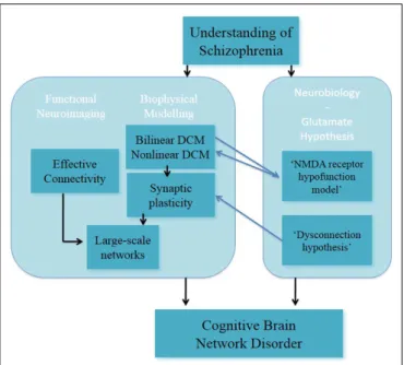
In a recent study, it has been reported that the combination of the DCM analysis of numerical “N-back” task in EST (128) and generative embedding resulted in the dissection of three subgroups of EST based on the mechanistically inferred DCM findings (139). This exemplary study showed that DCM can be applied as a generative model of large-scale networks in indi-viduals with schizophrenia. In summary, DCM is a promising approach for modeling synaptic plasticity; nevertheless in its cur-rent form it cannot reflect the full complexity in the processing required for the implementation of tasks such as working memory (Figure 3).
UNDERSTANDING OF SCHIZOPHRENIA IN DEVELOPMENT
Our understanding of schizophrenia is in continuous development and with more preclinical and clinical findings being published this understanding will advance further. A critical aspect of this under-standing is the facilitation of multidisciplinary approach between preclinical and clinical research in schizophrenia.
The original understanding of schizophrenia as a brain disorder stems from observational clinical work, which led onto preclinical
FIGURE 3 | Understanding of schizophrenia as a cognitive brain network disorder – verbal/numeric “N-back” task.