HAL Id: tel-01865143
https://tel.archives-ouvertes.fr/tel-01865143
Submitted on 31 Aug 2018HAL is a multi-disciplinary open access archive for the deposit and dissemination of sci-entific research documents, whether they are pub-lished or not. The documents may come from teaching and research institutions in France or abroad, or from public or private research centers.
L’archive ouverte pluridisciplinaire HAL, est destinée au dépôt et à la diffusion de documents scientifiques de niveau recherche, publiés ou non, émanant des établissements d’enseignement et de recherche français ou étrangers, des laboratoires publics ou privés.
Hepatic potential of Reversed-age Mesenchymal Stem
Cells and Endodermal Progenitors : Contribution of
LGR5 and Cdc42 cell signaling pathways
Diana Chaker
To cite this version:
Diana Chaker. Hepatic potential of Reversed-age Mesenchymal Stem Cells and Endodermal Progen-itors : Contribution of LGR5 and Cdc42 cell signaling pathways. Cellular Biology. Université Paris Saclay (COmUE); Université libanaise, 2017. English. �NNT : 2017SACLS562�. �tel-01865143�
NNT
:
20
17
SACLS
56
2
Hepatic potential of reversed-age
Mesenchymal stem cells and endodermic
progenitors: Contribution of Cdc42 and
LGR5 signaling pathways
Thèse de doctorat de l'Université Paris Saclay et
l'Université Libanaise préparée à l'Université Paris Sud
(Châtenay-Malabry)
Ecole doctorale n°569: innovation thérapeutique du
fondamental à l'appliqué (ITFA)
Spécialité du doctorat: Immunologie et Biothérapie
École doctorale en Sciences et Technologie-EDST
Spécialité: Biotechnologie
Thèse présentée et soutenue à Villejuif, le 18 décembre 2017, par
Diana Chaker
Christian POUS
Professeur, Université Paris Sud- Paris Saclay Président Hélène GILGENKRANTZ
Professeur, Université Paris Descartes Rapporteur
Rihab NASR
Professeur, Americain University of Beyrouth Rapporteur Marc KARAM
Professeur associé, Université de Balamand, Liban Examinateur Noushin DIANAT
Chargé de recherche, ESPCI, Paris Examinateur
Franck GRISCELLI
Professeur, Université Paris Descartes Directeur de thèse Ziad FAJLOUN
Professeur, Université Libanaise, Liban Directeur de thèse Nehman MAKDISSY
Acknowledgments
First, I would like to thank the members of my jury. I Thank PR Christian POUS for honoring me to chair this jury. PR Hélène GILGENKRANTZ and PR Rihab NASR, thank you for agreeing to be the reporters of this thesis. I would also like to thank DR Noushin DIANAT and DR Marc KARAM for agreeing to be the reviewers of this manuscript.
Also, my appreciation to Pr. Annelise Bennaceur-Griscelli for giving me the opportunity throughout the PhD research at UMR 935 of INSERM Villejuif and for all your support to achieve adequately this project. Thanks to your willingness to share your vast experience and knowledge. Without you I Havn‟t got to the point where I am now. I‟m very thankful to provide me funding through Vaincre le Cancer and therefore for the association president MR Michel OKS and director MR Laurent BIERE.
Many Thanks to REVIVA center in Lebanon, for letting me do a part of my research in their lab. Mostly I would like to thank the CEO DR Albert Azar, to be so comprehensive and objective during my decision to continue my thesis in France. Thank you to be so reasonable and judicious concerning my professional choice and to give me a family support to complete satisfactorily my PhD. I‟m recognizing for all what you offer to me in REVIVA platform.
I Foremost would like to thank my supervisors for the opportunity to study my PhD under your guidance. I express my sincere gratitude to Frank, I really enjoyed our conversations concerning my work, they were challenging and exciting. Those open questions helped me a lot when I was writing. I‟m appreciative for the assistance you provided at all levels whenever it was required. Thank you also for your human and generous approaches.
Also, I show my gratitude to Ziad, I„m grateful for your continuous willingness to help me in achieving all the lab activities during my stay in Lebanon financially and humanely wises. Thank you for minimizing me always the difficulties by saying “kheir”, this word gave me a new perspective at each down-feeling.
Thank you Norman for believing in me and encouraging me to continue my university cursus. Today, I realize the positive impact you added to my way. You taught me the good organization and to have always a dream for tomorrow.
I‟m very grateful for Charbel Mouawad, for your caring about the progression of the thesis and the writing of the scientific paper. Thank you to be the good messenger for my Lebanese part of this project.
I would like to thank Pr. Ali Turhan for your smiley face and high respectability even I didn‟t work with you.
At Reviva, I would like to thank also my colleague, Charbel Khalil, especially for giving me all the support and to facilitate me the lab duties. Also many thanks for Rose Mary to offer me a lot of administrative services and for your sincere attitude. Also, I‟m thankful to Mrs. Lola Azar who gave me all the push and the opportunities to progress in MEIH and REVIVA and having always confidence in my competencies.
A distinct gratitude for Mrs. Sandy Hage, you are a special project manager and you gave me the good assistance, the respectable way to write and how to develop my scientific strategy. I‟m realizing that you are my guardian angel who helps me to progress in my Lebanese part. I‟m so attached to the approachable and special moments we spent together during our stay in Lebanon. I wish you all the best for your moving to Chicago. Also, I would like to thank Mrs Ghada Oreibi for receiving me and for making my work easier in the genetic department of MEIH. Thank you also, for your professional collaboration way.
I thank my colleagues at U935 Villejuif, Thank you:
Eva and Lucas for being so ready to lend a hand; Afag for your Kindness. Lucie for your empathy, we experienced together the stressful moments of the thesis; Olivier for your scientific and technical advices; Jinwook for your sympathy; Hervé for your professional way of communication and to have the good answers and advices at any time. Jerome, for the big things you learned to me through a smallest number of words, for your expressive serenity and for being so caring and friendly; Adlen for giving me a distinctive idea about the research passion; Carlos for being so friendly, supportive and nice collaborator; Patricia for your sympathetic approach and sincerity; Dominique for your fast order processing, you are a very kind mami for Mila. Sara and Gladys for your funny spirit pushing me up a lot of time. Masae, you are a perseverant and friendly person and Jean for learning me the good way to do my western blots, for your gentleness and your friendly attitude.
Thank you Tony for your nice accompany and honesty. We made a good team together, I‟m happy to have you a real friend. All the best for your new challenge. Thank you Noufissa to give me the right opinions and to encourage me in 2014 for my PhD decision. You are a peaceful and enjoyable person.
I also would like to show my gratitude to Albert Tasteyre, you have a special communication skills, serenity and professionalism. You were a source of encouragement for my doubts.
I thank also Eric Rubinstein and Philippe Leclerc for their technical support. I am very grateful for the funding provided for me by Cedres Campus de France and CRD. Those financial aids allowed me to continue my both internships without strong economic difficulties.
Thank you. Rima Haddad for your friendly approach and your caring. Thank you Rindalah for your soft and nice assistance. .
My sincere thanks also go to Bernard and Emilie, you were my second family. You touch me by your peacefulness and humanity. As usual, I‟ll be by your sides. RIP my Mamou, near or far, wherever you are, you are giving hope to my life and you will be always in my heart.
Noha, my dear friend, thank you for your prayer which was always a constant sounding board during the most difficult moments during my thesis.
My dear Pauline, you provide me a lot of sincere helps. I know you are going through difficult moments with your disease, but be sure all the shiny days are waiting for you after this hard period.
I would like to express my sincere grateful to Faty, for your support provided to me and my son.
Thank you Mira for being a faithful friend, I‟m forever grateful for your love and support. You were by my sides comprehensively in all the difficult moments I spent these recent years.
Finally, I would like to thank perhaps the most beautiful individual, a special friend who recently picked me up through the lowest of times. Your support has been unconditional of which I am eternally grateful, thank you Anthony and also for your family.
Thank you my little sis Zanzoun and my “Sohri“Hamid for your continuous support especially last year. I would like to show my gratitude to Chadi, Cynthia, Fadi and Melissa for always giving me positive energy and practical answers.
Last but not the least, I would like to thank my parents for supporting me spiritually throughout my life and my studies, and,
I dedicate this work to my precious son
David.
Sincerely, Diana
Papers Included
Paper 1 (Submitted)
Direct reprogramming of LGR5+ Liver Progenitors cells responding to both
gp130/JAK/STAT3 and Wnt/β-catenin the signaling pathways
Diana Chaker , Frank Griscelli, Tony Ernault, Nicolas Moniaux, Olivier Féraud, Noufissa Oudrhiri, Herve Le-Stunff, Lara Bellini, Guillaume Pourcher, Sylvia Sanquer, Jamila Faivre, Christophe Desterke, Ali G Turhan, Annelise Bennaceur-Griscelli
Paper 2 (Submitted)
ML141 reverses the negative impact of the RhoGTPase Cdc42-dependent
donor’s age on hepatogenic differentiation of hADSCs
Diana Chaker, Charbel Mouawad, Albert Azar, Didier Quilliot, Ibrahim El Achkar, Ziad Fajloun, Nehman Makdissy
ABBREVIATIONS
A
AAT anti-trypsin
ADSC Adipose derived Stem Cells AFP Alfa Foeto Protein
Ag Antigen
Akt Protein kinase B (PKB) Alb Albumin
AMP Adenosine monophosphate
APAP acetyl-para-aminophenol (paracetamol) APC Adenomatous Polyposis Coli
aPKC Par6 atypical PKC ATP Adenosine TriPhosphate
D
DA Donor Age
DDR proteins DNA damage response proteins Dex Dexamethasone
DLK 1 Protein delta homolog 1 DP Dental Pulp
DP Doubling Period DSH disheveled
E
EAE Experimental Autoimmune Encephalomyelitis mice EB embryoid body
EC Endothelial Cells
E-cadherin Epithelial Cadherin ECM Extra Cellular Matrix
EGFR epidermal growth factor receptor EMT Epithelial to Mesenchymal Transition EpCAM Epithelial Cell Adhesion Molecule ERK Extracellular signal–regulated kinase ESC Embryonic Stem Cells
EST Estrogen
F
Fah fumarylacetoacetate hydrolase FGF Fibroblast Growth Factor Foxa Forkhead Box A Fzd Frizzled
B
β-FGF Basic fibroblast growth factor BM Bone Marrow
BMSC Bone Marrow Derived Mesenchymal Stem Cell BMP Bone Morphogenetic Protein
53-BP1 p53-binding protein 1
C
Cdc42 Cell division cycle 42 CFU-F Colony Forming Unit CK1 casein kinase 1 CK14 Cytokeratin 14 CK14 Cytokeratin 14
CLiP chemically induced Liver Progenitor
Cre/loxP Cyclization recombinase - locus of X-over P1 CREB cAMP responsive element binding protein CTGF connective tissue growth factor
cyp450 Cytochrome P450
H
HBG human hepatoma cell HBL Hepatoblast
HCC HepatoCellular Carcinoma HDAC Histone deacetylase
HepG2 hepatoblastoma-derived cell line HepPar Hepatocyte Paraffin
HGF Hepatocyte Growth Factor
HHEX Hematopoietically-expressed homeobox protein hiHep human induced hepatocyte
HLA-DR Human Leucocyte Antigen -Antigen D related HLC Hepatocytes - Like Cell
hMSC human Mesenchymal Stem Cells HNF1-β Hepatocyte nuclear factor 1β HNF4-α Hepatocyte nuclear factor 4α
HPLSC Human Pluripotent resident Liver Stem Cell HSC Hematopoeitic Stem Cells
HSP60 Heat shock protein 60
hTERT human Telomerase Reverse Transcriptase Huh7 hepato cellular carcinoma cell
G
G6Pc Glucose 6 phosphatase GAD GTPase Activating Protein GATA4 GATA binding protein GSK-3 glycogen synthase kinase-3 GTP Guanosine-5'-triphosphate
GDIs Guanine nucleotide dissociation inhibitors GEFs Guanine nucleotide exchange factors
I
IGF Insulin-like growth factor iHep Induced Hepatocytes-like IL 6/10/2 Interleukin 6/10/2
iMPC induced multipotent progenitor cell INFγ Interferon gamma
IRS Insulin Receptor Substrate
J
JAK Janus kinase
JNK c-Jun N-terminal kinase
K
KRT18 Cytokeratin 18 KRT19 Cytokeratin 19 KRT8 Cytokeratin-8 KC Kupffer CellsL
LB Liver Bud LDL Low-density lipoproteinLEF lymphoid enhancer binding protein
LGR5 Leucine-rich repeat-containing G-protein coupled receptor 5
LIFR Leukemia inhibitory factor Receptor Lin28 PAS D"ABBREVIATION
LP Liver Progenitor LR Leptin Receptor LT Liver Transplantation
M
MAPK Mitogen-Activated Protein Kinase MEF mouse embryonic fibroblast mEpiSC mouse epiblast stem cell
N
NANOG homeobox gene NO ABBREVIATION N-cadherin Neural Cadherin
NFκB Nuclear factor κB NK Natural Killer
NOD/SCID Nonobese diabetic/severe combined immunodeficiency
NPC Non-Parenchymal Cells
P
p16 cyclin-dependent kinase inhibitor p21 cyclin-dependent kinase inhibitor p38 mitogen-activated protein kinase p53 Tumor suppressor protein 53 Pak4 Serine/threonine-protein kinase 4 PBSC mobilized peripheral blood cell PDGF Platelet-derived growth factor PDT Population Doubling Time
PEDF pigment epithelium-derived factor PH Partial Hepatectomy
PHH Primary Human Hepatocytes PI3K PhosphoInositol 3 Kinase
pIR phosphorylated intramembrane Insulin receptor PKA Cyclic Camp protein kinase A
PKS Cyclic Camp Protein Kinase A PSC Pluripotent Stem cell
T
TAT tyrosine amino transferase TCF T-cell factor
TERT telomerase reverse transcriptase TGF α Transforming Growth Factor alpha TGF-β Transforming Growth Factor β TNF Tumor Necrosis Factor
TSA TrichoStatin A
Twist1 Twist-related protein 1
U
UCB Umbilical Cord Blood
UCMSC Umbilical Cord Derived Mesenchymal Stem cell
V
VCAM Vascular cell adhesion protein VEGF Vascular Endothelial Growth Factor
MET Mesenchymal to Epithelial Transition MMP Matrix MetalloProteinase
O
OC OnecutOCT4 Octamer-binding transcription factor 4 POU5F1 POU Domain, Class 5, Transcription Factor 1 OPN Osteopontin
OSM Oncostatin M OV6 Oval Cell Marker6
R
Rb Retinoblastoma Protein RhoA Ras-like GTP-binding protein 1 RNA ribonucleic acid
ROS Reactive Oxygen Species production RSPO Rspondins
RUNX 2 Runt-related transcription factor 2
S
SA- β gal Senescence-associated beta-galactosidase SASP Senescence-Associated Secretory Phenotype Sca-1 stem cell antigen 1
SCF Stem cell factor
SDF-1 Stromal cell-Derived Factor 1
smad 2/3 Mothers against decapentaplegic homolog 2/3 SOX17 sex determining region Y-box 17
SOX2 sex determining region Y-box 2 SOX9 sex determining region Y-box 9 SSEA-4 Stage-specific embryonic antigen 4
STAT3 Signal transducer and activator of transcription 3 SVF Stromal Vascular Fraction
LIST OF FIGURES
Figure1: Prometheus’s Greek... 0
Figure 2: Anatomy of the Liver ... 12
Figure 3: The Complexity of liver cell types and their interactions ... 14
Figure 4: Segregation of Hepatoblasts into hepatocytes and cholangiocytes. ... 15
Figure 5: The cell lineage steps during hepatic development ... 16
Figure 6: Contribution to the human Cytochrome P450 enzymes (CYP) isoforms to drug metabolism.. ... 19
Figure 7: Hepatocyte cell cycle phases ... 23
Figure 8: A schematic for Signaling pathways modulating hepatocyte proliferation in injured liver. ... 25
Figure 9: Weakness of liver regeneration in elderly people. ... 26
Figure 10: Schematic view of the bipotent progenitor’s location in the liver ... 27
Figure 11: Stem cells lineage relatives in adult liver and pancreas. ... 28
Figure 12: Regulation of ESCs pluripotency by LIFR and Wnt pathways ... 31
Figure 13: Generation of hepatocytes from iPSCs. Modeling of human liver disease in vitro ... 33
Figure 14: Schematic for hepatocytes dedifferentiation into bipotent progenitors. ... 36
Figure 15: Generation of liver bipotent progenitors mediated by chemical small molecules. ... 37
Figure 16: WNT pathway activation by LGR 5 ... 38
Figure 17: Crosstalk between Stat3/ LIFR and LGR5/Wnt in LGR5+ stem cells. ... 40
Figure 18: In vitro Human Lgr5+ organoids generation without Hepatocytes reprogramming process. ... 42
Figure 19: Change in distribution of donor age in recent years. Source: United Network for Organ Sharing reports, 2015 ... 92
Figure 20 : Evolution of liver transplants frequency in Europe between 1968 and 2013; From ELTR registry, 2016 ... 93
Figure 21: Morphological and molecular mechanisms involved in MSCs senescence ... 97
Figure 22: Age-Senescent Biomarkers in human MSCs. ... 98
Figure 23: Morphological and molecular considerations within MSC differentiation toward hepatocytes ... 106
Figure 24: Main molecular mechanisms involved in MSCs trans-differentiation into hepatocytes ... 107
Figure 25: Schematic of Cdc42 activity mode.. ... 110
Figure 26: Multiple signaling pathways controlling the cellular polarity via Cdc42 activation. ... 111
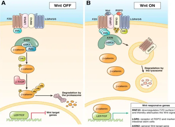
Figure 27: Wnt signaling pathways ... 112
Figure 28: Impact of CASIN on the polarity and epigenetic behaviors of aged HSCs related to Cdc42 GTP activity. ... 113
Figure 29: Regulation of Cdc42 by the MAPK signaling pathways. ... 115
Table of Contents
ABSTRACT ... ACKNOWLEDGMENTS ... PAPER INCLUDED ... ABBREVIATIONS ... ABBREVIATIONS ... LIST OF FIGURES ...CHAPTER 1 Thesis report structure ... 1
Part I: Lgr5+ Stem Cell Generation from Primary Murine Hepatocytes ... 2
Part 2: Impact of Cdc42 inhibition on aged-derived ADSCs behaviors ... 6
CHAPTER 2 Liver organogenesis, functions and diseases ... 11
2.1. Liver cellular composition ... 11
2.2. Ontogenesis of the liver ... 13
2.3. The mesenchymal to epithelial transition during liver development position ... 16
2.4. Liver functions ... 18
2.5. Liver diseases: The global health burden ... 20
CHAPTER 3 Mechanisms of liver regeneration: Contribution of bipotent Liver Progenitors ... 22
3.1. Overview ... 22
3.2. Molecular mechanisms implicated in proliferation of mature hepatocytes during liver regeneration process: Wnt, STAT3 and Cdc42 pathways ... 22
3.3. Age-associated mechanisms in the failure of liver stem cell regeneration ... 25
3.4. Primary hepatocytes senescence in vitro ... 26
3.5. Resident Liver bipotent Stem cells: Origin and limitation in liver regenerative potential ... 27
3.5.1. In vivo strategies for bipotent stem cells lineage tracing... 29
CHAPTER 4: Advances in Rerogramming of mature Hepatocytes toward Bipotent Liver Progrnitors (LPs) via the Activation of the WNT-LGR5 Pathway ... 30
4.1. Latest cues for Hepatocytes development in vitro ... 30
4.1.1. Limitations of Primary Human Hepatocytes (PHH) ... 30
4.1.2. Human Mesenchymal stem cells (hMSCs) ... 31
4.1.3. Generation of hepatocytes from human Pluripotent Stem cells (iPSC) ... 32
4.1.3.1. Human Embryonic Stem Cells (hESCs) ... 32
4.1.3.2. Human Induced Pluripotent Stem Cells ... 32
4.1.3.3. Limitation of Endoderm specification from hPSCs ... 34
4.1.4. Hepatocytes plasticity a new era in hepatology ... 35
4.2. LGR5-Wnt axis a new marker of liver stem cells in association with STAT3 pathway ... 38
4.3. Recent progresses resulting from the Lineage tracing of LGR5 stem cells ... 40
4.4. LGR5 a novel marker for long term culture and expansion of stem cells in 3D specter ... 41
4.5. Footprints ... 42
CHAPTER 5: Introduction to the scientific paper 1 ... 44
Scientific paper 1 ... 46
CHAPTER 6: Impact of Donor age (DA) on Mesenchymal Stem Cells functionalities ... 92
6.1. Impact of DA on liver transplantation ... 92
6.2. Age- related biomarkers of MSCs ... 93
6.2.1. Cellular activity: Proliferation and Apoptosis ... 93
6.2.2. Surface Antigen expression ... 94
6.2.3. Telomere length and Telomerase activity ... 94
6.2.4. Reactive Oxygen Species production (ROS) ... 95
6.2.5. The loss of multipotency and stemness ... 95
6.2.6. Age-related DNA damage and tumor suppression gene profile p53/p16 INK4A/p21 ... 95
6.3. DA impact on MSCs functionalities ... 96
6.3.1. Paracrine activity and niche aging: Role of MSCs derived exosomes ... 96
6.3.3. Migration and adhesion ability: Implication of Cdc42 in stem cell aging ... 99
CHAPTER 7: Hepatogenic potential of Adipose Tissue Mesenchymal Stem cells (ADSCs) ... 101
7.1. General debate about Hepatogenic potential of MSCs and their molecular characteristics ... 101
7.2. Mesenchymal stem cells: safe alternative for liver resident stem cells and induced hepatocytes from pluripotent stem cells ... 102
7.3. ADSCs as a potent source of human hepatocytes generation: Contribution of exosomes and Wnt pathway ... 103
7.4. Impact of DA on regeneration potential of ADSCs ... 105
7.5. Impact of DA on differentiation potential of ADSCs ... 105
7.6. Human MSCs to hepatocytes: the in vitro differentiation cues ... 106
CHAPTER 8: Cdc42: A novel biomarker of stem cell aging ... 109
8.1. Regulation of cdc42 GTPase protein ... 109
8.2. Cdc42 activation and stem cell aging (2001-2017) ... 109
8.3. Cdc42 and Wnt pathways in stem cell aging context ... 112
8.4. Cdc42 and Wnt pathways in the liver regeneration context ... 113
8.5. Main signaling pathways regulating Cdc42-GTPase activity ... 114
8.5.1. RAS pathway ... 114
8.5.2. Mitogen-activated protein kinase (MAPK) signalling pathway ... 114
8.5.3. Nuclear factor κB (NFκB) signalling pathway ... 115
8.5.4. The Phosphoinositol 3 kinase (PI3k) and Insuline receptor substrate pathways... 115
8.5.5. Cyclic Camp protein kinase A (PKA) ... 116
8.6. Pharmacological inhibition of activated Cdc42 ... 116
8.6.1. CASIN: Unspecific Cdc42 inhibitor ... 116
8.6.2. ML141 A novel spatial selective non-competitive specific Cdc42 inhibitors ... 117
8.6.3. Trichostatin A (TSA): Non-specific Cdc42 inhibitiors ... 118
8.6.4. Other pharmacological indirect Cdc42 inhibitors ... 119
CHAPTER 9: Introduction to the scientific paper 2 ... 121
Scientific paper 2 ... 124
CHAPTER 10: General discussion ... 153
Debate 1: The new technology of EndoPCs allowing the activation of Wnt/Lgr5 axis pathway in primary hepatocytes in absence of Wnt activators ... 153
Debate 2: Novel strategy to convert the age of hADSCs and their derived hepatocytes through ML141-Cdc42 inhibition ... 156
Debate 3: Reverse-aged hADSCs and EndoPCs technologies in the liver’s engineering and regenerative context ... 158
CONCLUSION ... 162
REFERENCES ... 163
ABSTRACT ... 191
….
And the soul is linked to the LIVER and one’s
rootedness in the local, circumscribed realities in which we
live our daily lives…. Thus the liver was considered to
contain the secret of fate and was used for fortune-telling….
The SELF associated to the memory function of the brain
that enables us to MAINTAIN a sense of awareness of
ourselves as being the self-same person over a lifetime,
despite changes in physical appearance due to AGING...
Et depuis longtemps…….
« Quant au rusé Prométhée il envoya contre lui un aigle aux ailes étendues qui rongeait
son
foie immortel
; il en renaissait durant la nuit autant ce que l'oiseau aux largesailes en avait dévoré pendant le jour…… »
Figure 1: Prometheus’s Greek myth oil on canvas painting by Peter
1 | P a g e
CHAPTER 1
Thesis report structure
The liver regenerative potential was hallowed millennia ago in the Greek mythology of Titan when Prometheus, who stole fire from Zeus and the gods and gave it to mortals, was punished by Zeus for his crime by having Prometheus bound to a rock, while an eagle ate his immortal liver on day after day (Fig. 1). This evokes invariably the hypothesis of the liver’s capacity for self-repair and regeneration. Since this myth and nowadays, researches were in progress and gave an irrevocable hope for a partial to advanced liver regeneration.
The liver may undergo permanent organ failure after severe injuries, chronic infection, cancer and aging. Hepatic failure leads to 1-2 million deaths per year and is classified as the 5th leading cause of death around the globe. Orthotopic liver transplantation is currently the effective therapy for end-stage liver diseases. However, the shortage of donors creates serious limitation related to this treatment. Thus, the patient’s waiting list to find donors has increased to more than 30% both in Europe and United States.
The availability of primary hepatocytes is a gold standard for the liver cell-base therapy. Human primary hepatocytes are not easy to be isolated from liver biopsies and have also a limited life span in culture with a progressive loss of cell function over the time. Therefore, one of the most curbs associated to primary hepatocytes’ liver therapy is the availability of a sufficient number of high quality and functional hepatocytes 2. Moreover, the isolation and culture of hepatocytes may cause major alterations in genes’ expression due to cell dedifferentiation under epithelial to mesenchymal transition (EMT) or due to cell apoptosis after only few days in culture3-6. In addition, in vitro culture models of the human liver cells are crucial for drug development and pathways mechanistic inquiries 7. To avoid human hepatocytes variability, an alternative would be to alter their in vitro short-life by inducing healthy hepatocytes for : (1) the establishment of high-throughput strategies allowing a better systems for susceptibility to drugs, (2) the modeling of liver development and diseases (3) a safe source for cell-based therapies and (4) the development of bio-artificial liver devices and (5) for a liver bio-printing approach 8, 9. Cell and tissue bioengineering of the liver earned high interests during the last decades and gained momentum through considerable progress in understanding the involvement and application of stem cells in liver regeneration.
Several studies show the existence of resident stem cells in the adult liver 10. Liver human progenitors have been observed during acute and chronic liver diseases but were found insufficient in number and were unable to perform complete liver regeneration. Stem cell- based strategies are consequently being explored as an attractive alternative approach to liver repair. Thus, various types of stem cells have been used in the recent era to produce a large number of functional hepatocytes in vitro. Several studies described the differentiation of the embryonic stem cells (ESC) into hepatocyte-like cells (HLCs) 11, 12. Despite the ESCs gold standard differentiation potential, their clinical application remains restrained considering the risk of teratoma formation and ethical limitations13. Later on, induced pluripotent stem cells (iPS) were introduced as an innovative approach in the field of liver cell therapy 14. However, iPSCs derived-HLCs have shown an incomplete differentiation status compared to primary hepatocytes15 since they maintain exogenous transgene expression which can interfere with differentiation protocols16.
2 | P a g e
Furthermore, the generation of induced Hepatocytes (iHep) from pluripotent stem cells presents high safety but ethical worries restricting their clinical uses for cell-base replacement regenerative medicine.
Thus, fundamental clinical studies attempt to open new challenges for hepatocytes generation from adult stem cells such as mesenchymal stem cells (MSCs). More recently, liver bipotent stem cells expressing the Wnt target gene called Lgr5 (leucine-rich-repeat-containing G-protein-coupled receptor 5) and deriving from hepatocytes dedifferentiation have emerged as a new platform for stem cell-based liver regenerative medicine; in fact,
Lgr5+ stem cells/progenitors actively contributed to liver regeneration via de novo generation of hepatocytes 17, involving a robust activation of Wnt signaling. Indeed, the contribution of the canonical and non-canonical Wnt signaling pathways governs a myriad of biological processes underlying the development and maintenance of adult tissue homeostasis, including regulation of stem cell self-renewal, cell proliferation, and instructing their differentiation fate into hepatocytes. Regulation of Wnts’ pathways can trigger multiple signaling cascades, some of them result in activation of small GTPase Rho, Rac, and Cdc42 18. Interestingly, mice lacking the RhoGTPase Cdc42 in their hepatocytes exhibited severe liver morphological defects indicating that Cdc42 acts in hepatocytes and might be required for liver function19. Recently, it has been reported in normal adult stem cells that LGR5 interacts with the IQGAP1-GTPase pathway which is an effector of Cdc42 to strengthen cell-cell adhesion. This interaction leads to activate the Cdc42/Rac complex and to crosslink the actin organization20 .Several reports discussed the role of Cdc42 in human MSC proliferation, migration and differentiation, and discussed in mice hematopeitic stem cells (HSCs) its involvement of aging21.
Currently, there is no published data showing the significance of the role of LGR5 in iPSCs derived-HLCs, neither the impact of the Cdc42 on the rejuvenation of human adult MSCs-derived HLCs; our strategy was based on identifying first the reprogramming process that may induce Hepatocytes differentiation (Hep-Dif) of mice LGR5+ iPSCs, and second the superlative Cdc42 inhibitor able to reverse the age effect on human MSCs and stimulate their Hep-Dif potential of adipose tissue derived stem cells.
In the following sections, the first part describes a new tool for liver bipotent stem cell generation via primary hepatocyte reprogramming process and intended to be differentiated into hepatocytes and cholangiocytes after long-term expansion in vitro, and the second part constitute a new strategy to reverse senescence of aged-derived MSCs before inducing their differentiation toward functional hepatocytes.
Part I: Lgr5+ Stem Cell Generation from Primary Murine Hepatocytes *
*Completed in France
Scientific Background # Part I
The liver presents two mechanisms for regeneration depending on the type of injury. Partial hepatechtomy (PH) involves the remained hepatocytes to restore the liver size. However, toxic injuries involve adult liver stem cells, called Oval cells in rodent, located in the biliary duct22 and Hering canal in humans 188, 189 .Those stem cells are able to give rise to both hepatocytes and ductal cells (cholangiocytes)23. Several pathways were shown to regulate the liver regeneration and hepatocytes proliferations such as canonical Wnt/β-catenin pathway, E-cadherin/ β-catenin membrane complex24, HGF/ β-catenin25, IL6/STAT-3 phosphorylation26, 27, and TGFβ 28. All these pathways are far to be efficient in human hepatocytes after PH and failed to complete liver regeneration due to the accumulations of reactive oxygen species (ROS) , the DNA damage in the remained hepatocytes and also to the non-sufficient
3 | P a g e
activation of liver stem cells 29. On the other side, these oval cells, are activated upon toxic injury when hepatocytes are prevented to proliferate and can give rise to hepatocytes and biliary duct cells (Cholangiocytes) 30, 31 . Although oval cells are activated upon injury, when liver insult is aggressive, involvement of this occasional population in liver regeneration is insufficient 32. Thus, the liver regenerative-based research was interested in two cover up to mimic the human in vivo mechanisms of liver regeneration. The first, primary human hepatocytes long term proliferation and the second, bipotent human stem cell isolation, expansion and activation. However, these two closes present strong limitations and are far to be maintained ex-vivo. In fact, primary hepatocytes isolation constituted a very complicated procedure as they do not proliferate easily on plastic dishes and enter quickly in senescence 33. On the other side, the Isolation of bipotent liver stem cells requires a molecular characterization of the surface markers. Unfortunately , these progenitors are not a distinct population of the liver since they express common markers with hepatoblasts markers (AFP and CK19) 34, 35 , hepatocytes markers (HNF4-β and Alblow) , adult multipotent stem cell markers (c-kit, Sca-1, Thy1 (CD90), and CD34) 36-38 and liver epithelial marker (EpCAM) 39. Furthermore, the better proliferative fraction of human Liver Progenitors (LP) in vitro was reported to constitute only 0.1% of total Epcam+ stem cell population40. Thereby, for long decades, regarding the multiple types of liver cells isolated from liver biopsies, the lack of oval cell- specific markers was limiting their lineage tracing.
The in vivo tracking of the bipotent liver stem cells was performed by using inducible-Cre promoters systems in transgenic mice such as promoters of Foxl1 , Sox9 and OPN which are reported to be expressed in only activated ductal stem cells 41, 42,43 . These progenitors were able to repopulate injured liver and to give rise to hepatocytes and cholangiocytes in vitro. These efforts have led to show that activated bipotent stem cells contribute to 2.5% of hepatocytes regeneration during recovery 43.
Thereby, the development of strategies to isolate and expand these progenitors in vitro appeared to be essential for the liver stem cell-based medicine.
The lookout moved backward to several years ago where hepatocyte showed a high plasticity upon injury and to give rise to both hepatocytes and biliary duct cells 44. Therefore, human hepatocyte lineage tracing in FAH-/- mice model showed a high capacity of hepatocytes to dedifferentiate into bipotent ductal stem cell expressing CK19+/EPCAM+/OPN+/SOX9+ upon injury and to give rise to both hepatocytes and biliary duct cells in suitable cell culture conditions45. Moreover, these hepatocytes derived from bipotent stem cells were shown to express leucine-rich-repeat-containing G protein-coupled receptor 5 (LGR5), a Wnt target gene which was reported to be highly expressed in dividing stem cells in several epithelial tissues such as small intestine and colon46, stomach47, hair follicles48. Simultaneously, LGR5 when fixed to its ligand Rspondin1 (Rspo1) promotes the activity of Wnt-Frizzled 49 mediated signaling via activation of IL6/STAT3 pathway50,51.
More interestingly, in a 3D system culture favorable for Wnt driving pathway, isolated single Lgr5+ stem cells formed organoids retaining the gene expression of the liver over several passages52.
In addition, these self-renewed Lgr5+ organoids were able to differentiate into functional hepatocytes when transplanted into FAH−/− mice.
Based on all these evidences , Huch et al, 2015 53 isolated EpCAM + stem cells directly from human liver biopsies . In 3D structure and in presence of Rspo1 and some small molecules inhibiting Notch, FGF4 and BMP pathways, EpCAM+ cells were able to form organoids expressing LGR5+. Hepatocytes derived from EpCAM+LGR5+ cells give rise to functional hepatocytes and regenerate damaged CCL4-Mouse Liver. And more recently, Katsuda et al, 2017 54
, found a combination of small molecules able to convert primary hepatocytes into LGR5+ bipotent LPs so-called chemically induced LPs (CLiPs) with high hepatocytic and cholangiocytic potentialities. This strategy skipped the complexity of EpCAM+ cells isolation and organoids formation in 3D structures. However, Rspo1 and other
4 | P a g e
canonical Wnt/β -catenin activators such as Wnt3a were essential to stably expand LGR5+ Stem cells before their differentiation. In contrast, organoids LGR5+ required inhibition of BMP, TGF-β and non-canonical Wnt in vitro.
Specific Aim(s) # Part I
Based on these outcomes, we centered in this first part on a new research tool for reprogramming mice primary hepatocytes into endodermic progenitors, so-called EndoPCs. Our general objective is to evaluate this novel technology of liver bipotent stem cells generation in vitro and to suggest it as a promising tool for stem cell-based liver therapy.
Our first aim is to assess the specific molecular identity of EndoPCs compared to pluripotent stem cells, Lgr5 oval stem cells and primary liver hepatocytes
Giving the authentical Lgr5 expression in EndoPCs, we intend to find the best Wnt activation conditions to derive specific EndoPCs-organoids and maintain their self-renewal upon withdrawal of Leukemia inhibitor factor (LIF) . In a next step we aim to evaluate the hepatic potential of EndoPCs within mice model of liver injury and their graft security. Furthermore we intend to study the crosstalk between Lgr5/Wnt and IL6/LIFR/STAT3 pathways within the EndoPCs under Wnt/Lgr5 activation cell culture conditions. Besides, we will study the hepatogenic functionnality of EndoPCS in two and three matrix dimentional structures.
Strategy # Part I
Our strategy was based on generating liver bipotent progenitors through primary hepatocytes reprograming towards endodermic lineage before reaching the pluripotency state of iPSCs.
For this end, C57BL/6 mice primary hepatocytes underwent partial reprogramming using non-replicative adenoviral vectors encoding for the four genes involved in pluripotency maintenance Oct4, Sox2, cMyc, and Klf4 as described with Crouzet J et al 55. Since these hepatocytes-derived progenitors expressed endodermic lineage genes such as Sox17, AFP and CK19, they are so-called endodermic progenitor’s cells (EndoPCs). EndoPCs are clonogenically stable in culture and show a specific molecular signature sharing with LGR+ progenitors liver stem cells. However, these endodermic progenitors were distinct from the above-mentioned progenitors by being non-dependent on Wnt/Rspo signaling and by the inhibition of Notch/BMP signaling. These EndoPCs expressing LGR5 were LIF dependent and showed stable long term self-renewal and expansion in vitro. They were able to differentiate into hepatocytes, cholangiocytes on collagen coated plates. Furthermore, they repopulate mice liver after partial hepatectomy. However, EndoPCs derived hepatocytes were partially functional in monolayer cell culture system. Thus, the optimization of the cell culture system was crucial to increase the hepatogenic potential of EndoPCs. Consecutively, the EndoPCs were differentiated into hepatocytes and cholangiocytes in 2D and 3D structures.
At a second step, we were interested to activate the Wnt/Rspo in the LGR5+ EndoPCs structures in presence of the same small molecules used by Sato et al, 2009 56 and involving Rspo-1, Noggin, EGF, HGF and Wnt3a. Canonical Wnt driven genes Lgr5, TCF4, beta-catenin and Axin2 are evaluated before and after Wnt activation. The target of this set of experiments was to assess if the WNT/LGR5 activation loop mechanism is maintained during the maintenance of multipotency of EndoPCs in culture.
5 | P a g e
Moreover, knowing that Wnt/LGR5 is upregulated by the STAT3 phosphorylation, activation of the JAK-STAT3 cascade via IL6 was performed in order to screen the effect of JAK2 inhibitor on EndoPCs proliferation and Wnt/ LGR5 downstream proteins and genes.
Results # Part I
Our results show that EndoPCs represent a potent source for hepatocytes and bile duct generation. 3D-system culture is crucial to boost their hepatogenic potential. Wnt activation is crucial to upregulate the LGR5 transcription and then to generate liver organoids from EndoPCs. Furthermore, it has been noted that the activation of STAT3 is able to upregulate LGR5 expression even in absence of Wnt components in the culture media which is a novel input in the research field of LGR5 bipotent stem cells. This strategy will be described and discussed while introducing our first scientific paper.
Structure # Part I
In the related chapters, further explanations will be elaborating the liver organogenesis, the mechanisms of liver regeneration and the advances in programming of adult hepatocytes in order to support the overall study performed in this project. This will be reviewed in 4 chapters:
Chapter 2. Liver organogenesis, functions and diseases. The liver anatomy is introduced in this chapter.
Also, the Mesenchymal epithelial transition associated to the hepatocytes specification is overviewed. Following, the liver functions and the related diseases were highlighted briefly.
Chapter 3. Mechanisms of liver regeneration; Contribution of bipotent Liver Progenitors (LPs). Two
mechanisms of regeneration are described. The cellular and molecular signaling pathways involved in the hepatocyte regeneration are largely detailed. In addition, limitations blocking the regenerative ability of the liver will be mentioned thoroughly. Since the liver regeneration is the main debate in this study, numerous topics have been highlighted such as Cdc42, Lgr5, aging, primary hepatocytes, Wnt/β-catenin and IL6/STAT3. Furthermore, liver stem cells are introduced as a potential source for cell-based liver regeneration science focusing on their isolation complexity and expansion methods for optimization. Lgr5 will be hosted as a novel bipotent liver stem cells marker crucial to activate Wnt pathway and to maintain long-term self-renewal of LGR5+ in 3D- in vitro atmosphere.
Chapter 4. Advances in Reprogramming of mature Hepatocytes towards Bipotent Liver Progrnitors (LPs) via the Activation of the WNT-LGR5 Pathway. The Lineage tractability of bipotent stem cells
strategies was stated in details. The human primary hepatocytes tractability will be described showing that hepatocytes plasticity can give rise to LGR5+ bipotent liver stem cells population in the biliary duct zone. Accordingly, new strategies were developed to induce the generation of LGR5+ population from liver hepatocytes which are expanded in vitro maintaining a stable system. Three strategies are reported: (i) Hepatocytes dedifferentiated towards bipotent progenies via pluripotency gene expression; (ii) Direct isolation of EpCAM cells from liver biopsies followed by LGR5+ induction via Wnt pathway activation. (iii) Chemically converting hepatocytes to bipotent liver stem cells.
Chapter 5. Introduction to the scientific paper 1.Our data presented in the first scientific paper entitled: ”Direct reprogramming of LGR5+ Liver Progenitors cells responding to both gp130/JAK/STAT3 and Wnt/β-catenin the signaling pathways”
6 | P a g e
Part 2: Impact of Cdc42 inhibition on aged-derived ADSCs behaviors**
**Completed in Lebanon
Scientific Background # Part 2
Many approaches highlighted a link between MSCs and the hepatic fate. Human MSCs (hMSCs), resident in various tissues and organs such as bone marrow , adipose tissue , muscle, liver, brain, periodontal tissue and pancreas, became recently a new hope of autologous cell therapy with low ethical problems and wide range of applications in chronic and acute liver diseases 57. Human Adipose Tissue-derived MSCs (ADSCs) showed greater proliferative potential, required less invasive procedure to be isolated and exhibited more potent immunomodulatory effects than bone marrow-derived MSCs (BMSCs) and others MSCs biological sources58-60. ADSCs were described as an ideal source in medical therapy for their safety and in vivo efficacy for degenerative repair 61. Recently, ADSCs have been described as a promising source of MSCs to be used as a regenerative medicine treatment for hepatic failure 62. Human ADSCs-derived hepatocytes are considered a potential tool to study drug toxicity by expressing hepatic genes involved in regulation of exogenous drug metabolism 63, 64. In addition, to date, several human clinical studies are conducted to explore the effects of ADSCs in patients with liver disease (NCT02705742/ NCT00913289/ NCT01062750). Furthermore, in recent studies, ADSCs derived exosomes are considered a new challenge in exosome-based therapies such as neurological disorders and interestingly for liver disease underlining the strong molecular mechanisms shared between ADSCs and liver environment 65 . “Why old cells are more vulnerable to pathology and disease than of young cells?" a question asked by Hayflik L. in 1998 just after the discovery of cell immortality after insertion of telomerase catalytic unit. At this century, observations stand at the fact that age changes occur intracellularly without any added explanations66. Today, aging science implicates knowledge of the molecular and cellular mechanisms of diseases, called age-related biomarkers in parallel to cancer research which preoccupied the researchers in the previous decades 67, 68 and is always expanding new hallmarks 68. Thus, researches on aging showed extraordinary advance over the last 15 years and defined new age molecular hallmarks. Nowadays, the aging is defined as the functional decline that affects living organisms and cell functionality associated to one or several of the following mechanisms: genomic instability, telomere attrition, epigenetic alterations, deregulated nutrient-sensing, mitochondrial dysfunction, cellular senescence, stem cell exhaustion, and altered intercellular communication. Also, each hallmark is considered when its inhibition shows a delay in the aging process and an increase of the cell lifespan and decrease of other upregulated age related biomarkers69.
Few months ago, Florian et al. , 2017 70, estimated that by 2050 the number of people over the age of 80 will triple
globally, proposing that special approaches will be established to accompany the aging diseases. In liver diseases and aging contexts , it was also discussed that the decline in hepatic progenitor cell population might be one of the reasons for impaired liver regeneration in aged donors for liver transplantation 71. Moreover, recent reports considered the donor age (DA) as a risk factor for transplant failure 72. Besides, BMSCs constitute the hallmark example of MSCs dysfunction in aged microenvironment 73, 74 where osteogenic potential is directed to adipocytes differentiation causing the osteoporosis phenomena 73, 75. Over the past few years, many studies reported the negative impact of adipose tissue DA on ADSCs regenerative ability in mice 76 and human disease 77, 78 demonstrated by a decrease in their differentiation potential into osteogenic, adipogenic and chondrogenic lineages 78-80. Other age-related biomarkers, such as the tumor suppressor genes p53, p16ink4a and p21, the telomerase activity and the proliferation rate were shown to be downregulated in aged donor’s derived-ADSCs
81-7 | P a g e
83
. Thus, the identification of age-related valid biomarkers will be a must to support stem cell - based therapies in elderly population.
Interestingly, hematopoietic stem cells aging was correlated with the high expression of a RhoGTPase protein called cell division cycle 42 (Cdc42) 84, 85 . Cdc42 was shown to strictly regulate cell migration and adhesion molecular mechanisms 86. As all the RhoGTPase proteins family, Cdc42 acts as a binary switch complex between the active form GTP-bound and the inactive form GDP- bound86-89. The Knockout of GTPase Activating Protein (GAP) , a negative regulator of the Cdc42 GTP complex, results in reducing significantly the expansion time, enhances the repair of DNA damaging and suppresses the p53, p16Ink4a, p21, and senescence-associated beta-galactosidase (SA-β-gal) expressions in several aged tissues in mice, such as heart, brain, lung, liver, spleen, kidney, and bone marrow . Consecutively, Cdc42 GTP-bound complex was suggested to regulate aging in mice stem cells 90
. Later on, with Carrillo-Garcia et al. (2012), the inhibition of Cdc42 activity was considered as the fountain of Youth for stem cells 91. Based on this hypothesis, Cdc42 activity was inhibited pharmacologically with a Cdc42 activity inhibitor (CASIN). CASIN worked as a histone deacetylase (HDAC) inhibitor, in another way, repaired the epigenetic DNA damage occurring during HSC aging and ROS intracellular accumulation 92 . Thereby, upon transplantation, CASIN-treated HSCs were shown to be potentially identical to HSCs isolated from young donors 21. To validate that Cdc42 activation can be a biomarker in human aging, the same group , Florian et al. (2017), reported on a cohort study of 196 aged human donors of HSCs a high correlation between the Cdc42-GTP expression and aging of hematopoietic cells 93.
Specific Aim(s) # Part II
Based on all these findings, our study will focus, in the second part of the project, on investigating the implication of the Cdc42 activity in human ADSCs aging and their hepatogenic potential. Since in vitro hMSCs senescence demonstrates phenotypes similar to biological aged MSCs, it was evident to support our ends by recent advances on in vitro aging of hMSCs. All the previous famous studies on Cdc42 and aging were done in hematopoietic stem cells (HSCs).
We hypothetized first that a certain imbalance may occur in the Cdc42 activity between MSCs derived from young and eldery subjects. Since the presence and function of the adipose tissue are extremely dependent of age, it was important to investigate the existing mechanisms of action in undifferentiated hADSCs, correlating Cdc42 activity particularly to MAPK and Wnt(s) signaling.
Next, we hypothetized whether any variations in the activity of Cdc42 occuring in undifferentatied hMSCs may affect the hepatocytes’ differentiation, thus higher activity could be a key determinant to inhibit hepatogenesis in vitro.
We aimed to correlate the reversibility of the Cdc42 activity by specific pharmacological inhibition to several stem cells related- age biomarkers and pathways and to evaluate if the down-regulation of Cdc42 in aged-hADSCs is essential to induce their hepatogenesis.
Strategy # Part II
Nowadays, there is no reported data showing the impact of the Cdc42 inhibition on reversing the age of elderly hADSCs and reducing their senescence aberrations. Thus, our strategy consisted of:
Recruitement of healthy subjects (n=61) with different ages (21 to 64 years old) for the collection of the stromal vascular fraction enriched with MSCs, followed by in vitro culture and differentiation of ADSCs into hepatocyte-like cells.
8 | P a g e
Different signaling pathways were studied such as key factors involved in hepatogenesis (endodermic, fetal liver, hepatic master and functional genes, Wnts canonical/non-canonical), aging (growth factors, transcription factors, inflammatory cytokines, kinases, others), cell’s properties (proliferation, adhesion, apopotosis) and cell’s functions (albumin and urea production, LDL uptake, exosomes release).
Identifying an efficient Cdc42 specific inhibitor (CASIN, TSA or ML141) in term of the higher ability to rejuvenate aged derived-hADSCs in vitro and by their assessment to compete to differentiate into hepatocyte-like cells when Cdc42-GTP activity returns to baseline levels.
Results # Part II
The data of this section describe that the aging-impact’s reversibility of human MSCs is possible by the use of pharmacological inhibitor of the small RhoGTPase Cdc42, ML141. Treatments of hADSCs-derived from AD with ML141 promote greater hepatogenic potential than young differentiated ADSCs counterparts. The inhibition of Cdc42-GTP activity might represent a novel target to rejuvenate ADSCs by modifying their immunomodulatory effects, decreasing apoptosis and improving the cell activity potential (adherence, proliferation, Hep-Dif and functionality). These mechanisms involved RAS/ERK/JNK MAPK pathways, CREB/NFkB/C/EBPα/PPARγ transcription factors, IGF/VEGF grow factors and Wnts signaling and exosomes release.
Structure # Part II
The supportive scientific data for the part II is elaborated in three different chapters as following:
Chapter 6. Impact of Donor age (DA) on Mesenchymal stem cells functionalities. In this section, I will
discuss the age-related biomarkers of human and animal MSCs subtypes. In addition, the mechanisms involved in the impairment of cell cycle and refracted differentiation potential of aged MSCs are stated. Next, the contribution of aged niches to the negative impact of donor age on MSCs functionality and their regenerative potential are detailed.
Chapter 7. Hepatogenic potential of Adipose Tissue MSCs (ADSCs). An overview on the hepatogenic
potential of hMSCs is first defined followed by a description on how aging impact negatively the hADSCs hepatic fate in vitro. In this context, it was evident to discuss some cell culture cues to be considered during
in vitro MET transition of ADSCs towards Hepatocytes. At the end of this chapter, Cdc42 is introduced as a
new marker for multipotent stem cells such as Hematopoeitic stem cells.
Chapter 8. Cdc42: A novel biomarker of cell aging. This chapter is crucial to support our aims; therefore I
will debate the mechanism of activation of Cdc42 over age and the signaling pathways regulating the Cdc42 cycles in stem cell aging. Focusing on Wnt signaling and Cdc42 activation is crucial since Wnt is implicated in MSCs cell cycle senescence and aging. To validate the role of Cdc42 in ADSCs aging, inhibition of Cdc42 remain our tactical step. Thus, the recent strategies targeting pharmacologically the inhibition of Cdc42 aged Stem cells are projected. Several Cdc42 inhibition mechanisms using small molecules in vitro are described. Indirect inhibition by Trichostatin A (TSA), CASIN and PI3K, JNK and MAPK pathways inhibitors or via a novel specific inhibitors called ML141 are particularly cited.
Chapter 9. Introduction to the scientific paper 2. Our data presented in the second scientific paper
entitled: “ML141 reverses the negative impact of the RhoGTPase Cdc42-dependent donor’s age on
9 | P a g e
Discussion and Perspectives
Chapter 10. The overall results completed as mentioned above will be described in details. Thereby, both
strategies will be evaluated compared to recent advances in liver stem cell-based regenerative medicine, liver derived cells in toxicology assays and novel strategies in liver bioprinting and bioengineering
10 | P a g e
.
PART I
11 | P a g e
CHAPTER 2
Liver organogenesis, functions and diseases
The fascinating regenerative potential of the Liver was hallowed millennia ago in the Greek myth of Prometheus whom Zeus punished by having his liver eaten every morning by an eagle. At nightfall Prometheus liver would grow back as to be ready for the eagle’s breakfast at the next sunrise 94 (Fig.1). However the liver may undergo permanent organ failure after severe injuries. To understand liver failure, a number of strategies to generate hepatocytes in vitro and mimicking the liver’s structure and composition have been proposed but unfortunately the success has so far been limited. In the next sections, the structure of the liver, its functions and its diseases will be presented. Strategies for treatment of liver failure will be discussed, followed by an overview of strategies mirroring the liver organization, its weaknesses and limitations.
2.1. Liver cellular composition
The liver is located in the upper right quadrant of the abdomen and is considered the largest organ in the human body and being crucial for life. The liver is divided into two main lobes with a right lobe being six times the size of the left one. The liver receives blood from the gastro-intestinal tract via the hepatic portal vein while the hepatic artery supplies oxygenated blood from the heart. Both of arterial and portal vein drain into the inferior vena cava and join the right heart atrium. The bile duct canals are combined with the branches formed by the portal vein and the hepatic artery. The bile produced in the liver is channeled to the bile ducts that join the gallbladder then exits the liver via the common bile duct (Fig.2).
2.1.1. Liver Endothelial sinusoidal cells
Understanding the liver micro-anatomic structure is the key to create liver model in vitro and to discuss liver bioengineering and liver signaling pathways .The basic functional unit of the liver is the lobule including canals, veins and different cell types95. The cells form a group around the central vein and are separated by sinusoids conducting the blood flow from the artery and the portal vein to the central vein. The sinusoidal barriers are lined by fenestrae of endothelial cells (ECs) which are essential to exchange metabolites between the liver cells, particularly the hepatocytes and the blood plasma. Liver ECs play also a role in the regulation of inflammation and immune responses and coordinate hepatocyte proliferation during liver regeneration96-98
2.1.2. Kupffer cells
The Kupffer cells (KCs) represent the liver macrophages which were described upon liver damage to secrete pro-inflammatory cytokines, activate liver ECs and attract immune cells to exacerbate the initial damage. KCs were shown to release TNF and IL6 for the priming of hepatocyte proliferation in mice liver regeneration model99-101. KCs
represent about 35% of the non-parenchymal liver cells in normal liver and 80–90% of the tissue macrophages present in the body102
. The KCs reside within the lumen of the liver sinusoids, adherent to the ECs. Together and with soluble compounds such as ECM proteins, KCs and ECs represent the principal liver non-parenchymal lobule structure.
12 | P a g e
Figure 2: Anatomy of the Liver, adapted from http://www.stanfordchildrens.org
2.1.3. The hepatic stellate cells
The hepatic stellate cells are located outside the sinusoid in the space of Disse. Different nominations were attributed such as vitamin A storing cells, lipocytes, interstitial cells, fat-storing cells and Ito cells103. They are liver-specific
mesenchymal cells that play critical roles in fibrogenesis. Recent studies in liver regeneration have increasingly focused on hepatic stellate cells and showed that this type of liver cells might play a critical role in liver regeneration 104
.
2.1.4. Pit cells
The Pit cells are located in the sinusoids and represent liver natural killer (NK) cells. They mediate cytotoxicity and produce cytokines to defend against liver cancer 105. Otherwise Pit cells were described to be part of liver adaptive immunity being implicated in acute and chronic liver diseases 106.
2.1.5. Hepatocytes
Hepatocytes constitute 60% of liver parenchymal cells (Fig.3), characterized by a polyhedral shape. The space between hepatocytes lines constitutes the bile canaliculi. The hepatocyte’s mitochondria provides energy and contributes along with ribosomes and endoplasmatic reticulum to the metabolic mechanisms of the liver covering glycogenesis, glycolysis, lipogenesis, ketogenesis, cholesterol synthesis, blood coagulation proteins synthesis,
ureagenesis and xenobiotics metabolism 107. Hepatocytes contribute actively in liver regeneration regardless the type of injury to compensate liver mass 108. The maturation of hepatocytes begins from the peri-portal region and is achieved in centri-lobular and peri-venular locations 109.
2.1.6. Cholangiocytes
The biliary tree is upholstered by epithelial cells called cholangiocytes. These cells are implicated in the transport of bile and production of factors preserving bile consistency. However the phenotype and the functionality are
13 | P a g e
increasingly variable within the cholangiocytes depending on their proximity from peri-biliary glands 110. The cholangiocytes represent 3% of the cellular liver composition (Fig.3).
2.1.7. Fibroblasts
Fibroblasts are associated with large vessels and biliary epithelium. Otherwise, liver fibroblasts contribute to biliary fibrosis, cirrhosis and particularly contribute to the chronic biliary disease where bile ducts become enveloped by thick layers of fibrosis111. Nerveless, liver fibroblasts participate to the maintenance of cholangiocytes notably via TGF-β signaling pathway 109, 112.
2.1.8. Liver local extracellular Matrix (ECM)
Comparing to other organs, the liver is not particularly rich in ECM components. Nevertheless this ECM plays a crucial role in maintaining the liver stem cells proliferation or maintaining their stemness. The liver ECM is constituted of basement membrane proteins consisting of laminin, collagen type I, III, IV and VI, perlecan, elastin which gradient composition differs with hepatocytes distribution. Hepatocytes are connected to other cells and lateral membrane by tight junctions 113. The laminin is required for appropriate expansion and differentiation of stem cells in response to injury. While the collagen type I have been shown to increase the proliferation of hepatic stem cells and to induce rapid differentiation of hepatoblasts (HBLs). Collagen III and IV have been shown to maintain the phenotype of hepatic stem cells and to slow their proliferation. Liver ECM is also supposed to play key roles in remodeling and chemokine/cytokine production, thus promoting differentiation into hepatocyte or cholangiocyte lineages or liver fibrosis depending on type of injury 112, 114, 115.
2.1.9. Liver Stem cells
Hepatic stem cells have been described to be present in the Canal oh Hering localized in the distal portion of the biliary tree 116 . Within the cholangiocytes carpet, the biliary tract contains a high proportion of cells expressing primitive endodermal stem cells markers (EndoSC). These progenitors have been shown to be multipotent in vitro giving rise to hepatocytic, biliary and pancreatic lineages117-119. To date, recent research highlights that all cells of the biliary tree seem to have a mark of stem/progenitor potential and plasticity explained by a multipotent fate potential 116, 120
. Liver Stem cells will be discussed in detail in Chapter 3.
2.2. Ontogenesis of the liver
2.2.1. Specification of definitive endoderm
Liver maturation is a cascade involving several complex regulatory mechanisms. In addition it concerns different cell type’s harmony. Thus, the understanding of these connections has critically enriched the researcher’s knowledge of stem cell maturation and improved our ability to culture, maintain and differentiate hepatocytes in
vitro121, 122. The Nodal signaling is necessary for the specification of the mesendoderm from the septum transversum. Later on, low expression of WNT and FGF4 specifies the ventral foregut definitive endoderm which forms the pancreas, liver, lung and thymus. Signals of FGF1/2 and BMPs assist in the development of hepatic competence and the raise of endothelial precursors between the hepatic buds and cells of the septum transversum 123. At this level, the key transcription factors HHEX and SOX17 become enriched in the hepatic diverticulum and activate hepatic genes such as Albumin (Alb) , AFP, HNF4, Gata4 and Gata6 124.
14 | P a g e Figure 3: The Complexity of liver cell types and their interactions. From Khetani and al, 2015125
2.2.2. Bipotent hepatoblasts and Biliary epithelium lineage (BEL) segregation
The endothelial progenitors play a key role in the patterning of the endodermal cells, where mesenchymal cells are engaged to form fibroblasts, stellate cells and connective tissue of the liver. Thereby these interactions and signaling between cell types are crucial to the designing of the adult lobules. In a followed step, the hepatic bud is occupied by primitive hematopoietic stem cells (HSC) originating from the umbilical cord (UC) veins and the vitelline. Sinusoidal cells arise from the earliest hepatic plates. The parenchyma is populated at this stage with a homogeneous population of bipotent HBLs giving rise to cholangiocytes in periportal liver and to hepatocytes in the centrilobular regions orchestrated by signals from ECs and mesenchyme of the portal regions. Consequently, the biliary epithelium tree patterning results from the HBLs adjacent to the portal vein where remaining HBLs commit to immature hepatocytes 123, 126, 127 .
2.2.3. Signals promoting hepatoblast migration
The Onecut-1 (OC-1, also known as Hnf6) and Onecut-2 (OC-2) regulate HBL delamination 128, 129 and the expression of extra cellular matrix (ECM) proteins and ECM remodeling enzymes such as matrix metalloproteinases (MMPs) 130, 131 . In absence of the cell-ECM interaction, the HBLs are unable to colonize the liver bud. Otherwise, the Small GTPases, well known for regulating cell migration, are shown to be closely involved in HBLs migration and maturation 132. Endothelial signals are essential also to delaminate HBLs and promote migration via the vascular endothelial growth factor receptor gene Vegfr-2 expression 133 . Thus, recent strategies generating hepatocytes in vitro rely on the combination of endothelial signals and the ECM composition to improve the functionality and stability of hepatocytes134-136 .
15 | P a g e Figure 4: Segregation of Hepatoblasts into hepatocytes and
cholangiocytes. From Stem cell book.org Aaron M. Zorn -2008
2.2.4. Hepatocytes maturation: Signaling pathways and gene expression
The bipotent HBLs express fetal liver genes (Afp) as well as markers of both hepatocytes (Albumin) and cholangiocytes (KRT19 or CK19). Opposite offence between signals of TGF-β and Wnt reinforce this lineage segregation. In the periportal mesenchyme the up regulation of these signals enhances the expression of cholangiocytes transcription factors (OC1, OC2, HNF1-β) in the adjacent HBLs, while in the parenchyma the repression of these signals up regulates the expression of hepatogenic factors (HNF4 and C/EBP) 137. The biliary ductal plate is modulated by a continuous signaling of Notch, EGF and HGF from the periportal mesenchyme 138 while other factors such as Oncostatin M (OSM), Dexamethasone (Dex), HGF and Wnt promote hepatocyte maturation. The HSCs cells in the liver secrete the OSM, which in combination with glucocorticoid hormones (Dex), HGF and Wnt promotes Hep-Dif139-143. OSM is crucial for hepatocytes metabolic maturation by activating the gp130 receptor and a JAK/STAT3 signaling pathway 144 and in the same time induces morphological maturation into polarized epithelium via K-ras and E-cadherin 145, 146 . On the other hand, hepatocyte growth factor (HGF), secreted by liver MSCs or ECs, supports the differentiation of fetal hepatocytes 139. These secreted factors upregulate the complex network of transcription factors C/EBPα, HNF1α, Foxa1–3, nuclear hormone receptors and HNF4α147, 148 (Fig. 4). In mice models, HNF4α is first expressed in HBLs and the KO of this gene results in a failure of expression of mature hepatic enzymes 149, 150. Furthermore HNF4α controls the mesenchymal to epithelial transition (MET) by regulating genes encoding the epithelial structure, cell adhesion and junctional proteins 151, 152 . Also, the functional hepatic maturation is achieved after birth under the continuous release of HGF by sinusoidal, stellate and ECs 143. To highlight the importance of OSM and HGF crucial roles in hepatocytes maturation and proliferation, several transgenic mice models defective for OSM receptor gp130 or C/EBPα and HNF1α showed respectively an impairement in hepatocytes maturity and a neonatal death resulting from hypoglycemia caused by impaired glycogen storage 153, 154 . Other strategies mediating HGF induced also hepatic gene expression140, 155, 156 .