HAL Id: inserm-00349133
https://www.hal.inserm.fr/inserm-00349133
Submitted on 22 May 2014HAL is a multi-disciplinary open access archive for the deposit and dissemination of sci-entific research documents, whether they are pub-lished or not. The documents may come from teaching and research institutions in France or abroad, or from public or private research centers.
L’archive ouverte pluridisciplinaire HAL, est destinée au dépôt et à la diffusion de documents scientifiques de niveau recherche, publiés ou non, émanant des établissements d’enseignement et de recherche français ou étrangers, des laboratoires publics ou privés.
inflammatory patterns.
Rachel Nadif, Valérie Siroux, Marie-Pierre Oryszczyn, Coralie Ravault,
Christophe Pison, Isabelle Pin, Francine Kauffmann
To cite this version:
Rachel Nadif, Valérie Siroux, Marie-Pierre Oryszczyn, Coralie Ravault, Christophe Pison, et al.. Het-erogeneity of asthma according to blood inflammatory patterns.. Thorax, BMJ Publishing Group, 2009, 64 (5), pp.374-80. �10.1136/thx.2008.103069�. �inserm-00349133�
Heterogeneity of asthma according to blood inflammatory patterns
Rachel Nadif12, Valérie Siroux34, Marie-Pierre Oryszczyn12, Coralie Ravault125, Christophe
Pison12, Isabelle Pin346, Francine Kauffmann12 on behalf of the Epidemiological Study on the
Genetics and Environment of Asthma (EGEA)
1
Inserm, U780, Epidemiology and Biostatistics, Villejuif, France
2
Univ Paris-Sud, IFR69, Villejuif, France
3Inserm, U823, Centre de Recherche Albert Bonniot, Epidemiologie des cancers et des
affections graves, La Tronche, France
4Univ Joseph Fourier, Grenoble, France 5
Present affiliation: Hôpital Gérontologique et Médico-Social de Plaisir-Grignon, Département d'Information Médicale, Plaisir, France
6
CHU Grenoble, pédiatrie, Grenoble, France
Corresponding author:
Rachel NADIF, PhD
Institut National de la Santé et de la Recherche Médicale Recherche en Epidémiologie et Biostatistique U780-IFR69 16 avenue Paul Vaillant Couturier
94807 Villejuif cedex, France
Phone number: 33 (0) 145 59 51 89 Fax number: 33 (0) 145 59 51 69 E-mail: [email protected]
Keywords: eosinophil; neutrophil; signs and symptoms; respiratory. Word count: 3032.
“The Corresponding Author has the right to grant on behalf of all authors and does grant on behalf of all authors, an exclusive licence (or non exclusive for government employees) on a worldwide basis to the BMJ Publishing Group Ltd and its Licencees, to permit this article (if accepted) to be published in [THORAX] editions and any other BMJPG Ltd products to exploit all subsidiary rights, as set out in our licence
ABSTRACT
Rationale. There is increasing interest regarding asthma heterogeneity in relation to
inflammatory patterns.
Objectives. To assess phenotypic characteristics, in particular clinical presentation of the
disease, in 381 well-characterized adult asthmatics from the French Epidemiological study on the Genetics and Environment of Asthma (EGEA) according to their blood inflammatory pattern.
Methods. Four blood inflammatory patterns were defined according to eosinophil (EOS) and
neutrophil (NEU) count cut-points. Samples with ≥ 250 EOS/mm3 were classified as EOShi
and those with ≥5000 NEU/mm3 as NEUhi. Clinical characteristics include typical asthma and COPD-like symptoms, as well as composite quantitative scores addressing the activity of the disease.
Measurements and Main Results. EOSlo pattern (<250 EOS/mm3) represented a substantial number of asthmatics (56.2%). Asthmatics with EOShi pattern had higher IgE, lower FEV1
and presented a more active asthma than those with EOSlo pattern. Among EOSlo, neutrophil inflammation (NEUhi) was related to less frequent positive skin prick test response (OR, 0.44; 95%CI, 0.20-0.96). Among EOShi, neutrophil inflammation did not explain current asthma or asthma activity, and was significantly related to nocturnal symptoms (OR, 5.21; 95%CI, 1.44-18.8) independently of age, sex, smoking and inhaled corticosteroid treatment. In non-smoker asthmatics, COPD-like symptoms, in particular chronic phlegm were more frequent in those with neutrophil inflammation, independent of eosinophil inflammation (OR, 2.35; 95%CI, 1.08-5.10).
Conclusions. Besides eosinophilia, neutrophil inflammation assessed in the blood is related to
specific characteristics of asthma. Considering simultaneously neutrophilic and eosinophilic inflammation may contribute to help to disentangle that complex disease.
INTRODUCTION
There is increasing interest of asthma heterogeneity in relation to inflammatory patterns. Reviews attracted the attention on non-allergic and on non-eosinophilic asthma, showing that only 50% of all asthma cases were attributable to eosinophilic airway inflammation.[1] Concomitantly, the negative results of anti IL-5 treatment in asthma also increased interest in asthmatics without eosinophilia.[2] Recent observations from the Epidemiological study on the Genetics and Environment of Asthma (EGEA) on the interrelationships of the three classical allergy markers used in epidemiology, skin prick test response, total IgE and eosinophils showed that eosinophils were significantly related to IgE and skin prick test response in children only,[3] increasing evidence that it is key to disentangle the phenotypic heterogeneity of asthma. In that context, Wenzel[4] suggested to consider “inflammatory phenotypes” i.e. eosinophil and neutrophil inflammation in adult persistent asthmas.
Few epidemiological reports have simultaneously considered associations of blood eosinophil and neutrophil inflammation with respiratory symptoms, including bronchitis, chronic phlegm, bronchial hyperresponsiveness, and lung function impairment.[5-6] None of these studies attempted to characterize asthmatics phenotypically regarding clinical symptoms, lung function, and bronchial hyperresponsiveness according to their inflammatory patterns.
The overall hypothesis of this paper is that phenotypic characteristics of asthmatics vary depending on the amount of circulating eosinophils and neutrophils. Using data from the EGEA study, the aim of the present study is to assess phenotypic characteristics among 381 well-characterized adult asthmatics according to their blood inflammatory pattern. Four inflammatory patterns were considered, depending on eosinophils and neutrophils, as those proposed by Simpson[7] in induced sputum. Respiratory symptoms were those typical of asthma and those typical of COPD, composite scores of symptoms potentially reflecting some clinical severity, lung function and bronchial hyperresponsiveness. Analyses took into
account the two other allergy markers skin prick test response and total IgE highly related to eosinophils, and smoking highly related to neutrophils.
METHODS Study design
The EGEA survey (1991-1995) combines a case-control study with a family study of asthmatic cases.[8] Briefly, asthmatic cases were recruited in chest clinics in five French cities, and asthmatic relatives were recruited from cases answered to a detailed questionnaire regarding respiratory symptoms, environment and treatment. The study was approved by the institutional review boards, and written informed consent was obtained from each subject. Of the 2047 subjects (904 children, 1143 adults), 217 were adult cases, and among the 794 adult relatives, 191 were asthmatics. The present study includes the 381 adult asthmatics (200 cases and 181 relatives) with available blood eosinophil and neutrophil cell counts.
Phenotypes
In cases, asthma was defined by a positive answer to four standardized questions: “Have you
ever had attacks of breathlessness at rest with wheezing?”, “Have you ever had asthma
attacks?”, “Was this diagnosis confirmed by a physician?” and “Have you had an asthma
attack in the last 12 months?”. In family members, asthma was defined by a positive answer
to the first or the second question. Symptoms typical of asthma (chest tightness, shortness of breath, cough and nocturnal symptoms), and symptoms typical of COPD (chronic cough, chronic phlegm, dyspnea grade 3) were recorded by standardized questionnaires.[8-9] Two composite scores already used in the litterature, and capturing different dimensions of the expression of the disease were studied.[10, 11] The first score,[10] varying from 1 to 4, based on 2002 GINA guidelines, combines clinical data (frequency of asthma attacks, persistent
symptoms between attacks, and hospitalisation in the past 12 months) and treatment (inhaled corticosteroids in the past 12 months) was labelled here asthma event score.[12] The second one, proposed by Pekkanen,[11] labelled here asthma symptomatic score, varying from 1 to 5, is based on the number of asthma symptoms (wheeze and breathlessness, woken with chest tightness, woken by attack of shortness of breath, attack of shortness of breath at rest, attack of shortness of breath after exercise). Total IgE, skin prick tests to 11 allergens, lung function test with methacholine challenge were performed.[8]
Inflammatory patterns
Subjects were asked to avoid smoking at least 1 hour and use of inhaler at least 4 hours prior to testing. Four inflammatory patterns were defined from white blood cell counts (WBC) according to eosinophil (EOS) and neutrophil (NEU) count cut-points. Samples with ≥250 EOS/mm3 were classified as EOShi. The cut-point for eosinophils, commonly used in epidemiology, corresponded to the 75th percentile in the 1356 adults from the EGEA study. In our population, only 27 asthmatics had neutrophil count equal or higher to the upper limit adult reference of 6700 cell/mm3.[13] Therefore, a cut-point corresponding also to the percentile 75th was chosen for neutrophils (5000 NEU/mm3), and samples with ≥5000 NEU/mm3 were classified as NEUhi. A cut-point corresponding to the percentile 90th (6040 NEU/mm3) was also studied (data not shown). Subjects were classified as EOSlo/NEUlo, EOSlo/NEUhi, EOShi/NEUlo and EOShi/NEUhi.
Statistical analysis
Phenotypic characteristics were first compared between subjects with EOSlo and those with EOShi pattern. Then, among subjects with EOSlo and EOShi pattern, phenotypic characteristics were compared regarding neutrophilic inflammation (EOShi/NEUlo vs EOShi/NEUhi, and EOSlo/NEUlo vs EOSlo/NEUhi respectively). For COPD-like symptoms, comparisons were
also performed between subjects with NEUlo and those with NEUhi pattern, according to current smoking status. Standard statistical tests (χ2 or Fisher exact test when appropriate, univariate and multivariate regression analyses adjusting for confounders) were performed. Due to the familial aggregation of the data, all multivariate analyses were conducted using generalized estimated equations (GEE) to take into account dependence between observations (GENMOD and MIXED procedures in SAS), unless there were less than 10 families included more than one subject (in such a situation, one individual was chosen at random and the analysis was redone with standard test). All statistical analyses were done using SAS version 9.1 (SAS Institute, Inc., Cary, NC).
RESULTS
The overall characteristics of the 381 adult asthmatics are summarized in Table 1. As expected, women reported more frequently dyspnea than men (odds ratio (OR) (95% confidence interval (CI)) (1.93 (1.17-3.18)). Eosinophil and neutrophil counts were significantly higher in inhaled corticosteroids (ICS) users than in non users (304 ± 218 vs 244 ± 174 cells/mm3, p=0.003, and 4362 ± 1914 vs 4029 ± 1330 cells/mm3, p=0.05 respectively). No significant association was found between eosinophil and neutrophil counts and hour of blood withdrawal, or use of concomitant medications such as antihistaminic drugs, phenothiazines or imipramine (not shown).
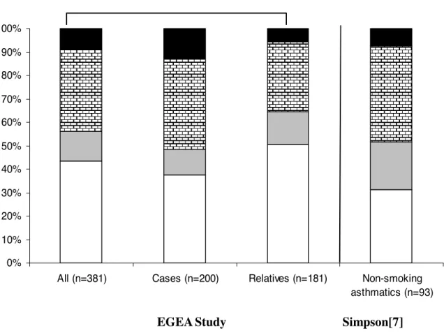
EOSlo/NEUlo, EOSlo/NEUhi, EOShi/NEUlo and EOShi/NEUhi patterns concerned 43.6, 12.6, 34.6 and 9.2% of the 381 adult asthmatics (Figure 1).
Table 1. -Characteristics of adult asthmatics included in the analyses Value All, n Asthmatic cases, n Asthmatic relatives, n 381 200 181
Age, year, mean ± SD 36.5 ± 13.1
Sex, women, % 49.6
Age of asthma onset, year, mean ± SD 18.0 ± 14.7
Total IgE, IU/ml, GM 166
Skin prick test positive response (any of 11 allergens), % 75.1
White blood cell counts
Eosinophils/mm3, mean ± SD
Neutrophils/mm3, mean ± SD 4202 ± 1667 275 ± 200
FEV1 % predicted, mean ± SD 93.5 ± 19.9
FEV1 < 80% predicted, % 21.4
Methacholine challenge, n* PD 20 ≤ 4 mg, %
185 75.1
Current asthma (asthma attacks in the last 12 months), % 72.3
Nocturnal symptoms (last 12 months), % Cough Chest tightness Shortness of breath 46.5 66.0 46.3
Asthma events score, last 12 months (1-4),[14] 2.36 ± 1.21
Asthma symptomatic score, last 12 months (1-5),[15] 2.96 ± 1.77
COPD-like symptoms, % Chronic cough Chronic phlegm Dyspnea grade 3 19.7 14.8 22.4 Smoking habits, % smokers ex-smokers non-smokers 21.7 26.8 51.5
Body Mass Index (BMI), kg/m2, mean ± SD 23.3 ± 16.2
Respiratory infection (last 3 weeks), % 14.7
Treatment (last 12 mo), % none
without inhaled steroids with inhaled steroids
27.3 20.5 52.2 *not performed if FEV1 < 80% pred.
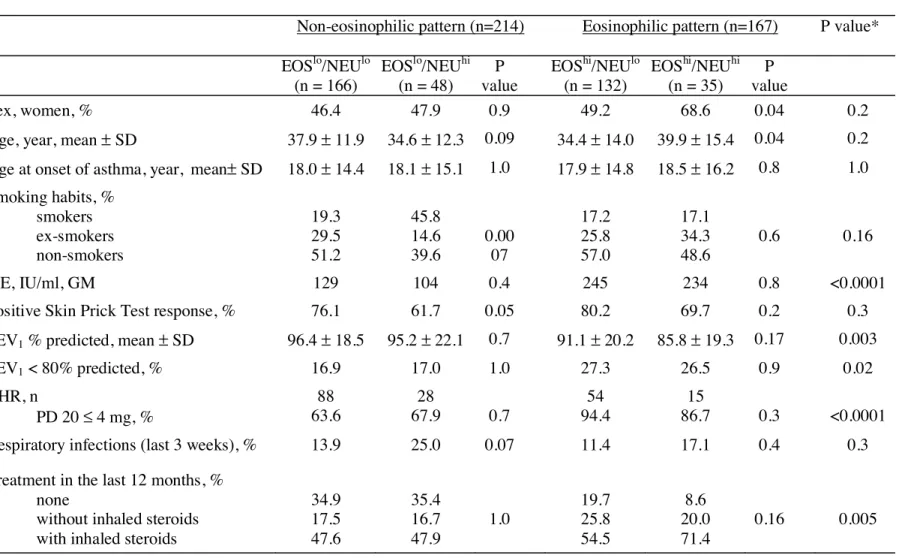
Table 2.- Characteristics of adult asthmatics according to their blood inflammatory pattern
Inflammatory patterns
Non-eosinophilic pattern (n=214) Eosinophilic pattern (n=167) P value*
EOSlo/NEUlo (n = 166) EOSlo/NEUhi (n = 48) P value EOShi/NEUlo (n = 132) EOShi/NEUhi (n = 35) P value Sex, women, % 46.4 47.9 0.9 49.2 68.6 0.04 0.2
Age, year, mean ± SD 37.9 ± 11.9 34.6 ± 12.3 0.09 34.4 ± 14.0 39.9 ± 15.4 0.04 0.2
Age at onset of asthma, year, mean± SD 18.0 ± 14.4 18.1 ± 15.1 1.0 17.9 ± 14.8 18.5 ± 16.2 0.8 1.0
Smoking habits, % smokers ex-smokers non-smokers 19.3 29.5 51.2 45.8 14.6 39.6 0.00 07 17.2 25.8 57.0 17.1 34.3 48.6 0.6 0.16 IgE, IU/ml, GM 129 104 0.4 245 234 0.8 <0.0001
Positive Skin Prick Test response, % 76.1 61.7 0.05 80.2 69.7 0.2 0.3
FEV1 % predicted, mean ± SD 96.4 ± 18.5 95.2 ± 22.1 0.7 91.1 ± 20.2 85.8 ± 19.3 0.17 0.003
FEV1 < 80% predicted, % 16.9 17.0 1.0 27.3 26.5 0.9 0.02 BHR, n PD 20 ≤ 4 mg, % 88 63.6 28 67.9 0.7 54 94.4 15 86.7 0.3 <0.0001
Respiratory infections (last 3 weeks), % 13.9 25.0 0.07 11.4 17.1 0.4 0.3
Treatment in the last 12 months, % none
without inhaled steroids with inhaled steroids
34.9 17.5 47.6 35.4 16.7 47.9 1.0 19.7 25.8 54.5 8.6 20.0 71.4 0.16 0.005
Comparison of asthma and COPD-like symptoms among asthmatics with eosinophilic and non-eosinophilic pattern
Asthmatics with EOShi pattern had significantly higher total IgE than those with EOSlo pattern (Table 2), which remained significant after adjustment for age, sex and smoking: 233 vs 117 IU/ml, p<10-4. They also had significantly lower FEV1, with adjusted values of 89.4 vs.
96.5%, (p=0.0003), and higher BHR than those with EOSlo pattern (Tables 2 and 5).
Asthmatics with EOShi pattern reported significantly more asthma attacks in the last 12 months, and more often being woken by an attack of shortness of breath or with chest tightness than those with EOSlo pattern (Tables 3 and 5). They also had significant higher asthma event score and symptomatic score than those with EOSlo pattern, with adjusted values of 2.64 vs 2.22, p=0.001, and 3.46 vs 2.69, p<10-4 respectively. Further adjustment for ICS treatment did not change the conclusion (not shown).
For COPD-like symptoms, asthmatics with EOShi pattern reported significantly more dyspnea
than those with EOSlo pattern (Table 4), association no longer significant in the multivariate analysis (Table 5). This association was similarly observed in men and in women.
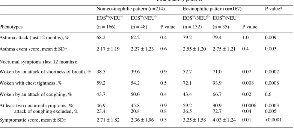
Table 3.- Clinical symptoms, and event and symptomatic scores in adult asthmatics according to their blood inflammatory pattern
Inflammatory patterns
Non-eosinophilic pattern (n=214) Eosinophilic pattern (n=167) P value*
Phenotypes EOSlo/NEUlo (n = 166) EOSlo/NEUhi (n = 48) P value EOShi/NEUlo (n = 132) EOSlo/NEUhi (n = 35) P value
Asthma attack (last 12 months), % 68.2 62.2 0.4 79.2 79.4 1.0 0.009
Asthma event score, mean ± SD† 2.17 ± 1.19 2.27 ± 1.23 0.6 2.55 ± 1.20 2.75 ± 1.21 0.4 0.003
Nocturnal symptoms (last 12 months):
Woken by an attack of shortness of breath, % 38.5 39.6 0.9 52.7 71.0 0.07 0.0002
Woken with chest tightness, % 59.2 54.2 0.5 72.1 93.9 0.008 0.0008
Woken by an attack of coughing, % 43.7 50.0 0.4 43.4 66.7 0.02 0.6
At least two nocturnal symptoms, % attack of coughing excluded, %
46.9 23.4 45.8 20.8 0.9 0.8 59.2 36.5 90.9 72.7 0.0006 0.04 0.0003 0.005
Symptomatic score, mean ± SD‡ 2.71 ± 1.82 2.36 ± 1.96 0.3 3.25 ± 1.58 4.03 ± 1.24 0.01 <0.0001
*Non-eosinophilic versus eosinophilic (crude P value).
†based on frequency of attacks, symptoms between attacks, hospitalisation taking treatment into account (see methods).
‡based on the number of asthma symptoms (wheeze and breathlessness, woken with chest tightness, woken by attack of shortness of breath, attack of shortness of breath at rest, attack of shortness of breath after exercise) (see methods).
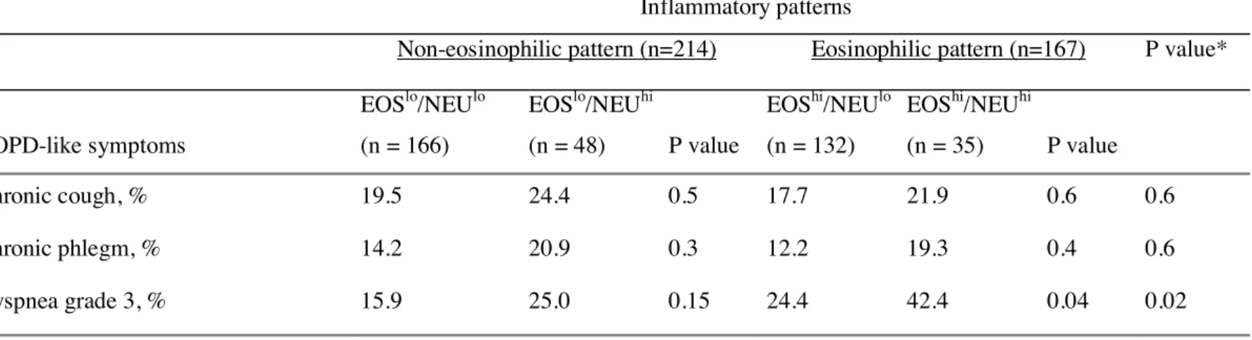
Table 4.- COPD-like symptoms in adult asthmatics according to their blood inflammatory pattern
Inflammatory patterns
Non-eosinophilic pattern (n=214) Eosinophilic pattern (n=167) P value*
COPD-like symptoms EOSlo/NEUlo (n = 166) EOSlo/NEUhi (n = 48) P value EOShi/NEUlo (n = 132) EOShi/NEUhi (n = 35) P value Chronic cough, % 19.5 24.4 0.5 17.7 21.9 0.6 0.6 Chronic phlegm, % 14.2 20.9 0.3 12.2 19.3 0.4 0.6 Dyspnea grade 3, % 15.9 25.0 0.15 24.4 42.4 0.04 0.02
Neutrophil inflammation among asthmatics with eosinophilic pattern
Asthmatics with the EOShi/NEUhi pattern were older and more frequently women (OR=2.25
(1.02-4.96)) than those with the EOShi/NEUlo pattern.
Despite a higher frequency of treatment with ICS, asthmatics with the EOShi/NEUhi pattern reported significantly more often being woken with chest tightness or by an attack of coughing than those with the EOShi/NEUlo pattern (Table 3), association which remained significant for chest tightness after adjustment (Table 5). Asthmatics with the EOShi/NEUhi pattern also reported more nocturnal symptoms considered together than those with EOShi/NEUlo pattern. Excluding woken by an attack of coughing from nocturnal symptoms did not change the conclusion. Asthmatics with the EOShi/NEUhi pattern have significant higher asthma symptomatic score than those with the EOShi/NEUlo pattern, independently of age, sex, and smoking (4.04 vs 3.36, p=0.03), association which became of borderline significance after further adjustment for ICS treatment (3.84 vs 3.31, p=0.07).
For COPD-like symptoms, asthmatics with the EOShi/NEUhi pattern reported significantly more dyspnea than those with the EOShi/NEUlo pattern, but the difference did not reach the level of significance after adjustment (Tables 4 and 5).
Neutrophil inflammation among asthmatics with non-eosinophilic pattern
Asthmatics with the EOSlo/NEUhi pattern were more often current smokers than those with the EOSlo/NEUlo pattern (OR=3.54 (1.78-7.04)) (Table 2). They had less positive skin prick test response than those with the EOSlo/NEUlo pattern (Tables 3 and 5). No other differences were observed.
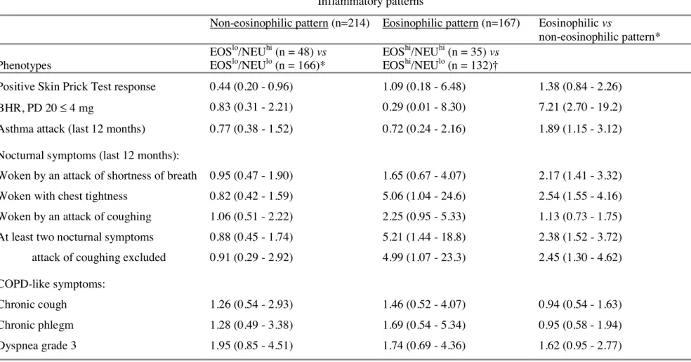
Table 5.- Associations between phenotypic characteristics and blood inflammatory patterns in adult asthmatics (multivariate analyses)
Inflammatory patterns
Non-eosinophilic pattern (n=214) Eosinophilic pattern (n=167) Eosinophilic vs
non-eosinophilic pattern* Phenotypes EOSlo/NEUhi (n = 48) vs EOSlo/NEUlo (n = 166)* EOShi/NEUhi (n = 35) vs EOShi/NEUlo (n = 132)†
Positive Skin Prick Test response 0.44 (0.20 - 0.96) 1.09 (0.18 - 6.48) 1.38 (0.84 - 2.26)
BHR, PD 20 ≤ 4 mg 0.83 (0.31 - 2.21) 0.29 (0.01 - 8.30) 7.21 (2.70 - 19.2)
Asthma attack (last 12 months) 0.77 (0.38 - 1.52) 0.72 (0.24 - 2.16) 1.89 (1.15 - 3.12)
Nocturnal symptoms (last 12 months):
Woken by an attack of shortness of breath 0.95 (0.47 - 1.90) 1.65 (0.67 - 4.07) 2.17 (1.41 - 3.32)
Woken with chest tightness 0.82 (0.42 - 1.59) 5.06 (1.04 - 24.6) 2.54 (1.55 - 4.16)
Woken by an attack of coughing 1.06 (0.51 - 2.22) 2.25 (0.95 - 5.33) 1.13 (0.73 - 1.75)
At least two nocturnal symptoms attack of coughing excluded
0.88 (0.45 - 1.74) 0.91 (0.29 - 2.92) 5.21 (1.44 - 18.8) 4.99 (1.07 - 23.3) 2.38 (1.52 - 3.72) 2.45 (1.30 - 4.62) COPD-like symptoms: Chronic cough 1.26 (0.54 - 2.93) 1.46 (0.52 - 4.07) 0.94 (0.54 - 1.63) Chronic phlegm 1.28 (0.49 - 3.38) 1.69 (0.54 - 5.34) 0.95 (0.58 - 1.94) Dyspnea grade 3 1.95 (0.85 - 4.51) 1.74 (0.69 - 4.36) 1.62 (0.95 - 2.77)
Results are expressed as odds ratios (ORs) (95% confidence interval (CI).
*adjusted for age, sex and smoking, and taking into account familial dependence of the subjects.
Comparisons of COPD-like symptoms among asthmatics with neutrophilic and non-neutrophilic pattern
Although chronic cough and chronic phlegm were more frequently reported by asthmatics with neutrophilic pattern (EOSlo/NEUhi or EOShi/NEUhi, n=48+35, NEUhi) than in those with non-neutrophilic pattern (EOSlo/NEUlo or EOShi/NEUlo, n=166+132, NEUlo) (Table 4), associations did not reach the level of significance with OR=1.32 (0.72-2.43) and 1.66 (0.85-3.22) for chronic cough and chronic phlegm respectively. Asthmatics with NEUhi pattern reported significantly more dyspnea than those with NEUlo pattern (OR=1.93 (1.12-3.35)). This association was similarly observed in men and in women, and remained significant after adjustment for age and smoking in women only with OR=2.08 (1.08 to 4.18), and OR=1.89 (0.74 to 4.81) in men.
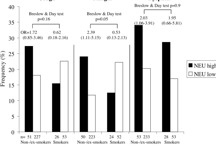
The relationships of COPD-like symptoms with neutrophilic pattern were examined by current smoking status (Figure 2). Only in non-smokers did asthmatics with NEUhi pattern report significantly more chronic phlegm and dyspnea than those with NEUlo pattern. The association remained significant after adjustment for age, sex and eosinophilic inflammation, and taking into account familial dependence of the subjects for chronic phlegm only with OR=2.35 (1.08-5.10), and OR=1.83 (0.95-3.53) and 1.79 (0.89-3.57) for dyspnea and chronic cough respectively. Adjusting for respiratory infections in the last three weeks did not change the conclusion (not shown).
Analyses done with the cut-point for neutrophils corresponding to the 90th percentile gave similar findings.
DISCUSSION
The present study shows marked differences in asthma phenotypic characteristics according to four blood inflammatory patterns defined by the amount of circulating eosinophils and neutrophils. Non-eosinophilic pattern (EOSlo) was present in 56% of the asthmatics, the EOSlo/NEUlo pattern being present in the majority (77%) of these asthmatics. Comparison of EOShi with EOSlo pattern confirms that EOShi had higher IgE, lower FEV1 and corresponds to
more active asthma (frequency of events and symptoms). Among EOSlo, neutrophil
inflammation (NEUhi) was related to less positive skin prick test response. Among EOShi, NEUhi did not explain current asthma or asthma event frequency, but was significantly related to nocturnal symptoms. In asthmatics without current smoking, COPD-like symptoms, in particular chronic phlegm were more frequent in those with NEUhi, independently of eosinophilic inflammation.
The first strength of this study was the possibility to study simultaneously the four inflammatory patterns in a way similar to that proposed by Simpson,[7] i.e. without overlap between the patterns, which was never done previously in epidemiology. Interestingly, the relative proportions of each pattern in our study were similar to those defined by Simpson[7] using induced sputum. The good characterization of asthmatics, and their heterogeneity regarding asthma, made of EGEA the ideal population to assess phenotypic characteristics according to blood inflammatory patterns. Limitations of our study are those commonly related to comparisons of groups with small sample sizes and those related to cross-sectional analyses of the data. Inflammatory patterns were defined according to eosinophil and neutrophil counts in blood, a fluid which may be considered as less reflecting lung inflammation than induced sputum, but is more easily accessible in clinical practice. Eosinophil count cut-point (250 cell/mm3) was commonly used in epidemiology, and a cut-point of 5000 cell/mm3 was chosen for neutrophils. Both cut-points corresponded to the
percentile 75th among the 1356 adults of EGEA, and the percentile 75th among the 901 non-asthmatic adults of EGEA corresponded to the same neutrophil cut-point. Increasing the neutrophil cut-point of 15% did not change the main conclusions. Further, correlations between inflammatory markers, including eosinophils and neutrophils, in induced sputum and peripheral blood have been scarcely studied.[14-18] Morphological and functional characteristics of bronchial eosinophils were similar to those of blood low-density eosinophils in patients with asthma,[14] and concomitent decreases in FeNO and blood neutrophil counts were observed among bar workers two months after a legislative ban on smoking in public places,[15] whereas no significant correlations between sputum and blood lymphocyte subsets in non-smoking adult asthmatics were reported.[16] In welders, only blood eosinophil count was related to the extend of welding,[17] whereas both sputum and blood eosinophils decreased in asthmatics after treatment with steroids.[18] These results suggest that blood and sputum eosinophils and neutrophils could respond differently to the same stimuli, and that the measurement of eosinophils and neutrophils in each of the specimen may give interesting complementary information.
Interestingly, in our asthmatic population, around 56% had a “non-eosinophilic asthma”, a result similar to those reported in general populations, accounting for around 30 to 70% of asthmatics depending on the studies.[1] Regarding eosinophil and neutrophil cut-points, 44% of asthmatics had a EOSlo/NEUlo pattern, suggesting that this pattern may represent another “type” of asthma in which blood inflammation is not a major feature, or that disease was not active at the moment of the study.
We confirmed the well-documented associations of blood eosinophilic pattern with high IgE, increased bronchial hyperresponsiveness (BHR) and lower FEV1 previously reported in
general or occupational populations.[5, 6, 19] Activated eosinophils are known to release several mediators which cause damages to the airway epithelium, leading to BHR due to
increased permeability.[20] We observed associations of eosinophilic pattern with more asthma attacks and more nocturnal symptoms reported in the last 12 months, and higher asthma event score and symptomatic score. These two scores should be considered as continuous variables reflecting the activity of the disease and also its severity,[10, 21] and peripheral eosinophil count has been suggested to be a marker of asthma activity.[22]
Among asthmatics with non-eosinophilic pattern, neutrophil inflammation was associated with less allergic sensitization, result which supports and extends previous reports,[23] suggesting a different pathogenesis than allergen-induced asthma, possibly more related to environmental exposure to various pollutants such as ozone or particulates,[1] and mediated through macrophages and epithelial cells rather than activated TH2 cells.[24]
Among asthmatics with eosinophilic pattern, neutrophil inflammation was clearly associated with more reports of dyspnea and nocturnal symptoms, and with a higher symptomatic score, suggesting a “more active” disease. No association was observed in asthmatics with non-eosinophilic inflammation, as previously found by Wenzel in induced sputum.[25] There is also growing evidence supporting that increased neutrophilic inflammation is present in “more severe” asthma.[26] Asthmatics who have severe disease and are resistant to corticosteroids, have raised neutrophil counts in their airways. Positive correlations between the concentrations of neutrophils and eosinophils in induced sputum from patients with severe asthma who are treated with drugs including corticosteroids have been reported.[27] As asthmatics with the EOShi/NEUhi pattern were more often treated with ICS, the high eosinophil and neutrophil counts could be at least partially a consequence of steroid treatment, known to enhance neutrophil survival.[28] Neutrophils may lead eosinophils to accumulate in the airways of patients with severe asthma and possibly aggravate the disease,[29] and it is unlikely that eosinophils regulate neutrophilic inflammation.[30] High eosinophil and neutrophil counts may also be related to exposure to specific environmental factors, in
particular ozone, recently found to promote an anti-apoptotic environment in allergen-primed animals.[31] Overall, relationships of neutrophil inflammation to asthma severity seem to depend upon the presence of eosinophil inflammation.
In our study, among asthmatics with eosinophilic pattern, those with neutrophil inflammation were older and more frequently women. Remodelling is known to increased with age and to be associated with neutrophilia.[32] Activated neutrophils may release inflammatory mediators, oxygen radicals and proteases which supports their involvement in the intense inflammation and remodelling found in severe asthma.[33] Regarding nocturnal asthma, intricate circadians variations in inflammation and in physiological manifestations have been reported,[34] including the proinflammatory hormone melatonin, and the hypothalamic-pituitary-adrenal axis. We previously found that in women with a history of premenstrual asthma, eosinophil counts were significantly higher than in other asthmatic women, an association that remained after adjustment for asthma severity.[35] Further, spontaneous neutrophil apoptosis is lower in healthy women as compared to men.[36] Hormone-related events may have an influence in the relationships of high eosinophil and neutrophil numbers with asthmatic symptoms. Despite the small sample size of the group of asthmatics with the EOShi/NEUhi pattern, all our results suggest that these asthmatics should be considered as having asthma with specific features different from those of asthmatics with eosinophilic inflammation alone.
Regarding COPD-like symptoms, our study revealed associations of neutrophilic pattern with more frequent reports of chronic cough, chronic phlegm and dyspnea. Significant associations of NEUhi with more frequent report of chronic phlegm and dyspnea occured in non- or ex-smokers, whereas no association was observed in smokers. The prevalence of reported chronic phlegm in our study was similar to that reported in previous epidemiological studies conducted in general populations from various contries,[37, 38] and even when considering
non-smokers only.[39] Results should be interpreted with caution as they were based on cross-sectional analyses. In non- or ex-smokers, results suggest that the association of hypersecretion with neutrophils reflects airway inflammation related to asthma and not to smoking. The lack of association in smokers may reflect a “healthy smoker effect” i.e. that asthmatic smokers stop smoking earlier than controls.[40] More attention should be paid to mucus hypersecretion in non-smokers. Analyses performed in our study did not allow to disentangle the association of EOShi/NEUhi with female sex and dyspnea which could be specifically considered in future epidemiological studies.
In conclusion, as suggested by Wenzel,[4] marked differences in phenotypic characteristics of asthma were evidenced according to blood inflammatory patterns. Besides eosinophilia, blood neutrophil inflammation is related to a different presentation of the asthmatic disease. Epidemiological studies on blood inflammatory patterns may provide additional information to physiological studies, and may contribute to help to disentangle this complex disease.
ACKNOWLEDGMENTS
The authors thank Benedicte Jacquemin and Jean Maccario for helpful discussions.
EGEA cooperative group
Coordination: F Kauffmann; F Demenais (genetics); I Pin (clinical aspects). Respiratory epidemiology: Inserm U 700, Paris M Korobaeff (EGEA1), F Neukirch (EGEA1); Inserm U
707, Paris : I Annesi-Maesano; Inserm U 780, Villejuif: F Kauffmann, N Le Moual, R Nadif, MP Oryszczyn; Inserm U 823, Grenoble: V Siroux. Genetics: Inserm U 393, Paris: J Feingold; Inserm U 535, Villejuif: MH Dizier; Inserm U 794, Evry: E Bouzigon, F Demenais; CNG, Evry: I Gut, M Lathrop. Clinical centers : Grenoble: I Pin, C Pison; Lyon: D Ecochard (EGEA1), F Gormand, Y Pacheco; Marseille: D Charpin (EGEA1), D Vervloet ; Montpellier: J Bousquet; Paris Cochin: A Lockhart (EGEA1), R Matran (now in Lille); Paris Necker: E Paty, P Scheinmann; Paris-Trousseau: A Grimfeld, J Just. Data and quality management: Inserm ex-U155 (EGEA1): J Hochez; Inserm U 780, Villejuif: N Le Moual, C Ravault; Inserm U 794, Paris: N Chateigner; Grenoble: J Ferran.
COMPETING INTEREST
CP has not received any research grants from companies but received reimbursement from Novartis France, GlaxoSmithKline France, Boehringer Ingelheim France, AstraZeneca France and Actélion France for attending several conferences : ISHLT, ERS, CPLF.
FUNDING
This research was funded in part by INSERM/Ministry of Research, ANR 05-SEST-020-02/05-9-97, ANR-06-CEBS, and the GA2LEN project, Global Allergy and Asthma European Network.
REFERENCES
1. Douwes J, Gibson P, Pekkanen J, et al. Non-eosinophilic asthma: importance and possible mechanisms. Thorax 2002;57:643-648.
2. Alam R, Busse WW. The eosinophil--quo vadis? J Allergy Clin Immunol 2004;113:38-42.
3. Oryszczyn M-P, Bouzigon E, Maccario J, et al. Interrelationships of quantitative asthma-related phenotypes in the Epidemiological Study on the Genetics and Environment of Asthma, Bronchial Hyperresponsiveness, and Atopy. J Allergy Clin Immunol 2007;119:57-63.
4. Wenzel SE. Asthma: defining of the persistent adult phenotypes. The Lancet
2006;368:804-813.
5. Kauffmann F, Neukirch F, Korobaeff M, et al. Eosinophils, smoking, and lung function.
An epidemiologic survey among 912 working men. Am Rev Respir Dis 1986;134:1172-1175.
6. Lewis SA, Pavord ID, Stringer JR, et al. The Relation Between Peripheral Blood Leukocyte Counts and Respiratory Symptoms, Atopy, Lung Function, and Airway Responsiveness in Adults. Chest 2001;119:105-114.
7. Simpson JL, Scott R, Boyle MJ, et al. Inflammatory subtypes in asthma: assessment and
identification using induced sputum. Respirology 2006;11:54-61.
8. Kauffmann F, Dizier M-H, Pin I, et al. Epidemiological Study of the Genetics and Environment of Asthma, Bronchial Hyperresponsiveness, and Atopy . Phenotype Issues.
Am. J. Respir. Crit. Care Med. 1997;156:123S-129.
9. Burney PG, Luczynska C, Chinn S, et al. The European Community Respiratory Health
Survey. Eur Respir J 1994;7:954-960.
10. Bouzigon E, Siroux V, Dizier MH, et al. Scores of asthma and asthma severity reveal new regions of linkage in EGEA study families. Eur Respir J 2007;30:253-259.
11. Pekkanen J, Sunyer J, Anto JM, et al. Operational definitions of asthma in studies on its aetiology. Eur Respir J 2005;26:28-35.
12. Global INitiative for Asthma. Global strategy for asthma management and prevention. . Bethesda (MD): National Heart, Lung, and Blood Institute, National Institute of Health.
1995 (updated 2006); NIH publication No. 95-3659.:URL:http://www.ginasthma.org
13. Swaanenburg JC, Rutten WP, Holdrinet AC, et al. The determination of reference values
for hematologic parameters using results obtained from patient populations. Am J Clin
Pathol 1987;88:182-191.
14. Gereng EA, Sukhodolo IV, Pleshko RI, et al. Comparative study of eosinophils in the blood and induced sputum during bronchial asthma. Bull Exp Biol Med 2004;137:50-52.
15. Menzies D, Nair A, Williamson PA, et al. Respiratory Symptoms, Pulmonary Function,
and Markers of Inflammation Among Bar Workers Before and After a Legislative Ban on Smoking in Public Places. JAMA 2006;296:1742-1748.
16. Pizzichini E, Pizzichini MM, Kidney JC, et al. Induced sputum, bronchoalveolar lavage and blood from mild asthmatics: inflammatory cells, lymphocyte subsets and soluble markers compared. Eur Respir J 1998;11:828-834.
17. Palmer KT, McNeill-Love R, Poole JR, et al. Inflammatory responses to the occupational
inhalation of metal fume. Eur Respir J 2006;27:366-373.
18. Bacci E, Cianchetti S, Ruocco L, et al. Comparison between eosinophilic markers in induced sputum and blood in asthmatic patients. Clin Exp Allergy 1998;28:1237-1243. 19. Jansen Desiree F, Rijcken B, Schouten Jan P, et al. The Relationship of Skin Test
Positivity, High Serum Total IgE Levels, and Peripheral Blood Eosinophilia to Symptomatic and Asymptomatic Airway Hyperresponsiveness. Am. J. Respir. Crit. Care
20. Gleich GJ, Flavahan NA, Fujisawa T, et al. The eosinophil as a mediator of damage to respiratory epithelium: a model for bronchial hyperreactivity. J Allergy Clin Immunol 1988;81:776-781.
21. Sunyer J, Pekkanen J, Garcia-Esteban R, et al. Asthma score: predictive ability and risk factors. Allergy 2007;62:142-148.
22. Ulrik CS. Peripheral eosinophil counts as a marker of disease activity in intrinsic and extrinsic asthma. Clin Exp Allergy 1995;25:820-827.
23. Simpson JL, Grissell TV, Douwes J, et al. Innate immune activation in neutrophilic asthma and bronchiectasis. Thorax 2007;62:211-218.
24. Pavord ID. Non-eosinophilic asthma and the innate immune response. Thorax
2007;62:193-194.
25. Wenzel SE, Schwartz LB, Langmack EL, et al. Evidence That Severe Asthma Can Be Divided Pathologically into Two Inflammatory Subtypes with Distinct Physiologic and Clinical Characteristics. Am. J. Respir. Crit. Care Med. 1999;160:1001-1008.
26. Holgate ST, Polosa R. The mechanisms, diagnosis, and management of severe asthma in
adults. The Lancet 2006;368:780-793.
27. Kikuchi S, Nagata M, Kikuchi I, et al. Association between neutrophilic and eosinophilic inflammation in patients with severe persistent asthma. Int Arch Allergy Immunol 2005;137 Suppl 1:7-11.
28. Sivertson KL, Seeds MC, Long DL, et al. The differential effect of dexamethasone on granulocyte apoptosis involves stabilization of Mcl-1L in neutrophils but not in eosinophils. Cellular Immunology 2007;246:34-45.
29. Kikuchi I, Kikuchi S, Kobayashi T, et al. Eosinophil Trans-Basement Membrane
Migration Induced by Interleukin-8 and Neutrophils. Am. J. Respir. Cell Mol. Biol. 2006;34:760-765.
30. Kobayashi T, Takaku Y, Kikuchi I, et al. Eosinophils do not enhance the trans-basement-membrane migration of neutrophils. Int Arch Allergy Immunol 2007;143 Suppl 1:38-43. 31. Kierstein S, Krytska K, Sharma S, et al. Ozone inhalation induces exacerbation of
eosinophilic airway inflammation and hyperresponsiveness in allergen-sensitized mice.
Allergy 2008;63:438-446.
32. Foley SC, Hamid Q. Images in allergy and immunology: neutrophils in asthma. J Allergy
Clin Immunol 2007;119:1282-1286.
33. Bousquet J, Jeffery PK, Busse WW, et al. Asthma . From Bronchoconstriction to Airways
Inflammation and Remodeling. Am. J. Respir. Crit. Care Med. 2000;161:1720-1745.
34. Sutherland ER. Nocturnal asthma. J Allergy Clin Immunol 2005;116:1179-1186.
35. Siroux V, Curt F, Oryszczyn MP, et al. Role of gender and hormone-related events on IgE, atopy, and eosinophils in the Epidemiological Study on the Genetics and Environment of Asthma, bronchial hyperresponsiveness and atopy. J Allergy Clin
Immunol 2004;114:491-498.
36. Molloy EJ, O'Neill AJ, Grantham JJ, et al. Sex-specific alterations in neutrophil apoptosis: the role of estradiol and progesterone. Blood 2003;102:2653-2659.
37. Bjornsson E, Plaschke P, Norrman E, et al. Symptoms related to asthma and chronic bronchitis in three areas of Sweden. Eur Respir J 1994;7:2146-2153.
38. Dodge RR, Burrows B. The prevalence and incidence of asthma and asthma-like
symptoms in a general population sample. Am Rev Respir Dis 1980;122:567-575.
39. Berglund DJ, Abbey DE, Lebowitz MD, et al. Respiratory Symptoms and Pulmonary Function in an Elderly Nonsmoking Population. Chest 1999;115:49-59.
40. Siroux V, Pin I, Oryszczyn MP, et al. Relationships of active smoking to asthma and asthma severity in the EGEA study. Epidemiological study on the Genetics and Environment of Asthma. Eur Respir J 2000;15:470-477.
Figure 1. Frequency of each inflammatory pattern in adult asthmatics.
Numbers of asthmatics are shown below each bar.
EOSlo/NEUlo EOSlo/NEUhi EOShi/NEU EOShi/NEUhi
Figure 2. Associations of COPD-like symptoms: chronic cough, chronic phlegm and dyspnea with neutrophilic pattern according to current smoking.Black boxes:
neutrophilic pattern (EOSlo/NEUhi or EOShi/NEUhi); gray boxes: non-neutrophilic patterns (EOSlo/NEUlo or EOShi/NEUlo).
Numbers of asthmatics are shown below each bar.
P values of Breslow and Day tests for interaction, which refers to the heterogeneity of odds ratios according to smoking habits.
Figure 1. Frequency of each inflammatory pattern in adult asthmatics.
Numbers of asthmatics are shown below each bar.
EOSlo/NEUlo EOSlo/NEUhi EOShi/NEUlo EOShi/NEUhi
0% 10% 20% 30% 40% 50% 60% 70% 80% 90% 100%
All (n=381) Cases (n=200) Relatives (n=181) Non-smoking
asthmatics (n=93)
Figure 2. Associations of COPD-like symptoms: chronic cough, chronic phlegm and dyspnea with neutrophilic
pattern according to current smoking.
Black boxes: neutrophilic pattern (EOS
lo/NEU
hior EOS
hi/NEU
hi); white boxes: non-neutrophilic patterns (EOS
lo/NEU
loor EOS
hi/NEU
lo). Numbers of asthmatics are shown below each bar.
Breslow and Day test for interaction refers to the heterogeneity of odds ratios according to smoking habits.
0
5
10
15
20
25
30
35
40
F
re
q
u
en
cy
(
%
)
NEU high
NEU low
n= 51 227 26 53 50 223 24 52 53 233 28 53 Non-/ex-smokers Smokers Non-/ex-smokers Smokers Non-/ex-smokers Smokers OR=1.72 0.62 2.39 0.53(0.85-3.46) (0.18-2.16) (1.11-5.15) (0.13-2.13) Breslow & Day test
p=0.16
Breslow & Day test
p=0.05 2.03 1.95 (1.06-3.91) (0.66-5.81) Breslow & Day test p=0.9