HAL Id: inserm-00137717
https://www.hal.inserm.fr/inserm-00137717
Submitted on 15 Jan 2010HAL is a multi-disciplinary open access archive for the deposit and dissemination of sci-entific research documents, whether they are pub-lished or not. The documents may come from teaching and research institutions in France or abroad, or from public or private research centers.
L’archive ouverte pluridisciplinaire HAL, est destinée au dépôt et à la diffusion de documents scientifiques de niveau recherche, publiés ou non, émanant des établissements d’enseignement et de recherche français ou étrangers, des laboratoires publics ou privés.
Ribosome-translocon complex mediates calcium leakage
from endoplasmic reticulum stores.
Fabien van Coppenolle, Fabien Vanden Abeele, Christian Slomianny,
Matthieu Flourakis, John Hesketh, Etienne Dewailly, Natalia Prevarskaya
To cite this version:
Fabien van Coppenolle, Fabien Vanden Abeele, Christian Slomianny, Matthieu Flourakis, John Hesketh, et al.. Ribosome-translocon complex mediates calcium leakage from endoplasmic retic-ulum stores.. Journal of Cell Science, Company of Biologists, 2004, 117 (Pt 18), pp.4135-42. �10.1242/jcs.01274�. �inserm-00137717�
Introduction
The endoplasmic reticulum (ER) plays an essential role in the folding and maturation of proteins and is also the largest calcium store (Berridge and Irvine, 1989; Pozzan et al., 1994). It has previously been demonstrated that the ER is a continuous calcium pool in mouse acinar pancreatic cells (Mogami et al., 1998; Park et al., 2000). Agonist-induced ER calcium release occurs through Ca2+ channels such as inositol triphosphate (IP3) and ryanodine receptors (Clapham, 1995) and may be potentiated by nicotinic acid adenine dinucleotide phosphate (NAADP) (Cancela et al., 2002). Calcium reuptake into intracellular stores occurs when the calcium release channels are closed (calcium negative feedback to the IP3 receptor) (Bezprozvanny et al., 1991) and is carried out by SERCA pumps (sarco-endoplasmic reticulum Ca2+-ATPases) (Pozzan et al., 1994). In the resting state, the Ca2+ content of the ER reflects a balance between this active uptake by SERCA and passive efflux or ‘basal leak’ through previously identified ‘leak channels’. This leakage is revealed when SERCA pumps are inhibited by thapsigargin or CPA (Hofer et al., 1996; Mogami et al., 1998). The physical structure of calcium leak channels has not yet been elucidated. Recently, interesting research (Heritage and Wonderlin, 2001; Roy and Wonderlin, 2003) has demonstrated that a polarized molecule could cross the endoplasmic membrane through the translocon (Simon and
Blobel, 1991), which is the complex involved in protein translocation. The translocon may therefore be permeable to calcium. In yeast and mammalian cells (Deshaies and Schekman, 1987; Gorlich et al., 1992b; Gorlich and Rapoport, 1993; Stirling et al., 1992), the translocon is formed by the association of heterotrimeric proteins Sec61α, Sec61β, and Sec61γ (Gorlich et al., 1992a) and other proteins including TRAM (translocation-associated membrane protein), BiP, calnexin, calreticulin, and Erp57 (for reviews, see Johnson and van Waes, 1999; Schnell and Hebert, 2003).
Electrophysiological studies have demonstrated that the translocon is a protein-conducting channel (Simon and Blobel, 1991), with a 4-6 nm pore diameter when bound to the ribosome (Hamman et al., 1997). The ribosome-free translocon has a 0.9-1.5 nm pore diameter (Hamman et al., 1998). This pore was visualized in electron microscopy using purified yeast Sec61 complex (Beckmann et al., 1997) with a 9.5 nm outer diameter. In mouse acinar pancreatic cells, we have demonstrated that puromycin induces a decrease in the endoplasmic reticulum calcium concentration ([Ca2+]
ER) (Lomax et al., 2002). Puromycin conformation is similar to the 3′ end of aminoacetylated tRNA. It terminates the peptide chain elongation and clears the protein from the translocon pore (Pestka, 1974; Pestova et al., 2001). At this point, the ribosome is still on the translocon.
Under resting conditions, the endoplasmic reticulum (ER) intraluminal free calcium concentration ([Ca2+]
ER) reflects a balance between active uptake by Ca2+-ATPases and passive efflux via ‘leak channels’. Despite their physiological importance and ubiquitous leak pathway mechanism, very little is known about the molecular nature of these channels. As it has been suggested that the open translocon pore complex of the ER is permeable to ions and neutral molecules, we hypothesized that the ribosome-bound translocon would be permeable to calcium after treatment with puromycin, a translation inhibitor that specifically releases polypeptide chains. At this time, the translocon channel is left open. We measured the fluctuations in cytoplasmic and luminal calcium concentrations using fluorescent dyes (fura-2 and mag-fura-2, respectively). The calcium release induced by
thapsigargin (a Ca2+-ATPase inhibitor) was lower after puromycin treatment. Puromycin also reduced the [Ca2+]
ER level when perfused into the medium, but was ineffective after anisomycin pre-treatment (an inhibitor of the peptidyl transferase). Puromycin had a similar effect in the presence of heparin and ryanodine. This puromycin-evoked [Ca2+]
ERdecrease was specific to the translocon. We conclude that the translocon complex is a major calcium leak channel. This work reveals a new role for the translocon which is involved in the control of the [Ca2+]
ER and could therefore supervise many physiological processes, including gene expression and apoptosis.
Key words: Prostate cancer, Translocon, Puromycin, Calcium leak, Endoplasmic reticulum, LNCaP cells
Summary
Ribosome-translocon complex mediates calcium
leakage from endoplasmic reticulum stores
Fabien Van Coppenolle1,*,‡, Fabien Vanden Abeele1,*, Christian Slomianny1, Matthieu Flourakis1,
John Hesketh2, Etienne Dewailly1 and Natalia Prevarskaya1
1Laboratoire de Physiologie Cellulaire, INSERM EMI 0228, Université de Lille 1, Bâtiment SN3, 59655 Villeneuve d’Ascq CEDEX, France 2School of Cellular and Molecular Biosciences, University of Newcastle, Agriculture Building, Kings Road, Newcastle-upon-Tyne, NE1 7RU, UK *These authors contributed equally to this work
‡Author for correspondence (e-mail: [email protected]) Accepted 19 April 2004
Journal of Cell Science 117, 4135-4142 Published by The Company of Biologists 2004 doi:10.1242/jcs.01274
4136
In this study, we demonstrate the role of the translocon as a calcium leak channel. Puromycin induces a calcium leak from the ER and decreases the thapsigargin response. Cycloheximide, an inhibitor of the elongation factor 2 (that does not release the nascent peptide chain) (Roy and Wonderlin, 2003), was unable to modify the [Ca2+]
ER. Thus, the puromycin-evoked calcium leak results from its direct effect on the ribosome-translocon complex rather than inhibition of protein synthesis.
We also verified that the decrease in the ER calcium content induced by puromycin, was specific to its action on the translocon process. Puromycin was unable to induce a calcium release after anisomycin application (an inhibitor of the peptidyl transferase) (Ioannou et al., 1998). Furthermore, we demonstrate that puromycin does not induce a calcium release through the IP3and ryanodine receptors. We also verified that the NAADP-induced calcium release was not affected by puromycin.
Using confocal microscopy on LNCaP cells, we observed a decrease in the colocalization between the 60 S ribosome subunit and the Sec61 protein in the translocon pore in cells treated with puromycin. We made similar observations in electron microscopy. This implies that even a small number of open translocon is sufficient to develop a calcium leak capable of decreasing the [Ca2+]
ER.
This study is the first to demonstrate that a passive calcium leak may occur specifically via the translocon during translation. This is a ubiquitous phenomenon and may influence the filling state of internal calcium stores, causing a modification in calcium signaling and cellular physiology.
Materials and Methods
Fluorescence measurements of [Ca2+]
c and [Ca2+]ER
Cytoplasmic calcium concentration ([Ca2+]c) was measured using
fura-2 using previously described methods (Vanden Abeele et al., 2002). The cells were continuously perfused with HBSS solution (120 mM NaCl; 5 mM KCl; 2 mM CaCl2; 2 mM MgCl2; 5 mM Glucose;
10 mM HEPES, pH 7.3) and chemicals were added via a whole-chamber perfusion system. The flow rate of the perfusion system was set to 1 ml/minute and the chamber volume was 500 µl.
For Ca2+imaging within the ER, LNCaP cells (ATTC, Rockville,
MD) were grown on glass cover slips and loaded with 2 µM of the AM-ester derivative of mag-fura-2 (Molecular Probes, Leiden, The Netherlands), or 5 µM of mag-fluo-4 AM (Molecular Probes, Leiden, The Netherlands) for 45 minutes at 37°C. After incubation with dye, the plasma membrane was then selectively permeabilized: cells were rinsed briefly in a high K+solution (125 mM KCl, 25 mM NaCl, 10
mM HEPES and 0.1 mM MgCl2, pH 7.2), exposed for 2 minutes to
an ‘intracellular buffer’ at 37°C after which 5 mg/ml digitonin was added. Permeabilized cells were continuously perfused with ‘intracellular buffer’ (the same solution without digitonin, supplemented with 0.2 mM MgATP, with free [Ca2+] clamped to 170
nM using Ca2+/EGTA buffers). Ratio imaging measurements of
mag-fura-2 fluorescence were made using a commercial imaging system (Princeton Instruments, Evry, France). Ratio imaging measurements of mag-fluo-4 were made using a confocal microscope (LSM 510, Zeiss, Le Pecq, France).
Confocal microscopy and colocalization
LNCaP cells were grown on cover slips and treated with puromycin as described below. Samples were fixed with cold acetone (–20°C) for
15 minutes and blocked with 1.2% gelatine in PBS (PBSG) for 30 minutes to avoid non-specific binding. They were subsequently incubated in a moist chamber for 1 hour at 37°C with the primary antibodies for Sec61 (goat polyclonal from Santa Cruz) and 60S ribosomal subunit (rabbit polyclonal) (Horne and Hesketh, 1990). After several washes in PBSG, the cover slips were incubated for 1 hour at 37°C with the corresponding secondary antibodies (donkey goat labelled with Texas Red X (Chemicon) and donkey anti-rabbit labelled with FITC, Jackson), washed in PBS and mounted in Mowiol. Fluorescence analysis was carried out using a Zeiss LSM 510 confocal microscope (488 nm excitation for FITC and 563 nm for Texas Red) connected to a Zeiss Axiovert 200 M with a ×63 1.4 numerical aperture oil immersion objective. Both channels were excited, collected separately and then merged to examine colocalization. The image acquisition characteristics (pinhole aperture, laser intensity, scan speed, etc.) were the same throughout the experiments to ensure the comparability of the results. Using confocal microscope software (AIM 3.2, Zeiss, Le Pecq, France) we calculated the colocalization coefficients as defined (Manders et al., 1993). This process calculates the sum of the colocalized pixels (labeled in red and green) divided by the total number of labeling pixels. The maximal coefficient of colocalization is therefore 1.
Electron microscopy
The cell pellets were fixed in 2.5% glutaraldehyde in 0.1 M cacodylate buffer, pH 7.4, for 30 minutes at 4°C, thoroughly washed in the same buffer, post-fixed in 1% osmium tetroxide in the same buffer for another 30 minutes at 4°C, washed and subsequently processed for embedding in Epon resin. The samples were cut on a Reichert Ultracut E and the section contrasted with lead citrate and uranyl acetate. The grids were then observed on a Hitachi H600 transmission electron microscope.
Results
Thapsigargin-induced calcium release is lower after puromycin treatment
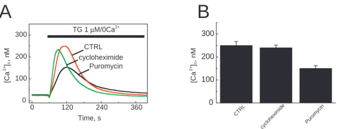
To study the possible involvement of translocon in calcium leakage, we first treated the LNCaP cells with puromycin (200 µM) or cycloheximide (1.8 mM) for 1 hour, or kept them in the medium alone (Fig. 1). In order to estimate the ER calcium content, we used thapsigargin (1 µM), which irreversibly blocks SERCA pumps and induces Ca2+ mobilization. We measured the passive calcium leak using the fura-2 (2 µM) calcium cytoplasmic probe in a Ca2+-free medium. The peak increase in cytoplasmic calcium concentration ([Ca2+]
c) induced by thapsigargin is therefore proportional to the initial calcium content of the endoplasmic reticulum. Fig. 1A shows the time course of a typical thapsigargin response in a calcium-free medium. The peak was 250 nM under control conditions and 230 nM after a 1-hour incubation with cycloheximide. Cells treated with puromycin showed a decrease in the thapsigargin response (150 nM). These mean differences are shown in Fig. 1B, which illustrates the peak value of the thapsigargin-evoked [Ca2+]
c increase. Under control conditions, the peak [Ca2+]
c value was 250±17.5 nM (n=34) and the response to thapsigargin of cycloheximide-incubated LNCaP cells was not statistically different (240±12 nM; n=35). In contrast, in puromycin-treated cells, the ER calcium content decreased by 40% compared to levels under control conditions and 37.5% compared to levels in cycloheximide-treated cells (150±12 nM; n=78).
Puromycin induces a luminal calcium leak through the translocon
The ER Ca2+ content was further investigated using the compartmentalized fluorescent Ca2+ indicator, mag-fura-2 AM, which we had previously used for direct measurements of [Ca2+]
ER in LNCaP cells (Vanden Abeele et al., 2002). Imaging experiments with mag-fura-2 AM were conducted after digitonin permeabilization. As shown in Fig. 2A, ionomycin (1 µM) induced a decrease in [Ca2+]
ER from 263
µM to 58 µM (–78%). The inset panels in Fig. 2A,B show the significant time course development of emission fluorescence from 340 nm and 380 nm excitation. Puromycin perfusion (200 µM) induced an 46% decrease in [Ca2+]
ER (Fig. 2B). After
puromycin treatment, the calcium store was almost empty but ionomycin induced a decrease, from 152 µM to 18 µM (Fig. 2B). Fig. 2C illustrates the cumulative data before and after puromycin perfusion. The mean decrease was 39.95±2.16% (n=26). A 20-µM puromycin concentration also induced a decrease in [Ca2+]
ER. Under these conditions the percentage of response was 30% with a mean decrease of 18.51±5.32% (n=15; data not shown).
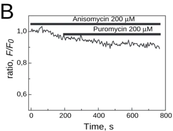
Anisomycin prevents puromycin-induced luminal calcium release
To be sure that puromycin induces a decrease in [Ca2+] ER by
A
B
0 120 240 360 0 100 200 300 cycloheximide Puromycin CTRL TG 1 µM/0Ca2+ ] a C[ + 2 c M , n ] a C[ + 2 c , n M Time, s 0 100 200 300 e d i m i x e h o l c y c n i c y m o r u P CTRLFig. 1. Puromycin treatment reduces the thapsigargin-induced calcium release from the ER. (A) Traces represent the [Ca2+]
c measured in 2 µM
fura-2 AM loaded LNCaP cells under control conditions or after a 1-hour incubation with 200 µM puromycin or 1.8 mM cycloheximide. Application of 1 µM thapsigargin (TG) produced an increase in [Ca2+]
cas a result of the inhibition of SERCA pumps. All measurements were
made at room temperature in Ca2+-free HBSS. (B) Means of the peak values of thapsigargin responses under control conditions (n=34), with
1.8 mM cycloheximide (n=35) and with 200 µM puromycin (n=78).
A
B
C
0 120 240 360 480 600 0 100 200 300 400 IM 1 µM Time, s 0 120 240 360 0 100 200 300 400 Time, s I380 I340 0 20 40 60 80 % of release I340 I380 100 CTRL Puromy cin 200 µM [C a 2 +] E R , µ M [C a 2 +] E R , µ M Puromycin 200 µM IM 1 µMFig. 2. Puromycin reduces the free Ca2+concentration within the ER
lumen. (A,B) Typical [Ca2+]ERtraces from mag-fura-2 AM loaded
LNCaP cells in response to 1 µM ionomycin (IM) (A) and 200 µM puromycin (B). The inset panels show individual fluorescence intensities at 340 nm (Ca2+-insensitive) and 380 nm (Ca2+-sensitive)
excitation wavelengths. Note that application of ionomycin results in a drop in the mag-fura-2 ratio. (C) Cumulative data (mean±s.e.m.) for control and puromycin-treated LNCaP cells on the percentage of calcium release from internal stores.
4138
maintaining the ribosome on the translocon, we used anisomycin. This second antibiotic inhibits the peptidyl transferase (Ioannou et al., 1998). Fig. 3A shows the ratio decrease (F/F0) of fluorescence after permeabilization of plasma membrane of mag-fluo-4 loaded LNCaP cells. The cytosolic dye was washed out after digitonin permeabilization. Puromycin at 200 µM induced a 35.2% decrease in [Ca2+]
ER after 260 seconds. Anisomycin at 200 µM was also used (Fig. 3B). During the dye loading and throughout the experiment, the cells were in the presence of this antibiotic. Puromycin was ineffective in generating a significant decrease in ratio (Fig. 3B). In this experiment, the gentle decrease in fluorescence results from the dye photobleaching. Only 6.5% of the cells responded to puromycin (200 µM) with a 24.95±3.51% (n=46) decrease in ratio (data not shown). The mean decrease in the ratio in the presence of both antibiotics was 5.33±2.34 (n=46). However, without anisomycin, puromycin induced a 30.73±2.69% (n=50) luminal calcium release in 78% of the cells (Fig. 3C).
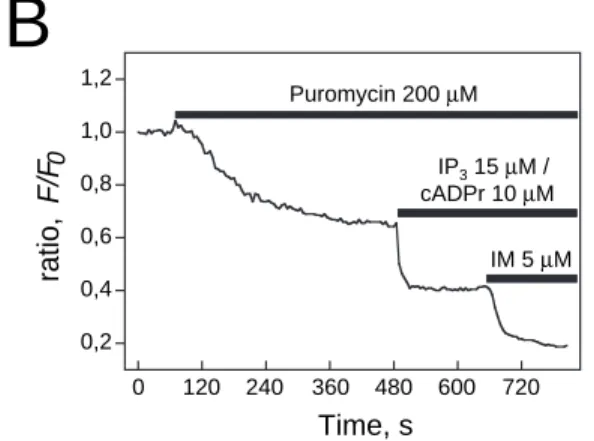
The puromycin-induced luminal calcium leak occurs through the translocon and not through the IP3 and ryanodine receptors activation
To exclude the hypothesis that the [Ca2+]
ERdecrease induced by puromycin was mediated by agonist-activated Ca2+release channels, we first perfused the IP3receptor inhibitor (heparin 500 µg/ml) and the ryanodine receptor inhibitor (ryanodine 20 µM). Fig. 4A illustrates the effect of puromycin on the ratio (–35.72%) in the presence of the inhibitors. Puromycin
decreased the mean ratio of 13 permeabilized cells by 31.21±5.76% in the presence of heparin and ryanodine (Fig. 4C). This value is significantly close to the puromycin effect without these inhibitors (–0.73±2.69%; P<0.01, paired Student’s t-test).
Secondly, we verified whether IP3(15 µM) and cADPr (10
µM) could have a cumulative effect after puromycin-induced calcium release. Puromycin evoked a 35% decrease in the ratio. Subsequent applications of IP3and cADPr induced a further 60% decrease (Fig. 4B). The mean decrease in the puromycin+IP3+ryanodine mixture was 51.76±8.17% (n=8). Ionomycin (5 µM) generated an additional fall in the ratio: 76.77±4.08% (n=8). Furthermore, puromycin (200 µM) did not inhibit NAADP (50 nM) induced calcium release (data not shown).
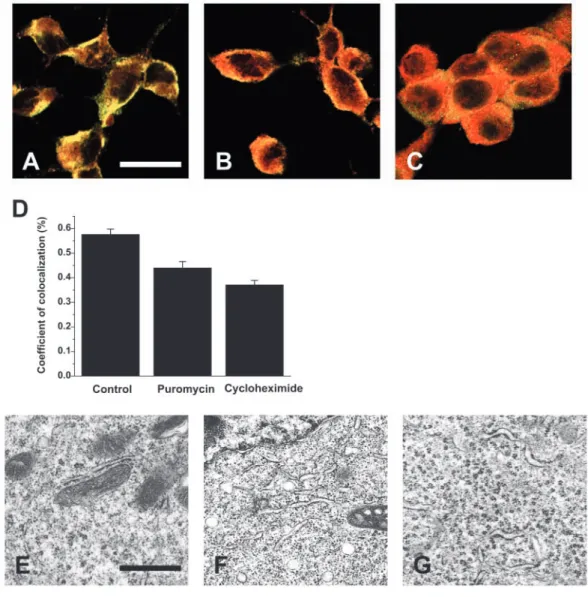
Colocalization between ribosome and translocon in confocal and electron microscopy
We first verified the effect of puromycin treatment on the subcellular distribution and colocalization of the 60S ribosomal subunit and the translocon. The 60S ribosomal subunit and the translocon protein, Sec61, were labeled in red and in green, respectively. We performed confocal microscopy under control conditions (Fig. 5A), after a 1-hour treatment with 200 µM puromycin (Fig. 5B) or with 1.8 mM cycloheximide (Fig. 5C). The colocalization was displayed in orange in the optical slices. Sec 61α and 60S ribosomal subunits were localized in the cytoplasm, both in the absence of puromycin and after puromycin treatment. The nucleus of
Journal of Cell Science 117 (18)
0 120 240 360 480 0,6 0,8 1,0 , oit ar F/ F 0 Time, s Puromycin 200 µM 0 200 400 600 800 0,6 0,8 1,0 Anisomycin 200 µM Puromycin 200 µM , oit ar 0 F/ F Time, s
A
B
C
0 10 20 30 40 % o f re le a se Puro myc in 200 µM Puro myc in 200 µM + Ani somyc inFig. 3. Anisomycin blocks the calcium leak from intracellular stores induced by puromycin. (A,B) The intracellular stores of LNCaP cells were loaded with the calcium indicator mag-fluo-4. Recordings of the leak from LNCaP cells, permeabilized with digitonin in two representative LNCaP cells in response to the application of puromycin are shown. Addition of puromycin induced a slow reduction in [Ca2+]ER(A) whereas anisomycin pre-treatment inhibits
passive Ca2+leakage through the ER in response to puromycin
application (B). (C) Cumulative data (mean±s.e.m.) for control and anisomycin-treated LNCaP cells of percentage of calcium released from internal stores.
LNCaP cells was not included in this localization (Fig. 5A-C). We calculated a colocalization coefficient for each cell (Fig. 5D) according to the method developed by Manders and colleagues (Manders et al., 1993).
Puromycin induced a 23.6% decrease in the colocalization coefficient. In the same way, cycloheximide reduced the colocalization by 35.6%. We obtained similar results with electron microscopy. We used control cells (Fig. 5E) and cells treated with 200 µM puromycin (Fig. 5F) or with 1.8 mM cycloheximide (Fig. 5G). In control conditions, the ribosomes are located on the ER membrane and inside the cytoplasm. In contrast, after a 1-hour treatment with puromycin or cycloheximide, most of the ribosomes were located inside the cytoplasm only. When treated with cycloheximide the ribosomes were aggregated into the cytoplasm in rosette-like polysomes.
Discussion
This work shows that the ubiquitous translocon complex in the endoplasmic reticulum membrane may act as a Ca2+ leak channel. The complex is not as tight as previously thought. Heritage and Wonderlin demonstrated that permeability of small polar molecules increased in cells treated with puromycin (Heritage and Wonderlin, 2001). Recently, it has been shown that the permeability of the ER to small polar molecules is coupled to translation (Roy and Wonderlin, 2003). In an initial study, we measured the effect of puromycin on the Ca2+ permeability of the ER membrane in mouse acinar
pancreatic cells (Lomax et al., 2002). In the present work, we demonstrate that puromycin acts on the translocon to induce a Ca2+ leak in LNCaP cells and also that a physiological calcium leak through the translocon is possible at the end of termination when the ribosome is still in place. Puromycin is a potent translation inhibitor, specifically blocking the ribosome on the translocon and clearing the nascent peptide chain (Pestova et al., 2001). Under these conditions, the translocon channel is maintained in open configuration (Johnson and van Waes, 1999) and calcium can be released by the ER.
We measured the effects of long term (1-hour) exposure to puromycin on the ER calcium content. Puromycin treatment (200 µM) of LNCaP cells loaded with fura-2 induced a significant decrease in the thapsigargin response, compared to control (–40%) and cycloheximide-treated cells (1.8 mM, 1 hour) (Fig. 1). This implies that Ca2+ was released from the store through translocon pores left open by puromycin before thapsigargin application. The thapsigargin response of LNCaP cells was similar under control conditions and in the presence of cycloheximide, which inhibits elongation (Roy and Wonderlin, 2003). Thus, the effect of puromycin on the thapsigargin-induced calcium release was not due to inhibition of protein synthesis, but may be produced by the fact that the ribosome-bound translocon complex is maintained in open configuration.
To observe the on-line effect of puromycin on calcium leakage, we used mag-fura-2 or mag-fluo-4 to measure changes in luminal calcium content (Fig. 2). All the following experiments were carried out in the absence of Ca2+-ATPase 0 200 400 600 800 1000 0,2 0,4 0,6 0,8 1,0 1,2 Ryanodine 20 µM + Heparin 500 µg/ml IM 5 µM Puromycin 200 µM , oit ar 0 F/ F Time, s 0 120 240 360 480 600 720 0,2 0,4 0,6 0,8 1,0 1,2 , oit ar F/ F 0 Time, s
A
B
C
0 20 40 60 80 100 % o f re lea se IM 5 µM Puromycin 200 µM IP3 15 µM / cADPr 10 µM Puro myc in 200 µM Puro myc in 200 µM + Ryanodine /Hep arin Puro myc in 200 µM + IP3/c AD Pr Puro myc in 200 µM + IP3 /cA DP r/IMFig. 4. Calcium leak from the intracellular stores induced by puromycin occurs independently of IP3and RyR stores.
(A) Time course of a typical experiment of the passive Ca2+leak
induced by puromycin in digitonin-permeabilized LNCaP cells treated with ryanodine and heparin. At the end of experiment, ionomycin was added to indicate the size of the total releasable pool. (B) Sequential application of puromycin and IP3 /cADPr to
indicate the size of each releasable pool. At the end of
experiment, ionomycin was also added to indicate the size of the total releasable internal calcium store. (C) Cumulative data (mean±s.e.m.) for the percentage of calcium released from internal stores.
4140
inhibitors, in order to measure the result of luminal Ca2+ influx (we did not block the Ca2+-ATPases) and efflux (due to passive leakage). It is known that ATP also modulates Ca2+leakage (Hofer et al., 1996). Under our experimental conditions, the permeabilized LNCaP cells were perfused with an ‘internal medium’, with the same ATP concentration throughout the experiment (200µM). [Ca2+]
ERwas stable before puromycin or ionomycin perfusion. Cycloheximide did not induce a decrease in [Ca2+]
ER(data not shown). As shown in Figs 2 and 3, puromycin triggered a calcium release from the ER. These results highlight the considerable impact of puromycin, as well as the fast kinetics of the translocon on the calcium leak. In physiological conditions, the pore diameter of the ribosome-free translocon is 0.9-1.5 nm (Hamman et al., 1998). During
translation, in ribosome-bound conditions, the pore aperture has a theoretical diameter of between 4 and 6 nm (Hamman et al., 1997). Recently ER permeability has been demonstrated to be coupled to protein synthesis (Roy and Wonderlin, 2003). Puromycin increased the 4-MalphaG (4-methyl-umbelliferyl-alpha-d-glucopyranoside) permeability, whereas cycloheximide did not. Furthermore, cycloheximide prevented the pactamycin-evoked 4-MalphaG permeability. They concluded that the permeation of 4-MalphaG is coupled to a gating mechanism, where the nascent protein chain locks the pore (Roy and Wonderlin, 2003). The results of our experiments are similar to these findings. Cycloheximide alone was unable to induce a Ca2+ release, unlike puromycin. The nascent protein needs to be released in order to let the Ca2+ Journal of Cell Science 117 (18)
Fig. 5. Colocalization of the 60S ribosomal subunit with translocon protein Sec 61. (A-C) Sec 61, recognized by polyclonal Sec 61 antibodies, was detected using FITC-labeled secondary antibodies. 60S ribosomal subunit, recognized by polyclonal antibodies, was detected using Texas-Red-labeled secondary antibodies. No staining was seen in the absence of primary antibodies (data not shown). Cells were analyzed using a Zeiss LSM 510 confocal laser scanning system connected to a Zeiss Axiovert 200 M. Images were collected separately for each channel (Texas-Red at 563 nm and FITC at 488 nm excitation) and merged as indicated. The pictures were taken after a 1-hour treatment under control conditions (A), with 200 µM puromycin (B) or with 1.8 mM cycloheximide (C). Bar, 20 µm. (D) Correlation coefficients calculated according to reported methods (Manders et al., 1993). Puromycin slightly reduced colocalization of the Sec61 and ribosome labels. (E-G) Transmitted electron micrographs of LNCaP cells in control conditions (E), or after a 1-hour treatment with 200 µM puromycin (F) or 1.8 mM
leak through the pore of the translocon. Furthermore, an interesting study (Potter and Nicchitta, 2002) has demonstrated that the ribosome remains in place on the translocon after translation. In these physiological conditions, Ca2+ release through the translocon probably occurs at this moment and could be a way for the cell to regulate the [Ca2+]
ER.
In our experiments, we generally applied 200 µM puromycin. Others have used similar concentrations to estimate the puromycin effect on ER permeability (Roy and Wonderlin, 2003; Lomax et al., 2002). With 20 µM puromycin, the [Ca2+]
ER decrease was lower (18.51±5.32%, n=15). Anisomycin (a peptidyl transferase inhibitor) inhibits the puromycin reaction (Ioannou et al., 1998). Anisomycin alone did not affect the ER calcium content (data not shown). In such conditions, when the peptidyl transferase is inhibited, the translocon is not permeable to calcium. Under the influence of anisomycin (200 µM), only 6.5% of the cells (n=46) responded to puromycin with a decrease in their ER calcium content. This percentage of response is low when compared to the 78% (n=50) puromycin response in cells untreated with anisomycin. Indeed, as anisomycin inhibits permeation by calcium via the translocon, the further action of puromycin (which releases the polypeptide chain and opens the translocon) could not be performed. This implies that puromycin specifically blocks the ribosome on the translocon, which is still open, thereby releasing calcium from the ER.
The calcium leak measured in the presence of puromycin does not involve NAADP, IP3 or ryanodine receptors. Puromycin induced the same calcium release with or without inhibitors of IP3or ryanodine receptors (Fig. 4). Hence, these experiments demonstrate that the puromycin-induced calcium leak does not occur through IP3 receptors and/or ryanodine receptors. Furthermore, as shown in Fig. 4B, we applied 15 µM IP3and 10 µM cADPr after puromycin perfusion. IP3and cADPr induced a further decrease in luminal calcium content after puromycin treatment. This cumulative effect emphasizes that the puromycin-induced calcium leak was not due to a non-specific action of puromycin on IP3channels and/or ryanodine receptors. We did similar experiments with 50 nM NAADP and still observed a NAADP response after puromycin perfusion.
To explore the putative role of translocon in calcium leakage further, it would be interesting to investigate whether the translocon is calcium permeable in a lipid bilayer. However, the mammalian translocon complex contains numerous subunits (Sec61α, Sec61β, Sec61γ, TRAM, Bip) and many associated proteins like SP, Calnexin or SRP (Johnson and van Waes, 1999). The stoichiometry of the translocon (especially the pore structure) is still unknown (Schnell and Hebert, 2003). The extraction of all the subunits of the translocon and their analysis in a lipid bilayer is not possible without impairing the structure of the native complex, thus altering its function. In addition, we planned to use siRNA or antisense to abolish the expression of at least one subunit of the translocon such as Sec 61α. However, as the translocon is essential for translation, this type of experiment induced the death of treated cells. Furthermore, due to the high number of different subunits, it is not possible to overexpress the whole translocon complex. Therefore, at the present time our experimental approach is the only one permitting the clear measurement within a living cell, of the decrease in [Ca2+]
ER that occurs through the translocon.
The luminal calcium concentration modulates many physiological processes, including gene expression and apoptosis (Jiang et al., 1994; Martikainen et al., 1991; McConkey, 1996; Putney and Ribeiro, 2000; Reynolds and Eastman, 1996; Wei et al., 1998). In LNCaP cells, calcium release from the ER has been shown to induce apoptosis (Skryma et al., 2000; Wertz and Dixit, 2000). Therefore, structures which are able to lower [Ca2+]
ER, such as Bcl-2 (Vanden Abeele et al., 2002), are involved in the control of apoptosis. This is the case for the translocon. As mentioned before, recent findings indicate that the ribosome remains bound to the ER membrane following the termination of protein synthesis (Potter and Nicchitta, 2000; Potter and Nicchitta, 2002; Seiser and Nicchitta, 2000). At this time, the ribosome-bound translocons may be calcium-permeable, as they are after treatment with puromycin and are thus able to release part of the calcium from the ER, with all the consequences previously described. We noticed a decrease in the colocalization between the translocon and the ribosome after lengthy exposure to puromycin (Fig. 5). These results were confirmed by electron microscopy. Nevertheless, we still had low ER calcium content after a 1-hour treatment with this antibiotic, as shown by the thapsigargin response in Fig. 1. This implies that a small number of open translocon is sufficient to induce a sustained ER calcium release. Seiser and Nicchitta did not observe a decrease in the ribosome binding to the endoplasmic reticulum membrane after 15 minutes of cycloheximide treatment (Seiser and Nicchitta, 2000). In our work, we evaluated the long-term effect of cycloheximide (1 hour) on the localization of the ribosome on the endoplasmic reticulum. In our experimental conditions, we measured a significant decrease in the colocalization coefficient between the ribosome and the translocon. These findings are confirmed by electron microscopy. These differences with the published work (Seisser and Nicchitta, 2000) are probably caused by the incubation time with cycloheximide. Cycloheximide inhibits translation, inducing a premature termination, which leads to the dissociation of the ribosome from the translocon.
For the first time, we have demonstrated that the puromycin-induced calcium release occurs through the translocon. This calcium leak probably occurs through the translocon at the end of the termination, when the polypeptide chain is released and during the time when the ribosome is still on the translocon. As this calcium leak is present in yeast and mammalian cells (Deshaies and Schekman, 1987; Gorlich and Rapoport, 1993; Stirling et al., 1992), our findings suggest that calcium release through the translocon is a common phenomenon in cell physiology.
We are grateful to Alexei Tepikin (University of Liverpool, UK) and to Helen Selliez-Vandernotte for reading the manuscript and for valuable discussions. This work was supported by grants from INSERM, the Ministère de l’Education Nationale, the Association pour la Recherche contre le Cancer and the Ligue Nationale contre le Cancer.
References
Beckmann, R., Bubeck, D., Grassucci, R., Penczek, P., Verschoor, A., Blobel, G. and Frank, J. (1997). Alignment of conduits for the nascent
polypeptide chain in the ribosome-Sec61 complex. Science 278, 2123-2126.
Berridge, M. J. and Irvine, R. F. (1989). Inositol phosphates and cell
4142
Bezprozvanny, I., Watras, J. and Ehrlich, B. E. (1991). Bell-shaped
calcium-response curves of Ins(1,4,5)P3- and calcium-gated channels from endoplasmic reticulum of cerebellum. Nature 351, 751-754.
Cancela, J. M., Van Coppenolle, F., Galione, A., Tepikin, A. V. and
Petersen, O. H. (2002). Transformation of local Ca2+spikes to global Ca2+
transients: the combinatorial roles of multiple Ca2+releasing messengers. EMBO J. 21, 909-919.
Clapham, D. E. (1995). Calcium signaling. Cell 80, 259-268.
Deshaies, R. J. and Schekman, R. (1987). A yeast mutant defective at an
early stage in import of secretory protein precursors into the endoplasmic reticulum. J. Cell Biol. 105, 633-645.
Gorlich, D. and Rapoport, T. A. (1993). Protein translocation into
proteoliposomes reconstituted from purified components of the endoplasmic reticulum membrane. Cell 75, 615-630.
Gorlich, D., Hartmann, E., Prehn, S. and Rapoport, T. A. (1992a). A
protein of the endoplasmic reticulum involved early in polypeptide translocation. Nature 357, 47-52.
Gorlich, D., Prehn, S., Hartmann, E., Kalies, K. U. and Rapoport, T. A.
(1992b). A mammalian homolog of SEC61p and SECYp is associated with ribosomes and nascent polypeptides during translocation. Cell 71, 489-503.
Hamman, B. D., Chen, J. C., Johnson, E. E. and Johnson, A. E. (1997).
The aqueous pore through the translocon has a diameter of 40-60 A during cotranslational protein translocation at the ER membrane. Cell 89, 535-544.
Hamman, B. D., Hendershot, L. M. and Johnson, A. E. (1998). BiP
maintains the permeability barrier of the ER membrane by sealing the lumenal end of the translocon pore before and early in translocation. Cell
92, 747-758.
Heritage, D. and Wonderlin, W. F. (2001). Translocon pores in the
endoplasmic reticulum are permeable to a neutral, polar molecule. J. Biol.
Chem. 276, 22655-22662.
Hofer, A. M., Curci, S., Machen, T. E. and Schulz, I. (1996). ATP regulates
calcium leak from agonist-sensitive internal calcium stores. FASEB J. 10, 302-308.
Horne, Z. and Hesketh, J. (1990). Immunological localization of ribosomes
in striated rat muscle. Evidence for myofibrillar association and ontological changes in the subsarcolemmal:myofibrillar distribution. Biochem. J. 268, 231-236.
Ioannou, M., Coutsogeorgopoulos, C. and Synetos, D. (1998). Kinetics of
inhibition of rabbit reticulocyte peptidyltransferase by anisomycin and sparsomycin. Mol. Pharmacol. 53, 1089-1096.
Jiang, S., Chow, S. C., Nicotera, P. and Orrenius, S. (1994). Intracellular
Ca2+signals activate apoptosis in thymocytes: studies using the
Ca(2+)-ATPase inhibitor thapsigargin. Exp. Cell Res. 212, 84-92.
Johnson, A. E. and van Waes, M. A. (1999). The translocon: a dynamic
gateway at the ER membrane. Annu. Rev. Cell Dev. Biol. 15, 799-842.
Lomax, R. B., Camello, C., Van Coppenolle, F., Petersen, O. H. and
Tepikin, A. V. (2002). Basal and physiological Ca2+ leak from the
endoplasmic reticulum of pancreatic acinar cells. Second messenger-activated channels and translocons. J. Biol. Chem. 277, 26479-26485.
Manders, E. M. M., Verbeek, F. J. and Alen, J. A. (1993). Measurement of
co-localization of objects in dual-color confocal images. J. Microsc. 169, 375-382.
Martikainen, P., Kyprianou, N., Tucker, R. W. and Isaacs, J. T. (1991).
Programmed death of nonproliferating androgen-independent prostatic cancer cells. Cancer Res. 51, 4693-4700.
McConkey, D. J. (1996). The role of calcium in the regulation of apoptosis.
Scanning Microsc. 10, 777-793.
Mogami, H., Tepikin, A. V. and Petersen, O. H. (1998). Termination of
cytosolic Ca2+signals: Ca2+reuptake into intracellular stores is regulated
by the free Ca2+concentration in the store lumen. EMBO J. 17, 435-442.
Park, M. K., Petersen, O. H. and Tepikin, A. V. (2000). The endoplasmic
reticulum as one continuous Ca(2+) pool: visualization of rapid Ca(2+) movements and equilibration. EMBO J. 19, 5729-5739.
Pestka, S. (1974). The use of inhibitors in studies on protein synthesis.
Methods Enzymol. 30, 261-282.
Pestova, T. V., Kolupaeva, V. G., Lomakin, I. B., Pilipenko, E. V., Shatsky, I. N., Agol, V. I. and Hellen, C. U. (2001). Molecular mechanisms of
translation initiation in eukaryotes. Proc. Natl. Acad. Sci. USA 98, 7029-7036.
Potter, M. D. and Nicchitta, C. V. (2000). Regulation of ribosome detachment
from the mammalian endoplasmic reticulum membrane. J. Biol. Chem. 275, 33828-33835.
Potter, M. D. and Nicchitta, C. V. (2002). Endoplasmic reticulum-bound
ribosomes reside in stable association with the translocon following termination of protein synthesis. J. Biol. Chem. 277, 23314-23320.
Pozzan, T., Rizzuto, R., Volpe, P. and Meldolesi, J. (1994). Molecular and
cellular physiology of intracellular calcium stores. Physiol. Rev. 74, 595-636.
Putney, J. W., Jr and Ribeiro, C. M. (2000). Signaling pathways between
the plasma membrane and endoplasmic reticulum calcium stores. Cell. Mol.
Life Sci. 57, 1272-1286.
Reynolds, J. E. and Eastman, A. (1996). Intracellular calcium stores are not
required for Bcl-2-mediated protection from apoptosis. J. Biol. Chem. 271, 27739-27743.
Roy, A. and Wonderlin, W. F. (2003). The permeability of the endoplasmic
reticulum is dynamically coupled to protein synthesis. J. Biol. Chem. 278, 4397-4403.
Schnell, D. J. and Hebert, D. N. (2003). Protein translocons. Multifunctional
mediators of protein translocation across membranes. Cell 112, 491-505.
Seiser, R. M. and Nicchitta, C. V. (2000). The fate of membrane-bound
ribosomes following the termination of protein synthesis. J. Biol. Chem.
275, 33820-33827.
Simon, S. M. and Blobel, G. (1991). A protein-conducting channel in the
endoplasmic reticulum. Cell 65, 371-380.
Skryma, R., Mariot, P., Bourhis, X. L., Van Coppenolle, F., Shuba, Y., Vanden Abeele, F., Legrand, G., Humez, S., Boilly, B. and Prevarskaya,
N. (2000). Store depletion and store-operated Ca2+current in human prostate
cancer LNCaP cells: involvement in apoptosis. J. Physiol. 527, 71-83.
Stirling, C. J., Rothblatt, J., Hosobuchi, M., Deshaies, R. and Schekman, R.
(1992). Protein translocation mutants defective in the insertion of integral membrane proteins into the endoplasmic reticulum. Mol. Biol. Cell 3, 129-142.
Vanden Abeele, F., Skryma, R., Shuba, Y., Van Coppenolle, F., Slomianny, C., Roudbaraki, M., Mauroy, B., Wuytack, F. and Prevarskaya, N.
(2002). Bcl-2-dependent modulation of Ca(2+) homeostasis and store-operated channels in prostate cancer cells. Cancer Cell 1, 169-179.
Wei, H., Wei, W., Bredesen, D. E. and Perry, D. C. (1998). Bcl-2 protects
against apoptosis in neuronal cell line caused by thapsigargin-induced depletion of intracellular calcium stores. J. Neurochem. 70, 2305-2314.
Wertz, I. E. and Dixit, V. M. (2000). Characterization of calcium
release-activated apoptosis of LNCaP prostate cancer cells. J. Biol. Chem. 275, 11470-11477.