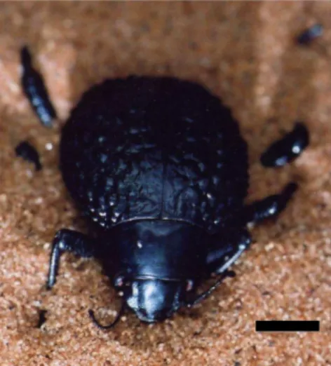
Dew condensation on desert beetle skin
Texte intégral
Figure

Documents relatifs
Toward this purpose, a preliminary in vitro study has been conducted with skin samples of mice, with and without inoculated melanoma tumor cells, in order to
Given a subshift Σ that contains communication channels, it is possible to code transfers of information through these channels thanks to a product and a finite type
From the top to the base, these formations are the Grès à Voltzia and the Couches Intermédiaires in the Upper Buntsandstein, the Poudingue de Sainte Odile, the Couches de Karlstal,
The pieces of amber containing the plant remains described herein were discovered in Cretaceous deposits from the “Plage de la Vierge” at Fouras – Bois Vert in the
Pour résumer, nous avons reproduit le renfor ement à la traversée du y lone de surfa e, observé notamment dans le as de la POI17, dans le adre idéalisé d'un modèle à deux ou
Il est co-auteur, avec Alain Beltran, d’une histoire de l’INRIA (His- toire d’un pionnier de l’informatique. 40 ans de recherche à l’INRIA, EDP Sciences, 2007)..
Phasico-tonic CBTs present a phasic firing, during release ramps, and develop a tonic discharge for the most released position of the CBCO strand. A: The first
Various cobalt mineralization styles on Earth can not be attributed to a single ore-forming model. At Bou Azzer, two types of ore bodies occur – “contact” and
