Resonance Raman Optical Activity Shows Unusual Structural
Sensitivity for Systems in Resonance with Multiple Excited States:
Vitamin B
12
Case
Ewa Machalska, Grzegorz Zajac, Anna Gruca, Fabio Zobi, Malgorzata Baranska, and Agnieszka Kaczor
*
ABSTRACT: In this work, cobalamins with different upper axial substituents and a cobalamin
derivative with a ring modification were studied using chiroptical spectroscopies, in particular
resonance Raman optical activity (RROA), to shed light on the influence of structural
modifications on RROA spectra in these strongly chiral systems in resonance with multiple
excited states at 532 nm excitation. We have demonstrated that for these unique systems
RROA possesses augmented structural specificity, surpassing resonance Raman spectroscopy
and enabling at the same time measurement of cobalamins at fairy low concentrations of∼10−5
mol dm−3. The enhanced structural specificity of RROA is a result of bisignate spectra due to
resonance via more than one electronic state. The observation of increased structural capability of RROA for cobalamins opens a new perspective for studying chiral properties of other biological systems incorporating d-metal ions.
R
aman optical activity (ROA) is a powerful structuralprobe of chirality for investigation of various forms of
matter. The specific sensitivity of ROA spectroscopy to the
chiral molecular environment makes it an efficient tool for
studying biomolecules, e.g., proteins, amino acids,
carbohy-drates, or supramolecular structures.1 SCP-ROA (scattered
circular polarization ROA) measures a small intensity di
ffer-ence in Raman scattering of right and left circularly polarized
light from optically active molecules.2Due to the fact that the
ROA effect is a fairly weak phenomenon, the intensities of
ROA signals are usually 3−4 orders of magnitude smaller than
Raman signals; therefore, methods providing ROA signal enhancement have been attracting a considerable amount of
attention.3−8 One of these methods is resonance ROA, a
chiroptical analogue of resonance Raman spectroscopy. A resonance ROA (RROA) spectrum, when derived from a single isolated electronic state, is monosignate and opposite in sign to the electronic circular dichroism (ECD) band of the resonant electronic transition and exhibits signals with the same relative intensities as those in the parent resonance
Raman (RR) spectrum.9In the simplest formulated theory of
resonance ROA, i.e., described above as single-electronic state
(SES) theory,9 where strong resonance with only one
electronic state is considered, only the Albrecht A-term of resonance Raman scattering contributes to the obtained
ROA.10An extension of the SES theory is the two-electronic
state (TES) theory,11where the resonance ROA signal appears
to be due to both A- and B-term mechanisms. As a result, bisignate RROA spectra are obtained due to resonance with
multiple excited electronic states.12Recently, ROA and other
chiroptical spectroscopies in conjunction with quantum-chemical calculations were applied to study the vibrational and electronic properties of chiral coordination complexes
showing that the presence of these metal ions significantly
increased the magnitude of the chiral signal due to resonance
via a single electronic state or multiple electronic states.13−15
Vitamins B12 (cobalamins) are biologically active
com-pounds incorporating a transition metal ion. These exogenous
compounds (Scheme 1) have a unique structure based on a
corrin ring encapsulating a low-spin Co3+ ion coordinated
equatorially by four pyrrolic nitrogen atoms and two axial ligands. 5,6-Dimethylbenzimidazole is covalently bound to the ring in one of its axial coordination positions (lower), and the other (upper) can be occupied by various substituents such as
cyano, hydroxyl, methyl, and adenosyl groups.16 Cobalamins
play an essential role in various biological processes, such as
nucleic acid metabolism and formation of red blood cells.17,18
Also, functionalized cobalamins, with a modified ring structure
or upper axial substituent, have diverse biological properties; for example, they can be used as a scaffold for the delivery of
antimalarial drugs to erythro- and hepatocytes.19
http://doc.rero.ch
Published in "The Journal of Physical Chemistry Letters 11(13): 5037–5043, 2020"
which should be cited to refer to this work.
The corrin ligand exhibits a single helical sense of absolute R
configuration of its stereocenters.20 The structure of the
macrocyclic corrin ring resembles the metalloporphyrin system, but with relevant differences. The most important is the presence of the additional side chains and the more reduced character of the corrin ring compared with that of the porphyrin one, which provides a greater degree of conforma-tional freedom of corrinoids. It was found that the increased flexibility of the corrin macrocycle plays a particular role in
Co−C bond activation by B12-dependent enzymes.
21−23
Ultraviolet−visible (UV−vis), ECD, and Raman
spectros-copy were applied to study structures of several corrinoids in
aqueous and organic solvents.24,25Strong absorption bands in
the visible and near-UV region, due to π−π* electronic
transitions, cause significant resonance enhancement in the
Raman spectra under visible light excitation. A few previous
works demonstrated that RR spectra of vitamin B12 and its
various analogues are strikingly similar, i.e., independent of the
nature of the upper axial substituent.24 The similarity of the
Raman spectra is not fully reflected in the similarity of the
absorption UV−vis spectra showing that the excited states of
the various derivatives of B12do differ.
24
On the contrary, ECD spectroscopy was shown to be more sensitive for replacement of an axial ligand of corrinoids, although due to the nature of the method (few broad features in the spectra) a detailed analysis of the structure using ECD is unavailable. It was
confirmed that distinct changes in the ECD and UV−vis
spectra were not related to chemical modifications of the corrin
ring, but could result from electronic or conformational
effects.25
Intense ECD spectra of cobalamins in the UV−vis range
were previously reported showing that cobalamins’ ECD
signals were quite sensitive to the substituent in the upper
axial position.25 Our study demonstrates (Figure 1) that
significant spectral differences in the ECD spectra are observed
when the atom linking the Co ion is different (CNCbl vs
OHCbl spectra), although some differences are also noticed
between CNCbl and HCC−Ph−CCCbl, where the cyano
group is modified by a bulky HCC−Ph−CC− group. On the contrary, for a studied derivative with the ring
modification, i.e., CNCbl-Br, the electronic spectra have
characteristics similar to those of the spectra of its nonmodified counterpart, CNCbl. A detailed consideration of the
assign-ment of the UV−vis and ECD spectra is presented in the
Supporting Information.
As cobalamins have multiple electronic transitions in the vicinity of the ROA laser excitation at 532 nm (green line in
Figure 1), we applied this method to study for thefirst time vibrational chiroptical properties of cobalamin derivatives. RR and RROA spectra of studied cobalamins recorded using an
excitation line of 532 nm are presented inFigure 2.
In agreement with previously published data,26RR spectra of
cobalamins with various upper axial substituents are strikingly
similar and reflect the structure of the macrocycle due to the
dominant contribution of the significantly resonantly enhanced
ring vibrations. Practically all intense RR bands have contributions from the corrin ring as demonstrated by the comparison of dicyanocobinamide RR spectrum (lacking a
bulky DMB group) with the spectra of cobalamins27and what
is reflected in our quantum-chemical calculations (Supporting
Information). Slight differences in the relative intensities and wavenumbers of the signals of all studied cobalamins result
most probably from different resonance conditions and
molecular (electronic) structure, respectively.
On the contrary, RROA signatures of analyzed cobalamins
exhibit significantly more pronounced variations in the
intensities of bands compared to RR spectra, which is clearly observed if the spectra are carefully investigated in the relevant
spectral ranges (Figure 2, insets). According to a detailed
analysis by Stich et al.25 and our quantum-chemical
calculations (Supporting Information), there are at least
three electronic states that might contribute to the resonance using 532 nm excitation for the considered cobalamins. In the case of CNCbl and OHCbl, there are two negative electronic states and one positive electronic state in the vicinity of the
excitation laser (Figure 1). An important consequence of this
Scheme 1. Molecular Structures of Cobalamins with Different Upper Axial Substituents, Including Cyanocobalamin (R = CN),
Hydroxocobalamin (R = OH), 1,4-Diethynylbenzenecobalamin (R = CC−Ph−CCH), and a Cobalamin Ring Derivative
[(C10)H = Br]
fact is that RROA spectra, although definitely recorded under the resonance conditions, are bisignate. This brings us to one of the most meaningful observations in this work. Our data
demonstrate that differences in the electronic structure due to
the different substituents scarcely affect RR spectra but result
in the modification of resonance ROA conditions and bring
significantly more pronounced variations in RROA intensities.
This means that in the studied case, the sensitivity of RROA to subtle changes in the molecular structure is increased compared to that of RR spectroscopy. To the best of our
knowledge, this is the first observation demonstrating
augmented structural sensitivity of RROA compared to RR
spectroscopy. For cobalamins with different upper axial
substituents, it originates from a highly chiral chromophore for which resonance enhancement takes place via multiple
electronic states that differ in energy and a sign of the rotatory
strengths. In the case of CNCbl, RROA signals are mostly positive due to the proximity of the laser excitation to the
negative ECD electronic transitions (first, 544 nm, second, 507
nm, and 483 nm band that is assigned to the vibrational
progression of the first electronic transition) and slightly
negative due to the third positive ECD transition at 433 nm.
For OHCbl, surprisingly, the proximity of both negative and positive ECD bands gives a similar, predominantly positive RROA spectrum. It demonstrates that the negative ECD (551
nm) electronic state with the dominant corrinoid ringπ → π*
transition (HOMO → LUMO) has the most pronounced
influence on the RROA spectra. This is contrary to the most
recent RROA study of Sgammato et al.7 in which even slight
changes in the intensity and energy of ECD bands resulted in the pronounced changes in RROA intensities. Additionally,
Sgammato et al.7reported a case in which RROA and RR are
highly sensitive to a different axial ligand of heme. In our study,
we highlight increased RROA specificity compared to RR in
systems, where different axial ligands do not affect the spin
state of the cobalt ion. According to our quantum-chemical calculations, molecular orbitals involved in the second (negative ECD) and third (positive ECD) electronic transitions of CNCbl are composed of not only the corrinoid
ringπ orbitals but also CN-π, DMB-π, and Co 3d orbitals. The
influence of that positive ECD transition on the sign of RROA
is clearly seen in the experimental RROA spectrum of CNCbl
and confirmed by the calculations, where the most negative
feature located in the experimental spectra at 497 cm−1 is
Figure 1.UV−vis and ECD spectra of cobalamins with different upper axial substituents: cyanocobalamin (R = CN), hydroxocobalamin (R = OH), 1,4-diethynylbenzenecobalamin (R = CC−Ph−CCH), and a cobalamin ring derivative [(C10)H = Br].
related mostly to the Co−CN bending, CN twisting, and
DMB ring breathing. The influence of the excitation of various
electronic states on the RROA spectrum is clearly visible in the
comparison of RROA spectra calculated using different
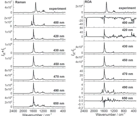
excitation lines (Figure 3).
The closer the excitation line is to thefirst electronic state
(443 nm, negative ECD), the more monosignate (positive) ROA is. For excitations at 400 and 420 nm, bisignate ROA is observed due to proximity of two electronic transitions (2 and 3) that have opposite sign rotatory strengths. Far from resonance (650 nm excitation line), CNCbl also gives the bisignate ROA spectrum.
A closer look at the recorded spectra demonstrates that alterations in the relative intensities of the RROA bands are
observed for all studied cobalamins with different upper axial
substituents. CNCbl and HCC−Ph−CCCbl signals at
∼1500 and ∼1205 cm−1, respectively, assigned to ring
vibrations have increased relative intensity in the spectra compared to those of OHCbl.
On the contrary, the RROA spectrum of OHCbl shows an increased relative intensity of several positive bands due to the
umbrella CH3bending vibrations in the range of 1300−1400
cm−1along with the signals in the range of 1100−1190 cm−1,
assigned mostly to the CH2 twisting and C−N and C−C
stretching modes. In the CNCbl and HCC−Ph−CCCbl
RROA spectra, signals in the aforementioned ranges are considerably less intense or have the opposite intensity. In
particular, in the HCC−Ph−CCCbl spectrum, a negative
band is observed at 1354 cm−1with the shoulder at ∼1370
cm−1, opposite in sign to signals observed in the OHCbl
spectrum. Negative RROA intensity is also clearly observed in
the range of 300−600 cm−1, where mostly bands associated
with the Co−R vibrations are predicted. In addition, this range
confirms that recorded RROA spectra of cobalamins with
different upper axial substituents are markedly different.
Calculated Raman and ROA spectra of CNCbl (Supporting
Information) are in good agreement with experimental results
and clearly confirm that the spectra are bisignate.
We investigated and characterized also a cobalamin with a
modified ring structure (Scheme 1), where the hydrogen atom
linked to C10was replaced with a bromine atom, to illustrate
the impact of a ring modification on RROA (Figure 2) spectra
of cobalamin.
Comparison of the CNCbl and CNCbl-Br spectra shows
that RR signatures are markedly affected by the ring
modification (Figure 2) due to different resonance conditions
[theα/β absorption band due to the π → π* transition along
the long C5−C15axis is red-shifted approximately 20−30 nm in
CNCbl-Br relative to CNCbl (Figure 1)] caused by the altered
electronic/geometric structure of the corrin ring resulting from
Br substitution. Due to the red-shift of the α/β absorption
band in CNCbl-Br, resonance conditions are altered compared to those of CNCbl and negative bands are observed in the CNCbl-Br RROA spectrum in various ranges. A characteristic
Figure 2.Raman and RROA spectra of cobalamins with different upper axial substituents: cyanocobalamin (R = CN), hydroxocobalamin (R = OH), 1,4-diethynylbenzenecobalamin (R = CC−Ph−CCH), and a cobalamin ring derivative [(C10)H = Br].
RROA feature of CNCbl-Br is also the band at 815 cm−1
[assigned to the nearly isolated C−C10−C(-H) vibrations
(Supporting Information)] absent from RR and RROA spectra of other corrinoids. The intensity of this band increases due to substitution with a heavy atom, i.e., a bromine atom. Although
ring modifications affect both RR and RROA spectra, for a
studied cyanocobalamin with a ring modification, RROA is still
a better probe of the molecular structure.
The obtained RROA/RR (CID) and ECD/UV−vis
(g-factor) ratios mostly do not obey the SES theory9 relation,
CID =−1/2g, which is not surprising in the multiple excited
state RROA. However, CIDs preserve the same order of magnitude and opposite signs as the g-factors of related
resonance transitions discussed above (Supporting
Informa-tion). For CNCbl-Br, the CIDs of a majority of positive RROA
bands equal approximately 10−4, while related negative ECD
transitions at 544, 507, and 483 nm possess g-factors close to
−10−4. A similar situation is observed for the negative RROA
band at 497 cm−1, where the CID value and the g-factor of
positive ECD at 433 nm are −4 × 10⁻³ and 5 × 10⁻³,
respectively. Furthermore, the most intense positive RROA
band of CNCbl (1501 cm−1) possesses a CID of 4.3× 10⁻⁴,
which is almost twice as high as the absolute value of the
g-factor at 544 nm (−2.4 × 10⁻⁴) and 9 times higher than the
absolute value of the g-factor at 507 nm (−0.5 × 10⁻⁴), but it is
surprisingly less than half of the g-factor at 483 nm (−8.5 ×
10−4). For other studied cobalamins, the CID and g-factor
values show analogous behavior.
Although ECD is a sensitive tool for studying molecules, it is inherently limited to chromophores and as such does not provide detailed or local information about the molecular
structure. VCD and ROA are significantly more informative in
this context as they refer to well-localized vibrational modes, but they are characterized by low sensitivity that practically excludes their application in the study of biological samples.
RROA provides significant enhancement of the signal and could occur as an interesting alternative for ECD for the study of the molecular properties of biologically relevant systems. Nevertheless, in line with the single-electronic state theory of
RROA,9 when the resonance occurs via a single electronic
state, monosignate RROA spectra are either identical to the respective RR spectra or mirror images of the RR signatures. It is clear that in such cases RROA spectra do not provide a variety of structural information, apart from possibly distinguishing between optical isomers and measuring analytes at low concentrations.
In general, bisignate RROA spectra may be a result of the
conformational freedom of the molecule,28weak enhancement,
and the presence of nonresonance bands along with the
resonantly enhanced ones6 or excitation via more than one
electronic state.11Recently, it has been proven that bisignate
RROA spectra can also be reproduced using a full quantum mechanical methodology, considering all RROA terms, along
with Franck−Condon and Herzberg−Teller mechanisms.29
For cobalamins, bisignate RROA spectra result from the proximity of more than one electronic transition to the excitation wavelength, in agreement with previous detailed
analysis of ECD spectra,25 which was also confirmed by
significant differences between RROA and RR spectra based
on various relative intensities of the bands and not only the sign of the spectrum. Our observation shows that for strongly chiral systems in resonance with multiple excited states, RROA
could be a method of augmented structural specificity,
surpassing RR spectroscopy and at the same time enabling
measurements of concentrations as low as 10−5mol dm−3. This
finding opens a new perspective for studying chiral properties of biological systems incorporating d-metal ions.
Figure 3.Comparison of experimental and calculated pre-resonance Raman and ROA spectra of CNCbl calculated at the CAM-B3LYP/6-31G(d)/ MDF10/6-31G(d)/PCM level of theory, using 400, 420, 430, 450, 470, 490, and 650 nm excitation lines.
■
ASSOCIATED CONTENT*
sı Supporting InformationThe Supporting Information is available free of charge at
https://pubs.acs.org/doi/10.1021/acs.jpclett.0c01218. Description of materials and methods, detailed descrip-tion of quantum-chemical calculadescrip-tions, and detailed
analysis of the spectra (PDF)
■
AUTHOR INFORMATION Corresponding AuthorAgnieszka Kaczor − Faculty of Chemistry and Jagiellonian Centre for Experimental Therapeutics (JCET), Jagiellonian
University, Krakow 30-387, Poland;
orcid.org/0000-0001-8337-8567; Email:agnieszka.kaczor@uj.edu.pl
Authors
Ewa Machalska − Faculty of Chemistry and Jagiellonian Centre for Experimental Therapeutics (JCET), Jagiellonian University, Krakow 30-387, Poland
Grzegorz Zajac − Jagiellonian Centre for Experimental Therapeutics (JCET), Jagiellonian University, Krakow 30-348,
Poland; orcid.org/0000-0003-4090-9334
Anna Gruca − Faculty of Chemistry and Jagiellonian Centre for Experimental Therapeutics (JCET), Jagiellonian University, Krakow 30-387, Poland
Fabio Zobi − Department of Chemistry, University of Fribourg, 1700 Fribourg, Switzerland
Malgorzata Baranska − Faculty of Chemistry and Jagiellonian Centre for Experimental Therapeutics (JCET), Jagiellonian University, Krakow 30-387, Poland
Complete contact information is available at:
https://pubs.acs.org/10.1021/acs.jpclett.0c01218
Notes
The authors declare no competingfinancial interest.
■
ACKNOWLEDGMENTSThis work was supported by the National Science Centre Poland (Projects 2017/25/B/ST4/00854 to A.K. and 2019/ 33/N/ST4/01986 to E.M.). This research was supported in part by PL-Grid Infrastructure.
■
REFERENCES(1) Barron, L. D.; Hecht, L.; McColl, I. H.; Blanch, E. W. Raman Optical Activity Comes of Age. Mol. Phys. 2004, 102 (8), 731−744. (2) Barron, L. D.; Bogaard, M. P.; Buckingham, A. D. Raman Scattering of Circularly Polarized Light by Optically Active Molecules. J. Am. Chem. Soc. 1973, 95 (2), 603−605.
(3) Vargek, M.; Freedman, T. B.; Lee, E.; Nafie, L. A. Experimental Observation of Resonance Raman Optical Activity. Chem. Phys. Lett. 1998, 287 (3−4), 359−364.
(4) Zajac, G.; Kaczor, A.; Pallares Zazo, A.; Mlynarski, J.; Dudek, M.; Baranska, M. Aggregation-Induced Resonance Raman Optical Activity (AIRROA): A New Mechanism for Chirality Enhancement. J. Phys. Chem. B 2016, 120 (17), 4028−4033.
(5) Dudek, M.; Machalska, E.; Oleszkiewicz, T.; Grzebelus, E.; Baranski, R.; Szcześniak, P.; Mlynarski, J.; Zajac, G.; Kaczor, A.; Baranska, M. Chiral Amplification in Nature: Studying Cell-Extracted Chiral Carotenoid Microcrystals via the Resonance Raman Optical Activity of Model Systems. Angew. Chem., Int. Ed. 2019, 58 (25), 8383−8388.
(6) Bogaerts, J.; Johannessen, C. On/off Resonance Raman Optical Activity of Human Serum Transferrin. J. Raman Spectrosc. 2019, 50 (5), 641−646.
(7) Sgammato, R.; Herrebout, W.; Johannessen, C. Resonance Raman Optical Activity of the imidazole-Myoglobin Complex: Titrating Enhancement. J. Raman Spectrosc. 2019, 50 (12), 1905− 1913.
(8) Haraguchi, S.; Hara, M.; Shingae, T.; Kumauchi, M.; Hoff, W. D.; Unno, M. Experimental Detection of the Intrinsic Difference in Raman Optical Activity of a Photoreceptor Protein under Preresonance and Resonance Conditions. Angew. Chem., Int. Ed. 2015, 54 (39), 11555−11558.
(9) Nafie, L. A. Theory of Resonance Raman Optical Activity: The Single Electronic State Limit. Chem. Phys. 1996, 205 (3), 309−322.
(10) Champion, P. M.; Albrecht, A. C. Resonance Raman Scattering: The Multimode Problem and Transform Methods. Annu. Rev. Phys. Chem. 1982, 33 (1), 353−376.
(11) Merten, C.; Li, H.; Nafie, L. A. Simultaneous Resonance Raman Optical Activity Involving Two Electronic States. J. Phys. Chem. A 2012, 116 (27), 7329−7336.
(12) Luber, S.; Neugebauer, J.; Reiher, M. Enhancement and de-Enhancement Effects in Vibrational Resonance Raman Optical Activity. J. Chem. Phys. 2010, 132 (4), 044113.
(13) Brichtová, E.; Hudecová, J.; Vršková, N.; Šebestík, J.; Bouř, P.; Wu, T. Binding of Lanthanide Complexes to Histidine-Containing Peptides Probed by Raman Optical Activity Spectroscopy. Chem. -Eur. J. 2018, 24 (34), 8664−8669.
(14) Autschbach, J.; Jorge, F. E.; Ziegler, T. Density Functional Calculations on Electronic Circular Dichroism Spectra of Chiral Transition Metal Complexes. Inorg. Chem. 2003, 42 (9), 2867−2877. (15) Le Guennic, B.; Hieringer, W.; Görling, A.; Autschbach, J. Density Functional Calculation of the Electronic Circular Dichroism Spectra of the Transition Metal Complexes [M(phen)3]2+(M = Fe,
Ru, Os). J. Phys. Chem. A 2005, 109 (21), 4836−4846.
(16) Proinsias, K.; Giedyk, M.; Gryko, D. Vitamin B12: Chemical
Modifications. Chem. Soc. Rev. 2013, 42 (16), 6605−6619.
(17) Fenech, M. Folate (Vitamin B9) and Vitamin B12and Their
Function in the Maintenance of Nuclear and Mitochondrial Genome Integrity. Mutat. Res., Fundam. Mol. Mech. Mutagen. 2012, 733 (1−2), 21−33.
(18) Wagner, C. Biochemical Role of Folate in Cellular Metabolism. Clin. Res. Regul. Aff. 2001, 18 (3), 161−180.
(19) Rossier, J.; Nasiri Sovari, S.; Pavic, A.; Vojnovic, S.; Stringer, T.; Bättig, S.; Smith, S. G.; Nikodinovic-Runic, J.; Zobi, F. Antiplasmodial Activity and In Vivo Bio-Distribution of Chloroquine Molecules Released with a 4-(4-Ethynylphenyl)-Triazole Moiety from Organo-metallo-Cobalamins. Molecules 2019, 24 (12), 2310.
(20) Senge, M. O.; Ryan, A. A.; Letchford, K. A.; MacGowan, S. A.; Mielke, T. Chlorophylls, Symmetry, Chirality, and Photosynthesis. Symmetry 2014, 6 (3), 781−843.
(21) Chowdhury, S.; Banerjee, R. Thermodynamic and Kinetic Characterization of Co-C Bond Homolysis Catalyzed by Coenzyme B12-Dependent Methylmalonyl-CoA Mutase. Biochemistry 2000, 39
(27), 7998−8006.
(22) Mebs, S.; Henn, J.; Dittrich, B.; Paulmann, C.; Luger, P. Electron Densities of Three B12Vitamins. J. Phys. Chem. A 2009, 113
(29), 8366−8378.
(23) Jensen, K. P.; Sauer, S. P. A.; Liljefors, T.; Norrby, P. O. Theoretical Investigation of Steric and Electronic Effects in Coenzyme B12Models. Organometallics 2001, 20 (3), 550−556.
(24) Stich, T. A.; Buan, N. R.; Brunold, T. C. Spectroscopic and Computational Studies of Co2+ corrinoids: Spectral and Electronic
Properties of the Biologically Relevant Base-on and Base-off Forms of Co2+cobalamin. J. Am. Chem. Soc. 2004, 126 (31), 9735−9749.
(25) Stich, T. A.; Brooks, A. J.; Buan, N. R.; Brunold, T. C. Spectroscopic and Computational Studies of Co3+-Corrinoids: Spectral and Electronic Properties of the B12 Cofactors and
Biologically Relevant Precursors. J. Am. Chem. Soc. 2003, 125 (19), 5897−5914.
(26) Park, K.; Brunold, T. C. Combined Spectroscopic and Computational Analysis of the Vibrational Properties of Vitamin B12
in Its Co3+, Co2+, and Co1+Oxidation States. J. Phys. Chem. B 2013, 117 (18), 5397−5410.
(27) Mayer, E.; Gardiner, D. J.; Hester, R. E. Resonance Raman Spectra of Vitamin B12and Dicyanocobalamin. Biochim. Biophys. Acta,
Gen. Subj. 1973, 297 (2), 568−570.
(28) Zajac, G.; Kaczor, A.; Chruszcz-Lipska, K.; Dobrowolski, J. C.; Baranska, M. Bisignate Resonance Raman Optical Activity: A Pseudo Breakdown of the Single Electronic State Model of RROA? J. Raman Spectrosc. 2014, 45 (10), 859−862.
(29) Vidal, L. N.; Giovannini, T.; Cappelli, C. Can the Resonance Raman Optical Activity Spectrum Display Sign Alternation? J. Phys. Chem. Lett. 2016, 7 (18), 3585−3590.