HAL Id: tel-01818522
https://tel.archives-ouvertes.fr/tel-01818522
Submitted on 19 Jun 2018HAL is a multi-disciplinary open access archive for the deposit and dissemination of sci-entific research documents, whether they are pub-lished or not. The documents may come from teaching and research institutions in France or abroad, or from public or private research centers.
L’archive ouverte pluridisciplinaire HAL, est destinée au dépôt et à la diffusion de documents scientifiques de niveau recherche, publiés ou non, émanant des établissements d’enseignement et de recherche français ou étrangers, des laboratoires publics ou privés.
Retinal waves : theory, numerics, experiments
Theodora Karvouniari
To cite this version:
Theodora Karvouniari. Retinal waves : theory, numerics, experiments. Bioinformatics [q-bio.QM]. COMUE Université Côte d’Azur (2015 - 2019), 2018. English. �NNT : 2018AZUR4014�. �tel-01818522�
!
Les ondes rétinnienes: thèorie,
numerique, expériences
!
!
Dora KARVOUNIARI
Inria, équipe Biovision
!
Présentée en vue de l’obtention du grade de docteur en Informatique de Inria, UCA
Dirigée par : Bruno Cessac Soutenue le : 15 mars 2018
!
Devant le jury, composé de :
Stephen Eglen, Reader, Cambridge University, Rapporteur
Evelyne Sernagor, Reader, University of Newcastle, Rapporteur
Mathieu Desroches, Chercheur CR1, Inria, Examinateur
Olivier Marre, Chercheur CR2, Institut de la Vision, Examinateur
Konstantina Nikita, Professeur, NTUA, Examinateur
Elisabath Pecou, Professeur, UCA, Examinateur Z. Jimmy Zhou, Professeur, Yale University, Examinateur
Bruno Cessac, Chercheur DR2, Inria, Directeur de thèse
Research Center Inria Sophia Antipolis - M´editerran´ee
PhD Thesis
Submitted in partial fulfillment of the requirements for the degree of doctor of the Universit´e Cˆote d’Azur
Specialized in: Informatics by
Dora Karvouniari
Retinal waves: theory, numerics,
experiments
Directed by:
Bruno Cessac, Research Director, Universit´e Cˆote d’Azur, Inria
To be defended on March 15th
2018 in front of the jury composed by:
Reviewers Stephen Eglen Reader, Cambridge University, UK Evelyne Sernagor Reader, University of Newcastle, UK
Examiners Mathieu Desroches Researcher CR1, Universit´e Cˆote d’Azur, Inria, France Olivier Marre Researcher CR2, Vision Institute, INSERM, France
Konstantina Nikita Professor, National Technical University of Athens, Greece Elisabeth Pecou Professor, Universit´e Cˆote d’Azur, France
Z. Jimmy Zhou Professor, Yale University, USA
rétiniennes
Résumé (1700 caractères maximum espaces compris)
Les ondes rétiniennes sont des bursts spontanées d'activité se propageant dans la rétine en
développement, jouant un rôle fondamental dans le façonnage du système visuel et des circuits
rétiniens. Ils disparaissent complètement à la maturation. Comprendre comment les ondes
rétiniennes sont initiées et se propagent dans la rétine pourrait nous permettre de concevoir des
protocoles pour déclencher de telles ondes rétiniennes dans la rétine adulte, s'attendant à
réintroduire une certaine plasticité dans le tissu rétinien et les projections dans le cerveau. Dans
ma thèse, je me suis concentré sur un stade spécifique de développement des ondes, appelé stade
II, induit par des cellules spécifiques (SAC) et médiée par le neurotransmetteur acétylcholine. Les
SAC immatures présentent un comportement d'éclatement spontané dû aux mécanismes
cellulaires intrinsèques, qui disparaissent complètement lors de la maturation. En outre, les SAC
immatures sont connectés par des connexions excitatrices, conduisant à des poussées d'activité en
propagation. L'esprit général de ce travail de thèse est de proposer un modèle pour les ondes
rétiniennes (i) suffisamment proche de la biophysique pour expliquer et proposer des expériences
et (ii) suffisamment bien posé mathématiquement pour analyser sa dynamique sur des paramètres
biophysiques variables. Dans ce contexte, nous avons voulu élucider les mécanismes qui font
éclater les SAC immatures et comment les ondes rétiniennes commencent, se propagent et
s'arrêtent. Nous avons proposé un modèle mathématique, fondé sur la biophysique, et à travers la
théorie des bifurcations, nous expliquons les mécanismes cellulaires sous-jacents possibles des
ondes rétiniennes, en soulignant les paramètres biophysiques pertinents contrôlant la propagation
et la disparition des ondes. En plus de cela, nous avons analysé comment l'évolution de la
conductance cholinergique due à la maturation des récepteurs nicotiniques modifie
considérablement les caractéristiques de l'onde rétinienne. En particulier, il existe un intervalle
très étroit de conductance de l'acétylcholine où la taille des ondes rétiniennes obéit à une
distribution de loi de puissance, suggérant un mécanisme spécifique (homéostatique) stabilisant
temporairement le réseau SAC dans cette gamme spéci
fique. En résumé, les résultats de cette
thèse sont principalement théoriques, mais ils conduisent également à des prédictions
Keywords
biophysics, modeling, simulations, bifurcations, retinal waves
Abstract (maximum 1700 prints including spaces)
Retinal waves are spontaneous bursts of activity propagating in the developing retina, playing a
fundamental role in shaping the visual system and retinal circuitry. They disappear completely
upon maturation. Understanding how retinal waves are initiated and propagate in the retina could
enable us to design protocols to trigger such retinal waves in the adult retina, expecting to
reintroduce some plasticity in the retinal tissue and the projections in the brain. In my thesis, I
have focused on a specific stage of development of waves, called stage II, induced by specific
cells (SACs) and mediated by the neurotransmitter acetylcholine. Immature SACs exhibit a
spontaneous bursting behavior due to intrinsic cellular mechanisms, which disappears completely
upon maturation. Also, immature SACs are connected by excitatory connections, leading to
propagating bursts of activity. The general spirit of this thesis work, is to propose a model for
retinal waves (i) sufficiently close to biophysics to explain and propose experiments and (ii)
suffciently well posed mathematically to analyse its dynamics upon varying biophysical
parameters. In this context, we wanted to ellucidate the mechanisms causing immature SACs to
burst and how retinal waves start, propagate and stop. We proposed a mathematical model,
grounded on biophysics, and through bifurcations theory we explain the possible underlying
cellular mechanisms of retinal waves, highlighting the relevant biophysical parameters controlling
waves propagation and disparition. On top of that, we analyzed how the evolution of cholinergic
conductance due to the maturation of nicotinic receptors dramatically changes the retinal wave
characteristics. Especially, there is a very narrow interval of acetylcholine conductance where
retinal waves size obey a power law distribution, suggesting a specific (homeostatic) mechanism
stabilizing temporarily the SACs network in this specific range. To sum up, this thesis results are
mainly theoretical, but they also lead to experimental predictions directly linked to biology.
Ithaka
”As you set out for Ithaka hope the voyage is a long one, full of adventure, full of discovery. Laistrygonians and Cyclops,
angry Poseidon don’t be afraid of them: you’ll never find things like that on your way as long as you keep your thoughts raised high, as long as a rare excitement
stirs your spirit and your body. Laistrygonians and Cyclops,
wild Poseidon you won’t encounter them unless you bring them along inside your soul, unless your soul sets them up in front of you. Hope the voyage is a long one.
May there be many a summer morning when, with what pleasure, what joy,
you come into harbors seen for the first time; may you stop at Phoenician trading stations to buy fine things,
mother of pearl and coral, amber and ebony, sensual perfume of every kind
as many sensual perfumes as you can; and may you visit many Egyptian cities
to gather stores of knowledge from their scholars. Keep Ithaka always in your mind.
Arriving there is what you are destined for. But do not hurry the journey at all.
Better if it lasts for years,
so you are old by the time you reach the island, wealthy with all you have gained on the way, not expecting Ithaka to make you rich. Ithaka gave you the marvelous journey. Without her you would not have set out. She has nothing left to give you now.
And if you find her poor, Ithaka won’t have fooled you. Wise as you will have become, so full of experience,
you will have understood by then what these Ithakas mean.” - C.P. Cavafy
Acknowledgements
Pursuing a PhD is a long journey, a life-changing experience that transformed my way of thinking, helping me evolve as a scientist but also as a person. There are times during a thesis where your limits and strength are tested, making you question ev-erything. For sure it is a bumpy ride full of battles, but it is also a wonderful one, a trip that I will always carry with me, thanks to the amazing people I met with who I shared some of the best years of my life.
First of all, I would like to thank my advisor Bruno Cessac, for being the best mentor one could ask during these years. Bruno is passionate about science but he is also passionate about supporting his students in every possible way, so for me he has been a supervisor, a mentor, a coach, a psychologist and also a good friend. I will always cherish these years working together, guiding and helping me in order to achieve the thesis I have dreamt of. He has been beyond any doubt a role model for me, as a scientist and as a person, and I will always appreciate deeply his constant support and care.
I was also extremely lucky to work with amazing collaborators, leading experts in many fields. I thank Lionel Gil, for his help, the extremely interesting discussions where he would always speak ”the voice of physics” and his restless spirit making me to push forward my theoretical point of view. I would like also to thank Olivier Marre, who has been giving me precious advice and guidance challenging me with his sharp spirit and observations, on neuroscience and how to work at the interface of mathematics and biology. Olivier, along with Serge Picaud, helped me realise one of my dreams for this thesis, as I was able towards the end to conduct experiments that our theoretical work predicted. For this opportunity I will be always grateful to them.
I would like to deeply thank Evelyne Sernagor, for being a pure inspiration for me. Evelyne is an incredible woman, and as her expertise in the domaine of retinal waves is world class, I consider myself highly privileged to have had her insight over the years. I will be forever grateful for her immediate and thoughtful responses to our biological questions, our enlightening discussions and her invaluable help. Also, I was glad to have her as a reviewer in my thesis, where her remarks are very educational and enriching.
I would also like to thank Stephen Eglen, for accepting to be a reviewer for my thesis and for his high-quality insightful report. My deepest gratitude goes also to the rest of the jury members: Mathieu Desroches, Konstantina Nikita, Elisabeth Pecou and Z. Jimmy Zhou for accepting to participate in my thesis defense and for their valueable insights as experts in various domains.
On the personal side, I was blessed with amazing friends and colleagues throughout the years in France all of whom made this experience unforgettable. Nikos and Katerina were the friends I chose for family, and the level of closeness we reached makes me
8 CONTENTS
always feel like home when I am around them. Loukas and Katt showed me how it is to have older siblings- I always call them uncles from America though. George, the only member of our Physics UoA clan in Cote d’Azur, showed me how a person can care about everything and nothing at the same time, with his pure child heart and his sharp intellect. Akis, is my awesome cool friend, a point of reference and I know he is the one I can always count on. Pavlos, who is never wrong and does always the right thing in the most effortless way.
Last year, while I thought that life gave me enough close friends already, I met Jenny, without who, my last year of PhD would not be as awesome, being always there for me discussing all our troubles away everyday, caring as well as laughing our hearts out. Nice now feels like home because of all my beloved friends: Christos, Konstantinos, Stefano, Milica, Alex, Teo, Penny, Pantelis, Stelios, Stelios, Bountouris, Maria, TTT, Konstantinos, Selma, Marco, Francois, Joss, Alicia and many more. I thank you all for those amazing years we shared.
In order to evolve we should never forget where we come from. For that, I am grateful to have the constant support and unconditional love of my mom, dad and two sisters Natasa and Ioanna, and my childhood friends Despina and Marieta, for all the endeav-ours I have embarked to in my life. However far, we are always connected through the deepest roots.
Last but most importantly, I thank my fiance, Panos, without whom, I can not imagine how my life would be. He always believes in me, even when I don’t and his limitless love makes me feel invincible. I really thank him for being the wind underneath my wings and the force that is always with me.
Contents
1 Introduction 7
1.1 Introduction to the visual system . . . 7
1.1.1 Visual system and structure . . . 7
1.1.2 The retina and its structure . . . 8
1.1.3 Starburst Amacrine Cells . . . 8
1.2 From neurons to neural networks . . . 10
Synapses. . . 10
Connectivity. . . 11
1.3 Retinal waves . . . 11
1.3.1 General mechanism of stage II retinal waves . . . 12
1.4 Modeling stage II retinal waves . . . 14
1.4.1 State of the art . . . 14
1.4.2 What is this thesis about? . . . 15
1.4.3 Methods and tools used in this thesis . . . 17
Bifurcations theory. . . 17
Numerical simulations. . . 17
2 Modeling the bursting activity of individual immature SACs 19 2.1 The Morris-Lecal model . . . 19
2.2 A biophysical model for bursting immature SACs . . . 20
2.3 Deriving sAHP dynamics . . . 22
Saturated calmodulin production. . . 22
Binding of calmodulin to SK terminals. . . 23
Calcium concentration . . . 23
Calcium concentration dynamics. . . 23
2.3.1 Full set of equations for the single SAC dynamics . . . 25
2.4 Rescaled equations and Multi-time scale analysis . . . 25
2.5 Parameters value and auxiliary functions . . . 25
Units. . . 25
Calibrating parameters from experiments . . . 26
Auxiliary functions. . . 26
Parameters. . . 26
2.6 Comparison with existing models . . . 26
2.7 Bifurcations analysis . . . 28
Slow-fast analysis. . . 28
Dynamical changes with respect to parameters variations. 28 Dynamically driven bursting. . . 29
Robustness with respect to parameters variations. . . 33 1
2.7.1 Variations of the bursting frequency depend on potassium and
calcium conductances. . . 33
2.8 Characterizing the effect of noise on the bursting activity . . . 37
2.9 Explaining the wide range of interburst intervals (IBI) across species by a unique mechanism . . . 39
2.9.1 How do the average interburst depend on the parameter VL. . . . 40
2.10 The role of the fast potassium conductance in bursting activity . . . 42
2.11 Exploring the role of the potassium conductance in the loss of SACs excitability upon maturation . . . 43
2.12 A conjecture on the role of the KV3 channels in the loss of SACs ex-citability upon maturation . . . 47
2.13 Discussion . . . 47
The role of inhibition . . . 48
Pharmacological control. . . 48
The role of bursting in the spatial structure of SAC . . . . 48
3 The cholinergic coupling: a 1-dimensional study 49 3.1 Modeling cholinergic coupling . . . 49
3.1.1 Cholinergic receptors . . . 49
Nicotinic acetylcholine receptors (nAChRs) . . . 50
3.1.2 Full set of equations . . . 52
Parameters. . . 52
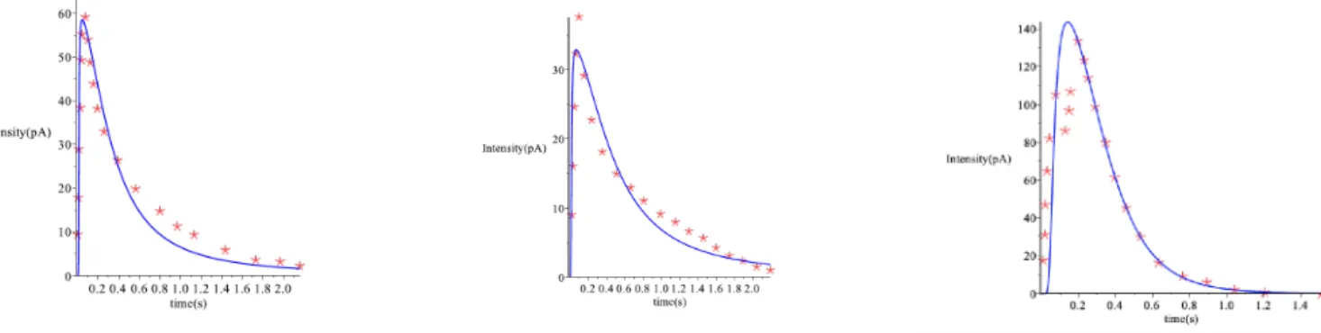
3.1.3 Tuning the parameters to fit experiments for acetylcholine dy-namics . . . 52
3.1.4 Bursting periods of immature SACs as a function of their bio-physical parameters . . . 52
The role of sAHP on bursting characteristics . . . 52
The role of cholinergic coupling on bursting characteristics 55 3.2 Bursting of an isolated cell and acetylcholine production. . . 55
Rest state . . . 55
Acetylcholine concentration dynamics. . . 57
Acetylcholine concentration in the rest state. . . 57
Ach concentration during a burst. . . 57
Ach concentration just after a burst. . . 58
Ach concentration during the hyperpolarization phase. . . 58
Numerical checks. . . 58
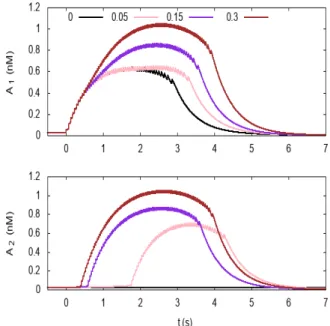
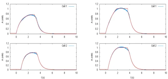
3.3 Bursting of 2 coupled cells. . . 59
Cells interaction. . . 59
Fitting the Ach profile. . . 59
The delay between bursts of cell 1 and cell 2. . . 60
Burst bifurcation. . . 61
Piecewise linear approximation for U . . . 62
Average Ach current in the rising phase. . . 63
Time of the next burst. . . 63
Calcium burst. . . 64
3.3.1 Remarks on how the waves propagate and stop . . . 64
Analytic characterization of the propagation in a 1-dimensional chain. . . 64
CONTENTS 3
3.4 Coupled Bursters in 1D (ring) . . . 65
3.4.1 Characterizing the dynamically driven bursting scenario in 1D . 66 3.4.2 Characterizing the noise driven bursting scenario in 1D . . . 67
3.5 How do SACs synchronize upon cholinergic coupling? . . . 73
3.5.1 Mechanism for two bursting cells synchronization . . . 73
Synchronization and coupled oscillators. . . 73
Synchronization in the dynamically driven bursting cells. . 74
Synchronization in the noise driven bursting cells. . . 76
3.6 What is the probability that one cell induces bursting to its neighbour? 76 3.6.1 Fluctuations about the rest state. . . 76
3.6.2 Ornstein-Uhlenbeck solution. . . 77
3.6.3 Bursting bifurcation. . . 77
3.7 Conclusion and Discussion . . . 77
How identical cells can display variability in behaviour. . . 78
4 Modeling and simulating stage II Retinal waves in 2D 79 4.1 Modeling a network of SACs . . . 79
4.1.1 Network structure . . . 79
4.2 How waves are generated in the developing retina? A proposed mecha-nism for waves triggering . . . 80
4.2.1 Scenario 1: Noise induced triggering. . . 80
4.2.2 Scenario 2: Dynamically driven triggering. . . 82
4.3 How do waves propagate? . . . 82
4.3.1 Propagation in a medium without friction . . . 83
4.3.2 Propagation in a medium with an sAHP landscape . . . 86
Analytic characterization of the wave propagation in a sAHP profile. . . 91
Example: R4(x, y) = ↵⇢, . . . 92
4.4 Distribution of waves near the critical point. . . 92
One wave. . . 93
Non direct interaction between waves. . . 93
4.5 Modeling the dynamical changes occuring within stage II retinal waves . 97 4.5.1 How does the cholinergic conductance evolves during development? 97 4.5.2 Different spatiotemporal patterns emerge within stage II retinal waves . . . 98
4.5.3 Spatial Correlations reveal a characteristic size for stage II waves 100 4.6 Characterizing the SACs population activity and the features of retinal waves . . . 100
4.6.1 Possible phase transition on the population firing rate . . . 100
4.6.2 Distribution of waves size and duration, Power laws, Criticality . 101 Waves statistical features . . . 101
4.7 Pattern formation in the dynamically driven bursting regime . . . 102
4.8 Conclusion and Discussion . . . 105
5 Towards a mesoscopic approach to analyse retinal waves 109 5.1 Transport equation . . . 110
5.1.1 Model and variables rescaling . . . 110
Fast dynamics of V, N . . . 111
Slow variables . . . 112
Medium scale variables . . . 112
5.1.3 Equation of transport for Ach . . . 112
Approximations . . . 112
Ach production on the lower branch. . . 113
Ach production on the upper branch. . . 113
Thresholding. . . 113
Variable Γ. . . 113
Piecewise linear approximation. . . 114
Laplacian approximation. . . 114
Singular diffusion. . . 114
Current. . . 115
5.2 Discussion . . . 115
Single neuron dynamics in the presence of a tunable sAHP and Ach currents . . . 115
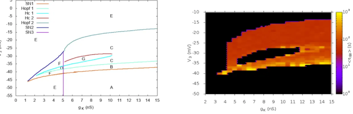
6 Confronting our model to experimental recordings 119 6.1 The role of potassium channels in waves characteristics . . . 119
6.2 Exploring the effect of the cholinergic transmission on the spatio-temporal patterns of stage II retinal waves . . . 125
Objective of the experiment. . . 125
Pharmacology. . . 125
Tissue preparation. . . 125
Recordings of retinal waves with MEA of 256 electrodes . 125 Analysis of the MEA recordings . . . 125
6.2.1 The effect of hexametonium in early stage II retinal waves . . . . 126
6.2.2 The effect of atropine in early stage II retinal waves . . . 129
6.2.3 Power laws and criticality in experimental recordings of retinal waves . . . 133
6.3 Conclusion and Discussion . . . 134
7 Conclusions and Perspectives 137 7.1 Reflecting on possible theoretical extensions . . . 137
Synaptic coupling versus volume diffusion. . . 137
The role of variability in SACs on waves generation. . . . 138
Extending our model towards a generic dynamical system for retinal waves . . . 139
!
!
!
Chapter 1
Introduction
In the natural world, all complex phenomena follow the laws of physics and for centuries physicists translate these physical rules in mathematical formalisms. For each physical system, one can try to decompose it to its parts and understand how these parts are connected to each other. Then, one can extract information, on which are the under-lying mechanisms, explaining what we observe. Imagine now, trying to understand something as complex as a network of electrically active biological cells in our eyes, which are connected to each other by highly complex mechanisms, working together to manifest a collective activity, which helps in fact the shaping of the visual system during development. All biological processes involved in this system, are simply too complex to be translated to a mathematical model that could be analysed. But what if we could find a simpler system, that has similar physical principles to our network of cells, and use it as an analogy in order to write down our equations. And so did Hodgkin and Huxley thought, that the electrical behaviour of a nerve, if we take into account only currents flowing in and out of a cell membrane, would be really similar to a classical electric circuit; and so they wrote their famous equations based on this idea [59]. Since then, mathematical modeling in biology and neuroscience has been vastly developed and used to understand the principles that govern neural networks. In this thesis, we are going to use the same approach, in order to describe mathematically how specific cells in the retina of the eye, exhibit propagating electrical activity in the process of shaping the visual system during development. In the following, as an introduction, we discuss the necessary elements for the reader, in order to smoothly follow the ideas of this work.
1.1
Introduction to the visual system
1.1.1 Visual system and structure
The visual system is part of the central nervous system and its main function is to process visual information. Although its functionality seems effortless, it carries out complex tasks including the reception of light and the formation of visual representa-tions, the identification and categorization of visual objects, computing distances to and between objects and guiding body movements in relation to the environment. The visual system includes basically the eyes, the optic nerve up to the Lateral Geniculate Nucleus (LGN) and the visual cortex. The neural signals, initially processed by the retina, travel via the axons of the ganglion cells through the optic nerves, dividing and
partially crossing over into the optic chiasm and then traveling via the optic tracts to the lateral geniculate nucleus (LGN). From the LGN, the signals continue to the primary visual cortex, where further visual processing takes place.
Figure 1.1: The visual system’s structure. A textbook representation of the visual system composed of the retina, the LGN and the visual cortex.
1.1.2 The retina and its structure
The retina is a light-sensitive tissue at the back of the eye that covers about 65 percent of its interior surface. In the middle of the retina is a small dimple called the fovea. It is the center of the eye’s sharpest vision and the location of most color perception. Moreover, retina is 0.5mm thick and it is composed of three layers of cell bodies and two layers of synapses.
Photosensitive cells, called photoreceptors (rods and cones), convert the incident light into electric pulses (spikes). A radial section of a portion of the retina reveals that the photoreceptors lie outermost in the retina against the pigment epithelium (see Fig 1.2). Light must, therefore, travel through the thickness of the retina before striking and activating the rods and cones. Subsequently the absorption of photons by the visual pigment of the photoreceptors is translated first into a biochemical message and then an electrical message that can stimulate all the adjacent neurons of the retina. The input from multiple photoreceptors is integrated and regulated by horizontal cells which are laterally interconnecting neurons with their cell bodies in the inner nuclear layer of the retina. Next, bipolar cells, existing between photoreceptors and ganglion cells, act directly or indirectly, to transmit signals from the photoreceptors to the ganglion cells. Amacrine cells are interneurons in the retina that are responsible for 70 per cent of input to retinal ganglion cells also. Ganglion cells (RGC), are the output neurons of the retina, and lie innermost in the retina, closest to the lens and front of the eye. Ganglion cells axons form the optic nerve, that carries the optic signal to the visual cortex.
1.1.3 Starburst Amacrine Cells
Starburst amacrine cells (SACs) are a subtype of retinal amacrine cells primarily iden-tified by the characteristic star-like shape of their dendritic arbor [28]. SACs have two
1.1. INTRODUCTION TO THE VISUAL SYSTEM 11
Figure 1.2: Retinal structure A textbook representation of the layered structure of the retina showing the different cell types: photoreceptors, bipolars, amacrines, horizontal, ganglion cells [19].
main functional roles are: i) their participation in the direction selectivity feature [6] and ii) generate spontaneous propagating activity in the developing retina [63]. SACs involvement in the direction selectivity feature in the retina computation, is explained by their morphological characteristics, such as the specific dentritic radial shape and a desymmetrised distribution of excitatory and inhibitory connections along the dendritic arbors [4, 60]. Also SACs, within the adult mammalian retina provide the critical inhibition that underlies the receptive field properties of direction-selective ganglion cells (DSGCs) [25].
Figure 1.3: Starburst amacrine cells. We clearly see their special shape of their dendritic arboring [63]
SACs, in the developing retina, exhibit autonomous rhythmic spontaneous activity, which generates propagating waves across the retina, called retinal waves, responsible to shape the visual pathways. SACs are the only cells in the retina that have been shown to release two neurotransmitters, acetylcholine and GABA [62]. In fact, SACs form a transient excitatory network, coupled by mutual cholinergic connections, gen-erating retinal waves. A detailed biophysical modeling of the spontaneous activity of SACs leading to propagating waves in the developing retina, is analyzed in detail in this thesis.
1.2
From neurons to neural networks
The overwhelming capacity of the retina to convert complex visual scenes into spike trains that send information to the visual cortex are largely due to its layered structure and to dynamical interactions between neurons in the retina. As a consequence and although it is very important to study the physiology and structure of individual neurons, in order to fully comprehend the functionality of the retina we need to study the system as a network, which processes the information via collective activity. Let us first though describe the components of a neural network in general.
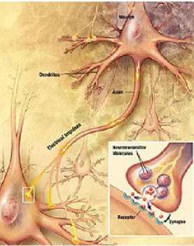
Synapses. Synapses are functional connections between neurons, or between neu-rons and other types of cells. A typical neuron may have several thousand synapses, although there are some types that make far fewer. Most synapses connect axons to dendrites, but there are also other types of connections, including axon-to-cell-body, axon-to-axon, and dendrite-to-dendrite. Synapses are generally too small to be rec-ognizable using a light microscope except at points where the membranes of two cells appear to touch, but their cellular elements can be visualized clearly using an elec-tron microscope. Chemical synapses are specialized junctions through which cells of the nervous system signal to one another and to non-neuronal cells such as muscles or glands 1.5. Chemical synapses allow the neurons of the central nervous system to form interconnected neural circuits. They are thus crucial to the biological compu-tations that underlie perception and thought. They also provide the means through which the nervous system connects to and controls the other systems of the body. Chemical synapses pass information directionally from a presynaptic cell to a postsy-naptic cell and are therefore asymmetric in structure and function. The presypostsy-naptic terminal, or synaptic button, is a specialized area within the axon of the presynaptic cell that contains neurotransmitters enclosed in small membrane-bound spheres called synaptic vesicles. Synaptic vesicles are docked at the presynaptic plasma membrane at regions called active zones. Electrical synapses are mechanical and electrically conduc-tive links between two adjacent neurons that are formed at a narrow gap between the pre- and postsynaptic neurons known as gap junctions. At gap junctions, such cells approach within about 3.5nm of each other, a much shorter distance than the 20 to 40nm distance that separates cells at a chemical synapse. In many animals, electrical synapse-based systems co-exist with chemical synapses.
Compared to chemical synapses, electrical synapses conduct nerve impulses faster, but, unlike chemical synapses, they lack gain since the signal in the postsynaptic neuron, is the same or smaller than that of the originating neuron. Electrical synapses are often found in parts of the neural systems that require the fastest possible response,
1.3. RETINAL WAVES 13
Figure 1.4: Chemical Synapse An artisitc representation of a chemical synapse (source Wikipedia).
such as defensive reflexes. An important characteristic of electrical synapses is that, most of the time, they are bidirectional. However, some gap junctions do restrict communication to only one direction [28].
Connectivity. The connections between neurons are made with chemical synapses and electrical gap junctions. One principle by which neurons work is neural sum-mation, i.e. potentials at the post synaptic membrane sum up in the cell body. If the depolarization of the neuron at the axon goes above threshold an action potential will occur that travels down the axon to the terminal endings to transmit a signal to other neurons. Excitatory and inhibitory synaptic transmission is realized mostly by inhibitory postsynaptic potentials and excitatory postsynaptic potentials. On the electrophysiological level, there are various phenomena which alter the response char-acteristics of individual synapses, (such as synaptic plasticity) and individual neurons, (intrinsic plasticity). Connections display temporal and spatial characteristics. Tem-poral characteristics refer to the continuously modified activity-dependent efficacy of synaptic transmission. It has been observed in several studies that the synaptic efficacy of this transmission can undergo short-term increase, called facilitation or decrease, ac-cording to the activity of the presynaptic neuron. The induction of long-term changes in synaptic efficacy, by long-term potentiation (LTP) or depression (LTD), depends strongly on the relative timing of the onset of the excitatory postsynaptic potential and the postsynaptic action potential.
1.3
Retinal waves
Long before the retina is responsive to light, in early development wave activity is observed. The so called retinal waves, observed in many vertebrate species - chicks
[31], ferrets [36], mice [53], turtles [55], macaques [69] etc. are spontaneous bursts of activity propagating in the developing retina and playing a fundamental role in shaping the visual system and retinal circuitry. They emerge due to the conjunction of intrinsic single-cell properties (excitability and long refractoriness) and network interactions [63]. In the developing retina, waves evolve during three consecutive stages, mainly characterized by different types of synaptic transmission. Stage I waves are mediated by gap junctions, travelling both horizontally across the RGC layer and vertically across the thickness of the retina, several days before synapse formation [56]. They are believed to play a role in the formation of the retinal circuitry but they are not yet fully understood [56]. Stage II waves are elicited by SACs through acetylcholine connections and propagate laterally accross the retina. Also, their propagation speed of is much slower than Stage I waves, they occur less frequently and their wave initiation points and trajectories are highly random [36, 56]. Functionally, they are found to refine the retinotopic mapping, which is the mapping of the visual input from the retina to the brain. In the next phase, stage III retinal waves switch their control from cholinergic to glutamatergic input [15], from the bipolar cells. These late waves are more localized, propagate vertically and disappear when vision is functional.
Figure 1.5: Stage II retinal waves. An experimental recording of stage II retinal waves in mice, measuring the local voltage, showing propagating activity [53].
1.3.1 General mechanism of stage II retinal waves
In the work of Zheng et al. [63], which is a benchmark on establishing the cellular mechanisms undelrying stage II retinal waves, they conclude that in order to gener-ate these waves, there are 3 necessary conditions (see Fig 1.6). The details of these conditions are developed throughout this thesis.
• (i) Fast repetitive bursts of spikes;
• (ii) Prolonged refractoriness through slow AfterHyperpolarisations (sAHP) • (iii) Synchronization via cholinergic connections of SACs
The fast repetitive firing is needed as a source of depolarization for wave initia-tion given that there is no external input (e.g. from visual stimulainitia-tion in the early
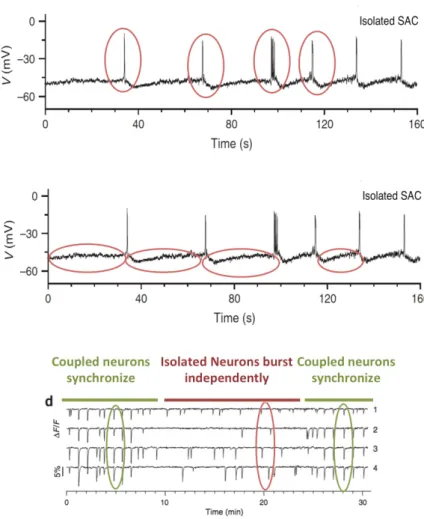
1.3. RETINAL WAVES 15
Figure 1.6: Illustrating the 3 necessary conditions to generate stage II retinal waves. Top and Middle. Patch clamp recording of an isolated SAC exhbiting bursting activity. In red, we mark the two components of bursting. Bottom. Calcium imaging to show that acetylcholine synchronizes the netowrk of SACs. All figures are from the work of [63].
retina). Thus, there must be some intrinsic mechanism by which neurons become ac-tive. As it is shown in [63], SACs exhibit fast oscillations due to their intrinsic cellular mechanisms, mainly controlled by voltage gated Ca+2 channels. Second, the long re-fractoriness of SACs, is needed to limit the spatial extent of waves and dictate the minimum interval between them. Finally, mutual cholinergic excitation, is essential for the synchronisation of SACs. When the multiple bursts occur in a neighbourhood of a neuron, SACs are synchronized, and they trigger a wave.
1.4
Modeling stage II retinal waves
The prevalence of retinal waves, observed in many different species and at different developmental stages, suggests that they are generated by generic collective and non linear mechanisms that still need to be unravelled. Developing mathematical models constitute a way to extract these underlying mechanisms. This strategy has been applied over the last twenty years, essentially for stage II, which are the most well studied waves.
1.4.1 State of the art
We briefly mention here previous models [34, 38, 54, 12], close to the spirit of the present thesis. For an extended review, see [50, 51].
Hennig et al. [54] model is, up to now the most elaborated model of stage II retinal waves with respect to biophysics. Especially, it capture the basis of the sAHP dy-namics and reproduces prominent statistical characteristics of waves experimentally observed in mice. As our own model is inspired by [54], we will widely comment it in the thesis. In this introduction we shortly expose our main criticism: (i) the equa-tions are not strictly bound to experimental findings and several parameter values are not biophysically justified. This holds for the other existing models [34, 38, 12]; (ii) this model requires an exogeneous shot noise current in order to induce the bursting activity triggering wave propagation: without this noise neurons stay at rest and it is not possible to reproduce the spontaneous depolarization and bursting observed in [63]. The same type of ”artificial” source of spontaneous depolarization is used by [34, 38, 12] as well. Moreover, the noise level needs to be fine tuned so as to reproduce experimental curves for waves size distribution; (iii) they do not provide a mathemat-ical analysis of their model. As we discuss in this thesis, such analysis is useful to unravel generic mechanisms for waves generation, propagation and shaping. Later, a dynamical system of a reaction diffusion type for stage II waves has been proposed by Lansdell et al. [12]. Their model is based on Hennig et al.’s with several important modifications: (i) The sAHP current is modeled in a less biophysical way; (ii) synaptic interactions are modeled by acetylcholine diffusion in a continuous medium in contrast to [54] where they use synaptic interactions. As in Hennig et al. model, SACs are at rest in spontaneous activity and waves trigerring requires a exogeneous noise. The authors perform a limited mathematical analysis.
The models [34, 38, 54] have compared their numerical results with experimental data and showed that they are able to reproduce some characteristics of retinal waves such as the waves sizes, duration, speeds and frequency. However, these models, are mostly able to reproduce already existing experimental results and not really predict new ones. Their approaches mainly lie on capturing phenomenologically the features of
1.4. MODELING STAGE II RETINAL WAVES 17 waves and do not model in detail the underlying biophysical mechanisms responsible for retinal waves generation.
1.4.2 What is this thesis about?
Having discussed the criticism of the state of the art, we have to say that the best model is the one that answers one’s questions and no single model can answer all of them. In other words, all models have their limitations as they are constructed so as to answer specific questions. So, in contrast to these existing models, our approach is based on the detailed biophysical modeling of the cellular mechanisms of the sponta-neous activity in immature SACs, addressing questions directly accessible and linked to pharmacological manipulations on retinal waves.
On theoretical grounds, it is natural to seek generic mechanisms (typically non linear instabilities) generating such waves and to extract from this analysis a few canonical parameters (e.g. conductances, characteristic times, reversal potentials etc.) con-trolling the generation of retinal waves as well as their characteristics. However, a conventional nonlinear analysis based on spatially extended dynamical systems would not be entirely appropriate to deal with the specific structure of the retina: discrete medium, specific connectivity via amacrine cells and electrical synapses, spiking gan-glion cells, conductances adaptation. Neurons are non linear systems producing a wide repertoire of dynamical activity, intrinsic, in response to a stimulus or due to change in physiological properties (e.g. conductances). They are connected in a complex way, because the graph of connectivity itself is complex, and evolve in time (synaptic plasticity), but also because the connectivity mechanism are themselves non linear, involving delays and memory. It’s also a multiscale problem, for the molecular scale to brain scale, from microseconds to years. As a consequence, the mathematical study of neuronal network is progressing slowly. Although there exist efficient methods to study theoretically extended neuronal networks in the cortex (mean-field, neural field, Fokker-Planck equation), the equivalent studies do not exist, to our best knowledge, in the retina. This work is an attempt to propose such an analysis, taking into account the specific characteristics of the retina (here amacrine cells and cholinergic coupling). An adaptation of the standard non linear analysis to these constraints is therefore done, integrating chemical synapses and a thorough mathematical analysis of their dy-namics. Our constructed set of equations constitutes therefore a solid basis to analyze non linear generation of waves in a neuronal network.
Using our model, we are able to answer several questions both on the single neuron as well as the network level. The detailed modeling of individual SACs dynamics as au-tonomous, rhythmic bursters and the mathematical analysis of our dynamical system using bifurcations theory, helps us identify the key parameters which control burst-ing in immature SACs. We propose a mathematical model, grounded on biophysics, which enables us to reproduce the bursting activity of SACs and to propose a plausible, generic and robust, mechanism that generates it. Based on a bifurcations analysis we exhibit a few biophysical parameters, especially regulating calcium and potassium ac-tivity, controlling bursting. We propose a testable experimental prediction on the role of voltage-dependent potassium channels on the excitability properties of SACs and on the evolution of this excitability along development. We also propose an explanation on how SACs across different species can exhibit a large variability in their bursting
periods, as observed experimentally, yet based on a simple, unique, mechanism. Extending the single neuron dynamics, we model in detail the mutual cholinergic synaptic connections between SACs, ending up exploring the mechanisms of SACs synchronization. We make a thorough analytic and numerical analysis to characterize how waves start, propagate and stop, providing analytic conditions that define the waves dynamics. We also provide an extension of our modeling towards a mesoscopic approach to study the propagation of waves in another spatial scale, that of acetyl-choline. Finally, we confront our model to experiments, performed by our collaborators, in order to validate our theoretical findings on the role of potassium channels and the cholinergic coupling on waves. Also, we use these experimental data to validate our predicted link to critical systems via power law manifestations for the waves charac-teristics.
Taken together, with this research we answer to the following questions with respect to our understanding of how retina works during development. Which is the biophysical mechanism that generates sustained periodically bursting in immature SACs and which are the parameters that control it? How can we link the biophysical parameters of our model directly with experimental measures, creating a framework where experiments can be reproduced by our model and our predictions could be tested experimentally? What is the mechanism underlying the loss of SACs excitability upon maturation? Is the mechanism of waves generation universal accross species given their variability in their characteristics? How do SACs synchronize in order to produce propagating waves? How do waves propagate and what is the type of their propagation? How do waves stop? How do the characteristics of waves depend on the biophysical parameters of their model and what can we predict for the network’s state? Is the network of SACs in a critical state and if so, what are the consequences of criticality on waves?
This work has possible future outcomes with respect to retina therapy as well. Un-derstanding how retinal waves are initiated and propagate in the retina could enable us to define protocols to trigger such retinal waves in the in vivo adult retina. In-ducing such waves is expected to reintroduce some plasticity in the retinal tissue and the projections in the brain. This induced plasticity could have important therapeutic applications to treat patients or stimulate regeneration of retinal ganglion cell axons following optic nerve crush.
The structure of the thesis is organized as follows: in Chapter 2, we study the dy-namics of a single SAC and we propose a mathematical model, grounded on biophysics, which enables us to reproduce the bursting activity of SACs and to propose a plausi-ble, generic and robust, mechanism that generates it. Based on a bifurcations analysis we exhibit a few biophysical parameters, especially regulating calcium and potassium activity, controlling bursting. In Chapter 3, we model the cholinergic connections and address mainly the questions ”How do waves start?” and ”How bursting cells synchro-nize?” in a 1-dimensional study. In Chapter 4, we perform the 2-dimensional study of retinal waves, characterizing the waves propagations as well as their statistical char-acteristics. In Chapter 5, we provide a derivation of a transport equation to study the spatiotemporal dynamics on a mesoscopic scale. In Chapter 6, we confront some of our theoretical findings to experiments. Finally, in Chapter 7, we conclude the thesis and give our perspectives for future extensions of this work.
1.4. MODELING STAGE II RETINAL WAVES 19
1.4.3 Methods and tools used in this thesis
Bifurcations theory. Many systems evolving in time are described by differential equations. Although it is tempting to try and find an analytic solution for these equations, this is, in most case not possible. Dynamical system theory is a branch of mathematics/physics, attempting to describe the behaviour of these solutions, with-out having an explicit representation of them. This is a powerful method, initiated by Poincare with a lot of applications in mathematics, physics, biology, chemistry, engineering, economics, and medicine. The mathematical study of changes in the qualitative behaviour of a set of non-linear equations, upon the continuous smooth variation of the parameters values, is called bifurcations theory. A bifurcation occurs when a dramatic sudden change in the behaviour of the dynamical system is caused by the small smooth change made to a parameter values [73]. In other words, there exist certain points, acting as ’thresholds’, at which the dynamics change abruptly, called bifurcation points. Bifurcations theory, provide us with tools to study the behaviour of complex dynamical systems depending on key parameters, using geometrical argu-ments, overcoming the difficulty to have direct solutions. It is a powerful tool, that helps us understand in depth how complex systems behave, allowing us to make predic-tions and extract mechanisms for the modeled systems. The name ”bifurcation” was first introduced by Henri Poincare in 1885 in the first paper in mathematics showing such a behavior [74].
Numerical simulations. In this work we use numerical methods done mostly in Python (we also use the Brian2 simulator [22] and MATLAB (we use the bifurcations software MATCONT [58]), but also methods programmed in C in order to simulate our model, perform bifurcations analysis and also characterize waves.
Chapter 2
Modeling the bursting activity of
individual immature SACs
Stage II retinal waves, are triggered by a transient network of neurons called Starburst Amacrine Cells (SACs) showing a bursting activity which disappears upon further maturation. The underlying mechanisms of the spontaneous bursting and the tran-sient excitability of immature SACs are not completely clear yet. While several models have tried to reproduce retinal waves, none of them is able to mimic the rhythmic au-tonomous bursting of individual SACs and understand how these cells change their intrinsic properties during development. Here, we propose a mathematical model, grounded on biophysics, which enables us to reproduce the bursting activity of SACs and to propose a plausible, generic and robust, mechanism that generates it. Based on a bifurcations analysis we exhibit a few biophysical parameters, especially regu-lating calcium and potassium activity, controlling bursting. We propose a testable experimental prediction on the role of voltage-dependent potassium channels on the excitability properties of SACs and on the evolution of this excitability along develop-ment. We also propose an explanation on how SACs across different species can exhibit a large variability in their bursting periods, as observed experimentally, yet based on a simple, unique, mechanism. As we discuss, these observations in the cellular level, have a deep impact on the retinal waves description.
This material is the subject of a paper ’A biophysical model explains the spontaneous bursting activity in the developing retina’, D. Karvouniari, L. Gil, O. Marre, S. Picaud, B. Cessac, currently under review in Nature Scientific Reports, [1].
2.1
The Morris-Lecal model
The Morris-Lecar model [13], is a conductance based model, and a simplified version of the Hodgkin-Huxley equations [59], since it does not describe the fast dynamics of sodium. The Morris-Lecar equations are particularly useful for modelling fast-spiking neurons, such as the pyramidal neurons of the neocortex. Morris-Lecar-type models may prove useful for studying scaling phenomena, such as showing how neural oscil-lators and oscillatory networks change as the cells grow during development. Finally, the Morris-Lecar model neuron has been applied to modeling networks of coupled neu-ral oscillators. Here the simple but realistic parameterization allows one to describe collective oscillations which depend on the inter-neuron coupling. A model employing
Morris-Lecar oscillators of different frequencies has been used to explain quite complex bursting phenomena of coupled neurons. All the above characteristics of the Morris-Lecar equations, make them the suitable candidate to model to describe the dynamics of autonomous bursters, such as immature SACs. Evidently, the basic Morris-Lecar model, as it is two dimensional, is not sufficient to model SACs dynamics, whose activity is generated by more complex currents. However, it is possible to extend Morris-Lecar equations by adding the additional currents involved in SACs bursting, as we will show in the following,
2.2
A biophysical model for bursting immature SACs
In the case of immature SACs, the two key biophysical mechanisms associated with the emergence of spontaneous bursting during early development are [63]:• (i) fast repetitive bursts of spikes mainly controlled by voltage-gated Ca+2 chan-nels;
• (ii) prolonged AHPs modulating fast oscillations, controlled by Ca+2-gated K+ channels.
Concerning (i), the fast repetitive firing during the active phase of bursting gen-erally results from the competition between a depolarizing and a hyperpolarizing cur-rent. Experiments in [63] on specifying the ionic channels involved in the spontaneous bursting of immature SACs, suggest voltage-gated Ca+2 channels for the depolarizing component. Note that [63] have shown that voltage-gated N a+ channels do not par-ticipate in the bursting mechanism of immature SACs (bursting activity of SACs was not altered upon tetrodoxin -TTX- application), thus, dynamics of N a+channels will not be considered in our modeling. The ionic channels related to the hyperpolariz-ing component of SACs bursthyperpolariz-ing have not yet been identified experimentally. In this work, we propose fast voltage-gated K+ channels play this role. This point is further developed in the following.
Concerning (ii), the long refractoriness in-between consecutive bursts is controlled by a slow After Hyper-Polarization (sAHP) K+ current, I
sAHP. It was observed by [63] that IsAHP is mediated by Ca+2-gated K+ channels, and that it resembles the sAHP observed by Abel et al. [11], generated by specific channels called SK. Follow-ing these tracks we propose a modelFollow-ing of ”SK”-like channel (as named in [63]) based on [54] for the structure of the equations and [44] for the calcium dynamics. The mechanism of the opening of Ca+2-gated ionic channels is analyzed in detail in the supporting information section. In order to simplify the cascade of chemical reactions taking place while opening the sAHP channels, we approximate the channel dynamics by reducing the process into two discrete steps: a) Four ions of Ca+2 bind to a second messenger protein called calmodulin, forming a saturated calmodulin complex, CaM; b) CaM binds to each of the four intracellular subunits of the channel to open it (see Fig 2.1). This process is mapped to our model through three variables: 1) the vari-able C which models the intracellular calcium concentration and mainly controls the gating variables of the sAHP channels, 2) the variable S which models the fraction of the saturated calmodulin and 3) the variable R which models the fraction of bounded terminals. This gating mechanism is sketched in Fig 2.1.
2.2. A BIOPHYSICAL MODEL FOR BURSTING IMMATURE SACS 23 From these observations, we model SACs activity with a conductance based model of the Morris-Lecar type [13] with additional currents featuring (i), (ii). The model involves 5 variables whose evolution is controlled by a set of non-linear differential equations (see Eq (2.2)-(2.8) ): V (t), the local membrane potential, N (t), the gating variable for fast voltage-gated K+ channels, R(t) and S(t), the gating variables for slow Ca2+-gated K+ channels and C(t), the intracellular Ca2+ concentration. All parameters values and the auxiliary functions involved are found in Methods.
The membrane voltage V (t) obeys: CmdV
dt = IL(V ) + IC(V ) + IK(V, N ) + IsAHP(V, R) + σ⇠t, (2.1) where Cm is the membrane capacitance, IL= −gL(V −VL) is the leak current, with gL leak conductance and VL leak reversal potential. ⇠t is a white noise whose amplitude is controlled by σ. The terms IC and IK, respectively corresponding to Ca+2 and K+ currents, are generating the fast Ca+2 oscillations. These currents are described by a Morris-Lecar model [13] where the voltage-gated Ca+2 current is:
IC(V ) = −gCM1(V )(V − VC). (2.2) gCM1(V ) is the voltage dependent conductance of the Ca+2 channel (see Eq (2.21) in Methods).
The fast voltage-gated K+ channel is modeled as:
IK(V, N ) = −gKN (V − VK). (2.3)
where the evolution of the fast voltage-gated K+channel gating variable N (t) is mod-eled as:
⌧N dN
dt = Λ(V )(N1(V ) − N), (2.4)
Λ(V ) and N1(V ) are given by Eq (2.22), (2.23) in Methods. Note that equations (2.1) - (2.4) with IsAHP = 0 and σ = 0, correspond to the Morris-Lecar model with a fast variable N.
The sAHP current takes the form:
IsAHP(V, R) = −gsAHPR4(V − VK), (2.5) where gsAHP is the maximum sAHP conductance. Indeed, 4 bound terminals are needed to open a Ca+2-gated K+ channel, thus the corresponding conductance is gsAHPR4, involving a fourth order nonlinearity.
Now, we model the gating mechanism of the Ca+2 gated K+ channels as follows. The gating variable R(t):
⌧R dR
dt = ↵RS(1 − R) − R, (2.6)
the fraction of saturated calmodulin concentration S(t); ⌧S
dS
dt = ↵SC
R
R
R
R
Activated
Ca-gated K+ channel
Ca 4 Calcium ions bind to each saturated
Calmodulin complex (CaM) Saturated Calmodulin molecule (CaM) binds
to each channel subunit
All 4 subunits R of the channel are bound to activate the channel
Gating mechanism
CaM
Ca CaCa CaCa CaCa CaCa CaCa CaCa CaCa
CaM CaM CaM
S
C
Figure 2.1: Schematic representation of the modeling of the gating mechanism of Ca+2-gated K+ channels. The correspondence between the channel’s activation steps and
the modeling state variables R, S, C is also indicated.
and the intracellular calcium concentration C(t); ⌧C dC dt = − ↵C HX C + C0− δCgCM1(V )(V − VC). (2.8) The derivation of the equations (2.6), (2.7), (2.8) is fully justified in Methods.
2.3
Deriving sAHP dynamics
To our best knowledge, the ionic channels type involved in sAHP for immature stage II SAC is not precisely known. However, Zheng et al. argue in [63] that these channels could share characteristics with SK channels, thoroughly studied for pyramidal neurons by Abel et al in [11]. On this basis we modeled SK channels dynamics. SK channels have four subunits associating to form a tetramer. The SK channel gating mechanism is controlled by intracellular calcium levels. The precise mechanism is: (i) calcium binds to the protein calmodulin forming the complex CaM where 4 ions Ca 2+ are fixed to calmodulin; (ii) CaM binds to a SK channel terminal to open it; (iii) 4 terminals must be open to let the SK channel open. We now model these different steps.
Saturated calmodulin production. The set of kinetic equations leading to CaM formation is widely described in M. Graupner’s work [43, 44]. This is a cascade of equations that we summarize in one kinetic equation, from free calmodulin, M , to the saturated one, CaM .
Let us call kass(M−4s−1) and kdiss(s−1) respectively association and dissociation con-stants of calmodulin. Set Kd4 = kdiss
kass. If we call M0 = [M ]+[CaM ] the total calmodulin
concentration, and S = [CaM ]M
0 the fraction of saturated calmodulin, we have [M ]
M0 = 1−S
and we obtain a kinetic equation: dS
dt = kassC
4(1 − S) − k dissS.
2.3. DERIVING SAHP DYNAMICS 25 where C is the intracellular calcium concentration. Setting ⌧S= kdiss1 and ↵S= kkassdis =
1 K4 d we arrive at: ⌧S dS dt = ↵SC 4 (1 − S) − S. (2.9)
Note equation (2.7) has a similar form to the one proposed by Hennig et al. in [54]. However, in their model our term C4 is replaced by K4C4
d+C4
which is hardly inter-pretable in terms of kinetics.
Binding of calmodulin to SK terminals. This corresponds to a reaction:
F + CaM
PF B ! PBF
B,
where F is the density of free terminals, B the density of bounded terminals, PF B (PBF) the transition rate from free to bound (bound to free).
Calling R the fraction of bounded terminals, ⌧R= PBF1 , ↵R= PPF BBF we end up with a kinetic equation:
⌧R dR
dt = ↵RS(1 − R) − R = ↵RS − (1 + ↵RS)R. (2.10) Equation (2.6), is similar to Eq (3) in [54] (⌧RdRdt = (↵C + S)(1 − R) − R), with a remarkable difference: in our model there is no direct dependence on calcium con-centration whereas the term ↵C in [54] corresponds to a direct binding of Ca2 to a terminal. Note that, taking the quite large value of the parameter ↵ ( 2400) considered by these authors, their equation is essentially equivalent to ⌧RdRdt = ↵ C(1 − R) − R = ↵ C − (1 + ↵ C)R with a steady state R = 1+α Cα C very close to 1 whenever ↵ C is quite larger than 1. In this case, S plays essentially no role.
Finally, since R is the probability that a terminal is open and since 4 terminals must be open to let the SK channel open, the sAHP conductance is gsAHPR4.
Calcium concentration Both variables R and S are driven by intracellular Ca2 concentration dynamics, given by:
⌧C dC dt = − ↵C HX C + C0− δCgCM1(V )(V − VC) (2.11) Equation (2.11) is a linear approximation of a more complex equation ((2.14) below). This equation is similar to Hennig et al (Eq (5)) with two notable differences: (1) We have added a rest concentration C0 avoiding unphysical situations where C can become negative; (2) the value of parameters are different.
Calcium concentration dynamics. The calcium current crossing a membrane sec-tion results from the opening of gates in ionic channels. Following [43] the equasec-tion for Ca concentration is (adapted with our notations):
dC dt = G nCaF IC S − JX(C) − Jp(C) + L " 1 1 +dCabound dC . (2.12)
Here G = VS = 6 µm−1= 6 ⇥ 105dm−1 is the surface to volume ratio that accounts for the localisation of the channels at the surface of the membrane, nCa= 2 is the calcium valence and F = 96500 C mol−1 is the Faraday number. dCabound
dC corresponds to a quasi-steady-state approximation for the calcium buffering where the bound calcium concentration (on calmodulin) Cabound is adapted instantaneously to the free calcium concentration C at each time. Since we have no way to estimate dCabound
dC we shall consider it is a constant and set 1
1+dCabounddC ⌘ Kbound. To alleviate notations we set: r = G Kbound
nCaF
. (2.13)
The first term in Eq (2.12), rIC
S corresponds to an increase of internal Ca2+ con-centration upon calcium influx (current IC(V )) generated by spikes or, in experiments, by voltage clamp. As VCa= 50mV this current is positive unless V > VCa.
The second term is −rJX(C), where JX(C) = ⇢XIX
C HX + C
,
is the efflux current density through sodium-calcium exchanger (NCX). It corresponds to an outward through NCX exchangers, contributing to restoring the initial Ca con-centration. Here, ⇢X is the surface density of the NCX membrane proteins. We take ⇢X = 100 µm−2 = 1012dm−2(from [44]). IX = 4.8⇥10−19C ms−1= 4.8⇥10−16C s−1 is the maximum ionic flux through a single NCX channel. This corresponds to 3 + charges (1.5 Ca2+) per ms; H
X = 1.8µM = 1.8 ⇥ 10−6M is the half activation con-centration [43].
Also, in (2.12), Jp(C) is the current density of Ca pumps. We shall neglect this term from now on. Finally, L is the leakage surface current density representing the residual conductivity of the plasma membrane. We have not been able to find its value in the literature. To summarize Eq (2.12) becomes: dC dt = r IC S − ⇢XIX C HX + C + L " . (2.14)
We also assume that NCX current term is approximated by a linear term. This is valid if one assumes that calcium concentration C ⌧ HX which is the case in our simulations.
In order to match (2.14) with the form (2.11) ((2.8) in the text), we set: ↵Ca ⌧Ca = r⇢XIX; (2.15) δCa ⌧Ca = r S; (2.16) C0 ⌧Ca = rL. (2.17) (2.18)
2.4. RESCALED EQUATIONS AND MULTI-TIME SCALE ANALYSIS 27
2.3.1 Full set of equations for the single SAC dynamics
To summarize, the full set of equations in our model with the 5 state variables V, N, C, S, R is the following: 8 > > > > > > > > > > > > > < > > > > > > > > > > > > > : CmdVdt = −gL(V − VL) − gCM1(V )(V − VC) − gKN (V − VK) − gsAHPR4(V − VK) ⌧NdNdt = Λ(V )(N1(V ) − N) ⌧CdCdt = −HαCXC + C0− δCgC(V )(V − VC) ⌧SdSdt = ↵S(1 − S)C4− S ⌧RdRdt = ↵RS(1 − R) − R (2.19)
2.4
Rescaled equations and Multi-time scale analysis
The dynamical system (2.19) has 3 characteristic times scales: fast variables V, N (of order ms); medium C (of order s) ; slow R, S (of order 10 s), fixed by the characteristic times given in the Table 2.1. In order to make explicit these time-scales separation we set ˜gX = |ggXL| for conductances (X = C, K, sAHP ); ⌧ = CgLm; ˜t = τt; ˜⌧X = ττX, where X = N, C, R, S. This gives: 8 > > > > > > > > > > > > > < > > > > > > > > > > > > > : ⌧LdVdt = −˜gL(V − VL) − ˜gCM1(V )(V − VC) − ˜gKN (V − VK) − ˜gsAHPR4(V − VK) ⌧NdNdt = Λ(V )(N1(V ) − N) ⌧CdCdt = −HαCXC + C0− δCgCM1(V )(V − VC) ⌧SdSdt = ↵S(1 − S)C4− S ⌧RdRdt = ↵RS(1 − R) − ROn the fast time scale, one uses the approximation τ1
X = 0, X = C, S, R, and
the variables C, S, R are constant. So, fast dynamics reduces to a Morris-Lecar model (here with a fast variable N ) in the presence of an additional current Iext = IsAHP (constant): 8 < : CmdVdt = −gL(V − VL) − gCM1(V )(V − VC) − gKN (V − VK) + Iext; ⌧NdNdt = Λ(V )(N1(V ) − N). (2.20)
2.5
Parameters value and auxiliary functions
Units. In all the thesis, physical quantities are expressed in the units displayed in table 2.1. Having integrated over all the surface of the membrane we omit the surface units.
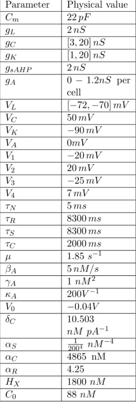
Calibrating parameters from experiments All parameters values are calibrated with respect to biophysics, found in the literature or fitted from experimental curves in [11], [62] and [63]. Morris-Lecar tuning parameters V1, V2, V3, V4 were calibrated (see Fig 2.11), so as to reproduce the experiment of [63] (Fig 4a), where the authors investigate the ionic mechanisms of the fast oscillations. Note that the bursting regime is robust to (small) variations of these parameters (results not shown). We tuned the sAHP parameters taking into account the analogy with SK channels studied in [11] (fit not shown). Also, we note that the intensity of sAHP observed by Abel et al. in pyramidal neurons (of order 150 pA) is quite bigger than in stage II SAC. In our model, this means a lower sAHP conductance gsAHP (gsAHP = 2 nS).
Physical quantity Dimension
Time ms Potential mV Capacitance pF Current pA Conductance nS Concentrations nM
Table 2.1: Dimensions of physical quantities used in the thesis.
Auxiliary functions. The dimensionless auxiliary functions involved in the dynam-ical equations appearing in the model definition are:
M1(V ) = 1 2[1 + tanh( V − V1 V2 )], (2.21) Λ(V ) = cosh(V − V3 2V4 ), (2.22) N1(V ) = 1 2[1 + tanh( V − V3 V4 )], (2.23)
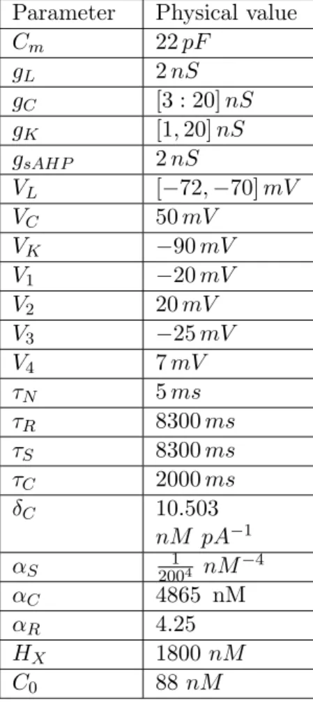
Parameters. The parameters used in the model are displayed in Table 3.1.
2.6
Comparison with existing models
In this section we shortly revisit models of SACs activity in the stage II and compare them to our model, ([34, 38, 54, 30, 12], for a review see [50, 51]). We would first like to remark that all models we know are devoted to describe wave activity and do not focus on thoroughly describing individual SACs dynamics. Especially, none of the models we know describe the biophysical mechanisms of SACs bursting activity and the role played by biophysical parameters. Instead the focus was more on having a relatively simple description of the cell activity with a minimal set of tunable parameters (a notable example is Butts et al model[34] which has two free parameters governing the waves properties).
The closest model to ours has been proposed by M. Hennig and collaborators [54] (referred as Hennig model) (see also the extension by Ford and Feller and the recent
2.6. COMPARISON WITH EXISTING MODELS 29 Parameter Physical value
Cm 22 pF gL 2 nS gC [3 : 20] nS gK [1, 20] nS gsAHP 2 nS VL [−72, −70] mV VC 50 mV VK −90 mV V1 −20 mV V2 20 mV V3 −25 mV V4 7 mV ⌧N 5 ms ⌧R 8300 ms ⌧S 8300 ms ⌧C 2000 ms δC 10.503 nM pA−1 ↵S 20014 nM−4 ↵C 4865 nM ↵R 4.25 HX 1800 nM C0 88 nM
Table 2.2: Range of values for the parameters used in the thesis.
paper of Xu et al. [9]). Actually, our model has been widely inspired by this work with several notable differences. As exposed in the Methods section our biophysical analysis of sAHP dynamics leads to equations and parameters values departing from Hennig model. Additionally, Hennig model does not consider a fast potassium dynamics and there is no fast oscillation. The mechanism that mimics SACs bursting is a switch from low membrane potential level to high one. This switch is determined by an exogeneous shot noise i.e. a voltage dependent rate modulated Poisson process with a slow decay. This activity is maintained long enough so that sAHP can be activated, enabling the cell to return to rest. In our model shot noise is not necessary to trigger activity. Instead, a cell can spontaneously switch to the bursting state, where it stays until the sAHP produced by its activity leads it back to the rest state. By spontaneous we mean literally happening or done in a sudden way, without any planning or without being forced/without premeditation. In our model this sudden switch is a bifurcation induced by the mere cells dynamics. The presence of a fast (Brownian) noise facilitates this transition, but, there is no need for a shot noise. The cell stays in the bursting state by its mere dynamics, even when it is isolated.
A similar modeling holds in Lansdell et al. model [12]. It is ruled as well by an excitable Morris Lecar model with a slow potassium variable linked to sAHP. There is no fast potassium. Here too cells do not burst. As in Henning the cell activity is
![Figure 1.3: Starburst amacrine cells. We clearly see their special shape of their dendritic arboring [63]](https://thumb-eu.123doks.com/thumbv2/123doknet/13203835.392783/21.892.309.585.810.1062/figure-starburst-amacrine-cells-clearly-special-dendritic-arboring.webp)