HAL Id: hal-03018338
https://hal.archives-ouvertes.fr/hal-03018338
Submitted on 22 Nov 2020HAL is a multi-disciplinary open access archive for the deposit and dissemination of sci-entific research documents, whether they are pub-lished or not. The documents may come from teaching and research institutions in France or abroad, or from public or private research centers.
L’archive ouverte pluridisciplinaire HAL, est destinée au dépôt et à la diffusion de documents scientifiques de niveau recherche, publiés ou non, émanant des établissements d’enseignement et de recherche français ou étrangers, des laboratoires publics ou privés.
Solubilization and stabilization of membrane proteins by
cycloalkane-modified amphiphilic polymers.
Anaïs Marconnet, Baptiste Michon, Christel Le Bon, Fabrice Giusti,
Christophe Tribet, Manuela Zoonens
To cite this version:
Anaïs Marconnet, Baptiste Michon, Christel Le Bon, Fabrice Giusti, Christophe Tribet, et al.. Solubilization and stabilization of membrane proteins by cycloalkane-modified am-phiphilic polymers.. Biomacromolecules, American Chemical Society, 2020, 21, pp.3459–3467. �10.1021/acs.biomac.0c00929�. �hal-03018338�
Solubilization and stabilization of membrane
proteins by cycloalkane-modified amphiphilic
polymers.
Anaïs Marconnet,1,2 Baptiste Michon,1,2 Christel Le Bon,1,2 Fabrice Giusti,1,2,† Christophe Tribet,3,* Manuela Zoonens1,2,*1
Université de Paris, Laboratoire de Biologie Physico-Chimique des Protéines Membranaires,
CNRS, UMR 7099, F-75005 Paris, France. 2
Institut de Biologie Physico-Chimique, Fondation Edmond de Rothschild pour le développement de la recherche Scientifique, F-75005 Paris, France. 3
P.A.S.T.E.U.R., Département de Chimie, École Normale Supérieure, PSL University, Sorbonne Université, CNRS, F-75005 Paris, France.
KEYWORDS: amphipols, extraction, membrane proteins, SMA co-polymers, stability, surfactants.
ABSTRACT: Membrane proteins need to be extracted from biological membranes and purified in their native state for most structural and functional in vitro investigations. Amphiphilic copolymers, such as amphipols (APols), have emerged as very useful alternatives to detergents for keeping membrane proteins water-soluble under their native form. However, classical APols, i.e. poly(acrylic acid) (PAA) derivatives, seldom enable direct membrane protein extraction. Poly(styrene-maleic anhydride) copolymers (SMAs), which bear aromatic rings as hydrophobic side groups, have been reported to be more effective extracting agents. In order to test the hypothesis of a role of cyclic hydrophobic moieties in membrane solubilization by copolymers, we have prepared PAA derivatives comprising cyclic rather than linear aliphatic side groups (CyclAPols). As references, APol A8-35, SMAs, and diisobutylene maleic acid (DIBMA) were compared with CyclAPols. Using as models the plasma membrane of Escherichia coli and the extraction-resistant purple membrane from Halobacterium salinarum, we show that CyclAPols combine the extraction efficiency of SMAs with the stabilization afforded to membrane proteins by classical APols such as A8-35.
Introduction
As gatekeepers of all membrane compartments in cells, membrane proteins (MPs) fulfill essential biological functions and are key targets for drugs.1,2
Their importance motivates structural and biophysical studies, which, however, critically depend on extrac-tion/purification procedures that keep MPs in their native state. Detergents can disrupt biological membranes and keep MPs water-soluble, but they often entail MP inactivation, in part due to their propensity to detach from MPs endogenous lipids or cofactors needed for activity. This has prompted the development of macromolecular amphiphiles such as
amphipols (APols),3–5
which were expected to be less aggressive dispersing agents and less prone to reach hidden hydrophobic regions in the protein. The prototypical APol, named A8-35, is a poly(acrylic acid) (PAA) polymer randomly modified with octylamine and isopropylamine side chains (Figure 1A). The mildness of APol association with MPs results
in an increased life-time of the native state of APol-complexed MPs.5
Another advantage of APols is the improvement of images in the fast growing field of single-particle electron
cryo-microscopy (cryo-EM) reconstitution.5,6
Conventional APols are however poor solubilizing
agents for biological membranes, by far less efficient than most detergents.7,8
They can break
liposomes (lacking MPs),9–11
but in practice MP/APol complexes can rarely be directly obtained from biological membranes and must be prepared by transferring the protein from detergent-solubilized extracts.5,12
Styrene maleic acid (SMA) co-polymers (Figure 1B) are APol variants with the distinctive property of enabling direct dispersion of proteolipidic
membranes with no need for detergent.13,14
They form lipid-protein-polymer mixed nanodiscs, called SMALPs. Spontaneous formation of lipid nanodiscs is not specific to SMAs as it has
been observed with lipids and diisobutylene/maleic acid copolymers (DIBMA, Figure 1C)15
and with copolymers similar to APols.16
Despite several limitations such as high UV
absorbance, due to the presence of aromatic groups, and polydispersity of the discs,17
of SMAs has developed in the field of MPs thanks to their solubilizing property. However, in the field of structural biology, the frequent need to use harsh extraction conditions may
explain the low number of high-resolution cryo-EM structures established using SMAs.6,18
To date, only a small number of single-particle cryo-EM structures of SMALPs have been obtained.6
In comparison, despite the need for surfactant exchange, classical APols (mainly
A8-35 and PMAL-C8)3,19,20 represent the most frequently used polymers.6 To circumvent the
need of surfactant exchange, improving the direct solubilization of membranes by APol-like polymers under mild experimental conditions would be highly desirable.
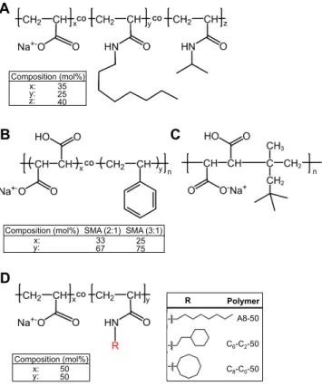
Figure 1. Chemical structures of amphipathic polymers used in this study. A) A8-35; molar
percentages of each unit are from ref.3
B) SMA co-polymers, with molar ratios of styrene to
maleic acid units of 2:1 or 3:1 (n ≈ 9).15
C) DIBMA co-polymer (n ≈ 37).15
D) PAA modified
with either linear or cyclic hydrocarbon groups (R) of 8 carbon atoms. The 𝑀! values for C6
-C2-50, C8-C0-50 and A8-50 (sodium salt form) were calculated using 𝐷𝑃! ≈ 35 for the parent
PAA21 and the degree of grafting, y, determined by 13C, 1H NMR and potentiometric titration
A D B x: 35 25 40 Composition (mol%) y: z: SMA (3:1) SMA (2:1) 33 67 25 75 y: x: Composition (mol%) C8-C0-50 C6-C2-50 A8-50 Polymer R x: 50 50 Composition (mol%) y: C
(~53 ± 1, ~51 ± 3, and ~56 ± 4 mol% for C6-C2-50, C8-C0-50 and A8-50, respectively; see
Figures S1 to S7 in SI and Table 1).
We hypothesized that replacing the flexible n-alkyl side groups of classical APols by cyclic hydrophobic groups might possibly mimic the role of SMA's aromatic side groups. Of note, introducing cyclic groups in the aliphatic tail of dodecylmaltoside results in
MP-stabiliz-ing analogs.22 We therefore examined whether cyclic hydrocarbon groups grafted onto PAA
might not i) improve the polymers’ ability to break up MP-containing membranes while ii) preserving the long-term stability of MPs. To this end, we prepared copolymers, named Cycl-APols, in which the linear octyl groups of A8-35-like APols were replaced by cyclic ones having the same number of carbon atoms (either cyclohexylethylamine or cyclooctylamine,
yielding polymers C6-C2-50 and C8-C0-50, respectively, Figure 1D). Cycloalkanes were
preferred over aromatic cycles in order to keep the CyclAPols UV-transparent. As references,
we compared CyclAPols with SMA (2:1) and (3:1) of relatively short length,14,23,24
DIBMA, and polyacrylate-based APols with linear side groups, namely A8-35 and A8-50 (an APol possessing a density of hydrophobic moieties of ~50%, i.e. equal to that of SMA (2:1) and CyclAPols).
We studied the membrane-solubilizing properties of CyclAPols using as a model the plasma membrane of Escherichia coli, a host cell widely used for producing recombinant MPs.25,26
We expressed two different MPs, YidC of E. coli fused to a green fluorescent protein (GFP), and the bacteriorhodopsin of Haloquadratum walsbyi (HwBR). To further examine the solubilizing properties of CyclAPols, we used the native purple membrane from
Halobacterium salinarum, which is well known for its resistance to solubilization by
polymers, be they SMA13,27
or classical APols.5
Finally, the thermal stability of BR from Hb.
Experimental Section
1) Chemicals
DIBMA and n-Dodecyl β-D-maltoside (DDM) were supplied by Anatrace. As indicated by the supplier, DIBMA was a purified version of Sokalan CP9 from BASF. SMA (2:1) and SMA (3:1) were obtained from ProFoldin (SMA21S) and Polyscope (Xiran SL25010 P20),
respectively. The number-average molecular masses (𝑀!) of DIBMA and SMA (3:1)
co-polymers were ~8,400 g·mol-1
and ~4,000 g·mol-1
, respectively.15
The lipid 1,2-dimyristoyl-sn-glycero-3-phosphocholine (DMPC) was purchased from Avanti Polar Lipids.
2) Synthesis of APols and composition determination
The synthesis of A8-35 was performed as previously reported.3
APol derivatives carrying either octylamine, cyclohexylethylamine or cyclooctylamine were obtained after a single step of poly(acrylic acid) (PAA) modification. Briefly, the hydrophobic side chains were randomly
grafted onto PAA of 𝑀!~ 5.5 kDa in N-methylpyrrolidone (NMP) in the presence of
dicyclohexylcarbodiimide (DCC). In each case, the resulting polymer obtained under the acidic form was purified by four cycles of precipitation/solubilization in water, performed at acidic and basic pH, respectively. The final polymer solution adjusted at pH 8-9 was dialyzed against MilliQ water and freeze-dried, yielding the expected APol. After synthesis, the final
yields of APols were 98% and 96% for CyclAPols C6-C2-50 and C8-C0-50, respectively, and
86% and 52% for APols of reference A8-35 and A8-50, respectively.
The grafting ratio of hydrophobic side chains was determined by 13
C and 1
H NMR spectroscopy (Figures S1 to S6) and potentiometric titration in a water/ethanol mixture
(20:80, v/v) (Figure S7). The 13
C NMR spectra of polymers, at 100 g·L-1
in deuterated methanol, were obtained after a long acquisition time (48-60h) due to the long delay between
each frequency pulse required for a complete relaxation of all carbon nuclei. The average degree of polymerization (𝐷𝑃!) of the commercial PAA used in this study for synthesizing
A8-35 and the APol derivatives was previously determined to be close to ~35 by size
exclusion chromatography (SEC) analysis in organic solvent.21
The number-average molar
masses (𝑀!) of sodium salts of APol derivatives were calculated with the 𝐷𝑃!value and the
grafting ratio (y) averaged from 13
C and 1
H NMR spectroscopy and pH-titration determination (Table 1). APol CO2 -(x1/x2, %) Hydrophobic groups (y1/y2, %) x (%) y (%) 𝑴𝐧 (g·mol-1 ) C6-C2-50 47/46 53/54 46.5 ± 0.5 53.5 ± 0.5 4919 C8-C0-50 52/46 48/54 49 ± 3 51 ± 3 4843 A8-50 47/40 53/60 43.5 ± 3.5 56.5 ± 3.5 5050
Table 1. Chemical compositions and number-average molecular masses of polyacrylate-based
APols. The percentages of grafts and free carboxylates were deduced from an average of 1
H and 13
C NMR analysis (yielding y1 and x1) and from acid-base titrations (yielding y2 and x2) for
each APol. The values were averaged, giving the final x and y ratios. For the APol of
reference A8-35, the averaged final ratios were x = 34%, y = 23% and z = 43%, and the 𝑀!
value was 4276 g·mol-1
.
3) Expression of YidC-GFP of E. coli and BR of Hq. walsbyi in E. coli strain and quantification of protein extraction using polymers
The expression vectors encoding for YidC of E. coli fused at its C-terminus to GFP (YidC-GFP) and BR of Hq. walsbyi (HwBR) were kindly provided by the laboratories of Jan Whilem and Anant Menon, respectively. The strains E. coli C41 and C43 (DE3) were transformed with each vector, respectively, and growth at 37°C in LB in the presence of either ampicillin or kanamycin. When the optical density at 600 nm reached 0.6-0.8, the cultures
were supplemented with IPTG. In the case of HwBR production, 5 μM all-trans retinal (Sigma) were also added. After induction, cells were harvested. The pellet of each culture was re-suspended with buffer containing 20 mM Tris-HCl, 150 mM NaCl, pH 8.0 and cell lysis was performed with a Cell Disruptor (Constant Systems). Unbroken cells and cellular debris were removed by centrifugation (20 min. at 10,000 × g). The membranes were harvested from supernatants by high-speed centrifugation (1h at 100,000 × g). The concentration of total MPs present in the membrane extracts was assessed with a calorimetric assay.
The solubilization of YidC-GFP from E. coli membranes was performed at a total MP concentration of 2 g·L-1
with either 1% w/v DDM, used as reference condition, or 2 g·L-1
polymers, corresponding to a total MP/polymer ratio of 1:1 (w/w). After 1 h incubation at 4°C, the samples were centrifuged 30 min. at 100,000 × g. The supernatants and the pellets, re-suspended in the initial volume of solubilization with buffer 20 mM Tris-HCl, 150 mM NaCl, pH 8.0 and 5% SDS, were loaded on 12-% acrylamide gels. After migration, the gels were washed with water and the fluorescence of GFP within the gel was detected with a Typhoon TLA 9500 (GE healthcare). The excitation and emission wavelengths were those of Alexa Fluor 488, i.e. 495 nm and 519 nm, respectively. The fluorescence of the band corresponding to YidC-GFP visible on the acrylamide gels was quantified using Image J software. The fluorescence of YidC-GFP measured for each supernatant was normalized with the fluorescence quantified for the 1%-DDM supernatant, which was defined as the maximum of solubilization. For the kinetic of YidC-GFP extraction, the solubilization was performed under the same experimental conditions except that aliquots taken off from samples were centrifuged 10 min. at 250,000 × g.
In the case of HwBR, the solubilization was carried out with both pure E. coli membranes and after the fusion of E. coli membranes with 100-nm extruded DMPC liposomes, prepared in 20 mM Tris-HCl, 150 mM NaCl, pH 8.0, by sonication in the dark for 30 min. using a bath
sonicator (VWR). The total MP/DMPC ratio was 1:0.5 (w/w), as previously reported.28
The membranes were then supplemented with either 0.2% w/v DDM or polymers at a final
concentration of 2 g·L-1
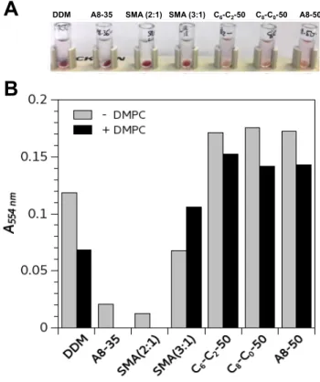
, i.e. a total MP/polymer ratio of 1:1 (w/w). After incubation for 4 h at room temperature, the samples were centrifuged 30 min. at 100,000 × g and HwBR present in the supernatants was assessed with absorbance at 554 nm.
4) Quantification of native BR extracted from DMPC-fused purple membrane by polymers
The purple membrane was purified from Hb. salinarum as described.29
The concentration of native BR of Hb. salinarum (HsBR) was adjusted to 5 g·L-1
with buffer 20 mM sodium phosphate, 100 mM NaCl, pH 7.0 and the membrane preparation was stored at -80 °C until usage.
Fusion of the native purple membrane with DMPC liposomes was performed as reported.13,27
Briefly, an aliquot of purple membrane was mixed with 100-nm extruded DMPC liposomes to reach a final HsBR/DMPC ratio varying from 1:3 to 1:6 (w/w). The samples, at
a HsBR concentration of 0.5 g·L-1
, were sonicated in the dark for 30 min. using a bath sonicator (VWR) and then stored at -80°C until usage.
Solubilization was initiated by mixing at a 1:1 volume ratio DMPC-fused purple membrane with polymer solutions to reach a final HsBR/polymer ratio varying from 1:3 to 1:9 (w/w). The samples were incubated overnight in the dark, at 25°C under shaking in a thermomixer. After an overnight incubation, all the samples were centrifuged at 4 °C for 20 min. at 200,000 × g. The UV-visible spectra of supernatants were recorded with a spectrophotometer (Hewlett Packard). The quantification of extracted HsBR was evaluated with the ratio of absorbance values at 554 nm measured before and after centrifugation.
Because APols and SMAs are polydisperse polymers, the representation of the molar ratios of HsBR/macromolecular chains depends on the weighted average of polymers mass.
Here, we used the number averaged experimental values of 𝑀!. For HsBR/polymer weight
ratios of 1:3 and 1:9 (w/w), the corresponding molar ratios of HsBR/C8-C0-50 are then of
~1:16.7 and ~1:50.3, respectively. For SMA (3:1), the average molar ratios of HsBR/SMA correspond to ~1:20.3 and ~1:60.8, respectively. At (w/w) HsBR/DMPC ratio fixed to 1:5, the (w/w) HsBR/polymer ratios of 1:3 and 1:9 correspond in addition to molar ratios of
DMPC/cycloalkane (from C8-C0-50) of ~1:1.6 and ~1:4.9, respectively. For SMA (3:1), the
molar ratios of DMPC/phenyl rings was similarly of ~1:2.8 and ~1:8.3, respectively.
5) Dynamic light scattering (DLS)
The scattering intensity was measured at 25°C and an angle of 90° in a BI-200SM Brookhaven Instrument system equipped with a 30 mW 637 nm laser and photomultiplier detection. Size distributions from all samples showed a monomodal distribution as analyzed
by CONTIN. Hydrodynamic diameters (DH) were calculated by fitting the correlation
functions by the cumulant analysis method at quadratic order. For the kinetics of
solubilization, a sample of DMPC-fused purple membrane (at 0.2 g·L-1
of HsBR) was mixed with a polymer solution (1:1 v/v) to reach a final HsBR/polymer ratio of 1:12.5 (w/w).
6) Size exclusion chromatography (SEC)
The solution dispersity of HsBR/DMPC/polymer complexes was characterized by SEC. After HsBR extraction, the samples were injected onto a Superose 12 10/300 GL column connected to an Äkta purifier-10 system (GE Healthcare). The sample and elution buffer was
20 mM sodium phosphate, 100 mM NaCl, pH 7.0. Elution was carried out at room temperature and UV absorbance monitored at three wavelengths (220, 280 and 554 nm).
7) Stability assay
Samples of HsBR (extracted directly from DMPC-fused purple membrane with polymers) were incubated at 50°C for 6h in the dark in a thermomixer. UV-visible spectra of samples were recorded each hour. The decrease of the absorbance value at 554 nm reflects the loss of retinal and a change in the native conformation of the protein. The absorbance values were normalized with the initial one, i.e. measured at 4°C.
Results
1. Synthesis of CyclAPols
The synthesis of the CyclAPol derivatives consists in the random grafting on a PAA back-bone of cycloalkane moieties via the formation of amide bonds. The average degree of grafting was controlled quantitatively by introduction of ad hoc percentages of cyclo-alkylamine reactants to reach values similar to the hydrophobic moieties in SMA (2:1), i.e. 50 mol% (within experimental error). The average percentages of grafting of
cyclohexylethylamine and cyclooctylamine, yielding C6-C2-50 and C8-C0-50, respectively,
were then determined by both 13
C and 1
H NMR spectroscopy and pH titration (Table 1). The resulting CyclAPols were highly soluble in aqueous solutions and UV-transparent (Figure S8).
The solubilizing properties of CyclAPols, assessed with biological membranes from two different origins, were compared with a set of five other polymers used as references: i) the commercially-available polymers SMA (2:1) and (3:1), which both carry cyclic aromatic rings as hydrophobic moieties with a density of 50 and 67 mol%, respectively, ii) the commercially-available polymer DIBMA, which carries short-length branched acyclic side groups present along the polymer at 33 mol%, and iii) the polyacrylate-based APols A8-35 and A8-50, which carry linear octyl chains grafted at 25 and 50 mol%, respectively. In contrast to A8-50, the archetypical APol A8-35 also comprises isopropyl moieties at 40
mol%, whose role is to reduce its charge density. The number-average molar masses (𝑀!) of
CyclAPol derivatives, A8-50, A8-35, and SMA (3:1) are comparable. In term of dispersity, CyclAPols, A8-50 and A8-35 were derived from the same PAA parent chain and, as a result, have the same polydispersity (Đ ≈ 2.0), whereas the polydispersity of SMAs is slightly
broader (typically 2.0-2.5).23 Regarding DIBMA, its 𝑀
! value is larger and its polydispersity
slightly narrower (Đ ≈ 1.8)30
than that of the other copolymers used in this study.
2. Solubilization by CyclAPols of E. coli plasma membrane fragments
The solubilizing properties of CyclAPols were tested with membrane fragments from E.
coli. Two different MPs were expressed and used as target MPs to estimate the extraction
effi-ciency of each polymer. The first protein was YidC of E. coli, an insertase whose
transmem-brane domain comprises six α-helices.31
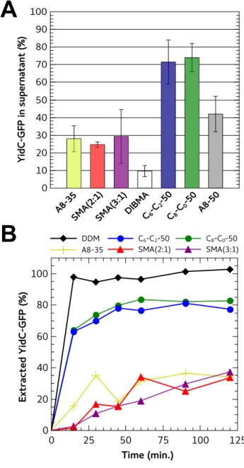
A version of YidC fused at its C-terminus with GFP (YidC-GFP) was used as reporter to quantify the solubilization yields. In Figure 2 are reported the amounts of polymer-extracted YidC-GFP recovered in the supernatants after 1 hour of incubation at 4°C at a fixed total MP/polymer ratio of 1:1 (w/w). Under these experimental conditions, the yields of YidC-GFP extracted with CyclAPols were the highest as compared to the references, i.e. A8-35, A8-50, the two SMAs and DIBMA (Figure 2A). The amounts of CyclAPol-extracted YidC-GFP were almost twice that obtained with A8-50 (~70-75% vs. ~40%). Monitoring the solubilization over 2-h incubation of the membranes with CyclAPols revealed a rapid increase of YidC-GFP fluorescence in supernatants before reaching a plateau after ~45 min. (Figure 2B). A 1-h incubation suffices to reach the maximal extraction (Figure 2A), whereas extraction by either one of the two SMAs or by A8-35 progresses more slowly (Figure 2B).
Figure 2. Fraction of YidC-GFP extracted from E. coli membranes upon addition of different polymers. A) Quantification of YidC-GFP extracted from E. coli membranes. Membranes were supplemented with each polymer stock solution to reach a final polymer concentration of 2 g·L-1
(i.e. a total MP/polymer (w/w) ratio of 1:1) and incubated for 1 h at 4°C. The buffer contained 20 mM Tris-HCl, 150 mM NaCl, pH 8.0. After centrifugation (30 min. at 100,000 × g), the protein content in the supernatants was analyzed by SDS-PAGE. The in-gel detection of YidC-GFP fluorescence was quantified with ImageJ. The fluorescence intensity of polymer-extracted YidC-GFP was normalized to the fluorescence intensity for YidC-GFP
A
solubilized with 1% w/v (10 g·L-1
) DDM, used as reference for a complete extraction. B) Kinetics of YidC-GFP extraction from E. coli membranes after addition of polymers. Over 2 h of incubation, aliquots were taken off from each sample and centrifuged (10 min. at 250,000 × g). The fluorescence of the supernatants was quantified in gel (Figure S9).
The second MP expressed in E. coli and used as reporter was the archaebacterial bacteriorhodopsin of Hq. walsbyi (HwBR) produced under its native conformation as indicated by its spectroscopic signature. This protein is a light-driven proton pump, whose transmembrane domain folds in a bundle of 7 α-helices.32
The amounts of HwBR extracted with A8-35 and SMA (2:1) were the lowest (Figure 3). In contrast to observations with YidC-GFP, the results obtained with HwBR revealed significant differences between A8-35 and the two SMAs. SMA (3:1) appeared to be more efficient at extracting HwBR from E. coli membranes than either SMA (2:1) or A8-35 (Figure 3). When solubilizing E. coli membranes with CyclAPols, the amounts of extracted HwBR were 2.5× higher than with SMA (3:1). As previously reported, the solubilization efficiency of SMA (3:1) can be improved by
modulating the bilayer characteristics with the addition of synthetic phospholipids DMPC.28
This treatment increased the amount of SMA (3:1)-solubilized HwBR (+50%), which remains nevertheless below the amount extracted by CyclAPols (Figure 3B). Intriguingly, no significant differences between CyclAPols and A8-50 were noted, in contrast to observations with YidC-GFP. Kinetics of solubilization over 4-h incubation of the E. coli membranes with polymers revealed that the maximal extraction of HwBR in CyclAPol-containing supernatants was reached in 1h, whereas extraction by SMA (3:1) progressed more slowly (Figure S10).
Figure 3. Extraction of Hq. walsbyi BR (HwBR) from E. coli membranes using various surfactants. The solubilization of E. coli membranes was carried out at room temperature at a
final polymer concentration of 2 g·L-1, corresponding to a total MP/polymer ratio of 1:1
(w/w). The reference condition in the presence of detergent was carried out with 0.2% w/v (2 g·L-1
) DDM. The buffer was 20 mM Tris-HCl, 150 mM NaCl, pH 8.0. After 4 h, the samples were centrifuged (30 min. at 100,000 × g) and the concentration of HwBR in the supernatants quantified by measuring the absorbance at 554 nm. The same experiment was performed with E. coli membranes fused with DMPC at a total MP/DMPC ratio of 1:0.5
(w/w) as described in ref.28
In the case of A8-35 and SMA (2:1), the absorbance values at 554 nm measured for supernatants collected after solubilization of DMPC-fused E. coli membranes were equal to 0.
3. Solubilization by CyclAPols of the purple membrane of Hb. salinarum
SMA (2:1) SMA (3:1) C6-C2-50 C8-C0-50 A8-50
A8-35
DDM A8-35 SMA (2:1) SMA (3:1) C6-C2-50 C8-C0-50 A8-50
DDM
A
To further investigate the membrane-solubilizing capacities of CyclAPols, we tested them on the purple membrane from Hb. salinarum, which represents both a simpler model than E.
coli membrane fragments, as it contains a single protein (HsBR), and a demanding one
because of the high density and tight packing of proteins (HsBR is accounting for three
quarters of the membrane mass).29
Indeed, purple membrane fragments proved resistant to solubilization by all of the polymers tested. Dilution of HsBR by fusion of the fragments with
DMPC lipids was thus used to lower this resistance to solubilization.13,27
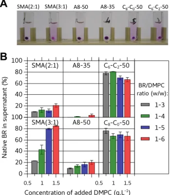
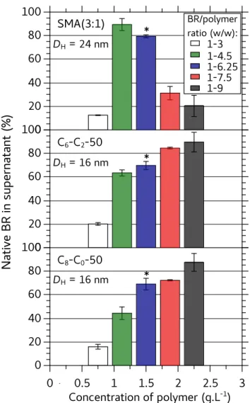
In order to be able to rank the polymers according to their solubilization efficiency, solubilization was assessed at various HsBR/DMPC ratios (Figure 4). A smaller pellet (Figure 4A) and higher recovery of native HsBR in supernatants (Figure 4B) indicated that SMA (3:1) was by far more efficient at extracting HsBR than SMA (2:1) and DIBMA (Figure S11). Under the same experimental conditions, A8-35 was unable to extract HsBR, confirming its poor detergency. Even the more hydrophobic A8-50, which differs from A8-35 by a two-fold higher density of n-octyl chains, did not perform better than SMA (2:1). In contrast, CyclAPols, with a similar degree of grafted side groups as A8-50, were efficient solubilizers. The amounts of native HsBR found in CyclAPol- and SMA (3:1)-containing supernatants were comparable (~80-85%). As expected, a decrease of extraction efficacy by SMAs was observed when lowering the DMPC content in proteoliposomes. In contrast, high percentages of solubilization were obtained with CyclAPols even at the lowest DMPC ratio, i.e. in the more demanding conditions (Figure 4B).
Figure 4. Solubilization by copolymers of DMPC-fused purple membrane at HsBR/polymer ratio of 1:6.25 (w/w) upon varying the amount of added DMPC. DMPC-containing proteo-liposomes were prepared by fusion between DMPC proteo-liposomes and purple membrane diluted at 0.5 g·L-1
[HsBR] (see Experimental section) before being mixed with an equal volume of polymer solution. Samples were incubated overnight at 25°C in the dark and then ultracentrifuged (20 min. at 200,000 × g). A) Photographs of samples after centrifugation (HsBR/DMPC ratio = 1:5 w/w). B) Percentage of native HsBR in the supernatants as quantified by the ratios of absorbance values at 554 nm measured before and after centrifugation.
To compare further CyclAPols and SMA (3:1), we assessed the effect of polymer con-centration on the DMPC-fused purple membrane (at a fixed HsBR/DMPC ratio of 1:5 w/w) and measured the size of the complexes formed in supernatants. The percentage of native,
A
SMA(2:1)& SMA(3:1)& A8#50& A8#35& C8#C0#50& C6#C2#50&solubilized HsBR decreased sharply at low polymer concentration (Figure 5), indicating that a polymer/HsBR ratio of 3:1 (w/w) is not sufficient to fully disperse all of the HsBR. All three copolymers however enabled up to 90% solubilization at higher polymer/HsBR ratios. Surpri-singly, the amount of SMA (3:1)-solubilized HsBR passed through a maximum, which is
confirmed by the size of the purple pellets (Figure S12). The hydrodynamic diameter (DH) of
the complexes (at a HsBR/polymer ratio of 1:6.25) were measured by DLS. Compared to
SMA (3:1), the DH of complexes formed with CyclAPols were smaller. The population of
solubilized objects was also analyzed by size exclusion chromatography (SEC), confirming that HsBR/DMPC/SMA (3:1) supernatants contained larger objects than those formed with CyclAPols (more material eluted in the void volume of the column; Figure S13). Therefore, whereas CyclAPols and SMA (3:1) can solubilize up to 90% HsBR, the complexes formed with CyclAPols appear smaller and more monodisperse.
Figure 5. Fraction of native HsBR extracted as a function of polymer concentration. The
HsBR/DMPC ratio was 1:5 (w/w) and the extraction procedure as described in the legend to
Figure 4. Asterisks point to samples used for DH measurement in supernatants by DLS at
25°C.
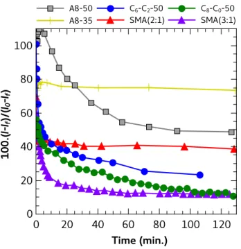
Next we assessed whether the copolymers solubilized the lipids along with HsBR. To this aim, kinetics were recorded by DLS at a HsBR/polymer ratio of 1:12.5 w/w (Figure 6). The scattered intensity reflects mainly evolution of proteoliposome size and concentration,
because the small particles (free polymer particles and MP/polymer complexes; DH < 40 nm)
immediately after supplementation of the HsBR/DMPC proteoliposomes with a solution of polymer (1:1 v/v, see Experimental section), and gradually reached a plateau (in < 60 min. in the slowest case, that of A8-50). First, this kinetics validates that the 24-h incubation used to quantify the amount of solubilized HsBR was sufficient to reach maximal extraction. Second,
the average DH as measured by DLS did not vary significantly with time and was about
130-200 nm (Table S1). This indicates that the proteoliposomes were the predominant scatterers during the whole solubilization. The absence of size variation, while the total intensity dropped down by up to 90%, rules out a gradual extraction of lipids or HsBR from all
membranes, and is thus incompatible with the “cookie-cutter” model proposed for SMAs.33
In contrast, it is the number of proteoliposomes that decreases, without affecting the mean diameter of the residual ones. This probably indicates that once a liposome is destabilized (possibly due to the polymer gaining access to its internal surface), it rapidly disperses into small particles rather than undergoing a progressive size decrease. Interestingly, the plateau measured at long times correlates with the percentage of solubilized HsBR. Namely, the plateau reached with A8-35 was the highest, revealing that ~80% of the initial proteoliposomes were left intact. In the presence of efficient solubilizers, either SMA (3:1) or CyclAPols, the scattered intensity decreased by up to 90%, which can be compared with the 90% HsBR solubilization in Figures 4 and 5.
Figure 6. Kinetics of disruption of DMPC/purple membrane proteoliposomes upon addition of various polymers. At time 0, one volume of polymer solution was mixed to one volume of proteoliposomes. Kinetics were monitored at 25°C by measuring the light scattering intensity,
I, at 90° angle (I0 is the initial intensity and If the intensity scattered by supernatants 24 h after
adding the polymers). The w/w ratios of HsBR/DMPC and HsBR/polymer were 1:5 and 1:12.5, respectively.
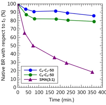
4. Stability of CyclAPol-extracted HsBR
HsBR carries a retinal chromophore whose spectroscopic properties are extremely
sensitive to the conformation of its binding pocket.34
Accordingly, the stability of HsBR was assessed by measuring its absorbance at 554 nm. The kinetics of degradation were accelerated by incubating the complexes at 50°C. As shown in Figure 7, a rapid decrease of the fraction of native HsBR is observed for SMA (3:1), whereas the spectral properties of HsBR are stable for hours in CyclAPol-solubilized samples.
Figure 7. Kinetics of inactivation of HsBR incubated at 50°C after extraction from DMPC-fused purple membrane with polymers. The (w/w) ratios of HsBR/DMPC and HsBR/polymer were 1:5 and 1:6.25, respectively. The rate of inactivation in SMA (2:1) is similar to that in SMA (3:1) but, because they bear on very small amounts, the data have not been plotted here.
Discussion
Detergent-free extraction of MPs using amphipathic polymers presents two main interests for structural and functional investigations of MPs. First, it avoids exposing the protein to detergent, which is a frequent cause of inactivation. Second, it increases the probability that the protein will retain lipids that are important to its function and stability. Among amphipathic polymers that can substitute for detergents, classical, polyacrylate-based APols (the prototype of which is A8-35) and SMA co-polymers are the most frequently used.4,5,14
Classical APols like A8-35 are very mild surfactants, so that MPs must generally be first extracted with detergents before being transferred to the polymer.12
SMAs present the
advantage of being more solubilizing, so that direct MP extraction is often possible.14
In the present study, improving the efficiency of polyacrylate-based APols at extracting MPs directly from membranes while preserving their stabilizing effect was attempted using newly designed APols carrying cycloalkane moieties (CyclAPols).
Although pure lipid liposomes can be dispersed with a comparable efficiency in the presence of many copolymers, such as A8-35, SMA (2:1) and CyclAPols (see Figure S14), this does not reflect their efficiency at extracting MPs. Biological membranes are indeed more complex systems than pure liposomes and the proteins to be extracted differ one from another. In the present study, the archaebacterial BR from two different organisms, that of Hb.
salinarum naturally present in the purple membrane and that of Hq. walsbyi produced as a
recombinant protein in E. coli were used as target protein to assess the efficiency of polymers at solubilizing MPs. The two BRs share 56% sequence identity, their transmembrane domain adopts the same fold, i.e. a bundle of 7 α-helices, and they bind the same cofactor, retinal, which, when bound to the native protein, gives BR its spectroscopic signature. By looking at the conditions required to extract the highest amounts of the two BR versions from their
respective membranes, i.e. the densely packed purple membrane and the more fluid E. coli membranes, the solubilizing properties of the polymers appear very similar. For instance, the efficiency of A8-35, SMA (2:1) and DIBMA at extracting BR is very limited regardless of the membrane used, whereas that of SMA (3:1) improves in the presence of exogenous lipids. The predominant lipid components of the plasma membrane of E. coli are the zwitterionic lipid phosphatidylethanolamine (75%) and the anionic lipids phosphatidylglycerol (20%) and cardiolipin.35
By fusing DMPC, a zwitterionic lipid, the overall charges of the membrane decreases and the fluidity of the membrane is modified. These parameters presumably affect the efficiency of SMA (3:1) at extracting HwBR. Under the same experimental conditions, i.e. at low polymer concentrations, CyclAPols show better capacities to extract HwBR than either A8-35, SMA (2:1) or SMA (3:1). In addition, solubilization by CyclAPols is not as dependent on the addition of exogenous lipids as that by SMA (3:1) is. When the reporter protein is YidC-GFP, the protein-extraction efficiency of CyclAPols remains high, which suggests that the solubilizing properties of CyclAPols may be protein-independent. Recovering the highest amount of MPs i) at low polymer concentrations over a short period of incubation (typically 1 h) and ii) without the need to add exogenous lipids presents two advantages. First, it minimizes the risk of disruption of fragile interactions in MP complexes (amphipathic polymers, despite their lower dissociating character as compared to detergents, can also be
disruptive when used at high concentrations).5,36,37
Second, the natural lipid composition in the vicinity of the MP to be extracted is not modified.
The comparison of CyclAPols with their counterpart A8-50 highlights the importance of the cyclic nature of the hydrophobic moieties. A8-50 and the two CyclAPols have the same density of hydrophilic and hydrophobic moieties, i.e. ~50 mol%. However, A8-50, which comprises linear aliphatic (octyl) tails, allows one to recover lower amounts of both HsBR from DMPC-fused purple membrane and YidC-GFP from E. coli membrane as compared to
CyclAPols. The comparison of CyclAPols with DIBMA, which carries ~33 mol% of acyclic short-length branched (diisobutyl), also shows a more efficient extraction with CyclAPols. The improvement of solubilization achieved with CyclAPols is therefore due to the cyclic nature of the hydrophobic side-groups. In the case of detergents, the few available data
suggest that cyclic hydrophobic groups markedly affect their solution behavior.38–40
In-tail cycles restrict packing possibilities, which is reflected in a lower propensity to self-assemble (up to 8 times higher critical micelle concentration compared to linear counterparts, lower
aggregation number, more hydrated core).38–40
A lower stability of intra-polymer hydrophobic association in CyclAPols likely makes these groups more available to interact with lipid bilayers, and thus to extract MPs. But other molecular parameters may also play a role. Not surprisingly, a higher density of hydrophobic side-groups in amphiphilic copolymers enhance
their interaction with liposomes,9,10
which is in line with their relative efficiency at solubilizing BR (SMA (2:1) < SMA (3:1) and A8-35 < A8-50). Overall, however, the density of side groups does not suffice to account for solubilization efficiency. Thus, the density of hydrophobic moieties along C8-C0-50 (or C6-C2-50) is the same as in A8-50, whereas these
polymers exhibit significant differences in extraction efficiency (C8-C0-50 >> A8-50).
Moreover, because cycloalkyl moieties expose less surface to water than linear chains, cyclic side groups are effectively less hydrophobic than linear ones, so that CyclAPols rank below A8-50 in term of effective hydrophobicity and could be expected to be less efficient solubilizers. A second possibly relevant parameter is the degree of ionization of the
copoly-mer chains.10
As APols are weak acids, ionization of polymer chains is reported to lower the detergency by markedly increasing hydrophilicity. Potentiometric titrations of APols and CylAPols revealed a subtle shift in the degree of ionization, at fixed pH, of the various polymers (Figure S15). The degree of ionization at neutral pH, however, ranks the polymers as follows: A8-50 < C8-C0-50 < A8-35, which does not correlate with extraction efficiency. It
seems reasonable to conclude that the cyclic nature of the side groups, rather than the hydrophilic/hydrophobic balance, plays the key role in improving MP extraction.
The relative efficiency of polymers with respect to MP extraction varies depending on the protein under consideration. For instance, the amounts of HwBR extracted from E. coli membranes with either A8-35 or SMA (2:1) were much lower than that obtained with SMA (3:1), whereas the amounts of YidC-GFP extracted with these three polymers were similar. A8-50 showed also some variability depending on the protein and/or the membrane used. Similarly, DIBMA showed a poor efficiency at extracting both YidC-GFP and BR, whereas it
has been reported to be able to extract other MPs.30,41
In the present series of experiments, the two CyclAPols showed little sensitivity to the nature of the proteins. In addition, an interesting observation is that HsBR is much more stable when extracted with CyclAPols than with SMAs: it retains 80-85% of its native conformation after 6 h at 50°C, vs. ~20% in SMAs. How general this effect is and which mechanism underlies it will require further investigations.
Conclusions
In conclusion, we have shown that cyclic hydrophobic moieties improve the copolymer ability to solubilize membranes as compared to linear aliphatic chains. Detergent-free solubilization of HsBR by CyclAPols was as efficient as with SMA (3:1) and the
CyclAPol-extracted HsBR was as stable as in A8-355
(a stability that SMAs failed to provide). The CyclAPols can also solubilize different MPs from E. coli membranes over a short period of incubation (typically 1h) and at low concentration. Recently, CyclAPols have been validated in structural studies of MPs using cryo-EM single particle analysis and
electrospray ionization mass spectrometry.42
UV-visible spectroscopic measurements (Figure S8). By ranking the various copolymers according to their detergent-like efficiency, it appears that differences between CyclAPols and classical APols correlate neither with their hydrophobicity nor with chain ionization, and thus likely reflect specific properties of cycloalkane side-chain self-assemblies and/or binding to membranes.
ASSOCIATED CONTENT
Supporting Information. Polymer characterization by 1
H, 13
C NMR spectroscopy and potentiometric titrations, UV-visible spectra of polymers, SDS-PAGE analysis of YidC-GFP, kinetics of HwBR extraction, photographs of tubes containing polymer-extracted HsBR after ultracentrifugation, size exclusion chromatography profiles, hydrodynamic diameters of
HsBR/DMPC proteoliposomes upon addition of polymer solutions, solubilization of pure
liposomes by polymers, potentiometric titrations of polymers in the presence of DMPC liposomes.
AUTHOR INFORMATION
Corresponding Author
* Correspondence should be addressed to Christophe Tribet ([email protected]) and Manuela Zoonens ([email protected]).
Present Addresses
†ICSM, Univ Montpellier, CEA, CNRS, ENSCM, Marcoule, France.
MZ led the project. AM, BM, FG, CT and MZ designed the experiments. AM synthesized and characterized the CyclAPols with the help of FG. AM and MZ performed the quantification and kinetics of YidC-GFP solubilization. MZ carried out the HwBR extraction experiments. AM and BP assisted by MZ performed the HsBR extraction and stabilization experiments. BM investigated the effects of HsBR/DMPC and HsBR/polymer ratios. CLB performed the SEC analysis. CT performed and analyzed the DLS experiments. MZ and CT wrote the manuscript. All authors have given approval to the final version of the manuscript.
ACKNOWLEDGMENT
We thank Elodie Point for helping in purple membrane preparation, Oana Ilioaia for producing YidC-GFP in Escherichia coli, and Jean-Luc Popot for constructive comments on the manuscript. This work was supported by the Centre National de la Recherche Scientifique (CNRS), with specifically an “80PRIME” grant, the University Paris-7 (Université de Paris), and the “Initiative d’Excellence” program from the French State (Grant “DYNAMO”, ANR-11-LABX-0011-01).
ABBREVIATIONS
APols, amphipols; CyclAPols, amphipols bearing cycloalkane side groups; DDM, n-Dodecyl
β-D-maltoside; DIBMA, diisobutylene maleic acid copolymer; DLS, dynamic light
scattering; GFP, Green Fluorescent Protein; HsBR, bacteriorhodopsin of Halobacterium
salinarum; HwBR, bacteriorhodopsin of Haloquadratum walsbyi; MPs, membrane proteins;
PAA, poly(acrylic acid); SEC, size exclusion chromatography; SMAs, styrene maleic acid copolymers.
REFERENCES
(1) Landry, Y.; Gies, J.-P. Drugs and Their Molecular Targets: An Updated Overview.
Fundam. Clin. Pharmacol. 2008, 22 (1), 1–18.
(2) Rucevic, M.; Hixson, D.; Josic, D. Mammalian Plasma Membrane Proteins as Potential Biomarkers and Drug Targets. Electrophoresis 2011, 32 (13), 1549–1564.
(3) Tribet, C.; Audebert, R.; Popot, J. L. Amphipols: Polymers That Keep Membrane Proteins Soluble in Aqueous Solutions. Proc. Natl. Acad. Sci. U.S.A. 1996, 93 (26), 15047–15050.
(4) Zoonens, M.; Popot, J.-L. Amphipols for Each Season. J. Membr. Biol. 2014, 247 (9– 10), 759–796.
(5) Popot, J.-L. Membrane Proteins in Aqueous Solutions: From Detergents to Amphipols; Springer: New York, 2018.
(6) Le Bon, C.; Michon, B.; Popot, J.-L.; Zoonens, M. Amphipathic Environments for Determining the Structure of Membrane Proteins by Single-Particle Electron Cryo-Microscopy. submitted for publication.
(7) Champeil, P.; Menguy, T.; Tribet, C.; Popot, J.-L.; Maire, M. le. Interaction of
Amphipols with Sarcoplasmic Reticulum Ca2+
-ATPase. J. Biol. Chem. 2000, 275 (25), 18623–18637.
(8) Picard, M.; Duval-‐Terrié, C.; Dé, E.; Champeil, P. Stabilization of membranes upon interaction of amphipathic polymers with membrane proteins. Protein Sci. 2004, 13 (11), 3056–3058.
(9) Ladavière, C.; Toustou, M.; Gulik-Krzywicki, T.; Tribet, C. Slow Reorganization of Small Phosphatidylcholine Vesicles upon Adsorption of Amphiphilic Polymers. J.
Colloid Interface Sci. 2001, 241 (1), 178–187.
(10) Vial, F.; Rabhi, S.; Tribet, C. Association of Octyl-Modified Poly(Acrylic Acid) onto Unilamellar Vesicles of Lipids and Kinetics of Vesicle Disruption. Langmuir 2005, 21 (3), 853–862.
(11) Tribet, C.; Vial, F. Flexible Macromolecules Attached to Lipid Bilayers: Impact on Fluidity, Curvature, Permeability and Stability of the Membranes. Soft Matter 2008, 4 (1), 68–81.
(12) Le Bon, C.; Marconnet, A.; Masscheleyn, S.; Popot, J.-L.; Zoonens, M. Folding and Stabilizing Membrane Proteins in Amphipol A8-35. Methods 2018, 147, 95–105. (13) Knowles, T. J.; Finka, R.; Smith, C.; Lin, Y.-P.; Dafforn, T.; Overduin, M. Membrane
Proteins Solubilized Intact in Lipid Containing Nanoparticles Bounded by Styrene Maleic Acid Copolymer. J. Am. Chem. Soc. 2009, 131 (22), 7484–7485.
(14) Stroud, Z.; Hall, S. C. L.; Dafforn, T. R. Purification of Membrane Proteins Free from Conventional Detergents: SMA, New Polymers, New Opportunities and New Insights.
Methods 2018, 147, 106–117.
(15) Oluwole, A. O.; Klingler, J.; Danielczak, B.; Babalola, J. O.; Vargas, C.; Pabst, G.; Keller, S. Formation of Lipid-Bilayer Nanodiscs by Diisobutylene/Maleic Acid (DIBMA) Copolymer. Langmuir 2017, 33 (50), 14378–14388.
(16) Yasuhara, K.; Arakida, J.; Ravula, T.; Ramadugu, S. K.; Sahoo, B.; Kikuchi, J.-I.; Ramamoorthy, A. Spontaneous Lipid Nanodisc Fomation by Amphiphilic Polymethacrylate Copolymers. J. Am. Chem. Soc. 2017, 139 (51), 18657–18663.
(17) Jamshad, M.; Grimard, V.; Idini, I.; Knowles, T. J.; Dowle, M. R.; Schofield, N.; Sridhar, P.; Lin, Y.; Finka, R.; Wheatley, M.; Thomas, O. R. T.; Palmer, R. E.; Overduin, M.; Govaerts, C.; Ruysschaert, J.-M.; Edler, K. J.; Dafforn, T. R. Structural Analysis of a Nanoparticle Containing a Lipid Bilayer Used for Detergent-Free Extraction of Membrane Proteins. Nano Res. 2015, 8 (3), 774–789.
(18) Autzen, H. E.; Julius, D.; Cheng, Y. Membrane Mimetic Systems in CryoEM: Keeping Membrane Proteins in Their Native Environment. Curr. Opin. Struct. Biol. 2019, 58, 259–268.
(19) Nagy, J. K.; Kuhn Hoffmann, A.; Keyes, M. H.; Gray, D. N.; Oxenoid, K.; Sanders, C. R. Use of Amphipathic Polymers to Deliver a Membrane Protein to Lipid Bilayers.
FEBS Lett. 2001, 501, 115–120.
(20) Gorzelle, B. M.; Hoffman, A. K.; Keyes, M. H.; Gray, D. N.; Ray, D. G.; Sanders II, C. R. Amphipols Can Support the Activity of a Membrane Enzyme. J. Am. Chem. Soc.
2002, 124, 11594-11595.
(21) Giusti, F.; Rieger, J.; Catoire, L. J.; Qian, S.; Calabrese, A. N.; Watkinson, T. G.; Casiraghi, M.; Radford, S. E.; Ashcroft, A. E.; Popot, J.-L. Synthesis, Characterization and Applications of a Perdeuterated Amphipol. J. Membr. Biol. 2014, 247 (9), 909– 924.
(22) Hovers, J.; Potschies, M.; Polidori, A.; Pucci, B.; Raynal, S.; Bonneté, F.; Serrano-Vega, M. J.; Tate, C. G.; Picot, D.; Pierre, Y.; Popot, J.-L.; Nehmé, R.; Bidet, M.; Mus-Veteau, I.; Bußkamp, H.; Jung, K.-H.; Marx, A.; Timmins, P. A.; Welte, W. A Class of Mild Surfactants That Keep Integral Membrane Proteins Water-Soluble for Functional Studies and Crystallization. Mol. Membr. Biol. 2011, 28 (3), 171–181.
(23) Dörr, J. M.; Scheidelaar, S.; Koorengevel, M. C.; Dominguez, J. J.; Schäfer, M.; van Walree, C. A.; Killian, J. A. The Styrene–Maleic Acid Copolymer: A Versatile Tool in Membrane Research. Eur. Biophys. J. 2016, 45 (1), 3–21.
(24) Korotych, O.; Mondal, J.; Gattás-Asfura, K. M.; Hendricks, J.; Bruce, B. D. Evaluation of Commercially Available Styrene-Co-Maleic Acid Polymers for the Extraction of Membrane Proteins from Spinach Chloroplast Thylakoids. Eur. Polym. J. 2019, 114, 485–500.
(25) Zoonens, M.; Miroux, B. Expression of Membrane Proteins at the Escherichia Coli Membrane for Structural Studies. Methods Mol. Biol. 2010, 601, 49–66.
(26) Dilworth, M. V.; Piel, M. S.; Bettaney, K. E.; Ma, P.; Luo, J.; Sharples, D.; Poyner, D. R.; Gross, S. R.; Moncoq, K.; Henderson, P. J. F.; Miroux, B.; Bill, R. M. Microbial Expression Systems for Membrane Proteins. Methods 2018, 147, 3–39.
(27) Orwick-Rydmark, M.; Lovett, J. E.; Graziadei, A.; Lindholm, L.; Hicks, M. R.; Watts, A. Detergent-Free Incorporation of a Seven-Transmembrane Receptor Protein into Nanosized Bilayer Lipodisq Particles for Functional and Biophysical Studies. Nano
Lett. 2012, 12, 4687–4692.
(28) Broecker, J.; Eger, B. T.; Ernst, O. P. Crystallogenesis of Membrane Proteins Mediated by Polymer-Bounded Lipid Nanodiscs. Structure 2017, 25 (2), 384–392.
(29) Oesterhelt, D.; Stoeckenius, W. Rhodopsin-like Protein from the Purple Membrane of
Halobacterium Halobium. Nature New Biol. 1971, 233, 149–152.
(30) Oluwole, A. O.; Danielczak, B.; Meister, A.; Babalola, J. O.; Vargas, C.; Keller, S. Solubilization of Membrane Proteins into Functional Lipid-Bilayer Nanodiscs Using a Diisobutylene/Maleic Acid Copolymer. Angew. Chem. Int. Ed. Engl. 2017, 56 (7), 1919–1924.
(31) Kumazaki, K.; Kishimoto, T.; Furukawa, A.; Mori, H.; Tanaka, Y.; Dohmae, N.; Ishitani, R.; Tsukazaki, T.; Nureki, O. Crystal Structure of Escherichia Coli YidC, a Membrane Protein Chaperone and Insertase. Sci. Rep. 2014, 4, 7299.
(32) Hsu, M.-F.; Fu, H.-Y.; Cai, C.-J.; Yi, H.-P.; Yang, C.-S.; Wang, A. H.-J. Structural and Functional Studies of a Newly Grouped Haloquadratum Walsbyi Bacteriorhodopsin Reveal the Acid-Resistant Light-Driven Proton Pumping Activity. J. Biol. Chem. 2015,
(33) Parmar, M. J.; Lousa, C. D. M.; Muench, S. P.; Goldman, A.; Postis, V. L. G. Artificial Membranes for Membrane Protein Purification, Functionality and Structure Studies.
Biochem. Soc. Trans. 2016, 44 (3), 877–882.
(34) Váró, G. Analogies between Halorhodopsin and Bacteriorhodopsin. Biochim. Biophys.
Acta 2000, 1460 (1), 220–229.
(35) Raetz, C. R.; Dowhan, W. Biosynthesis and Function of Phospholipids in Escherichia
Coli. J. Biol. Chem. 1990, 265 (3), 1235–1238.
(36) Popot, J.-L.; Althoff, T.; Bagnard, D.; Banères, J.-L.; Bazzacco, P.; Billon-Denis, E.; Catoire, L. J.; Champeil, P.; Charvolin, D.; Cocco, M. J.; Crémel, G.; Dahmane, T.; de la Maza, L. M.; Ebel, C.; Gabel, F.; Giusti, F.; Gohon, Y.; Goormaghtigh, E.; Guittet, E.; Kleinschmidt, J. H.; Kühlbrandt, W.; Le Bon, C.; Martinez, K. L.; Picard, M.; Pucci, B.; Rappaport, F.; Sachs, J. N.; Tribet, C.; van Heijenoort, C.; Wien, F.; Zito, F.; Zoonens, M. Amphipols from A to Z. Annu. Rev. Biophys. 2011, 40, 379–408.
(37) Sverzhinsky, A.; Qian, S.; Yang, L.; Allaire, M.; Moraes, I.; Ma, D.; Chung, J. W.; Zoonens, M.; Popot, J.-L.; Coulton, J. W. Amphipol-Trapped ExbB–ExbD Membrane Protein Complex from Escherichia Coli: A Biochemical and Structural Case Study. J.
Membr. Biol. 2014, 247 (9), 1005–1018.
(38) Maiti, S.; Chatterji, P. R. Comparison of the Aggregation Behavior of Di- and Triblock Nonionic Amphiphiles with Linear and Cyclic Hydrophobic Tails. Langmuir 1997, 13 (19), 5011–5015.
(39) Varadaraj, R.; Bock, J.; Zushma, S.; Brons, N. Influence of Hydrocarbon Chain Branching on Interfacial Properties of Sodium Dodecyl Sulfate. Langmuir 1992, 8 (1), 14–17.
(40) Sugiyama, K.; Esumi, K. Micellar Properties of Sodium Cyclododecyl Sulfate in Aqueous Solution. Langmuir 1996, 12 (10), 2613–2615.
(41) Barniol-Xicota, M.; Verhelst, S. H. L. Stable and Functional Rhomboid Proteases in Lipid Nanodiscs by Using Diisobutylene/Maleic Acid Copolymers. J. Am. Chem. Soc.
2018, 140 (44), 14557–14561.
(42) Higgins, A.; Flynn, A.; Marconnet, A.; Postis, V. L. G.; Lippiat, J.; Chung, C.; Ceska, T.; Zoonens, M.; Sobott, F.; Muench, S. P. Cycloalkane-Modified Amphiphilic Polymers for High Resolution Single Particle Cryo-EM and Mass Spectrometry. in
Supporting Information
Solubilization and Stabilization of Membrane Proteins by
Cycloalkane-Modified Amphiphilic Polymers
Anaïs Marconnet1,2 , Baptiste Michon1,2 , Christel Le Bon1,2 , Fabrice Giusti1,2,† Christophe Tribet3, *, Manuela Zoonens1,2, * Affiliations: 1
Université de Paris, Laboratoire de Biologie Physico-Chimique des Protéines Membranaires, CNRS, UMR 7099, F-75005 Paris
2
Institut de Biologie Physico-Chimique, Fondation Edmond de Rothschild pour le développement de la recherche Scientifique, F-75005 Paris, France
3
P.A.S.T.E.U.R., Département de Chimie, École Normale Supérieure, PSL University, Sorbonne Université, CNRS, F-75005 Paris, France
*Correspondence should be addressed to Christophe Tribet ([email protected]) or
Supplementary Figures:
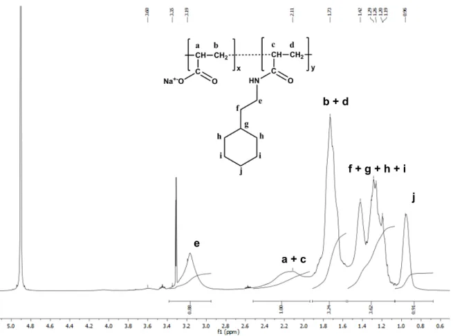
1. Chemical composition of C6-C2-50 analyzed by
1H and 13C NMR spectroscopy Proton Number of proton per monomer δ (ppm) Theoretical value e 2 3.16 2y a + c 1 + 1 1.97-2.51 x + y b + d 2 + 2 1.56-1.91 2(x + y) f + g + h + i 11 0.70-1.55 11y j 2 0.96 2y Figure S1. 1
H NMR analysis of C6-C2-50 and signal assignments.
! ! a + c b + d f + g + h + i e j
Carbon Number of carbon per monomer δ (ppm) Theoretical value Free carboxylate 1 183.78 x Carboxamide 1 177.46 y e + f 2 36.17-41.52 2y g + h 3 33.47-35.52 3y i + j 3 26.63-28.38 3y Figure S2. 13
C NMR analysis of C6-C2-50 and signal assignments.
! ! -CO2-Na+ -CONH- e + f g + h i + j
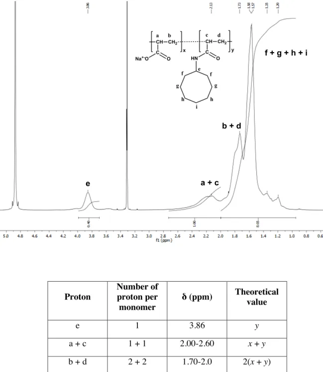
2. Chemical composition of C8-C0-50 analyzed by 1 H and 13 C NMR spectroscopy Proton Number of proton per monomer δ (ppm) Theoretical value e 1 3.86 y a + c 1 + 1 2.00-2.60 x + y b + d 2 + 2 1.70-2.0 2(x + y) f + g + h + i 14 1.05-1.70 14y Figure S3. 1H NMR analysis of C
8-C0-50 and signal assignments.
! ! ! a + c b + d f + g + h + i e
Carbon Number of carbon per monomer δ (ppm) Theoretical value Free carboxylate 1 183.62 x Carboxamide 1 176.22 y e 1 50.57 y f 2 33.13 2y g + h + i 5 24.94-29.70 5y Figure S4. 13
C NMR analysis of C8-C0-50 and signal assignments.
! -CO2-Na+ -CONH- ! g + h + i f e
3. Chemical composition of A8-50 analyzed by 1 H and 13 C NMR spectroscopy Proton Number of proton per monomer δ (ppm) Theoretical value e 2 2.86-3.30 2y a + c 1 + 1 2.00-2.55 x + y b + d + f 2 + 4 1.43-1.98 2(x + 2y) g + h + i + j + k 10 1.07-1.41 10y l 3 0.80-0.98 3y Figure S5. 1
H NMR analysis of A8-50 and signal assignments. ! ! e a + c b + d + f g + h + i + j l
Carbon Number of carbon per monomer δ (ppm) Theoretical value Free carboxylate 1 184.31 x Carboxamide 1 177.70 y e 1 40.66 y f + g + h + i + j 5 28.38 – 33.07 5y k 1 23.73 y l 1 14.50 y Figure S6. 13
C NMR analysis of A8-50 and signal assignments.
! ! e -CO2-Na+ -CONH- f + g + h + i + j k l
4. Potentiometric titrations of polymers in H2O/Ethanol
Figure S7. Acid-base titrations of polymers. The titrations of free carboxylate groups of C6
-C2-50 (A), C8-C0-50 (B), and A8-50 (C) were performed at 20 °C in a mixture of
water/ethanol (80:20, v/v).
5. UV-visible compatibility of CyclAPols
Figure S8. UV-visible spectra of copolymers used in the study. Solutions of polymers were
prepared at 1 mg/mL in deionized water. Spectra were recorded using a 10-mm path length cuvette. 0 2 4 6 8 10 12 3 4 5 6 7 8 9 10 11 pH Volume (mL) 0 2 4 6 8 10 12 3 4 5 6 7 8 9 10 pH Volume (mL) 0 2 4 6 8 10 12 3 4 5 6 7 8 9 10 11 pH Volume (mL)
A
B
C
Volume (mL) Volume (mL) Volume (mL)
pH pH pH 0 0,5 1 1,5 2 2,5 3 3,5 4 200 250 300 350 400 Absorbance Wavelength (nm) SMA (3:1) C6-C2-50 C8-C0-50 SMA (3:1) C6-C2-50 C8-C0-50 0.5 1.5 2.5 3.5
6. SDS-PAGE analysis of YidC-GFP extracted from the plasma membrane of E. coli
Figure S9. SDS-PAGE analysis of YidC-GFP extracted from E. coli membranes using
polymers. Over 2-h incubation, aliquots were taken off from samples and centrifuged (10 min. at 250,000 ×g). Supernatants (S) and pellets (P) were loaded on 12-% acrylamide gels. After migration, the band of YidC-GFP was visualized on the gels upon exposition to excitation and emission wavelengths (495 and 519 nm, respectively) revealing the GFP fluorescence at the expected position of YidC-GFP.
SMA (3:1) SMA (2:1) DDM A8-35 C6-C2-50 C8-C0-50 P 15 min S Ref: DDM – 1h 30 min 45 min 1h 1h30 2h P S S P S P S P S P
7. Kinetics of solubilization of Haloquadratum walsbyi BR (HwBR) from either pure or
DMPC-fused plasma membrane of E. coli
Figure S10. Kinetics of extraction of HwBR from pure membrane or DMPC-fused membrane
of E. coli with polymers. Extruded DMPC liposomes with a diameter of 100 nm were prepared in buffer 20 mM Tris-HCl, 150 mM NaCl, pH 8.0 and fused by sonication to E. coli membranes at a total MP/DMPC ratio of 1:0.5 (w/w). The solubilization was carried out at
room temperature in the presence of polymers at 2 g·L-1
. Aliquots of each sample were taken off and centrifuged (30 min. at 100,000 ×g). The UV-visible spectra of supernatants were measured and the absorbance values at 554 nm plotted as function of time.
8. Comparison of SMA vs. DIBMA at solubilizing Halobacterium salinarum BR (HsBR) from the DMPC-fused purple membrane
Figure S11. Photographs of samples of the DMPC-fused purple membrane supplemented
with either SMA (3:1) or DIBMA after overnight incubation at 25°C, in the dark and
0 0,04 0,08 0,12 0,16 0,2 0 1 2 3 4 5 A 554 nm Time (h) SMA (3:1) C6-C2-50 C8-C0-50 SMA (3:1) C6-C2-50 C8-C0-50 + DMPC: C6-C2-50 C6-C2-50 C8-C0-50 C8-C0-50 - 0.04 0.08 0.12 0.16 0.2
SMA(3:1) DIBMA
centrifugation (20 min. at 200,000 ×g). The HsBR/DMPC ratio was 1:5 (w/w) and the
HsBR/polymer ratio used for solubilization was 1:6.25 (w/w).
9. Effect of polymer concentrations on HsBR extraction from the DMPC-fused purple membrane
Figure S12. Photographs of samples of the DMPC-fused purple membrane supplemented
with polymers after overnight incubation at 25°C, in the dark and centrifugation (20 min. at 200,000 ×g). The HsBR/DMPC ratio was 1:5 (w/w) and the HsBR/polymer ratios used for solubilization were either 1:3 or 1:9 (w/w) as indicated.
10. Size exclusion chromatography (SEC) analysis of HsBR/lipid/polymer particles after extraction.
Figure S13. SEC profiles of HsBR/lipid/polymer particles. The DMPC-fused purple
membrane was solubilized overnight, in the dark at room temperature with either SMA (3:1),
C6-C2-50, or C8-C0-50. After centrifugation (20 min at 200,000 ×g), each sample was injected
onto a Superose 12 10/300 GL column connected to an Äkta purifier-10 system. The sample
C
6-C
2-50
C
8-C
0-50
SMA (3:1)
1:3
1:9
and elution buffers contained 20 mM sodium phosphate, 100 mM NaCl, pH 7.0. V0 and VT
stand for the void volume and total volume, respectively.
11. Evolution over time of the hydrodynamic diameters (DH) of HsBR/DMPC
proteoliposomes upon addition of polymer solutions
Table S1. Average hydrodynamic diameters (DH) as measured by DLS along the kinetics
after supplementation of HsBR/DMPC proteoliposomes with a solution of polymer (1:1 v/v). The size measurements, performed at two incubation times, i.e. 2.7 h and 24 h (before and after centrifugation), are given with a standard error of +/- 15 nm. The (w/w) ratios of
HsBR/DMPC and HsBR/polymer were fixed to 1:5 and 1:12.5, respectively.
Polymers) a+er)2.7)h) 0 + A8035 210 202 110 A8050 153 120 51 SMA)(2:1) 170 180 32 SMA)(3:1) 220 214 nd C60C2050 127 75 30 C80C0050) 206, nd, nd, centrifugation after 24 h DH (nm)
12. Solubilization of pure DMPC liposomes in the presence of polymers
Figure S14. Kinetics of disruption of pure DMPC liposomes upon addition of polymers.
Extruded DMPC liposomes with a diameter of 100 nm were prepared in buffer 20 mM sodium phosphate, 100 mM NaCl, pH 7.0. Solubilization was initiated by mixing one volume of liposomes to one volume of polymer solutions to reach a final lipid/polymer (w/w) ratio of 1:1.25. Kinetics were monitored by measuring light scattering intensity, I, at 90° angle and 25°C.
13. Potentiometric titrations of polymers in the presence of DMPC liposomes
Figure S15. Acid-base titrations of polymer solutions mixed with 100-nm extruded DMPC
liposomes. Solutions of polymers were supplemented with 100-nm extruded DMPC
0 100 200 300 400 500 600 700 800 0 1 2 3 4 Sc atte re d in te n si ty (c p s) Time (min) Vesicles alone + A8-35 + SMA (2:1) + C8-C0-50 C8-C0-50 0 0,2 0,4 0,6 0,8 1 5 6 7 8 9 Io n iza ti o n s ta te pH A8-35 A8-50 C8-C0-50 SMA (2:1) SMA (3:1) C8-C0-50 0.8 0.6 0.4 0.2
liposomes in deionized water to reach a final polymer/DMPC ratio of 1:10 (w/w). This ratio ensures that neutralized polymer chains (prone to precipitate at low pH in water) can bind to liposomes, thus maintaining their solubility up to the end of titration. The polymers, at a final concentration of 0.25 mg/mL, were titrated with a 0.016-M HCl solution added dropwise in the sample while stirring. The pH values were measured with a nano-electrode (Inlab Nano, Mettler Toledo) at 25 °C after each addition of HCl.