HAL Id: hal-02077548
https://hal.archives-ouvertes.fr/hal-02077548
Submitted on 22 Mar 2019
HAL is a multi-disciplinary open access
archive for the deposit and dissemination of
sci-entific research documents, whether they are
pub-lished or not. The documents may come from
teaching and research institutions in France or
abroad, or from public or private research centers.
L’archive ouverte pluridisciplinaire HAL, est
destinée au dépôt et à la diffusion de documents
scientifiques de niveau recherche, publiés ou non,
émanant des établissements d’enseignement et de
recherche français ou étrangers, des laboratoires
publics ou privés.
rebound median and no HIV DNA rebound following
cART interruption in a phase I/II randomized
controlled clinical trial
Erwann Loret, Albert Darque, Elisabeth Jouve, Elvenn Loret, Corinne
Nicolino-Brunet, Sophie Morange, Elisabeth Castanier, Josiane Casanova,
Christine Caloustian, Charleric Bornet, et al.
To cite this version:
Erwann Loret, Albert Darque, Elisabeth Jouve, Elvenn Loret, Corinne Nicolino-Brunet, et al..
In-tradermal injection of a Tat Oyi-based therapeutic HIV vaccine reduces of 1.5 log copies/mL the
HIV RNA rebound median and no HIV DNA rebound following cART interruption in a phase I/II
randomized controlled clinical trial. Retrovirology, BioMed Central, 2016, 13 (1),
�10.1186/s12977-016-0251-3�. �hal-02077548�
RESEARCH
Intradermal injection of a Tat Oyi-based
therapeutic HIV vaccine reduces of 1.5 log
copies/mL the HIV RNA rebound median
and no HIV DNA rebound following cART
interruption in a phase I/II randomized
controlled clinical trial
Erwann P. Loret
1*, Albert Darque
1,3, Elisabeth Jouve
4, Elvenn A. Loret
1, Corinne Nicolino‑Brunet
1,
Sophie Morange
2, Elisabeth Castanier
2, Josiane Casanova
2, Christine Caloustian
2, Charléric Bornet
3,
Julie Coussirou
3, Jihen Boussetta
3, Vincent Couallier
5, Olivier Blin
4, Bertrand Dussol
2and Isabelle Ravaux
1Abstract
Background: A Tat Oyi vaccine preparation was administered with informed consent to 48 long‑term HIV‑1 infected
volunteers whose viral loads had been suppressed by antiretroviral therapy (cART). These volunteers were rand‑ omized in double‑blind method into four groups (n = 12) that were injected intradermally with 0, 11, 33, or 99 µg of synthetic Tat Oyi proteins in buffer without adjuvant at times designated by month 0 (M0), M1 and M2, respectively. The volunteers then underwent a structured treatment interruption between M5 and M7.
Results: The primary outcomes of this phase I/IIa clinical trial were the safety and lowering the extent of HIV RNA
rebound after cART interruption. Only one undesirable event possibly due to vaccination was observed. The 33 µg dose was most effective at lowering the extent of HIV RNA and DNA rebound (Mann and Whitney test, p = 0.07 and p = 0.001). Immune responses against Tat were increased at M5 and this correlated with a low HIV RNA rebound at M6 (p = 0.01).
Conclusion: This study suggests in vivo that extracellular Tat activates and protects HIV infected cells. The Tat Oyi vac‑
cine in association with cART may provide an efficient means of controlling the HIV‑infected cell reservoir.
Keywords: HIV, Tat, Vaccine, Clinical trial, ART interruption
© 2016 Loret et al. This article is distributed under the terms of the Creative Commons Attribution 4.0 International License (http://creativecommons.org/licenses/by/4.0/), which permits unrestricted use, distribution, and reproduction in any medium, provided you give appropriate credit to the original author(s) and the source, provide a link to the Creative Commons license, and indicate if changes were made. The Creative Commons Public Domain Dedication waiver (http://creativecommons.org/ publicdomain/zero/1.0/) applies to the data made available in this article, unless otherwise stated.
Background
The quest for an efficient vaccine against HIV has been a major issue since the initial cloning and identification of HIV-1 as the causative agent of AIDS [1, 2]. Although there is consensus that HIV eradication will require
elimination of HIV infected cells that are mainly CD4 T lymphocytes (CD4), no cART and/or vaccine approaches have been able to reduce significantly the level of HIV infected cells in peripheral blood until now [3]. Different reasons to explain this lack of efficacy include the pos-sibility that HIV variability is too high for the immune system [4]. The development of cART from 1996 showed that even with a low HIV-1 variability, the immune sys-tem is still unable to eradicate HIV infected cells [5]. The reservoir of HIV infected cells in the peripheral blood
Open Access
*Correspondence: erwann.loret@univ‑amu.fr
1 ETRAV Laboratory, Faculty of Pharmacy, Centre National de la Recherche
Scientifique (CNRS), Aix Marseille University, 27 Boulevard Jean Moulin, 13385 Marseille, France
is made of cells at the latent state, which is remarkably stable since its eradication even with successful cART would require at least 70 years [6]. Why cytotoxic or CD8 T Lymphocytes (CTL) can not eliminate HIV infected cells remains controversial and it has been proposed that the central nervous system (CNS) could be a sanctuary (CTL have no access to CNS due to the blood–brain bar-rier) that would refresh peripheral blood with activated HIV infected cells [7]. However, the development of HIV DNA measurements points out CNS as a minor reservoir, while guts and rectal tissues called GALT and RALT are the major tissue reservoirs and CTL have access to theses tissues [8, 9]. Only GALT and RALT have HIV sequences compatible with the refreshment of peripheral blood with activated HIV infected cells [8]. Even patients with undetectable viraemia show evidence of ongoing viral replication in these tissue reservoirs and there is equilib-rium with the peripheral blood compartment [10]. It was shown recently that HIV infected cells become rapidly resistant to CTL following HIV infection [3]. It could be possible that the capacity for HIV infected cells to survive in an environment containing CTL is due to the secretion from HIV infected cells of a HIV protein called Tat [11]. The presence of extracellular Tat in peripheral blood has been demonstrated in different clinical studies showing the presence of antibodies against Tat [11]. Extracellular Tat goes on to be secreted under ART and might protect HIV infected cells from CTL dues to its capacity to cross CTL membranes to trigger apoptosis [11]. Furthermore, extra cellular Tat might activate latent HIV infected cells and explain the rapid HIV rebound when cART is stopped [11]. However, there is a paucity of informa-tion on the role that extracellular Tat plays in infected subjects.
A single individual termed the “Berlin patient”, who was infected by HIV in 1995 is acknowledged to have been cured of HIV after having received a hematopoi-etic stem cell transplantation in 2007 from a compatible donor possessing the delta 32 mutation in CCR5 [12]. HIV DNA was no longer detectable in his peripheral blood in 2009 [13]. The complete eradication of HIV in this patient was demonstrated in 2012 by a retro serocon-version that is characterized by a significant decrease of antibodies against HIV [13]. A similar retro seroconver-sion was reported for the child termed the “Mississippi baby” at age of three [14]. HIV-1 antibodies were not detected in the child at 24, 26, and 28 months of age, but were found when she was 4 years old in 2014 due to a viral rebound [15]. Retro seroconversion has never been observed in patients receiving cART with no detectable viraemia and is therefore considered to be a marker for the elimination of HIV infected cell [11, 13]. In contrast,
low-level viraemia that is below the limit of detection of viral RNA can be enough to sustain a high level of anti-bodies against HIV in spite of efficient ART [8].
Retro seroconversions were also observed after a sur-vey of 2 years in 23 of 25 women who were seropositive in 1986 in Gabon but at the time this was interpreted as due to mutations in the HIV envelope protein [16]. A HIV strain termed HIV-1 Oyi was cloned from one of these women while she was still seropositive [16]. It was later shown that HIV Oyi had envelope proteins similar to other HIV strains and a correlation was established between the retro seroconvertion of these 23 women and the capacity for Tat Oyi to have immune proper-ties never observed in other Tat variants [17]. This led to the testing of a Tat Oyi vaccine on rhesus macaques that were challenged by mucosal inoculation with a het-erologous recombinant SHIV. The results showed that SHIV infected CD4 cells were no longer detectable in the peripheral blood 2 months after SHIV challenge in all macaques vaccinated with Tat Oyi and one of these ani-mals retro seroconverted [18]. The efficacy of Tat Oyi was recently confirmed in another heterologous SHIV chal-lenge [19]. It was also possible to identify a highly con-served surface on Tat variants and Tat Oyi has specific mutations that transform this highly conserved surface in a 3D épitope [20]. Tat Oyi can generate neutralizing antibodies against Tat variants whatever their mutations establishing a rational for testing Tat Oyi as active princi-ple of a therapeutic vaccine in human trials [11].
This study describes the first clinical trial with Tat Oyi in a phase I/II double blinded and randomized controlled clinical trial. The primary outcome of phase I was the absence of serious adverse events (SAE) due to vaccina-tion. The primary outcome for phase II was the capacity to control HIV RNA rebound in a vaccine group follow-ing treatment interruption. The secondary outcome for phase I and phase II was a specific immune response against Tat related to the vaccine and characterized by amplification and/or cross recognition of six Tat vari-ants (including Tat Oyi) that are representative of the five main HIV-1 subtypes [11].
Results
Study design and eligibility criteria
The hypothesis was that neutralizing antibodies against Tat induced by this vaccine may help CTL to elimi-nate HIV infected cells. The Tat Oyi vaccine rationale is not to develop a Tat specific CTL response since HIV infected cells have GP120 at their surfaces and not Tat. Free extracellular Tat may constitute a screen that pro-tects HIV infected cells from CTL. Neutralizing antibod-ies against Tat may destroy this screen. We proposed to
test the Tat Oyi vaccine to HIV-1 infected volunteers who were virologically suppressed for at least 1 year while on ART. Three double blinded intradermal injec-tions (500 µL) at Month 0 (M0), M1 and M2 of a solu-tion having respectively 0, 11, 33 or 99 µg in four groups of volunteers. The vaccine was composed essentially of a synthetic Tat Oyi in 100 mM Phosphate buffer pH 4.5 and 9 g/L NaCl. Three intradermal injections were car-ried out in each group and the only difference with the placebo group was the absence of Tat Oyi. No adjuvant or other compounds were added. Volunteers agreed to stop cART for 2 months between M5 and M7, but cART could be resumed if a volunteer was >100 copies/mL HIV RNA at M6. Observance of cART and cART interrup-tion between M5 and M7 were monitored by detecinterrup-tion of antiretroviral levels in blood samples over the course of the study.
Volunteers had been recruited from among long term suppressed HIV infected patients who did not experience cART in early infection. cART interruption of 2 months is considered to be safe for these HIV infected patients with a CD4 counts >350 cells/mm3 and in whom a limited
decrease of about 65 CD4 cells/mm3 has been observed
[21, 22]. HIV RNA rebound, following cART interruption for these patients is characterized by the appearance of detectable HIV RNA at day 15 that increases until stabi-lization (up to 30 days) with a mean 10,000 (log 4) HIV RNA copies/mL [21]. This dynamic can be different for HIV patients who received cART during primary infec-tion [15, 23, 24], perhaps because early initiation of cART may reduce the size of the HIV DNA reservoir [<1.5 log HIV DNA copies per 106 peripheral blood mononuclear
cells (PBMC)] potentially influencing HIV RNA rebound when cART is interrupted [23]. Levels of HIV DNA in peripheral blood of patients receiving cART (without early initiation) since <10 years appear to be constant with a median of 1.9 log HIV DNA copies per 106 PBMC
and a mean difference of 0.2 log copies per 10 years of effective cART [25, 26].
Base line and safety
Fifty-one volunteers were initially included and filled a written informed consent but only 46 followed the pro-tocol to the end. The mean and median age was 46 years old, 27 % (n = 13) were women, they had no co infec-tion with hepatitis viruses, they have CD4 >350 cells/ mm3 and were never under 200 CD4 cells/mm3 since
12 months (see Table 1 for base line and EVATAT in clinicaltrial.gov for the complete inclusion and exclu-sion criteria). Only one SAE (a facial neuralgia for 1 week) that occurred 11 months after vaccination with three 11 µg vaccine doses was considered as pos-sibly related to vaccination (Table 2). The participant
flow diagram shows that four other patients had a Seri-ous SAE between first assessment in March 2013 and when the last patient left the trial at M12 in December 2014 (Fig. 1). One of these four SAE occurred between the time of assessment and the first vaccination (M0) and another SAE was a chirurgical intervention pro-grammed before vaccination (Table 2). The two other SAE were tuberculosis and a diarrhea related to ART and it is not possible to establish a relationship with vaccination.
Table 1 Base line
Volunteers characteristics Variable Total (n = 46) Age (years) Mean 46.4 (±9.7) Median (Q1–Q3) 47.0 [42.0–51.5] Sexe n (%) woman 13 (27 %)
HIV infection diagnosis (years)
Mean 12.4 (±7.9)
Median (Q1–Q3) 12.0 [4.8–20.3]
Years since HIV RNA <40 copies/mL
Mean 6.0 (±4.3) Median (Q1–Q3) 4.6 [2.1–9.7] ART Nucleoside base 1 (2.1 %) Non nucleosidic 28 (58.3 %) Protease 15 (31.3 %) Integrase 4 (8.3 %)
HIV DNA (copies/106 PBMC)
Mean 86.7 (±117.2)
Median (Q1–Q3) 49.5 [1.0–111.0]
Log HIV DNA
Mean 1.7 (±0.5) Median (Q1–Q3) 1.7 [0.0–2.1] CD4 (cells per µL) Mean 692.4 (±259.6) Median 666.0 [515.0–791.5] CD8 (cells per µL) Mean 688.3 (±309.8) Median (Q1–Q3) 654.0 [475.0–753.0] CD4/CD8 Mean 1.1 (±0.4) Median (Q1–Q3) 1.1 [0.8–1.4]
Nadir (CD4 <350 cells per µL) (years)
Mean 5.8 (±4.9)
Median (Q1–Q3) 4.4 [2.0–8.3]
Nadir CD4 (cells per µL)
Mean 336.3 (±114.0)
HIV RNA
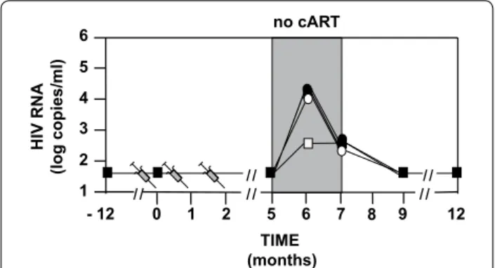
The 46 volunteers did not have detectable HIV RNA for at least 1 year before to stop cART at M5 (Fig. 2). Ten volunteers did not resume cART at M6. The primary outcome of the phase IIa study was reached with 33 µg showing a lowering of the extent of HIV RNA rebound
median from 4.1 to 2.6 log copies/mL (Fig. 2) with eight volunteers (66 %) being <3.5 log copies/mL (Fig. 3). Four volunteers (33 %) in the 33 µg group were able to main-tain their viremia <2 log copies/mL after cessation of cART (Fig. 3) and did not resume their treatment at M6. In the three other groups (including the placebo group) two volunteers did not resume cART at M6. We observe in Table 3 that the analysis of the four groups confirmed that 33 µg gave the best control of RNA rebounds with the lowest median. A one sided Mann and Whitney test that compared the placebo and the 33 µg group showed that differences between the two were borderline sig-nificant (p = 0.07), while the two other vaccine groups yielded none significant differences (p > 0.1).
CD4 and CD8 cell
The 11 µg group seemed to yield a significant increase (variation >65 CD4 cells/mm3) from 554 (total range or
TR was 418–1067) to 636 (TR 502–1124) CD4 cells/mm3
Table 2 Serious adverse events (SAE) (phase I primary end point)
a Time is expressed in months (M) after the first injection or in days (D) before
the first injection
Volunteers Groups (µg) Events Timea Due to vaccine
No 2 11 Facial neuralgia M11 Possible
No 24 99 Diarrhea M3 Doubtful
No 36 99 Tuberculosis M3 Doubtful
No 41 33 Hernia hiatus repair M9 Not possible
No 47 33 Haemorrhoids D‑7 Not possible
Assessed for eligibility (n= 51)
Excluded (n=3)
¨ Detectable viremia (n=1) ¨ Declined to participate (n=2)
Analysed (n= 12)
Excluded from analysis (n=0) Lost to follow-up (n= 0) Discontinued intervention (n= 0)
PLACEBO (Phos. Buffer 100 mM pH 4.5, NaCl 9g/l)
Allocated to intervention (n= 12) ¨ Received allocated intervention (n= 12) ¨ Did not receive allocated intervention (n=0)
Lost to follow up (n=0) Tuberculosis (n=1) (99 µg)
Declined to participate (n=1) (99 µg) Discontinued intervention (n=2)
Tat Oyi (11, 33 or 99 µg with Phos Buffer 100 mM pH 4.5, NaCl 9g/l) Allocated to intervention (3x12 n=36)
Received allocated intervention (n= 36) Did not receive allo cated intervention (n= 0)
Analysed (n= 34)
Excluded from analysis due to discontinuated Intervention (n=2) related to no treatment interruption between M5 and M7 Randomized (n= 48)
at M5 (Fig. 4a). Mann and Whitney test (M5 vs. M0) con-firmed that 11 µg had a significant increase (p = 0.001) compared to 33 µg (p = 0.22) and 99 µg (p = 0.92) in a Mann and Whitney test. After treatment interrup-tion, the four groups had a similar decrease from 671 (TR 533–773) to 519 (TR 453–677) CD4 cells/mm3 for
n = 46. No effect of the vaccine was detectable on CD4 levels following treatment interruption. However, the 33 µg group at M12 had an increase compared to M0 from 706 (TR 542–783) to 784 (TR 554–837) CD4 cells/ mm3, while the placebo group had a decrease between
M0 and M12 going from 615 (TR 436–781) to 476 (TR
436–677) CD4 cells/mm3 (Fig. 4a). Higher variability
was observed with CD8 levels, especially for the placebo group CD8 level with the 11 µg group being significantly higher at M5 (p = 0.004). The CD8 level with the 33 and 99 µg groups appeared to be higher after treatment inter-ruption (Fig. 4b). The median of the CD4/CD8 ratio was <1 for the 12 months of the study excepted at M6, where it was 1.25 with 33 µg and 0.96 with 99 µg, while it was 0.75 with the placebo and 0.88 with 11 µg.
HIV DNA
Cell-associated total HIV DNA measured in this study can not be considered as a true measurement of HIV-1-infected cells that persist in peripheral blood during cART, which are the source of rebound viremia upon stopping therapy. This HIV DNA measurement count 2 3 4 - 12 0 1 2 5 6 7 9 12 6 5 1 TIME (months) HIV RNA (log copies/ml ) 8 no cART
Fig. 2 Design of the study and HIV RNA Rebound. HIV‑1 infected volunteers (n = 46) were randomized into four groups having at month 0 (M0), M1 and M2 double blinded intradermal injections with respectively 0 (n = 12‑black square), 11 µg (n = 12‑black circle), 33 µg (n = 12‑white square) or 99 µg (n = 10‑white circle) of a synthetic pro‑ tein called Tat Oyi in a saline buffer without adjuvant. The volunteers stopped their antiviral treatment between M5 and M7. Volunteers had undetectable HIV RNA since at least 1 year before vaccination and treatment interruption. HIV RNA rebound is displayed as the median for each group on a logarithm scale. Four volunteers did not resume ART at M6 in the 33 µg group while only two volunteers resumed ART in the other groups. HIVRNA at M7 is depicted for vol‑ unteers who resumed ART at M6 (n = 36) and at M7 (n = 10)
2 3 4 6 5 1 HIV RNA (log copies/ml) 7 0 11µg 33µg 99µg
Fig. 3 HIV RNA at M6 for each volunteer. The HIV RNA level (copies/ mL) is displayed on a logarithmic scale. For each group, the median is shown as a grey line
Table 3 Vaccine efficacy to prevent HIV RNA rebound (phase II primary end point)
HIV RNA at M6 after cART interruption at M5
* p values were determined for each vaccine group versus placebo with a one sided Mann and Whitney test without adjustment. Undetectable viraemia (<40 copies/ml) were replaced by 39 (1.6 log) copies/ml
Tat Oyi 0 (n = 12) 11 µg (n = 12) 33 µg (n = 12) 99 µg (n = 10) Medians (cop‑ ies/ml) 12,921 20,706 442 9475 Total range 39–0.9 × 10⁶ 39–1.2 × 10⁶ 39–0.4 × 10⁶ 81–0.1 × 10⁶ Log medians 4.1 4.3 2.6 4.0 p values* 0.38 0.07 0.50 600 800 CD4 T cells (cells/µ L) 400 600 800 5 0 3 4 6 7 9 12 TIME (months) CD8 T cells (cells/µ L) 8 400
a
b
Fig. 4 Levels of CD4 and CD8 lymphocytes. a Median CD4 count for each group. b Median CD8 count for each group. Group 1 is depicted as a black square, group 2 as a black circle, group 3 as a white square and group 4 as a white circle
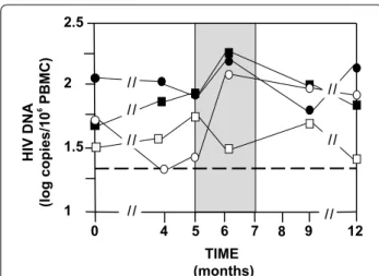
both integrated HIV DNA in HIV infected cells and free HIV DNA resulting from HIV infected cells desegrega-tion. The most significant decrease in HIV DNA was observed with the 99 µg group (n = 10) between M0 and M4 (Fig. 5). Median decreases from 1.8 (TR 1.3–2.5) to 1.3 (TR 1.3–2.6) log copies HIV DNA/106 PBMC were
observed in six volunteers having HIV DNA <20 copies at M4. However, an equivalent increase occurred between M4 and M5 (before treatment interruption) and contin-ued after treatment interruption at M6. Furthermore, the difference between M0 and M4 with 99 µg is not signifi-cant (p = 0.27). Only the 33 µg group did not show a HIV DNA rebound after treatment interruption at M6 with five volunteers having HIV DNA <20 copies. The absence of HIV DNA rebound is significant with a Mann and Whitney test (p = 0.001) compared to 99 µg (p = 0.1) and 11 µg (p = 0.49). The HIV DNA decrease with 33 µg was less spectacular but was prolonged from M0 to M12 (Fig. 5). At M12, the 33 µg group has a median of 1.4 log copies/mL that is very close of the undetectable level, but this difference is not significant (p > 0.1) compared to M0. Four volunteers with 33 µg and three volunteers with 99 µg were still having HIV DNA <20 copies at M12. This result suggests that extracellular Tat probably protects latent HIV infected cells.
IgG anti Tat response
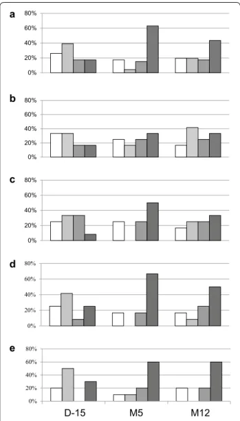
Immune responses against Tat were based on the capac-ity to recognize and/or increase recognition of six dif-ferent Tat variants representative of the five main HIV-1 subtypes in a sandwich ELISA test [11]. Four categories were observed with recognition of none, recognition of one Tat variant (low responder), recognition of two Tat variants (moderate responder), and recognition of three to six Tat variants (high responder). Prior to vaccination, specific responses against Tat were mostly low (39 %) or non-existent (26 %) (Fig. 6). Only 17 % were high responder but this increased to 63 % (n = 29) at M5 after vaccination. Five volunteers who had no Tat response before vaccination never developed an immune response against Tat in spite of three injections of either 11 µg (n = 2), 33 µg (n = 2) and 99 µg (n = 1). Vaccination in two volunteers had an immune response against Tat unchanged before and after vaccination, one who receive 11 µg while the other received the 99 µg dose. Therefore, 7/34 volunteers (20 %) did not respond to Tat Oyi injec-tion. Four volunteers who where high responders prior to vaccination did not display responsiveness after injection of 33 µg (n = 1) and 99 µg (n = 3) of Tat Oyi. The high-est increase in Tat immune response at M5 was seen with the 33 µg dose since eight patients (67 %) have a high immune response against Tat (Fig. 6d). To try to deter-mine the role of anti Tat immune responsiveness in HIV RNA rebound, we compared at M5 the high respond-ers (n = 28) with the three other groups (n = 18). The high responders median was 3.3 while the median for the three other groups was 4.6 log copies/mL (p = 0.01). This finding suggests that the anti Tat immune response may prevent the ability of extracellular Tat to activate latently HIV infected cells.
Discussion
The only successful phase II vaccine clinical trial against HIV used a recombinant virus triggering envelope pro-teins (RV144), and demonstrated an efficacy of about 30 % at preventing HIV infection [27]. Two previous phase II clinical trials were performed with Tat fragments (TUTI-16) [28] or with recombinant Tat (ISST-02) [29]. Interestingly, in these two clinical trials as in this one, a dose of 30 µg of Tat yielded the best results [28, 29]. Three years after vaccination in the ISST-02 protocol, a signifi-cant increase in CD4 cells and a HIV DNA decrease of 0.2 log copies/106 PBMC occurred but there was no
pla-cebo group to determine if these results were related to the vaccine [30]. Another phase II clinical trial with the same vaccine (ISST-03) is ongoing in South Africa [30].
The Tat Oyi vaccine is entirely synthetic and no adju-vant or compounds such as interleukins were added to amplify the immune response. The results presented
1.5 2 5 0 4 6 7 9 12 TIME (months) HIV DNA (log copies/1 0 6 PBMC ) 8 1 2.5
Fig. 5 HIV DNA in peripheral blood. HIV DNA medians for each group are shown from M0 to M12. Volunteers with undetectable HIV DNA (<20 copies/106 PBMC) were counted as having respectively 19 copies, which is 1.3 log copies/ml (dashed line). The placebo group is depicted as a black square, the 11 µg group as a black circle, the 33 µg group as a white square and the 99 µg group as a white circle. Median decreases in HIVDNA levels were observed at doses of 99 µg (n = 10) from 1.8 (TR 1.3–2.5) to 1.3 (TR 1.3–2.6) log copies HIV DNA/106 PBMC between M0 and M4 and is due to six volunteers with HIV DNA cop‑ ies <20. The absence of HIVDNA rebound at M6 with the 33 µg group is highly significant in a Mann and Whitney test (p = 0.001). At M12, the 33 µg group has a median of 1.4 log copies/ml that is very close of the undetectable level. However, this difference is not significant (p > 0.1) compared to M0
in this study are therefore due only to Tat Oyi, and the differences observed between the three different doses employed and the placebo are related only to the quan-tity of Tat Oyi injected. HIV RNA rebound following treatment interruption was chosen as a primary out-come to evaluate vaccine efficacy in phase II (M5 to
M12). Tat Oyi is the first vaccine to demonstrate effi-cacy in the context of cART interruption, even though 21 other trials have tried and failed to show differences from the placebo group [31]. It is important to out-line that lowering the extent of HIVRNA rebound to <0.5 log copies is considered as significant [31]. Since we have 8 volunteers <3.5 log copies/mL in the 33 µg group, it suggests an efficacy of 66 % for the Tat Oyi vaccine regarding the HIV RNA rebound. The immune response against Tat was monitored by ELISA test using Tat variants representative of the five main HIV-1 sub-types [11]. Previously, we showed using this ELISA test that an immune response against Tat variants was pre-sent for at least 50 % of HIV infected patients in African and European cohorts due to endogenous extracellu-lar Tat [11]. Furthermore, we showed that this anti-Tat immune response could often be transient [11]. This study shows that anti-Tat immunity can help to control HIV RNA rebound and that the anti-Tat Oyi response may help to lower HIV DNA.
This study confirms in humans the important role of extracellular Tat in activating latent cells. Latency is a way for HIV, as other defective viruses, to maximize gene expression while avoiding global T cell activa-tion that would involve the rapid eliminaactiva-tion of HIV infected cells since activated T cells can survive for only a few days [4]. Different molecular mechanisms of HIV latency have been recently reviewed including Tat and other transcription factors such as NF-κB, the cyclin dependent kinases CDK13 and CDK11, and the posi-tive transcription elongation factor b (P-TEFb) [32]. It is interesting that different strategies called “shock and kill” have been proposed to cure AIDS with agents that activate these proteins or release P-TEFb [32]. An anti-Tat compound such as Dihydro-cortistatin A can suppress HIV transcription but also inhibits HIV-1 mediated neuro inflammation [33]. The blood–brain barrier appears to be damaged in HIV infected patients, and extracellular Tat may be involved in this process [33]. This damage may be due to CTLs that are present in the CNS in the mouse model [34]. This recent discov-ery does not fit well with the role of sanctuary that the CNS is thought by some to play for HIV infected cells. The Tat Oyi vaccine may help to diminish HIV-associ-ated neurocognitive disorders.
Conclusion
The intradermal administration of the Tat Oyi vaccine was safe and could attain the effects described without adjuvant. The lack of efficacy of the vaccine at a dose of 99 µg from M5 may be due to a collapse of the Tat immune response before M5. Tat Oyi is the first thera-peutic vaccine to show a measure of success in regard 0% 20% 40% 60% 80% 0% 20% 40% 60% 80% a b c d e 0% 20% 40% 60% 80% 0% 20% 40% 60% 80% 0% 20% 40% 60% 80% D-15 M5 M12
Fig. 6 Evolution of the IgG anti Tat immune response in ELISA test. Data are shown before vaccination (D‑15), after vaccination at M5 (just before ART interruption) and at M12 (5 months after ART was resumed). The volunteers were classified in four categories: The white bar corresponds to no recognition of Tat variants. The different shades of grey correspond to the capacity for volunteers to recognize one (low responder), two (moderate responder) or three to six (high responder) Tat variants. a Evolution of the Tat immune response for all volunteers (n = 46). b Evolution of the Tat immune response in the placebo group (n = 12). c Evolution of the Tat immune response in the 11 µg group (n = 12). d Evolution of the Tat immune response in the 33 µg group (n = 12). e Evolution of the Tat immune response in the 99 µg group (n = 10)
to both HIV RNA and DNA in a phase II clinical trial. The Tat Oyi vaccine appears to reduce HIV DNA and potentially the numbers of HIV infected cells in periph-eral blood. Preliminary results show that three volunteers who received 33 or 99 µg are still HIV DNA undetectable at M24. The Tat Oyi vaccine together with cART may provide a means of control of HIV infection.
Methods
Participants
The protocol was entitled: “Evaluation in seropositive patients of a synthetic vaccine targeting the HIV Tat pro-tein” with the acronym: “EVATAT”. The protocol was reg-istered in the European Clinical trial data base (EudraCT) with the ID number: 2012-000374-36. The protocol got a favorable advice from an ethic committee (CPP SudMed 2) on November 9th, 2012 and an authorization from the French drug agency (ANSM) on January 14th, 2013 and was registered in clinicaltrial.gov with the reference NCT01793818. A written informed consent was obtained from all participants. This was a monocentric clinical trial located in the University Hospital Center “la Conception” in Marseille (Provence). All the volunteers were French citizens living in different cities in Provence, the average age was 46 years old with the oldest being 64 years old and the youngest 32 years old. The main inclusion crite-ria were: Age from 18 to 64 years old, on effective ART for at least 12 months with a viral load <40 copies/mL. The main exclusion criteria: Patients in HIV-1 primo infection, HIV-2 infected, having antibodies against HBV, HCV or HTLV-1 viruses, chronic active infections, immunosuppressive therapy, cancer, pregnancy and breastfeeding. Adverse events were monitored and clini-cal examination performed at each visit, with blood sam-pling for safety (general biochemistry and hematology). HIV RNA was analyzed by conventional methods with respectively a cut off of 40 copies per mL and HIV DNA HIV-1 DNA levels were measured by using the Generic HIV® assay (Biocentric, Bandol, Provence) with a cut off of 20 copies/million of PBMC. CD4+ and CD8+ T cell counts were analyzed by conventional methods.
Vaccine preparation
The active principle is a synthetic protein of 101 amino acid residues termed Tat Oyi [11]. Tat Oyi was synthe-sized in the ETRAV laboratory (Faculty of Pharmacy, Marseille, Provence) in Fmoc solid phase synthesis with a synthesizer APPLIED 433A in one run as previously described [35]. Two clinical lots (Freeze dried 33 µg Tat Oyi and Phosphate buffer 100 mM pH 4.5 NaCl 9 g/L) were produced by the pharmaceutical company ELI-APHARM at Sofia Antipolis (Provence). The two clinical lots were sterile and stable for 3 years at 4 °C. The vaccine
was reconstituted by the CHU Conception hospital phar-macy on the day of administration by mixing freeze dried Tat Oyi (0, 11, 33 or 99 µg) with the buffer.
Immune response against Tat
The antibody response against Tat was monitored with an ELISA test using Tat variants representative of the five main HIV-1 subtypes and Tat Oyi [11] in the ETRAV laboratory. For each volunteer, six blood samplings were carried out at M0, M2, M5, M7, M9 and M12 and sera were extracted and stored at—25 °C. For each patient and for each blood sampling the presence of IgG against six Tat variants was tested on six sera dilutions (1/3, 1/9, 1/27, 1/81, 1/243, 1/729, 1/2181, 1/6561). The ELISA tests were made in 2 days and the first day the six Tat variants were coated on 96 wheels NUNC plates in duplicate. We had six sera from each patient at M0, M2, M5, M7, M9 & M12 and one plate per serum was used. The second day, the six sera of a same volunteer were added with the same dilutions (one sera for one plate) and the asso-ciation Tat variants/Human IgG in the six dilutions was revealed with peroxide conjugate Goat anti Human IgG and the colorimetric ABTS reaction at 433 nm, meas-ured with a BIOTEK spectrophotometer. The six plates were measured the same day with a delay of 45 min cor-responding to the time of incubation for each plate with ABTS. The intra assay variability of the ELISA test was monitored at the end of the second day with OD values that had to have a standard deviation <0.2 OD between a same Tat variant and a same dilution in the duplicate of a same plate. Inter assay variability was monitored with ELISA test that had to be reproducible at least twice for each volunteer with a delay of 6 months between two experiments. After vaccination, a specific response due to the vaccine was characterized by the apparition of an immune response against Tat variants for volunteers who had no response before vaccination. For volunteers who had an immune response against Tat before vaccination, a specific response due to the vaccine was characterized by recognition at a lower dilution of the same Tat variant and/or recognition of new Tat variants.
Randomization and masking
It was a mono centric clinical study and it was double-blind with respect to vaccine dose assignment. The injec-tions occurred in the “Centre d’Investigation Clinique” (CIC) in the UCH “la Conception”. The volunteers and the investigators in the CIC were unaware of the content in the syringes. Pharmacists, from the “Pharmacie à Usage Interne” (PUI) in the UCH “la Conception”, prepared and distributed the syringe the morning of the arrival of each volunteer in the CIC according to a schedule pre-viously planned with the volunteer and in agreement
with the EVATAT protocol. Volunteers (n = 48) were randomly assigned (1:1:1:1) to receive either 0, 11, 33 or 99 µg of a synthetic Tat Oyi complemented with a saline buffer. Randomization was carried with volunteers strati-fied in four block size named A, B, C and D and the vac-cine doses corresponding to each letter was not known. The randomization scheme was prepared by Dr. Albert Darque in the PUI and securely stored with restricted access. No request to unmask study participants was asked for EVATAT study and the randomization was unmasked only on September 15th, 2015 after the “freez-ing” of EVATAT data from all investigators.
Statistical analysis
The sample sizes have not been evaluated with a statisti-cal methodology including power analysis. Nonparamet-ric testing procedures were planned, and multifactorial analysis of RNA, DNA, CD4, CD8 was the core of the study. It was not possible to use standard tools to deter-mine sample sizes according to a pre-specified power of the analysis (that requires a given value for the detectable difference). A total amount of 48 participants divided in four groups was accepted by authorities for a phase I/ IIa clinical trial, and ensured enough sample sizes for an exploratory statistical analysis. Volunteers with undetect-able HIV RNA (<40 copies/mL) or HIV DNA (<20 cop-ies/106 PBMC) were counted according to the “worst
case scenario”, which were 39 copies for HIV RNA and 19 copies for HIV DNA (respectively 1.6 log and 1.3 log. The non parametric Mann and Whitney test [36] was used as a standard test. All p values are one-sided because of an a priori knowledge of the expected variation between pla-cebo and vaccine groups. To compare vaccinated groups with a placebo require multiple comparisons that may induce type I family wise errors when non adjusted p values are used to detect significant differences between groups [37–39]. This is especially the case for large num-ber of groups and unforeseen comparisons. Our analy-ses do not suffer from this problem because only three groups are compared to the placebo. Moreover, these comparisons were planned in the statistical analysis plan of the study and have not to be considered as unplanned subgroup comparisons. Thus, despite the fact that the sample sizes was most of the time not sufficient enough to ensure a statistical significance of the observed differ-ences by adjusting for multiples comparisons, we pro-vided non adjusted p values for one-sided Mann and Whitney tests as elements for discussion. An overall Kruskal–Wallis test would not have revealed a statisti-cally significant difference among groups, mainly due to a lack of power in comparison of one control group ver-sus the three other groups with 48 patients. The Consort 2010 [40] warns against the risk of spurious findings, that
is why these statistical data must be seen as an explora-tory analysis.
Authors’ contributions
EPL and IR conceived the clinical trial, IR was the main investigator, EPL wrote the manuscript. AD made the randomization. AD, CB, JC and JB prepared the syringes, JC and CC made the injections, EPL and EAL made ELISA test to study the Tat response. CNB made the data management. EJ and VC made statistical analyses. SM, OB and BD, oversaw the clinical trial and the manuscript. All authors read and approved the final manuscript.
Author details
1 ETRAV Laboratory, Faculty of Pharmacy, Centre National de la Recherche
Scientifique (CNRS), Aix Marseille University, 27 Boulevard Jean Moulin, 13385 Marseille, France. 2 Centre d’Investigation Clinique, Assistance
Publique –Hôpitaux de Marseille (AP‑HM), University Hospital Center (UHC) «la Conception», 147 Bd Baille, 13385 Marseille, France. 3 Pharmacie Usage
Interne, AP‑HM, UHC «la Conception», 147 Bd Baille, 13385 Marseille, France.
4 Centre de Pharmacologie Clinique et Evaluations Thérapeutiques (AP‑HM),
UHC «la Timone», 28 Boulevard Jean Moulin, 13385 Marseille, France. 5 Unité
Mixte de Recherche CNRS 5251, Institut de Mathématique de Bordeaux, CNRS, Bordeaux 2 University, 33000 Bordeaux, France.
Acknowledgements
This manuscript is dedicated to the memory of Roger Treger, first president of BIOSANTECH. BIOSANTECH is the sponsor of this study and is the exclusive licensee of patents covering the use of TAT‑Oyi for preventing or treating aids and promoter of clinical trials aiming the development of an anti‑HIV vaccine using TAT‑Oyi. The authors thank all the volunteers who participate to the clinical trial. EPL thanks Dr. Catherine Tamalet, Dr. Jean de Mareuil, and Dr. Karim Boulhimez for fruitful discussions. EPL thanks the members of the EVATAT watching committee Dr. Martine Barré, Dr. Frank Tollinchi and Dr. Jean Gugliotta. EPL thanks Estelle Charles for the data management. EPL thanks the students of Pharmacy and Biotechnology for their help to carry out ELISA tests to determine the Tat immune response.
Competing interests
The authors declare that they have no competing interests. Role of the funding source
The sponsor of the study was not involved in study design, data analysis, data interpretation and writing of this manuscript. The corresponding author had fully access to all the data in the study and had final responsibility for the deci‑ sion to submit the manuscript.
Received: 1 February 2016 Accepted: 13 March 2016
References
1. Barré‑Sinoussi F, Chermann JC, Rey F, Nugeyre MT, Chamaret S, Gruest J, Dauguet C, Axler‑Blin C, Vézinet‑Brun F, Rouzioux C, Rozenbaum W, Mon‑ tagnier L. Isolation of a T‑lymphotropic retrovirus from a patient at risk for acquired immune deficiency syndrome (AIDS). Science. 1983;220:868–71. 2. Gallo RC, Salahuddin SZ, Popovic M, Shearer GM, Kaplan M, Haynes BF,
Palker TJ, Redfield R, Oleske J, Safai B, et al. Frequent detection and isola‑ tion of cytopathic retroviruses (HTLV‑III) from patients with AIDS and at risk for AIDS. Science. 1984;224:500–3.
3. Deng K, Pertea M, Rongvaux A, Wang L, Durand CM, Ghiaur G, Lai J, McHugh HL, Hao H, Zhang H, Margolick JB, Gurer C, Murphy AJ, Valen‑ zuela DM, Yancopoulos GD, Deeks SG, Strowig T, Kumar P, Siliciano JD, Salzberg SL, Flavell RA, Shan L, Siliciano RF. Broad CTL response is required to clear latent HIV‑1 due to dominance of escape mutations. Nature. 2015;517:381–5.
4. Ho DD, Neumann AU, Perelson AS, Chen W, Leonard JM, Markowitz M. Rapid turnover of plasma virions and CD4 lymphocytes in HIV‑1 infection. Nature. 1995;373:123–6.
5. Chun TW, Davey RT Jr, Engel D, Lane HC, Fauci AS. Re‑emergence of HIV after stopping therapy. Nature. 1999;401:874–5.
6. Siliciano JD, Kajdas J, Finzi D, Quinn TC, Chadwick K, Margolick JB, Kovacs C, Gange SJ, Siliciano RF. Long‑term follow‑up studies confirm the stability of the latent reservoir for HIV‑1 in resting CD4+ T cells. Nat Med. 2003;9:727–8.
7. Hellmuth J, Valcour V, Spudich S. CNS reservoirs for HIV: implications for eradication. J Virus Erad. 2015;1:67–71.
8. Bruner KM, Hosmane NN, Siliciano RF. Toward an HIV‑1 cure: measuring the latent reservoir. Trends Microbiol. 2015;23:192–203.
9. Chun TW, Nickle DC, Justement JS, Meyers JH, Roby G, Hallahan CW, Kottilil S, Moir S, Mican JM, Mullins JI, Ward DJ, Kovacs JA, Mannon PJ, Fauci AS. Persistence of HIV in gut‑associated lymphoid tissue despite long‑term antiretroviral therapy. J Infect Dis. 2008;197:714–20.
10. Imamichi H, Degray G, Asmuth DM, Fischl MA, Landay AL, Lederman MM, Sereti I. Lack of compartmentalization of HIV‑1 quasi species between the gut and peripheral blood compartments. J Infect Dis. 2011;204:309–14. 11. Loret EP. HIV extracellular tat: myth or reality ? Curr HIV Res. 2015;13:90–7. 12. Allers K, Hütter G, Hofmann J, Loddenkemper C, Rieger K, Thiel E, Sch‑
neider T. Evidence for the cure of HIV infection by CCR5Delta32/Delta32 stem cell transplantation. Blood. 2011;117:2791–9.
13. Yukl SA, Boritz E, Busch M, Bentsen C, Chun TW, Douek D, Eisele E, Haase A, Ho YC, Hütter G, Justement JS, Keating S, Lee TH, Li P, Murray D, Palmer S, Pilcher C, Pillai S, Price RW, Rothenberger M, Schacker T, Siliciano J, Siliciano R, Sinclair E, Strain M, Wong J, Richman D, Deeks SG. Challenges in detecting HIV persistence during potentially curative interventions: a study of the Berlin patient. PLoS Pathog. 2013;9:e1003347.
14. Persaud D, Gay H, Ziemniak C, Chen YH, Piatak M Jr, Chun TW, Strain M, Richman D, Luzuriaga K. Absence of detectable HIV‑1 viraemia after treat‑ ment cessation in an infant. N Engl J Med. 2013;369:1828–35.
15. Hill AL, Rosenbloom DI, Fu F, Nowak MA, Siliciano RF. Predicting the out‑ comes of treatment to eradicate the latent reservoir for HIV‑1. Proc Natl Acad Sci USA. 2014;111:13475–80.
16. Huet T, Dazza MC, Brun‑Vézinet F, Roelants GE, Wain‑Hobson S. A highly defective HIV‑1 strain isolated from a healthy Gabonese individual pre‑ senting an atypical western blot. AIDS. 1989;3:707–15.
17. Opi S, Péloponèse JM Jr, Esquieu D, Campbell G, de Mareuil J, Walburger A, Solomiac M, Grégoire C, Bouveret E, Yirrell DL, Loret EP. Tat HIV‑1 primary and tertiary structures critical to immune response against non‑ homologous variants. J Biol Chem. 2002;277:35915–9.
18. Watkins J, Esquieu D, Campbell GR, Lancelot S, de Mareuil J, Opi S, Annappa S, Salles JP, Loret EP. Reservoir cells no longer detectable after a heterologous SHIV challenge with the synthetic HIV‑1 Tat Oyi vaccine. Retrovirology. 2006;3:8–18.
19. Lakhashe SK, Byrareddy SN, Zhou M, Bachler BC, Hemashettar G, Hu SL, Villinger F, Else JG, Stock S, Lee SJ, Vargas‑Inchaustegui DA, Cofano EB, Robert‑Guroff M, Johnson WE, Polonis VR, Forthal DN, Loret EP, Rasmus‑ sen RA, Ruprecht RM. Multimodality vaccination against clade C SHIV: partial protection against mucosal challenges with a heterologous tier 2 virus. Vaccine. 2014;32:6527–36.
20. Mediouni S, Baillat G, Darque A, Ravaux I, Loret E. Identification of a highly conserved surface on Tat variants. J Biol Chem. 2013;288:19072–80. 21. Davey RT Jr, Bhat N, Yoder C, Chun TW, Metcalf JA, Dewar R, Natarajan
V, Lempicki RA, Adelsberger JW, Miller KD, Kovacs JA, Polis MA, Walker RE, Falloon J, Masur H, Gee D, Baseler M, Dimitrov DS, Fauci AS, Lane HC. HIV‑1 and T cell dynamics after interruption of highly active antiretroviral therapy (HAART) in patients with a history of sustained viral suppression. Proc Natl Acad Sci USA. 1999;96:15109–14.
22. El‑Sadr WM, Lundgren J, Neaton JD, Gordin F, Abrams D, Arduino RC, Babiker A, Burman W, Clumeck N, Cohen CJ, Cohn D, Cooper D, Darbyshire J, Emery S, Fätkenheuer G, Gazzard B, Grund B, Hoy J, Klingman K, Losso M, Markowitz N, Neuhaus J, Phillips A, Rappoport C, Strategies for manage‑ ment of antiretroviral therapy (SMART) study group. CD4+ count‑guided interruption of antiretroviral treatment. N Engl J Med. 2006;355:2283–96. 23. Skiest DJ, Morrow P, Allen B, McKinsey J, Crosby C, Foster B, Hardy RD.
It is safe to stop antiretroviral therapy in patients with preantiretrovi‑ ral CD4 cell counts >250 cells/microL. J Acquir Immune Defic Syndr. 2004;37:1351–7.
24. Assoumou L, Weiss L, Piketty C, Burgard M, Melard A, Girard PM, Rouzioux C, Costagliola D, ANRS 116 SALTO study group. A low HIV‑DNA level in peripheral blood mononuclear cells at antiretroviral treatment
interruption predicts a higher probability of maintaining viral control. AIDS. 2015;29:2003–7.
25. Hurst J, Hoffmann M, Pace M, Williams JP, Thornhill J, Hamlyn E, Meyerow‑ itz J, Willberg C, Koelsch KK, Robinson N, Brown H, Fisher M, Kinloch S, Cooper DA, Schechter M, Tambussi G, Fidler S, Babiker A, Weber J, Kelle‑ her AD, Phillips RE, Frater J. Immunological biomarkers predict HIV‑1 viral rebound after treatment interruption. Nat Commun. 2015;6:8495–503. 26. Ruggiero A, De Spiegelaere W, Cozzi‑Lepri A, Kiselinova M, Pollakis G,
Beloukas A, Vandekerckhove L, Strain M, Richman D, Phillips A, Geretti AM, ERAS Study Group. During stably suppressive antiretroviral therapy integrated HIV‑1 DNA load in peripheral blood is associated with the frequency of CD8 cells expressing HLA‑DR/DP/DQ. Ebiomedicine. 2015;2:1153–9.
27. Rolland M, Edlefsen PT, Larsen BB, Tovanabutra S, Sanders‑Buell E, Hertz T, deCamp AC, Carrico C, Menis S, Magaret CA, Ahmed H, Juraska M, Chen L, Konopa P, Nariya S, Stoddard JN, Wong K, Zhao H, Deng W, Maust BS, Bose M, Howell S, Bates A, Lazzaro M, O’Sullivan A, Lei E, Bradfield A, Ibitamuno G, Assawadarachai V, O’Connell RJ, deSouza MS, Nitayaphan S, Rerks‑Ngarm S, Robb ML, McLellan JS, Georgiev I, Kwong PD, Carlson JM, Michael NL, Schief WR, Gilbert PB, Mullins JI, Kim JH. Increased HIV‑1 vaccine efficacy against viruses with genetic signatures in Env V2. Nature. 2012;490:417–20.
28. Goldstein G, Damiano E, Donikyan M, Pasha M, Beckwith E, Chicca J. HIV‑1 Tat B‑cell epitope vaccination was ineffectual in preventing viral rebound after ART cessation. Hum Vaccin Immunother. 2012;8:1425–30.
29. Bellino S, Tripiciano A, Picconi O, Francavilla V, Longo O, Sgadari C, Panic‑ cia G, Arancio A, Angarano G, Ladisa N, Lazzarin A, Tambussi G, Nozza S, Torti C, Focà E, Palamara G, Latini A, Sighinolfi L, Mazzotta F, Di Pietro M, Di Perri G, Bonora S, Mercurio VS, Mussini C, Gori A, Galli M, Monini P, Cafaro A, Ensoli F, Ensoli B. The presence of anti‑Tat antibodies in HIV‑infected individuals is associated with containment of CD4+ T‑cell decay and viral load, and with delay of disease progression: results of a 3‑year cohort study. Retrovirology. 2014;11:49–57.
30. Ensoli F, Cafaro A, Casabianca A, Tripiciano A, Bellino S, Longo O, Fran‑ cavilla V, Picconi O, Sgadari C, Moretti S, Cossut MR, Arancio A, Orlandi C, Sernicola L, Maggiorella MT, Paniccia G, Mussini C, Lazzarin A, Sighinolfi L, Palamara G, Gori A, Angarano G, Di Pietro M, Galli M, Mercurio VS, Castelli F, Di Perri G, Monini P, Magnani M, Garaci E, Ensoli B. HIV‑1 Tat immuni‑ zation restores immune homeostasis and attacks the HAART‑resistant blood HIV DNA: results of a randomized phase II exploratory clinical trial. Retrovirology. 2015;12:33–62.
31. Pollard RB, Rockstroh JK, Pantaleo G, Asmuth DM, Peters B, Lazzarin A, Garcia F, Ellefsen K, Podzamczer D, van Lunzen J, Arastéh K, Schürmann D, Clotet B, Hardy WD, Mitsuyasu R, Moyle G, Plettenberg A, Fisher M, Fätkenheuer G, Fischl M, Taiwo B, Baksaas I, Jolliffe D, Persson S, Jelmert O, Hovden AO, Sommerfelt MA, Wendel‑Hansen V, Sørensen B. Safety and efficacy of the peptide‑based therapeutic vaccine for HIV‑1, Vacc‑4x: a phase 2 randomised, double‑blind, placebo‑controlled trial. Lancet. 2014;14:291–300.
32. Cary DC, Fujinaga K, Peterlin BM. Molecular mechanisms of HIV latency. J Clin Invest. 2016;126:448–54.
33. Mediouni S, Jablonski J, Paris JJ, Clementz MA, Thenin‑Houssier S, McLaughlin JP, Valente ST. Didehydro‑cortistatin A inhibits HIV‑1 Tat medi‑ ated neuroinflammation and prevents potentiation of cocaine reward in Tat transgenic mice. Curr HIV Res. 2015;13:64–79.
34. Richards MH, Narasipura SD, Seaton MS, Lutgen V, Al‑Harthi L. Migra‑ tion of CD8+ T cells into the central nervous system gives rise to highly potent anti‑HIVCD 4dimCD8 bright T cells in a Wnt signaling‑dependent manner. J Immunol. 2016;196:317–27.
35. Péloponèse J‑M, Collette Y, Gregoire C, Bailly C, Campèse D, Meurs E, Olive D, Loret EP. Full peptide synthesis, purification and characterization of six Tat variants: differences observed between HIV‑1 isolates from Africa and other continents. J Biol Chem. 1999;274:11473–9.
36. Siegel S, Castellan NJ. Nonparametric statistics for the behavioral sci‑ ences. New York: McGraw‑Hill; 1988. p. 399.
37. Benjamini Y, Hochberg Y. Controlling the false discovery rate: a practical and powerful approach to multiple testing. J R Stat Soc B. 1995;57:289. 38. Hochberg Y. A sharper Bonferonni procedure for multiple tests of signifi‑
• We accept pre-submission inquiries
• Our selector tool helps you to find the most relevant journal
• We provide round the clock customer support
• Convenient online submission
• Thorough peer review
• Inclusion in PubMed and all major indexing services
• Maximum visibility for your research Submit your manuscript at
www.biomedcentral.com/submit
Submit your next manuscript to BioMed Central
and we will help you at every step:
39. Holm S. A simple sequentially rejective multiple test procedure. Scand J Stat. 1979;6:65–70.
40. Moher D, Hopewell S, Schulz KF, Montori V, Gøtzsche PC, Devereaux PJ, Altman DG. CONSORT 2010 explanation and elaboration: updated guidelines for reporting parallel group randomised trials. J Clin Epidemiol. 2010;63:e1–37.
41. Moher D, Hopewell S, Schulz KF, Montori V, Gøtzsche PC, Devereaux PJ, Elbourne D, Egger M, Altman DGCONSORT. Explanation and Elaboration: updated guidelines for reporting parallel group randomised trials. Int J Surg. 2010;2012(10):28–55.
![Table 1 Base line Volunteers characteristics Variable Total (n = 46) Age (years) Mean 46.4 (±9.7) Median (Q1–Q3) 47.0 [42.0–51.5] Sexe n (%) woman 13 (27 %)](https://thumb-eu.123doks.com/thumbv2/123doknet/13610297.424700/4.892.455.807.159.863/table-base-volunteers-characteristics-variable-total-years-median.webp)
![Fig. 1 EVATAT flow diagram. This flow diagram was constructed according to CONSORT recommendation [41]](https://thumb-eu.123doks.com/thumbv2/123doknet/13610297.424700/5.892.88.439.174.293/fig-evatat-diagram-diagram-constructed-according-consort-recommendation.webp)


