HAL Id: hal-00796520
https://hal.archives-ouvertes.fr/hal-00796520
Submitted on 4 Mar 2013
HAL is a multi-disciplinary open access
archive for the deposit and dissemination of
sci-entific research documents, whether they are
pub-lished or not. The documents may come from
teaching and research institutions in France or
abroad, or from public or private research centers.
L’archive ouverte pluridisciplinaire HAL, est
destinée au dépôt et à la diffusion de documents
scientifiques de niveau recherche, publiés ou non,
émanant des établissements d’enseignement et de
recherche français ou étrangers, des laboratoires
publics ou privés.
Chlamydomonas reinhardtii.
Guillaume Allorent, Ryutaro Tokutsu, Thomas Roach, Graham Peers, Pierre
Cardol, Jacqueline Girard-Bascou, Daphné Seigneurin-Berny, Dimitris
Petroutsos, Marcel Kuntz, Cécile Breyton, et al.
To cite this version:
Guillaume Allorent, Ryutaro Tokutsu, Thomas Roach, Graham Peers, Pierre Cardol, et al.. A Dual
Strategy to Cope with High Light in Chlamydomonas reinhardtii.. The Plant cell, American Society
of Plant Biologists (ASPB), 2013, 25 (2), pp.545-57. �10.1105/tpc.112.108274�. �hal-00796520�
A Dual Strategy to Cope with High Light in
Chlamydomonas reinhardtii
WGuillaume Allorent,
a,b,c,d,1Ryutaro Tokutsu,
e,1Thomas Roach,
f,2Graham Peers,
gPierre Cardol,
hJacqueline Girard-Bascou,
iDaphné Seigneurin-Berny,
a,b,c,dDimitris Petroutsos,
a,b,c,dMarcel Kuntz,
a,b,c,dCécile Breyton,
jFabrice Franck,
kFrancis-André Wollman,
iKrishna K. Niyogi,
lAnja Krieger-Liszkay,
fJun Minagawa,
eand Giovanni Finazzi
a,b,c,d,3aCentre National Recherche Scientifique, Unité Mixte Recherche 5168, Laboratoire Physiologie Cellulaire et Végétale, F-38054
Grenoble, France
bCommissariat à l’Energie Atomique et Energies Alternatives, l’Institut de Recherches en Technologies et Sciences pour le Vivant,
F-38054 Grenoble, France
cUniversité Grenoble 1, F-38041 Grenoble, France
dInstitut National Recherche Agronomique, Unité Mixte de Recherche 1200, F-38054 Grenoble, France eDivision of Environmental Photobiology, National Institute for Basic Biology, 444-8585 Okazaki, Japan
fCommissariat à l’Energie Atomique et Energies Alternatives Saclay, Institute of Biology and Technology-Saclay, Centre National de la
Recherche Scientifique, Unité Mixte de Recherche 8221, Service de Bioénergétique, Biologie Structurale et Mécanisme, 91191 Gif-sur-Yvette cedex, France
gDepartment of Biology, Colorado State University, Fort Collins, Colorado 80523-1062
hLaboratoire de Génétique des Microorganismes Département des Sciences de la Vie, Université de Liège, B-4000 Liege, Belgium iUnité Mixte de Recherche 7141, Centre National de la Recherche Scientifique/Université Pierre et Marie Curie Institut de Biologie
Physico Chimique, F-75005 Paris, France
jUnité Mixte de Recherche 5075, Centre National de la Recherche Scientifique/Commissariat à l’Energie Atomique/Université
Grenoble 1, Institut de Biologie Structurale, F-38054 Grenoble, France
kLaboratoire de Bioénergétique, Département des Sciences de la Vie, Université de Liège, B-4000 Liege, Belgium
lHoward Hughes Medical Institute, Department of Plant and Microbial Biology, University of California and Physical Biosciences
Division, Lawrence Berkeley National Laboratory, Berkeley, California 94720-3102
Absorption of light in excess of the capacity for photosynthetic electron transport is damaging to photosynthetic organisms. Several mechanisms exist to avoid photodamage, which are collectively referred to as nonphotochemical quenching. This term comprises at least two major processes. State transitions (qT) represent changes in the relative antenna sizes of photosystems II and I. High energy quenching (qE) is the increased thermal dissipation of light energy triggered by lumen acidification. To investigate the respective roles of qE and qT in photoprotection, a mutant (npq4 stt7-9) was generated in Chlamydomonas reinhardtii by crossing the state transition–deficient mutant (stt7-9) with a strain having a largely reduced qE capacity (npq4). The comparative phenotypic analysis of the wild type, single mutants, and double mutants reveals that both state transitions and qE are induced by high light. Moreover, the double mutant exhibits an increased photosensitivity with respect to the single mutants and the wild type. Therefore, we suggest that besides qE, state transitions also play a photoprotective role during high light acclimation of the cells, most likely by decreasing hydrogen peroxide production. These results are discussed in terms of the relative photoprotective benefit related to thermal dissipation of excess light and/ or to the physical displacement of antennas from photosystem II.
INTRODUCTION
When photosynthetic organisms are exposed to light intensity overwhelming their photosynthetic capacity, they initiate a variety
of mechanisms to protect their photosynthetic machinery from photodamage. All these processes are associated with photosys-tem II (PSII) and are collectively referred to as nonphotochemical quenching (NPQ) (Horton et al., 1996). This term comprises (1) state transitions (i.e., the reversible association of chlorophyll binding complexes with PSII [in state 1] and photosystem I [PSI; in state 2]) (Bonaventura and Myers, 1969; Allen, 1992); (2) high energy– dependent quenching (qE) (i.e., enhanced thermal energy dissipa-tion occurring when the chloroplast is energized by a high rate of photosynthetic electron flow); and (3) photoinhibition, dismantling the photosystems photodamaged by high light.
State transitions are regulated by protein phosphorylation catalyzed by a membrane-bound kinase, STATE TRANSITION7
1These authors contributed equally to this work.
2Current address: Institute of Botany, University of Innsbruck, Sternwartestrasse 15, A-6020 Innsbruck, Austria.
3Address correspondence to giovanni.finazzi@cea.fr.
The author responsible for distribution of materials integral to the findings presented in this article in accordance with the policy described in the Instructions for Authors (www.plantcell.org) is: Giovanni Finazzi (giovanni.finazzi@cea.fr).
WOnline version contains Web-only data.
www.plantcell.org/cgi/doi/10.1105/tpc.112.108274
(STT7 in Chlamydomonas reinhardtii or STN7 in higher plants; Rochaix, 2007). The kinase is activated in response to redox changes in the electron flow chain. Reduction of the plastoqui-none (PQ) pool, either by unbalanced PSII/PSI activity (in favor of the former) or by the chlororespiratory chain, activates the ki-nase through a mechanism that requires plastoquinol2binding to the cytochrome b6f complex (Wollman, 2001). The
phosphor-ylated light-harvesting complex II (Pi-LHCII) is disconnected from PSII and becomes a PSI antenna. The process is reversible be-cause the kinase is inactivated by oxidation of the plastoquinol (i.e., when the PSII/PSI balance is modified in favor of the latter). This results in Pi-LHCII dephosphorylation by a phosphatase
(Pribil et al., 2010; Shapiguzov et al., 2010), which seems to have constitutive but low activity. Dephosphorylated LHCII moves back to PSII, leading to state 1.
The second phenomenon is qE, the pH-dependent component of NPQ, which corresponds to an increased thermal deactivation of the PSII antenna. In higher plants and green algae, qE is related to the accumulation of two carotenoids (antheraxanthin and zeaxanthin), produced by deepoxidation of violaxanthin during the so-called xanthophyll cycle (Yamamoto et al., 1962). More-over, qE in plants is facilitated by the PSII subunit Photosystem II subunit S (PSBS), which acts as a pH-driven amplifier of carotenoid-mediated energy quenching (Niyogi, 1999). The last component of NPQ, photoinhibition, represents a slowly reversible quenching, generally ascribed to the occurrence of photoinhibition (i.e., the damage to the PSII reaction center [Aro et al., 1993] that occurs when the other photoprotective responses cannot suc-cessfully alleviate the light stress). More recently, it has been proposed that this phase also reflects a long-lasting quenching process, possibly related to conformational changes in the PSII antenna (Dall’Osto et al., 2005).
In higher plants, qE is by far the most prominent photoprotective response (Horton et al., 1996). Conversely, quenching due to state transitions (qT) does not provide significant protection from light stress, as indicated by the comparative analysis of the fitness of Arabidopsis thaliana mutants lacking state transitions and/or qE under variable environmental conditions in the field (Frenkel et al., 2007). This is likely because state transitions are of limited am-plitude in higher plants. They are thought to adjust the relative absorption capacity of the two photosystems to optimize light use (Allen, 1992). On the other hand, state transitions are a prominent phenomenon in C. reinhardtii, where up to 80% of the light-harvesting complexes can reversibly migrate between PSII and PSI following redox-driven phosphorylation (Delosme et al., 1996). This difference can be explained by the observation that only a few LHCIIs are phosphorylated in plants, while both the minor (CHLOROPHYLL PROTEIN [CP]) and major (LIGHT HARVESTING COMPLEX II [LHCII]) antennas are targeted in C. reinhardtii (Minagawa, 2011). Because of the large change in the PSII and PSI absorption capacity, state transitions in C. reinhardtii do not only serve the purpose of balancing the absorption of the two photosystems. It has been proposed instead that they may play another role in this alga (i.e., enhancing PSI activity in order to favor cyclic electron flow around PSI to increase ATP production) (Vallon et al., 1991; Cardol et al., 2009). Consistent with this hypothesis, a supercomplex containing PSI, the cytochrome b6f complex, FERREDOXIN NADP REDUCTASE, and PROTON
GRADIENT REGULATOR5 LIKE1, a protein involved in cyclic flow (Dal Corso et al., 2008; Tolleter et al., 2011), was recently purified from state 2 acclimated cells (Iwai et al., 2010).
An additional role for state transitions can be envisaged in this alga. The psbS gene is present but not expressed under normal growth conditions (Bonente et al., 2008). In C. reinhardtii, the extent of qE is modulated by LHCSR3 (Peers et al., 2009; Bonente et al., 2011; Petroutsos et al., 2011), a member of the light-harvesting complex stress-related (LHCSR) protein family, formerly known as LI818 (Savard et al., 1996). LHCSR genes are only found in algae and mosses (Elrad and Grossman, 2004; Nymark et al., 2009). In C. reinhardtii, LHCSR3 is induced after a prolonged exposure (several hours) to high light stress (Peers et al., 2009). During this period, photodamage can take place because of the lack of photoprotection. State transitions, which develop in a few minutes (Delepelaire and Wollman, 1985), could play an active role in counterbalancing high light stress in this alga by transferring a large part of the PSII antenna to PSI, which is a very efficient quencher of absorbed energy.
To investigate this possibility, and to assess the respective roles of state transitions and qE in high light acclimation in C. reinhardtii, we employed a genetic approach to generate a double mutant lacking both state transitions and qE. By comparing fluorescence quenching, photosynthetic activity, and reactive oxygen species (ROS) production in the wild type, single mutants deficient in state transitions (stt7; Depège et al., 2003) and qE (npq4; Peers et al., 2009), and in the double mutant (npq4 stt7-9), we show that both qE and qT act together during the development of NPQ in this alga. In particular, while qE could be the major photo-protective response in steady state (as in higher plants), state transitions seem to play a prominent role during the early phase of NPQ induction, probably by preventing hydrogen peroxide (H2O2) production, but also provide some protection in steady state conditions. We interpret this result in terms of the possible benefit provided by the reduced size of the PSII antenna in state 2, but also by the diminished reactivity of the PSI-generated electron with molecular oxygen due to the generation of the cyclic PSI supercomplex (Iwai et al., 2010).
RESULTS
Generation of a Double Mutant Impaired in State Transitions and qE in C. reinhardtii
To address the relative role of NPQ-qE and NPQ-qT in light ac-climation of C. reinhardtii, we produced a double mutant impaired in both qE and state transitions. This was done by crossing two mutants. One was the state transition–deficient strain stt7-9, a clone that bears the same mutation as the stt7 mutant (Depège et al., 2003) but that has a higher mating capacity (Cardol et al., 2009). The second was npq4, a mutant in which the qE response is largely impaired, due to the disruption of the LHCSR3.1 and LHCSR3.2 genes (Peers et al., 2009). We isolated five clones (npq4 stt7-9, clones #2, #4, #7, #14, and #32) lacking both state transitions and qE, as detected by an in vivo fluorescence screen, and their characteristics are presented in Supplemental Figures 1 and 2 online. Two of them (clones #2 and #7) were further analyzed. To assess state transition capacity, maximum
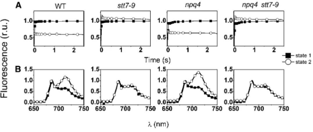
fluorescence yield was recorded at room temperature upon a 2.5-s illumination on cells acclimated in state 1 and in state 2 (Figure 1; see Supplemental Figure 1 online). State 1 was induced by placing the cells in darkness under strong aeration and state 2 by adding the ionophore carbonylcyanide-p-trifluoromethoxyphenyl hydra-zine (FCCP; 5 µM) to the incubation medium in the dark. This compound uncouples respiration, thereby lowering the cellular ATP content. This leads in turn to the activation of glycolysis and, therefore, to the accumulation of NADPH, which can reduce the PQ pool via the chlororespiratory chain (reviewed in Wollman, 2001). A reduced PQ pool activates the STT7 kinase, triggering the transfer of a large fraction of Pi-LHCII to PSI, which acts as a strong
energy quencher (Delosme et al., 1996). Ultimately, this results in a drop in the maximal fluorescence yield in state 2 conditions (Figure 1A; see Supplemental Figures 1A and 1C online), which was present in the two strains containing an active kinase (the wild type and npq4) but absent in stt7-9 (as expected because of the dis-ruption of STT7) and in the five npq4 stt7-9 clones. In these strains, a small fluorescence increase was observed in state 2 conditions, a phenomenon already observed upon reduction of the PQ pool in strains impaired in state transitions (Hohmann-Marriott et al., 2010). Measurements of fluorescence emission at cryogenic temper-atures confirmed the state transition phenotype of the selected clones (Figure 1B; see Supplemental Figures 1B and 1D online). Unlike in state 1, the fluorescence spectrum was dominated by PSI emission (l = 715 nm) in the wild-type and npq4 strains under state 2 promoting conditions. This reflects the increased absorption capacity of this complex, induced by LHCII binding (Bonaventura and Myers, 1969). On the other hand, in the same conditions, the PSII emission bands (l = 685 and 695 nm) were predominant in the stt7-9 strain, where LHCII remained associated
with PSII, and in the selected clones, confirming their lack of state transition phenotypes.
In parallel, progeny from the stt7 3 npq4 cross were analyzed for their capacity to develop qE in high light (Figure 2; see Supplemental Figure 2 online). Cells were grown mixotrophically in a Tris-acetate-phosphate (TAP) medium at low light (50 µmol photons m2 2s21) to prevent the accumulation of LHCSR3
(Peers et al., 2009). They were harvested in the exponential phase, resuspended in a minimum high-salt (HS) medium (without CO2supply), and then transferred to high light (500 µmol photons m22s21) to induce LHCSR3 accumulation (Peers
et al., 2009). Fluorescence quenching was measured at different times during high-light exposure, along with the accumulation of LHCSR3, which was evaluated by immunoblotting with a spe-cific antibody. Figure 2 shows that quenching was induced after a few hours of high-light exposure in the wild type and the stt7-9 strains, in parallel with the accumulation of LHCSR3. A small NPQ was seen in the npq4 as well as in the putative npq4 stt7-9 clones (Figure 2A; see Supplemental Figure 2A online), in which LHCSR3 did not accumulate in high light (Figure 2B; see Supplemental Figure 2B online). In these clones, the absence of qE was not due to impaired carotenoid deepoxidation (Figure 3; see also Peers et al., 2009 for npq4). In fact, a slightly higher deepoxidation was observed in the double mutant at the end of illumination (Figure 3A). In both the npq4 and npq4 stt7-9 mu-tants, a lower intensity band cross-reacting with the anti-LHCSR antibody was observed at the end of the illumination period (Figure 2B). Because no quenching increase was associated with the induction of this protein, and because of its apparent lower molecular weight (Peers et al., 2009; Bonente et al., 2011), we tentatively ascribed this band to LHCSR1, an LHCSR
Figure 1. State Transition Phenotype of the Different Strains.
Cells were harvested in the exponential phase and resuspended in minimum HS medium at a concentration of 2 3 107cells mL21. r.u., relative units. (A) Fluorescence induction curves. Traces were recorded in the presence of 10 µM DCMU, in the wild type (WT), stt7-9 (lacking the STT7 kinase), npq4 (lacking LHCSR3), and npq4 stt7-9 clones. Strains were placed either in state 1 (solid squares) or in state 2 (open circles) conditions by placing them in darkness under strong aeration (for state 1) or by incubating them with 5 µM FCCP in the dark for 20 min (for state 2). A decrease of the Fm level under state 2 conditions is indicative of a decrease in the size of the PSII antenna due to LHCII migration to PSI. Similar results were obtained when cells were placed in state 1 by illumination in the presence of 20 µM DCMU.
(B) Low-temperature (77 K) fluorescence emission spectra of cells under state 1 (closed squares) and state 2 (open circles) inducing conditions. A high ratio between the fluorescence emission band at 715 nm (PSI) and at 685 nm + 695 nm (PSII) is indicative of a transition to state 2 due to enhanced energy collection by PSI, following LHCII migration from PSII. PSII emission was normalized to 1 in all genotypes.
isoform that seems to play a minor role in qE development in cells fully acclimated to high light (Peers et al., 2009).
Fluorescence Quenching Dynamics in C. reinhardtii Wild Type and Mutants Impaired in State Transitions and qE To further evaluate the effect of this treatment on NPQ, fluores-cence quenching dynamics were assayed in cells that were ex-posed to high light for 4 h and then vigorously aerated in the dark for 15 min. Normally, this treatment is sufficient to allow for a complete relaxation of NPQ and reversion of cells to state 1 (Figure 1). However, this was not the case for wild-type cells that had been subjected to a shift from mixotrophic conditions in low light to photoautotrophy in high light. We found that CP26, CP29, and LHCII type I (LhcbM3/-4/-6/-8/-9) were heavily phosphor-ylated in wild-type cells (as shown by immunoblotting using an antiphosphothreonine antibody; Figure 4A). In parallel, we observed changes in the 77K fluorescence emission spectra, which con-firmed the occurrence of state 2 in the wild-type and npq4 strains (Figure 4B).
When the light was switched on, we observed NPQ (Figure 5) in agreement with earlier results (Peers et al., 2009; see also Figure 2). However, the fluorescence quenching pattern observed after high-light treatment turned out to be extremely complex (Figure 5) and different from the one normally seen in plants. In high light–treated wild-type cells, light exposure first leads to the development of a fast fluorescence quenching (phase I, Figure 5), which is completed in ;30 s. This phase is followed by a fluo-rescence recovery (phase II, Figure 5) that continues for around 3 min, and then by a second slower fluorescence quenching (phase III, Figure 5), until a steady state level is reached. When the light is switched off, quenching relaxation is observed (phase IV, Figure
5), and this is replaced by a slower decline (phase V, Figure 5), down to the initial dark level. In cells constitutively locked in state 1 (stt7-9), the fluorescence quenching profile is different: both the fluorescence rise (phase II) in high light and its decline in the dark (phase V) are abolished (Figure 5). Therefore, illumination led to the onset of a fast quenching (phase I), which rapidly reached a steady state level. The cells almost completely recover in the dark (phase IV). In the qE-deficient npq4 strain, phases II and V are maintained, while phases I, III, and IV are largely depressed (Figure 5). In the double mutant, all the phases observed in the wild type and in the single mutants are largely abolished. This indicates that the complex kinetics measured in wild-type cells is due to the superimposition of qE and state transitions (Figure 5). The comparison between the different genotypes suggests that phases I and IV, which are observed only in the strains capable of accumulating LHCSR3 (the wild type and stt7-9), should correspond to the development of qE in high light and to its relaxation in the dark, respectively. Phases II and V are inhibited by the stt7 mutation, suggesting a role for state tran-sitions. Fluorescence changes are associated with the phos-phorylation state of LHCII proteins, including the minor CPs (Allen, 1992; Takahashi et al., 2006; Rochaix, 2007). Thus, we tested the occurrence of phosphorylation-associated qT during the dark–high light–dark incubation by immunoblot-ting wild-type cells with an antiphosphothreonine antibody. We found that the phosphorylation level of LHCII slowly decreased upon illumination, confirming the occurrence of a state 2-to-1 transition during phase II (see Supplemental Figure 3A online). However, LHCII dephosphorylation also continued during phase III. When the light was switched off, LHCII phosphorylation recovered with a time course largely correlating with phase V.
Figure 2. NPQ Induction in Wild-Type, stt7-9, npq4, and npq4 stt7-9 Strains.
Cells were harvested in the exponential phase and resuspended in minimum HS medium at a concentration of 2 3 107cells mL21. WT, the wild type. (A) NPQ efficiency. Cells were exposed to high light (500 µmol photons m22s21, white light) without external carbon dioxide addition for the indicated times and then briefly (10 min) dark adapted. NPQ capacity was evaluated from the (Fm 2 Fm9)/Fm9 parameter (Bilger and Björkman, 1990) using a fast imaging setup. Values represent mean 6SE(n = 4 biological replicates).
(B) LHCSR accumulation. Cells were harvested at the indicated times, and samples were analyzed by immunoblotting with an anti-LHCSR antibody. ATPB (b-subunit of the CF0FiATPase) is shown as loading control. One microgram of chlorophyll was loaded in each lane.
We also employed chemical inhibitors to investigate the nature of NPQ in C. reinhardtii. qE is selectively suppressed by the dis-sipation of the DpH using the K+/H+exchanger nigericin (Horton
et al., 1996). Therefore, it is possible to exploit the different DpH sensitivity of these two processes to deconvolute the qE (i.e., of the nigericin sensitive) and qT (i.e., of the nigericin insensitive) components of NPQ starting from fluorescence traces measured in wild-type cells under control or nigericin poisoned conditions (see Supplemental Figure 3B online). We found a strong correla-tion between the kinetics of qT estimated by this approach (see Supplemental Figure 3C online) and the changes in the phos-phorylation profile measured by immunoblotting. Therefore, we conclude that phases II and V correspond to a state 2-to-1 transition in the light and to the recovery of state 2 in the dark, respectively. In agreement with previous assignments based on the comparative analysis of fluorescence kinetics in the four genotypes (Figure 5), we also confirm that phase I represents the
onset of qE-type quenching, while phase IV corresponds to its relaxation in the dark. To further investigate if phase II and IV could be due to state transitions, we employed the respiratory-deficient mutant dum22, which has been previously shown to be in state 2 in the dark (Cardol et al., 2003; see Supplemental Figure 4 online). Phase II is clearly observed in low light TAP-grown
Figure 3. Pigment Composition of the Different Strains in the Dark and after High-Light Exposure.
(A) Carotenoids and a-tocopherol content. C. reinhardtii cells were grown as described in Methods and harvested either in the dark or after high-light (500 µmol photos m22s21, white light) exposure for 4 h. The cells were centrifuged and the pellet was resuspended in methanol. After three consecutive extractions, the supernatant was used to estimate pigment content by HPLC analysis. DES indicates the deepoxidation ratio ([zeaxanthin] + 1/2 [antheraxanthin])/([zeaxanthin] + [antheraxanthin] + [violaxanthin]). a-Tocopherol content is expressed as relative units (r.u.; after normalization to the lutein content). Values represent mean 6SE(n = 3 biological replicates). WT, the wild type.
(B) Cellular chlorophyll (Chl) a+b content in dark and high light–treated cells. Values represent mean 6SE(n = 4 to 6 biological replicates). Statistical comparison was performed using one-way analysis of vari-ance (ANOVA) followed by the Tukey multiple comparison test (P < 0.05). Symbols in the graph denote significant differences: #, between light and dark; §, from the wild type, npq4, and stt7-9.
Figure 4. Protein Phosphorylation and State Transition Phenotype in Wild-Type, stt7, npq4, and npq4 stt7-9 Mutant Cells upon High-Light Treatments.
Cells were harvested in the exponential phase and resuspended in mini-mum HS medium. They were exposed to high light (500 µmol of photons m22s21, white light) for 4 h and then briefly (15 min) dark adapted. WT, the wild type.
(A) Phosphorylation status of the PSII antennae, including CP26, CP29, and LHCII type I (LhcbM3/-4/-6/-8/-9) in high light–treated cells. Protein phos-phorylation was measured by immunoblotting with antiphosphothreonine antibody. Cells were shock frozen at the time of sampling and pelleted by centrifugation before protein extraction. Total proteins from the 2 3 106cells were loaded in each lane.
(B) Low-temperature (77 K) fluorescence emission spectra in high light– treated cells. A high ratio between the fluorescence emission band at 715 nm (PSI) and at 685 nm + 695 nm (PSII) is indicative of a transition to state 2 due to enhanced energy collection by PSI, following LHCII mi-gration from PSII. PSII emission was normalized to 1 in all genotypes. r.u., relative units.
dum22 (see Supplemental Figure 4A online) and is absent in the wild-type cells grown in a similar fashion. This provides another piece of evidence that the phase II pattern observed in high light–grown cells is due to the cells being in state 2 in the dark. Altogether, the data of Figures 4 and 5 and Supplemental Figures 3 and 4 online show that exposure of cells for several hours to high light, as required to induce LHCSR3, not only provides cells with the capacity to develop qE but also induces a transition to state 2 in the dark as previously observed in the green alga Dunaliella tertiolecta (Casper-Lindley and Björkman, 1998). Both phenomena (qE and transition to state 2 in the dark) were largely absent in cells that were exposed to low light (see Supplemental Figure 5 online).
Phase III of fluorescence quenching is particularly interesting. This phase is seen in high light–treated cells (Figure 5) but not in low light–acclimated ones (see Supplemental Figure 5 online). It is also evident in low light–grown dum22 cells (see Supplemental Figure 4A online), where its appearance correlates with increased LHCSR3 accumulation (see Supplemental Figure 4C online) and xanthophyll deepoxidation (see Supplemental Figure 4D online), when compared with the wild-type cells in the same conditions. Phase III is abolished by the npq4 mutation (Figure 5) as well as by nigericin incubation to dissipate the DpH in both high light– treated cells (see Supplemental Figures 3B and 6 online) and low light–treated dum22 cells (see Supplemental Figure 4B online). Thus, this phase should correspond to a qE type of NPQ. How-ever, according to our deconvolution of the kinetics of qT, and to measurements of the dynamics of LHCII phosphorylation, this
phase also corresponds to the end of the state 2-to-1 transition, which had already started during phase II. Phase III therefore comprises both a qT and a qE type of quenching. The obser-vation of different qE phases (phases I and III) in relation to the occurrence of state transitions suggests that the quenching capacity of C. reinhardtii could be different in states 1 and 2. To understand the relationship between the two phenomena, we analyzed possible links between qT and LHCSR3. Since this protein is presumably a part of the PSII antenna, we reasoned that it could reversibly migrate between the two photosystems during state transitions. This hypothesis was tested by measuring changes in the association of this protein with the photosystems in state 1 and state 2 conditions. We first induced LHCSR3 synthesis in cells containing His-tagged versions of PSII or PSI by exposing them to high light for 4 h as in Figure 2. Then, we acclimated these cells to either state 1 or state 2 (state 1: light + DCMU; state 2: FCCP). PSI and PSII supercomplexes were then isolated and analyzed for the presence of LHCSR3 by immu-noblot analysis. Figure 6 shows that a significant amount of LHCSR3 was associated with PSI in state 2 as well as the PSII antenna, including the major trimeric LHCII (LhcbM6), the minor monomeric LHCII proteins (CP26 and CP29), and the cyto-chrome b6f complex, as previously shown in the case of the
cyclic PSI supercomplex (Iwai et al., 2010). Conversely, LHCSR3 was almost exclusively bound to PSII in state 1 as well as the PSII antenna, including major and minor LHCII proteins. This
Figure 5. Fluorescence Dynamics in High Light–Treated C. reinhardtii Cells.
Cells were harvested in the exponential phase and resuspended in minimum HS medium at a cell concentration of 2 3 107mL21. They were exposed to high light (500 µmol of photons m22s21, white light) for 4 h and then briefly (15 min) dark adapted before measuring their fluores-cence dynamics upon exposure to actinic light (700 µmol photons m22 s21, l = 520 nm). Closed box, dark; open box, actinic light on. Spikes represent maximum fluorescence emission achieved upon illumination with saturating pulses. Black bars indicate phases I through V. See text for more details. r.u., relative units; WT, the wild type.
Figure 6. LHCSR3 Migrates between PSII and PSI during State Tran-sitions.
Cells were placed either in state 1 (S1; 20 µM DCMU) or in state 2 (S2; 5 µM FCCP in the dark) conditions immediately after high-light incubation (500 µmol photons m22s21, white light) for 4 h. PSI and PSII super-complexes were isolated as described in Methods, and their protein composition was tested by immunoblotting with the specific antibodies. PSII-His and PSI-His supercomplex proteins (0.5 µg) were loaded in the respective lanes. Cyt, cytochrome.
indicates that LHCSR3 is also a part of the mobile fraction of the PSII antenna during state transitions. Its migration between the two photosystems could therefore modulate energy quenching in PSII, by changing the number of quenching effectors bound to this complex.
Role of State Transitions and qE in Photoprotection of C. reinhardtii
A major role of NPQ in photosynthetic organisms is to protect the components of the electron flow chain, and PSII in particular, from light damage. By showing that both LHCSR3-dependent quenching and state transitions are induced in response to high light, our data suggest that both processes could be involved in photoprotection in this alga. This possibility was investigated by measuring changes in PSII activity before and after the high-light treatment required to induce LHCSR3 accumulation. We ob-served that the variable fluorescence/maximum fluorescence parameter, which reflects the maximum quantum efficiency of PSII, was decreased to a greater extent in the single mutants than in the wild type after exposure to 500 µmol photons m22
s21for 4 h (Figure 7A). This suggests that some photoinhibition
was occurring in stt7-9 and npq4 during this treatment. The decline of the Fv/Fm was even more pronounced in the double mutant, indicating an increased light sensitivity in this strain. Assessment of PSII and PSI activities by measuring the elec-trochromic shift (ECS; Bailleul et al., 2010a) confirmed this ob-servation. In the dark, a PSII:PSI ratio of ;1 was found in all strains, in agreement with previous estimates (Cardol et al., 2009). Conversely, a stronger decrease of the PSII photochemical activity was seen in npq4 stt7-9 when compared with the other genotypes after incubation in high light (Figure 7B). This indicates
that PSII was more susceptible to high-light stress in the double than in the single mutants and the wild type. Consistent with this, besides changes due to state transitions, a lower PSII:PSI ratio was observed in npq4 compared with the wild type and npq4 stt7-9 compared with stt7-9 at cryogenic temperatures after high-light exposure (Figure 4B). The deleterious effects of the double mutation were confirmed when the analysis of the effects of high light on the photosynthetic properties of four strains was extended to 12 h (see Supplemental Figure 7 online). In this case, we observed some additional accumulation of LHCSR3, confirming the role of this protein in the long-term photoprotection of C. reinhardtii, as well as some increased photoinhibition, which was enhanced in the double mutant up to 8 h of high-light exposure. Under conditions where the electron flow capacity is saturated, photoinhibition is triggered by the accumulation of ROS, which are produced at different levels in the photosynthetic electron flow chain (Krieger-Liszkay and Trebst, 2006). To understand the reasons for the increased photosensitivity of the npq4 stt7-9 mutant, we measured ROS levels in the wild type, the single mutants, and in the double mutant during the first hours of high-light exposure. We first evaluated the accumulation of H2O2in the four genotypes and found that this species was produced in all strains during exposure to high light (Figure 8). However, differences were seen between the different strains in both the rate and the extent of H2O2accumulation. The amount
of H2O2 was lower in wild-type than in npq4 and stt7-9 cells. Conversely, H2O2accumulation was increased in the double
mu-tant, possibly explaining its higher photosensitivity. Moreover, we observed that stt7-9 and the double mutant accumulated more H2O2than the npq4 and wild-type strains at the beginning of
il-lumination. This suggests that state transitions may be of particular importance in preventing H2O2generation during the first phase of
high-light exposure (i.e., before the induction of qE). To provide a more complete scenario of ROS production in C. reinhardtii, we also tried to measure 1O
2generation in the different
geno-types analyzed in this work using the specific dye DanePy oxalate (Fischer et al., 2007). Unfortunately, the sensitivity of this dye was insufficient to detect1O
2in vivo unless enhancers
(bromoxynil or methylene blue) were added to the cells (see Supplemental Figure 8 online). Nonetheless, we detected lower amounts of a-tocopherol in high light–treated npq4 stt7-9 cells when compared with the other genotypes (Figure 3A). a-Tocopherol acts as an efficient scavenger of intracellular1O
2(Krieger-Liszkay
and Trebst, 2006; Li et al., 2012). It may be that its lower concen-tration in this strain is due to its rapid consumption to counteract the increased generation of this ROS. The observation of some chlorophyll bleaching after high-light exposure (Figure 3B) is also consistent with the idea that ROS production was increased in the double mutant.
DISCUSSION
In this work, we studied the development of state transitions and of qE during high-light acclimation of the green alga C. reinhardtii and their respective roles in protecting the photosynthetic ma-chinery from photodamage. Using a combined biochemical, biophysical, and genetic approach, we first demonstrated that high-light treatment for several hours (without any additional
Figure 7. PSI and PSII Activities in Wild-Type, npq4, stt7-9, and npq4 stt7-9 Cells.
Cells were harvested in the exponential phase and resuspended in mini-mum HS medium at a cell concentration of 2 3 107mL21. WT, the wild type. Black columns, dark-adapted cells; gray columns, cells exposed to high light (500 µmol photons m22s21, white light) for 4 h and then shortly (10 min) dark adapted. Values represent means 6SE(n = 3 to 6 biological replicates). Statistical comparison was performed using one-way ANOVA followed by the Tukey multiple comparison test (P < 0.05). Symbols in the graph denote significant differences: #, from the wild type; §, from the wild type, npq4, and stt7-9.
(A) PSII efficiency. PSII quantum yield was monitored as Fv/Fm using an imaging fluorescence setup.
(B) Functional PSII:PSI ratio from the ECS signal. PSII and PSI activities were measured as described in Methods.
carbon dioxide source) not only induces qE (Figure 2), in agreement with previous data (Peers et al., 2009; Bonente et al., 2011; Petroutsos et al., 2011), but also results in transition to state 2 that is not reversed by strong agitation (oxygenation) in the dark for 15 min after the treatment (Figures 4 and 5; see Supplemental Figure 3 online). This suggests that the two pro-cesses are linked in C. reinhardtii. Consistent with this possi-bility, cells that are maintained in low light cannot develop qE and state 2 (see Supplemental Figure 5 online). Next, we revealed that both state transitions and qE are involved in protecting C. reinhardtii cells from high-light stress. This conclusion is sup-ported by the increased light sensitivity of the npq4 stt7-9 double mutant, which displays a higher PSII photosensitivity (Figure 7; see Supplemental Figure 7 online), possibly because of en-hanced ROS production during the high-light exposure (Figure 8). The occurrence of significant light stress in this strain is also confirmed by the slight increase in its deepoxidation state (Figure 3A), the reduced a-tocopherol content in high light (Figure 3A), possibly to counteract1O
2accumulation, and the
bleaching of chlorophyll (Figure 3B). The observation of a tight link between qE and state transitions seems different from pre-vious data reported in C. reinhardtii (Peers et al., 2009). However, this apparent contradiction can be explained by the fact that the cells were systematically exposed to 5 min of far-red light to ensure transitions into state 1 before the NPQ measurements (Peers et al., 2009). This may have prevented the observation of fluorescence changes related to state transitions during illumination, in contrast with the data shown in this work (Figure 5).
Metabolic Links between qE and qT in C. reinhardtii The concomitant occurrence of qE and qT in high light suggests that both responses could be induced by the same metabolic signal(s) in the cell. Information concerning the nature of the photosynthetic step responsible for the induction of qE and qT in high light can be obtained from the kinetic analysis of the NPQ response presented in Figure 5. There, we show that high light– treated cells are in state 2 in the dark (Figure 5; see also Figure 4 and Supplemental Figure 3 online) and revert to state 1 upon illumination (phase II). This behavior can be rationalized based on previous knowledge of the mechanisms of state transition. The first step in the activation of state 2 transition is the re-duction of the PQ pool (Wollman, 2001), either via PSII photo-chemistry or via a nonphotochemical (chlororespiratory) process. A net reduction of the PQ pool is expected in high light because the rate of PQH2generation by PSII becomes faster than the rate of PQH2 oxidation by the cytochrome b6f complex at this
in-tensity. Thus, high-light exposure should promote the transition to state 2 instead of the transition to state 1 as observed in Figure 5. On the other hand, previous work has shown that the state 2 transition is inhibited by high light (Rintamäki et al., 2000; Vink et al., 2004), either due to the negative redox control exerted by the ferredoxin/thioredoxin system on STT7/STN7 kinase (Rintamäki et al., 2000) or to a conformational change within the PSII an-tenna that prevents its phosphorylation by the active kinase (Vink et al., 2004). Both mechanisms can equally account for the rapid recovery of state 1 observed upon illumination of the cells (phase II). No such inhibitory effect is expected upon dark ad-aptation, since both mechanisms are light dependent (Aro and Ohad, 2003). Therefore, both hypotheses are compatible with the high phosphorylation levels seen in wild-type and npq4 cells after high-light exposure (Figure 4; see Supplemental Figure 3 online). High-light treatments in the absence of any exogenous carbon source, as required to induce LHCSR3 (Peers et al., 2009), should lead to a limitation of photosynthetic activity by carbon as-similation at the level of the Calvin cycle, thereby promoting NADPH accumulation. After illumination, this NADPH can be consumed by the chlororespiratory pathway in reducing the PQ pool and thereby promoting a rapid state 2 transition. This could explain the origin of phase V. Consistent with this, exposure of C. reinhardtii cells to high light enhances the capacity for nonphotochemical reduction of the PQ pool at the expense of stromal reducing equivalents (Houyoux et al., 2011).
qE and qT Dynamics during Induction of NPQ in C. reinhardtii
The tight relationship between state transitions and qE in high light–treated cells is not only evident in the dark but also during illumination. This is shown by the existence of two fluorescence quenching phases in high light–treated wild-type cells: The first one (phase I) likely corresponds to the development of qE in state 2 cells, while the second one (phase III) most likely rep-resents an additional quenching phase linked to the attainment of state 1. Indeed, this phase can be ascribed to a qE type of quenching, based on its absence in the qE-lacking strain npq4 (Figure 5) and on its sensitivity to the dissipation of the DpH by the addition of the ionophore nigericin (see Supplemental Figure 6
Figure 8. H2O2Production during High-Light Exposure of C. reinhardtii Cells.
Cells were harvested in the exponential phase and resuspended in mini-mum HS medium at a concentration of 5 3 106cells mL21. They were exposed to high light (500 µmol of photons m22 s21, white light), and samples were collected at different time points. H2O2production was as-sessed as described in Methods. Values represent means 6SE(n = 4 to 6 biological replicates). Statistical comparison was performed using one-way ANOVA followed by the Tukey multiple comparison test (P < 0.05). Symbols in the graph denote significant differences: #, from stt7-9 and npq4-stt7-9; §, from the wild type and npq4 stt7-9; @, from the wild type, npq4, and npq4 stt7-9. WT, the wild type.
online). However, phase III also comprises a state 2-to-1 transi-tion, as indicated by measurements of protein phosphorylation kinetics in the light (see Supplemental Figure 3 online). The ob-servation that this phase is slower than phase I (i.e., qE onset in state 2) may suggest that qE development is slower in state 1 than in state 2. However, this possibility is not consistent with the ob-servation that the rate of fluorescence quenching at the onset of illumination is similar in wild-type and stt7 cells, which are in states 2 and 1, respectively. Moreover, the different rates of phases I and III cannot be explained by a different accumulation of LHCSR3 and zeaxanthin, which accumulated to a maximum extent during the high-light treatment that precedes the measurement of NPQ (Figures 2 and 3, respectively). Under these conditions, lumen acidification becomes the only time-limiting process for qE onset (Joliot and Finazzi, 2010), leading in principle to a very fast quenching response (Johnson et al., 2009). To explain the different qE kinetics measured in phases I and III, we propose instead that phase III represents the signature of the dynamics of LHCSR3 binding to PSII during the state 2-to-1 transition. LHCSR3 re-versibly binds PSI in state 2, possibly because of its phosphory-lation by STT7 (Bonente et al., 2011) (i.e., the same kinase involved in PSII antenna phosphorylation) (Depège et al., 2003). Therefore, the observation that qE quenching increases (phase III) at the end of the antenna movement to PSII (state 1 transition, phase II + phase III) suggests that the reassociation of LHCSR3 to PSII during a state 2-to-1 transition could be slower than the binding of the antenna complexes to the reaction center. This would explain why the fluorescence emission is first increased by the state 1 transition (phase II) due to the increased PSII absorption capacity, and then decreased (phase III) due to the reassociation of LHCSR3 with PSII. This latter process should amplify fluorescence quenching in PSII by increasing the number of quenching effectors bound to the PSII supercomplex, as already shown in the case of the plant qE ef-fector PSBS (Li et al., 2002).
Photoprotective Role of State Transitions in C. reinhardtii As discussed above, the phenotypic analysis of the npq4 stt7-9 double mutant suggests that both qE and qT counteract photo-damage in high light. In particular, measurements of ROS production suggest that state transitions play a photoprotective role during the early phase of light stress (i.e., when qE has not yet developed). This role is illustrated by the enhanced H2O2production (Figure 8)
observed in the two strains lacking STT7 (stt7-9 and npq4 stt7-9) during the first hours of illumination. This observation is in contrast with the kinetic analysis of NPQ (Figure 5), which shows that both wild-type and npq4 cells rapidly reach state 1 upon high-light ex-posure (phase II). Accordingly, no differences are expected be-tween the four genotypes, which should all be in state 1 after a few minutes of illumination (either because of the inactivation of STT7 or because of the inhibition of LHCII phosphorylation by the mecha-nisms discussed above). However, this incoherence can be tackled by considering the fact that recovery of state 1 is not complete in high light. In steady state photosynthesis, a fraction of the photo-synthetic chains is always in state 2 (Forti et al., 2003), possibly to ensure the proper balance between NADPH and ATP production. The existence of this fraction, which is only expected in the STT7-containing strains (the wild type and npq4), could explain why only
a lower amount of H2O2is produced by these strains during the early phase of high-light exposure (i.e., before the induction of qE). The observation that the impairment of state transitions enhan-ces H2O2production is intriguing. The opposite would be expected
because PSI turnover is increased in state 2, owing to the largely enhanced absorption capacity. This higher turnover capacity should translate into an increased H2O2production under the
conditions employed here, where the availability of CO2should be limiting to photosynthesis. This apparent contradiction can be explained considering (1) that a part of the H2O2generated in the chloroplast is produced by PSII. This is consistent with re-cent data showing that this ROS can be produced directly by PSII, although to a very small extent (Pospíšil, 2012). Therefore, transition to state 2 may reduce the generation of H2O2by
re-ducing the excitation pressure on this complex. On the other hand, (2) recent data have shown that the state 2 transition in C. reinhardtii leads to the formation of supercomplexes containing PSI, FERREDOXIN NADP REDUCTASE, PROTON GRADIENT REGULATOR5 LIKE1, and the cytochrome b6f (Iwai et al., 2010).
These supercomplexes should be able to sequester the soluble electron carriers (plastocyanin and ferredoxin) within a thermo-dynamically confined space, to enhance the efficiency of cyclic electron flow around PSI (Iwai et al., 2010). By doing so, it could also lower the probability of side reactions between PSI acceptors and molecular oxygen, thereby directly reducing the yield of H2O2
generation. Removal of both mechanisms in strains locked in state 1 could enhance the production of H2O2, thereby increasing
the probability of damaging the photosynthetic apparatus. This negative effect could be amplified in a genetic background where production of other ROS species is also enhanced, such as npq4. Although the detection limits of DanePy in the absence of bro-moxynil are too low to reliably measure1O
2, other cellular
re-sponses (pigment bleaching, a-tocopherol consumption, and photoinhibition) suggest that this is the case at least in the npq4 stt7-9 strain. This is in agreement with the notion that by accumulating a large amount of excited state in the antenna, decreased qE could increase the concentration of the dan-gerous triplet state of chlorophylls, which is the main source of1O
2(Krieger-Liszkay and Trebst, 2006). Consistent with this
hypothesis, data obtained in intact chloroplasts from Arabi-dopsis indicate that1O
2production is higher in a mutant lacking
PSBS (Roach and Krieger-Liszkay, 2012).
Physiology of State Transitions in C. reinhardtii Revisited Previous data in higher plants have documented that state transitions do not play any relevant role in the high-light stress response (Frenkel et al., 2007) but are only involved in the short-term acclimation to a changing low light environment (Bellafiore et al., 2005). The different results observed in higher plants and in C. reinhardtii likely reflect the observation that state transitions are by far larger in this alga than in plants. The reasons for the different extent of qT between the two types of organism have been largely debated (reviewed in Wollman, 2001; Finazzi, 2005; Rochaix, 2007; Minagawa, 2011). In the frame of the observations presented here, it is tempting to speculate that one possible reason for the larger capacity of state transitions in C. reinhardtii is its involvement in photoprotection. As discussed above, induction
of the qE response during a low-light to high-light transition is a much slower process in this alga than in plants, owing to the longer time required to accumulate LHCSR3 during high-light exposure (Peers et al., 2009, Figure 2; this work). The occur-rence of a state 2 transition during this period may alleviate high-light pressure on PSII by reducing H2O2production (likely via the establishment of the PSI-cytochrome b6f supercomplex) but
also by reducing its light-harvesting capacity (via the transfer of a large fraction of the PSII antennas to the rather efficient energy quencher reaction center of PSI). Consistent with this possibility, the LHCSR proteins that modulate the qE response are con-stitutively expressed in other microalgae, such as the diatom Phaeodactylum tricornutum (Nymark et al., 2009; Bailleul et al., 2010b; Zhu and Green, 2010), while state transitions are absent (Ting and Owens, 1993). In the moss Physcomitrella patens, both PSBS and LHCSR3 contribute to qE (Alboresi et al., 2010), but the LHCSR isoform that modulates quenching is also con-stitutively expressed.
Altogether, it appears that a large state transition capacity may have evolved to provide photosynthetic organisms with a high degree of flexibility in coping with abiotic stresses. Indeed, the possibility of modulating the PSI and PSII absorption to a large extent (by displacing most of the LHCII) and to regulate the ATP/NADPH synthesis capacity via changes in the linear versus cyclic electron flow ratio (Cardol et al., 2009; Iwai et al., 2010) can provide a clear benefit in high-light conditions when the electron flow chain is overreduced. The same system could also be useful under nutrient deprivation. Indeed, a systematic transition to state 2 is observed upon nutrient starvation in C. reinhardtii (i.e., a con-dition where the electron flow capacity is restricted and light is absorbed in excess), paving the way to photoinhibition (reviewed in Finazzi, 2005). By providing benefits in terms of ROS production (Figure 8), the large displacement of LHCII between PSII and PSI could mediate the first algal responses to stress and contributes to some protection also in steady state (see Supplemental Figure 7 online). On the other hand, qE would maintain an essential role in protecting cells from high light, in agreement with earlier results (Peers et al., 2009). Interestingly, LHCSR3 transcripts also accumu-late under nutrient stress conditions in C. reinhardtii (Zhang et al., 2004; Naumann et al., 2007), suggesting that both layers of photo-protection are required to boost the fitness of the cells under non-permissive conditions.
METHODS
Cells and Growth Conditions
Several Chlamydomonas reinhardtii wild-type strains were studied in this work. The 137c and 222+ strains are derived from strain 137C. 4A+ is also derived from 137C (gift from J.-D. Rochaix, University of Geneva). The npq4 strain is a mutant lacking a functional copy of LHCSR3.1 and LHCSR3.2 (Peers et al., 2009). stt7-9 is a clone allelic to stt7 (Depège et al., 2003), which can be easily crossed, unlike the original strain (gift from J.-D. Rochaix; Cardol et al., 2009). The double mutant npq4 stt7-9 clones were obtained by crossing the npq4 mt2mutant with a stt7-9 mt+ mutant using standard procedures. Five npq4 stt7-9 clones were isolated (see Supplemental Figures 1 and 2 online). The dum22 strain is a mito-chondrial DNA deletion mutant lacking the left telomere, cob, and part of nd4 (Cardol and Remacle, 2008). Genetically modified C. reinhardtii
strains PSII-His (Cullen et al., 2007) and PSI-His (Gulis et al., 2008), carrying a 6x His-tag at the C terminus of psbH and the N terminus of psaA, respectively, were used to isolate thylakoid membranes. C. reinhardtii cells were routinely cultivated at 50 µmol photons m22s21in mixotrophic (in a TAP medium) conditions.
Fluorescence Measurements
To evaluate state transitions (Figure 1; see Supplemental Figure 1 online), algae were grown mixotrophically, harvested in the exponential phase, and resuspended to a cell density of 2 3 107cells mL21in HS medium (Sueoka, 1960). Two milliliters was loaded onto a multiwell plate (24 wells) and subjected to continuous shaking (by putting a glass bead into each well and depositing the plate on a shaker) in the dark or illuminated in the presence of the PSII inhibitor DCMU (20 µM) to obtain state 1. State 2 was achieved by incubating the cells with FCCP (5 µM) for 20 min (Bulte et al., 1990). Fluorescence emissions were measured at 77K using a Speedzen MX fluorescence imaging setup (JBeamBio) or a charge-coupled device spectrophotometer (JBeamBio).
Fluorescence Quenching Measurements
Algae in minimum HS medium were exposed to high-light intensity (500 µmol of photons m22s21) white light provided by a WH 100 light-emitting diode panel (JBeamBio) to induce the accumulation of LHCSR3. The NPQ response was measured using a Speedzen MX fluorescence imaging setup (JBeamBio). Excitation was done in the blue range (l = 450 nm) using short pulses (10 µs). Emission was measured in the near far red. Saturating pulses (duration 250 ms) were provided by a green (l = 520 nm) light-emitting diode array. Measurements were done 15 min after high-light exposure to ensure relaxation of the NPQ induced by the preillumination. The actinic light (l = 520 nm) intensity was 700 µmol of photons m22s21. NPQ was calculated as (Fm 2 Fm9)/Fm9 (Bilger and Björkman, 1990), where Fm and Fm9 are the maximum fluorescence emission level in the dark and light, respectively, measured after exposure to a saturating pulse of light, the intensity of which was 2700 µmol of photons m22s21.
ROS Production
For H2O2measurements, cells were exposed to 500 µmol photons m22s21 provided by a 125-W cool fluorescent lamp. One milliliter of culture was diluted onefold and combined with 10 units of horseradish peroxidase and 0.5 µM Amplex Red (Invitrogen) forming the fluorescent resorufin product (excitation, 518 nm; emission, 583 nm). C. reinhardtii cells were removed by brief centrifugation before measurement. H2O2was quantified against a standard curve. Measurements at time 0 were subtracted from all other time points. DanePy fluorescence was measured in the same conditions as those of Fischer et al. (2007).
Spectroscopy Analysis
Spectroscopy analysis was performed in vivo using a JTS-10 spectro-photometer (BioLogic). Changes in the amount of functional photosyn-thetic complexes were evaluated measuring the ECS spectral change, a shift in the pigment absorption bands that is linearly correlated to the number of light-induced charge separations within the reaction centers (Witt, 1979). Functional PSI and PSII content was estimated from changes in the amplitude of the fast phase of the ECS signal (at 520 to 546 nm) upon excitation with a saturating laser flash (520 nm, 5-ns duration). PSII contribution was calculated from the decrease in the signal amplitude upon the addition of DCMU (20 µM) and hydroxylamine (2 mM) to irre-versibly block PSII charge separation. Conversely, PSI was estimated as the fraction of the signal that was insensitive to these inhibitors (Bailleul et al., 2010a).
Biochemical Analysis
Whole-Cell Analysis
C. reinhardtii cells were shock frozen at the time of sampling and pelleted later by centrifugation at 16,000g for 2 min. Proteins were extracted by resuspending the pellet in 100 mL of extraction buffer (60 mM DTT, 60 mM Na2CO3, 2% [v/v] SDS, and 12% [w/v] Suc) and collected in the super-natant after centrifugation at 16,000g for 10 min. Proteins were separated on 12.5% denaturing polyacrylamide gels (Laemmli, 1970), transferred onto nitrocellulose membrane (0.45 mm; Whatman), and revealed by immunodetection with the ECL+ detection kit (GE Healthcare).
Isolation of Thylakoid Membranes and Purification of PSII and PSI Supercomplexes
C. reinhardtii cells were harvested after an illumination at 500 µmol photons m22s21in HS medium for 4 h and disrupted twice by BioNeb (Glas-col) at 7.5 kilogram force cm22. Thylakoid membranes were isolated as described previously (Tokutsu et al., 2009) with a slight modification: The buffer used for preparing thylakoid membranes contained 25 mM MES, 0.33 M Suc, 5 mM EDTA, and 1.5 mM NaCl, pH 6.5. Purification of PSII supercomplex and PSI supercomplex was done using the respective His-tagged strains as described previously by Iwai et al. (2008, 2010), except that n-dodecyl-a-D-maltoside (a-DM; Anatrace) was used to solubilize the thylakoid membranes. Thylakoid membranes were adjusted to 0.4 mg chlorophyll mL21in thylakoid prepa-ration buffer and solubilized with 1.0% (w/v) a-DM on ice for 5 min in the dark (Tokutsu et al., 2012). The solubilized membranes were applied to a column with ProBond resin (Invitrogen), which was preequilibrated with the same buffer supplemented with 0.02% (w/v) a-DM. The column was washed with the same buffer supplemented with 15 mM imidazole and 0.02% (w/v) a-DM until the flow-through solution became clear. The photosystem super-complexes were eluted with the same buffer supplemented with 250 mM imidazole and 0.02% a-DM. SDS-PAGE and immunoblotting were conducted as described previously (Tokutsu et al., 2009).
Antibodies
Antibodies used to detect the polypeptides in the supercomplexes were as described previously (Iwai et al., 2010). Anti-LHCSR antibody was a kind gift of M. Guertin (Université Laval, Canada) to K.K.N., antiphosphothreonine antibodies were from Zymed Lab, and anti-ATPB was obtained from Agrisera.
Pigment Analysis
The method used to analyze and quantify carotenoids is detailed by Fraser et al. (2000). C. reinhardtii cells were frozen and resuspended in methanol. After three consecutive extractions, the supernatant was used to estimate pigment content by HPLC analysis using a C30 reverse-phase column (250 3 4.6 mm) manufactured by YMC Co. and purchased from Interchim. The following mobile phases used were methanol (A), water/ methanol (20/80 by volume) containing 0.2% (w/v) ammonium acetate (B), and tert-methyl butyl ether (C). The gradient used was 95% A/5% B isocratically for 12 min, a step to 80% A/5% B/15% C at 12 min, followed by a linear gradient to 30% A/5% B/65% C by 30 min (Fraser et al., 2000). Identification of pigments was achieved by comparing retention times and spectral properties of authentic standards. Quantification was achieved by evaluating the area below each peak using software provided by the manufacturer. Tocopherols were measured using the same method as for pigments, using a fluorescence detector instead of a diode array detector. Accession Numbers
Sequence data from this article can be found in the Arabidopsis Genome Initiative or GenBank/EMBL databases under the following accession
numbers: STN7, AAO63768.1; STT7, AAO63768.1; and LCCSR3.2, EDP01087.1.
Supplemental Data
The following materials are available in the online version of this article. Supplemental Figure 1. State Transition Phenotype of the Different Strains.
Supplemental Figure 2. High-Energy Quenching Phenotype of the Different Strains.
Supplemental Figure 3. Dynamics of Protein Phosphorylation and Fluorescence Quenching in High Light–Treated C. reinhardtii Wild-Type Cells. Supplemental Figure 4. Fluorescence Quenching Dynamics in dum22 Cells.
Supplemental Figure 5. Fluorescence Dynamics in C. reinhardtii Cells in Low Light.
Supplemental Figure 6. Sensitivity of Fluorescence Quenching to the Presence of a Proton Gradient in C. reinhardtii.
Supplemental Figure 7. Long-Term High-Light Acclimation in C. reinhardtii.
Supplemental Figure 8. In Vivo Detection of 1O
2 Production by DanePy in C. reinhardtii.
ACKNOWLEDGMENTS
G.A., G.F., M.K., C.B., D.P., and D.S.-B. thank financial support from Agence Nationale de la Recherche Grant “phytadapt” NT09_567009. G.F. and J.M. thank the Japanese Society of Technology–Centre National de la Recherche Scientifique cooperative program on Marine Genomics and Marine Biology for support. R.T. and J.M. acknowledge financial support from the Research Fellowship for Young Scientists (21001384) and the NEXT Program initiated by the Council for Science and Technology Policy Grant (GS026), respectively, K.K.N. and G.P. were supported by a grant from the Chemical Sciences, Geosciences, and Biosciences Division, Office of Basic Energy Sciences, Office of Science, U.S. Department of Energy (Field Work Proposal number 449B). K.K.N. is an investigator of the Howard Hughes Medical Institute and the Gordon and Betty Moore Foundation. P.C. and F.F. are junior and senior research associates of the Fonds de la Recherche Scientifique, respectively. This work was also supported by Fonds de la Recherche Scientifique grants (MIS F.4520 and FRFC 2.4597) and by ANR-09-BLAN-0055-01 to A.K.-L. T.R. was supported by the European Union FP7 Marie Curie Initial Training Network HARVEST (FP7 Project 238017). This study was carried out under the National Institute for Basic Biology Cooperative Research Program for the Okazaki Large Spectrograph number 12-513. We thank James Connorton (Commissariat à l’Energie Atomique) for critical reading of the article and Marion Fargier for help during the first phase of this study. AUTHOR CONTRIBUTIONS
G.A., R.T., T.R., G.P., P.C., J.G.-B., D.S.-B., M.K., C.B., F.F., F.-A.W., K.K.N., A.K.-L., J.M., and G.F. designed the research. G.A., R.T., T.R., G.P., P.C., J.G.-B., D.S.-B., M.K., C.B., A.K.-L., and G.F. performed the research. G.A., R.T., T.R., P.C., M.K., D.P., C.B., A.K.-L., J.M., and G.F. analyzed data. A.K.-L., J.M., and G.F. wrote the article.
Received December 7, 2012; revised January 20, 2013; accepted January 28, 2013; published February 19, 2013.
REFERENCES
Alboresi, A., Gerotto, C., Giacometti, G.M., Bassi, R., and Morosinotto, T. (2010). Physcomitrella patens mutants affected on heat dissipa-tion clarify the evoludissipa-tion of photoprotecdissipa-tion mechanisms upon land colonization. Proc. Natl. Acad. Sci. USA 107: 11128–11133. Allen, J.F. (1992). Protein phosphorylation in regulation of
photo-synthesis. Biochim. Biophys. Acta 1098: 275–335.
Aro, E.M., and Ohad, I. (2003). Redox regulation of thylakoid protein phosphorylation. Antioxid. Redox Signal. 5: 55–67.
Aro, E.M., Virgin, I., and Andersson, B. (1993). Photoinhibition of photosystem II. Inactivation, protein damage and turnover. Biochim. Biophys. Acta 1143: 113–134.
Bailleul, B., Cardol, P., Breyton, C., and Finazzi, G. (2010a). Elec-trochromism: A useful probe to study algal photosynthesis. Pho-tosynth. Res. 106: 179–189.
Bailleul, B., Rogato, A., de Martino, A., Coesel, S., Cardol, P., Bowler, C., Falciatore, A., and Finazzi, G. (2010b). An atypical member of the light-harvesting complex stress-related protein family modulates diatom responses to light. Proc. Natl. Acad. Sci. USA 107: 18214–18219.
Bellafiore, S., Barneche, F., Peltier, G., and Rochaix, J.D. (2005). State transitions and light adaptation require chloroplast thylakoid protein kinase STN7. Nature 433: 892–895.
Bilger, W., and Björkman, O. (1990). Role of the xanthophyll cycle in photoprotection elucidated by measurements of light-induced ab-sorbance changes, fluorescence and photosynthesis in leaves of Hedera canariensis. Photosynth. Res. 25: 173–186.
Bonente, G., Ballottari, M., Truong, T.B., Morosinotto, T., Ahn, T.K., Fleming, G.R., Niyogi, K.K., and Bassi, R. (2011). Analysis of LhcSR3, a protein essential for feedback de-excitation in the green alga Chlamydomonas reinhardtii. PLoS Biol. 9: e1000577. Bonente, G., Passarini, F., Cazzaniga, S., Mancone, C., Buia, M.C.,
Tripodi, M., Bassi, R., and Caffarri, S. (2008). The occurrence of the psbS gene product in Chlamydomonas reinhardtii and in other photosynthetic organisms and its correlation with energy quench-ing. Photochem. Photobiol. 84: 1359–1370.
Bonaventura, C., and Myers, J. (1969). Fluorescence and oxygen evolution from Chlorella pyrenoidosa. Biochim. Biophys. Acta 189: 366–383.
Bulte, L., Gans, P., Rebeille, F., and Wollman, F.A. (1990). ATP control on state transitions in vivo in Chlamydomonas reinhardtii. Biochim. Biophys. Acta 1020: 72–80.
Cardol, P., Alric, J., Girard-Bascou, J., Franck, F., Wollman, F.A., and Finazzi, G. (2009). Impaired respiration discloses the physio-logical significance of state transitions in Chlamydomonas. Proc. Natl. Acad. Sci. USA 106: 15979–15984.
Cardol, P., Gloire, G., Havaux, M., Remacle, C., Matagne, R., and Franck, F. (2003). Photosynthesis and state transitions in mito-chondrial mutants of Chlamydomonas reinhardtii affected in respi-ration. Plant Physiol. 133: 2010–2020.
Cardol, P., and Remacle, C. (2008). The mitochondrial genome. In The Chlamydomonas Source Book 3, Vol. 2, Organellar and Meta-bolic Processes, D. Stern and E.E. Harris eds (Kidlington, UK; Bur-lington, MA; San Diego, CA: Academic Press, Elsevier), pp. 445–468. Casper-Lindley, C., and Björkman, O. (1998). Fluorescence quenching in four unicellular algae with different light-harvesting and xanthophyll-cycle pigments. Photosynth. Res. 56: 277–289.
Cullen, M., Ray, N., Husain, S., Nugent, J., Nield, J., and Purton, S. (2007). A highly active histidine-tagged Chlamydomonas reinhardtii photosystem II preparation for structural and biophysical analysis. Photochem. Photobiol. Sci. 6: 1177–1183.
DalCorso, G., Pesaresi, P., Masiero, S., Aseeva, E., Schünemann, D., Finazzi, G., Joliot, P., Barbato, R., and Leister, D. (2008). A
complex containing PGRL1 and PGR5 is involved in the switch between linear and cyclic electron flow in Arabidopsis. Cell 132: 273–285. Dall’Osto, L., Caffarri, S., and Bassi, R. (2005). A mechanism of
nonphotochemical energy dissipation, independent from PsbS, re-vealed by a conformational change in the antenna protein CP26. Plant Cell 17: 1217–1232.
Delepelaire, P., and Wollman, F.A. (1985). Correlations between fluorescence and phosphorylation changes in thylakoid membranes of Chlamydomonas reinhardtii in vivo: A kinetic analysis. Biochim. Biophys. Acta 809: 277–283.
Delosme, R., Olive, J., and Wollman, F.A. (1996). Changes in light energy distribution upon state transitions: An in vivo photoacoustic study of the wild type and photosynthesis mutants from Chlamy-domonas reinhardtii. Biochim. Biophys. Acta 1273: 150–158. Depège, N., Bellafiore, S., and Rochaix, J.D. (2003). Role of
chlo-roplast protein kinase Stt7 in LHCII phosphorylation and state transition in Chlamydomonas. Science 299: 1572–1575.
Elrad, D., and Grossman, A.R. (2004). A genome’s-eye view of the light-harvesting polypeptides of Chlamydomonas reinhardtii. Curr. Genet. 45: 61–75.
Finazzi, G. (2005). The central role of the green alga Chlamydomonas reinhardtii in revealing the mechanism of state transitions. J. Exp. Bot. 56: 383–388.
Fischer, B.B., Krieger-Liszkay, A., Hideg, E., Snyrychová, I., Wiesendanger, M., and Eggen, R.I.L. (2007). Role of singlet oxy-gen in chloroplast to nucleus retrograde signaling in Chlamydo-monas reinhardtii. FEBS Lett. 581: 5555–5560.
Forti, G., Furia, A., Bombelli, P., and Finazzi, G. (2003). In vivo changes of the oxidation-reduction state of NADP and of the ATP/ ADP cellular ratio linked to the photosynthetic activity in Chlamy-domonas reinhardtii. Plant Physiol. 132: 1464–1474.
Fraser, P.D., Pinto, M.E., Holloway, D.E., and Bramley, P.M. (2000). Technical advance: Application of high-performance liquid chro-matography with photodiode array detection to the metabolic pro-filing of plant isoprenoids. Plant J. 24: 551–558.
Frenkel, M., Bellafiore, S., Rochaix, J.D., and Jansson, S. (2007). Hierarchy amongst photosynthetic acclimation responses for plant fitness. Physiol. Plant. 129: 455–459.
Gulis, G., Narasimhulu, K.V., Fox, L.N., and Redding, K.E. (2008). Purification of His(6)-tagged photosystem I from Chlamydomonas reinhardtii. Photosynth. Res. 96: 51–60.
Hohmann-Marriott, M.F., Takizawa, K., Eaton-Rye, J.J., Mets, L., and Minagawa, J. (2010). The redox state of the plastoquinone pool directly modulates minimum chlorophyll fluorescence yield in Chlamydomonas reinhardtii. FEBS Lett. 584: 1021–1026.
Horton, P., Ruban, A.V., and Walters, R.G. (1996). Regulation of light harvesting in green plants. Annu. Rev. Plant Physiol. Plant Mol. Biol. 47: 655–684.
Houyoux, P.A., Ghysels, B., Lecler, R., and Franck, F. (2011). In-terplay between non-photochemical plastoquinone reduction and re-oxidation in pre-illuminated Chlamydomonas reinhardtii: A chlo-rophyll fluorescence study. Photosynth. Res. 110: 13–24. Iwai, M., Takahashi, Y., and Minagawa, J. (2008). Molecular remodeling
of photosystem II during state transitions in Chlamydomonas reinhardtii. Plant Cell 20: 2177–2189.
Iwai, M., Takizawa, K., Tokutsu, R., Okamuro, A., Takahashi, Y., and Minagawa, J. (2010). Isolation of the elusive supercomplex that drives cyclic electron flow in photosynthesis. Nature 464: 1210–1213. Johnson, M.P., Pérez-Bueno, M.L., Zia, A., Horton, P., and Ruban,
A.V. (2009). The zeaxanthin-independent and zeaxanthin-dependent qE components of nonphotochemical quenching involve common conformational changes within the photosystem II antenna in Arabi-dopsis. Plant Physiol. 149: 1061–1075.