HAL Id: hal-01608182
https://hal.archives-ouvertes.fr/hal-01608182
Submitted on 26 May 2020
HAL is a multi-disciplinary open access archive for the deposit and dissemination of sci-entific research documents, whether they are pub-lished or not. The documents may come from teaching and research institutions in France or abroad, or from public or private research centers.
L’archive ouverte pluridisciplinaire HAL, est destinée au dépôt et à la diffusion de documents scientifiques de niveau recherche, publiés ou non, émanant des établissements d’enseignement et de recherche français ou étrangers, des laboratoires publics ou privés.
aspartylglucosaminidases secreted in venom of the
parasitoid wasps Asobara tabida and Leptopilina
heterotoma
Quentin Coulette, Séverine Lemauf, Dominique Colinet, Geneviève Prevost,
Caroline Anselme, Marylène Poirie, Jean-Luc Gatti
To cite this version:
Quentin Coulette, Séverine Lemauf, Dominique Colinet, Geneviève Prevost, Caroline Anselme, et al.. Biochemical characterization and comparison of aspartylglucosaminidases secreted in venom of the parasitoid wasps Asobara tabida and Leptopilina heterotoma. PLoS ONE, Public Library of Science, 2017, 12 (7), pp.e0181940. �10.1371/journal.pone.0181940�. �hal-01608182�
Biochemical characterization and comparison
of aspartylglucosaminidases secreted in
venom of the parasitoid wasps Asobara tabida
and Leptopilina heterotoma
Quentin Coulette1, Se´verine Lemauf2, Dominique Colinet2, Geneviève Pre´vost1, Caroline Anselme1, Marylène Poirie´2, Jean-Luc Gatti2
*
1 Unite´ “Ecologie et Dynamique des Systèmes Anthropise´s” (EDYSAN, FRE 3498 CNRS-UPJV), Universite´ de Picardie Jules Verne, Amiens, France, 2 Universite´ Coˆte d’Azur, INRA, CNRS, ISA, Sophia Antipolis, France
*jean-luc.gatti@inra.fr
Abstract
Aspartylglucosaminidase (AGA) is a low-abundance intracellular enzyme that plays a key role in the last stage of glycoproteins degradation, and whose deficiency leads to human aspartylglucosaminuria, a lysosomal storage disease. Surprisingly, high amounts of AGA-like proteins are secreted in the venom of two phylogenetically distant hymenopteran para-sitoid wasp species, Asobara tabida (Braconidae) and Leptopilina heterotoma (Cynipidae). These venom AGAs have a similar domain organization as mammalian AGAs. They share with them key residues for autocatalysis and activity, and the matureα- andβ-subunits also form an (αβ)2structure in solution. Interestingly, only one of these AGAs subunits (αfor
AtAGA andβfor LhAGA) is glycosylated instead of the two subunits for lysosomal human AGA (hAGA), and these glycosylations are partially resistant to PGNase F treatment. The two venom AGAs are secreted as fully activated enzymes, they have a similar aspartylglu-cosaminidase activity and are both also efficient asparaginases. Once AGAs are injected into the larvae of the Drosophila melanogaster host, the asparaginase activity may play a role in modulating their physiology. Altogether, our data provide new elements for a better understanding of the secretion and the role of venom AGAs as virulence factors in the para-sitoid wasps’ success.
Introduction
More than 60% of the 115,000 described hymenopteran species [1] have a parasitoid lifestyle [2,3]. They oviposit in (endoparasitoids) or on (ectoparasitoids) other arthropod hosts that they use as a source of nutrients during larval development, ultimately leading to host death. Parasitoid wasps thus play an important role in controlling field arthropod populations and are use as biological auxiliaries against agricultural pests. Oviposition of endoparasitoid eggs into insect hosts induces an immune response resulting in egg melanotic encapsulation.
a1111111111 a1111111111 a1111111111 a1111111111 a1111111111 OPEN ACCESS
Citation: Coulette Q, Lemauf S, Colinet D, Pre´vost
G, Anselme C, Poirie´ M, et al. (2017) Biochemical characterization and comparison of
aspartylglucosaminidases secreted in venom of the parasitoid wasps Asobara tabida and Leptopilina heterotoma. PLoS ONE 12(7): e0181940.https:// doi.org/10.1371/journal.pone.0181940
Editor: Erjun Ling, Institute of Plant Physiology and
Ecology Shanghai Institutes for Biological Sciences, CHINA
Received: June 5, 2017 Accepted: July 10, 2017 Published: July 24, 2017
Copyright:© 2017 Coulette et al. This is an open access article distributed under the terms of the
Creative Commons Attribution License, which permits unrestricted use, distribution, and reproduction in any medium, provided the original author and source are credited.
Data Availability Statement: All relevant data are
within the paper and its Supporting Information files.
Funding: This work received support from the
French National Research Agency (PARATOXOSE project, ANR-09-BLAN-0243-01), the INRA Division for Plant Health and Environment (SPE) and the “Investments for the Future” LABEX SIGNALIFE: program reference
ANR-11-LABX-Wasps have thus evolved strategies to evade or overcome this defense as well as to regulate the host physiology, the most prevalent being the injection of venom containing virulence mole-cules at oviposition [4–7]. Recent venomics (transcriptomics/proteomics) studies suggest that endoparasitoid wasps’ taxa–even closely related species or strains–can rely on different venom protein cocktails to achieve parasitism success. In contrast, similar proteins have been identi-fied as major venom components in phylogenetically distant species, as exempliidenti-fied by the aspartylglucosaminidase (AGA) (N4-(beta-N-acetylglucosaminyl)-L-asparaginase; also named glycosylasparaginase; EC 3.5.1.26) abundant in the venom of both the braconidAsobara tabida
(AtAGA) [8,9] and the figitidLeptopilina heterotoma (LhAGA) [10,11]. These species have commonDrosophila hosts, including Drosophila melanogaster, for which they compete in the
field, but they differ by their immuno-evasive (A. tabida) versus immuno-suppressive (L. het-erotoma) strategy [12,13]. Interestingly, however, both species induce a transient paralysis of theD. melanogaster host larvae at oviposition [14–16].
Aspartylglucosaminidase is found from bacteria to mammals. In mammals, it is described as a ubiquitous lysosomal amidohydrolase that catalyzes the last stage of degradation of glyco-sylated proteins, i.e. the cleavage of the glycosidic bond between the sugar chain and the side chain of L-asparagine [17,18]. Its functional deficiency in humans is responsible for a lyso-somal storage disease, aspartyl-glucosaminuria (AGU, OMIM entry 208400), that leads to mental retardation [19]. Human AGA is produced as a single protein chain that undergoes self-cleavage to generate anα- and a β-subunit. The mature enzyme occurs as a heterotetramer of twoα- (~25-kDa) and two β- (~18-kDa) subunits, whose active residue, a threonine, is exposed at the N-terminus of theβ-subunits. Human AGA can also act as an asparaginase by hydrolyzing L-asparagine to L-aspartic acid and ammonia [20–23].
Here, we describe and compare the molecular features and biochemical activities of AGAs secreted in the venom ofA. tabida and L. heterotoma. We notably demonstrate that they have
an efficient asparaginase activity in addition to being aspartylglucosaminidases, which brings novel hypotheses regarding their potential role in wasps’ parasitism success.
Materials and methods
Biological material
TheL. heterotoma Gotheron and A. tabida A1 strains originate from populations collected in
Gotheron and Sainte-Foy-les-Lyon (France), respectively. They were reared at 25˚C on a sus-ceptibleD. melanogaster strain (Gif stock 1333) and adult individuals were kept at 18˚C with
water and honey. All experiments were performed with 3- to 7-day-old parasitoid females.
Venom collection
Venom was collected by dissecting either the venom reservoir (L. heterotoma) or the
multi-lobed venom gland (sinceA. tabida reservoir contains low amounts of venom) and
dilacerat-ing tissues in Rdilacerat-inger’s saline (KCl 182 mM; NaCl 46 mM; CaCl23 mM; Tris-HCl 10 mM)
sup-plemented with a protease inhibitor cocktail (Sigma). Venom extracts were then centrifuged for 5 min at 5,000 g to remove residual tissues and cellular debris and kept on ice or stored at -20˚C until use.
AGA sequences analysis
The cDNA sequence ofA. tabida venom AGA (ACX94224) was previously reported [8,9].L. heterotoma venom AGA (KP888635) was identified from venom apparatus transcriptomics
data [10]. TheA. tabida (AtAGA), L. heterotoma (LhAGA), and Homo sapiens (hAGA,
0028. Quentin Coulette PhD was funded by a grant from the French Ministry for Research. The funders had no role in study design, data collection and analysis, decision to publish, or preparation of the manuscript.
Competing interests: The authors have declared
P20933) predicted AGA protein sequences were aligned with MAFFT [24]. Signal peptide and N-glycosylation sites prediction were performed with SignalP (http://www.cbs.dtu.dk/ services/) and NetNGlyc (http://www.cbs.dtu.dk/services/NetNGlyc/), respectively. Pfam (http://pfam.janelia.org/) was used to search for protein domains. Protein disulfide bonds were predicted using DIpro (http://scratch.proteomics.ics.uci.edu/).
Molecular modeling of mature AtAGA and LhAGA was done with the Phyre server using default parameters (http://www.sbg.bio.ic.ac.uk/phyre2/) [25]. For both proteins, the model with highest confidence (100%) and coverage (76% and 75% for AtAGA and LhAGA, respec-tively) was obtained using the crystal structure with PDB code 1P4V as template. Model quality was assessed by the QMEAN server (http://swissmodel.expasy.org/qmean/) [26]. Briefly, QMEAN score is a global reliability score with values ranging between 0 (lower accuracy) and 1 (greater accuracy). The associated Z-score relates this QMEAN score to the scores of a non-redundant set of high-resolution X-ray structures of similar size with ideal values being close to 0. Visualization of AtAGA and LhAGA model structures, superposition with the hAGA crystal structure (PDB code: 1APY) [27], and root mean square deviations were obtained using PyMOL v0.99 (http://www.pymol.org).
SDS-polyacrylamide gel electrophoresis
Proteins were separated on 12.5% or 15% SDS-PAGE. For reducing and non-reducing condi-tions, samples were mixed with Laemmli buffer [28] in presence or absence of 2.5% β–mercap-toethanol, respectively. For reducing SDS-PAGE, samples were heated for 10 min at 95˚C before loading. Gels were silver stained according to Morrissey [29]. Average molecular weights of AGA subunits were estimated from four independent SDS-PAGE experiments.
Immunoblotting
Following SDS-PAGE electrophoresis, proteins were transferred onto a nitrocellulose mem-brane [30]. The membrane was blocked with 2% non-fat milk in TBS-Tween and incubated overnight at 4˚C with one or a combination of the following primary rabbit polyclonal anti-bodies: anti-hAGA, raised against theα-subunit of the human AGA (anti-24; Generous gift from the Department of Medical Genetics, National Public Health Institute, Helsinki, Fin-land); anti-P30 and anti-P18, raised against theα- and β-subunits of AtAGA, respectively [9]; anti-LhAGA produced against a mix of synthetic peptides, one designed from theα-subunit and the other from theβ-subunit of LhAGA. Antibodies directed against AtAGA and LhAGA were species-specific while the anti-hAGA cross-reacted with both AtAGA and LhAGA. Pri-mary antibodies were diluted at 1:1,000 except for anti-LhAGA (1:5,000). Following washing with TBS-Tween, membranes were incubated 2 hrs at room temperature with a goat anti-rab-bit IgG conjugated to horseradish peroxidase (A6154 Sigma; 1:25,000).
For the lectin assay, Western blots were blocked with 3% BSA in TBS-Tween and incubated overnight with 2μg/mL of different biotinylated lectins (Con A, DBA, PNA, RCA120, SBA,
UEA I, WGA; Kit I, Vector Laboratories). After washing in TBS-Tween, blots were incubated with Streptavidin-peroxidase (Sigma; 1:2,000 in TBS-Tween 3% BSA). Reactive bands were detected using a chemiluminescent substrate (Immobilon Western, Millipore) and imaged with a cooled digital camera.
N-deglycosylation assay
Venom was collected in 10 mM sodium phosphate buffer (pH 7.5) and supplemented with 0.2% SDS and 0.5%β-mercaptoethanol. Samples were heated at 95˚C for 10 min, cooled on ice and incubated overnight at 37˚C with increasing amounts (from 6.25 to 100 U) of PNGase F
(Biolabs, UK). Reaction was stopped by heating at 95˚C for 5 min. Carbohydrate removal was visualized by the migration shift of protein bands on SDS-PAGE after silver staining and immunoblotting with anti-AGA antibodies or by the presence/absence of signal with the bioti-nylated Con A. Specificity of the Con A binding was demonstrated by signal loss in presence of 200 mM of methyl-α-D-glucopyranoside.
Enzyme activity assays
Aspartylglucosaminidase activity was measured at room temperature using the fluorogenic substrate AspAMC [L-Aspartic acidβ-(7-amido-4-methylcoumarin)] (A1057, Sigma) in 50 mM Tris-HCl buffer (stock solution: 10 mM in ethylene glycol) [31]. The optimum pH for activity was assessed using the same buffer at a 4–10 pH range, with 0.5 mM AspAMC and venom extracts from 7 venom apparatus per assay. Vmaxand Kmwere identified using 0.03,
0.06, 0.09, 0.18, 0.27 mM substrate concentrations, and 1.4μg and 0.3 μg of FPLC-purified AtAGA and LhAGA, respectively. The release of the fluorescent 7-amino-4-methylcoumarin was measured continuously after substrate addition (Xenius XM fluorometer, SAFAS;λexc350
nm andλem450 nm), and the initial speed of the reaction was estimated during the first hour
(linear part of the curve). As venom extracts or AGA-containing FPLC fractions had a similar activity whether freshly prepared or stored at -20˚C, frozen and fresh samples were used indifferently.
Since L-asparagine inhibits the aspartylglucosaminidase activity of human AGA [21], we tested whether it acted similarly onA. tabida and L. heterotoma venom. The AspAMC assay
was performed using 0.5 mM substrate and venom extracts from 2 venom apparatus, with var-ious concentrations of L-asparagine (0.5, 1, 2 and 5 mM) or 5 mM glutamine, an amino acid with similar properties to L-asparagine, to test for enzyme specificity.
In order to determine the L-asparaginase Vmaxand Kmof the FPLC-purified AtAGA and
LhAGA, we used an enzyme-coupled assay [32]. asparaginase catalyzes the hydrolysis of L-asparagine to L-aspartate and glutamic oxaloacetic transaminase (G2751, Sigma) subsequently catalyzes the transamination of L-aspartate andα-ketoglutarate to oxaloacetate and L-gluta-mate. Oxaloacetate is then reduced to malate in the presence of malic dehydrogenase (M2634, Sigma) with oxidation of NADH to NAD+. The change over time in absorbance of NADH at 340 nm, monitored on a microplate reader (Spectramax plus, Molecular Devices), thus allows direct evaluation of the sample asparaginase activity. All assays were done in Tris buffered saline at pH 7.4, following a first calibration with increasing amounts of theE. coli asparaginase
(100–300 units/mg; Sigma). For Vmax/Kmdetermination, L-asparagine concentrations were
0.03, 0.06, 0.09, 0.17, 0.26 mM and quantities of FPLC-purified AtAGA and LhAGA were 0.9μg and 0.7 μg, respectively. The initial speed was estimated during the first hour of the reac-tion (linear part of the curve).
In both aspartylglucosaminidase and asparaginase assays, Vmaxand Kmwere obtained from
Michaelis-Menten Substrate-Velocity Curves fitted using the GraphPad Prism module (V5;
www.graphpad.com), and average values were calculated from two independent experiments. Similar values were found using the nonlinear least-squares fitting (Microsoft Excel Solver pack) [33].
FPLC purification of AGA
Size fractionation of venom proteins was carried out by fast protein liquid chromatography (FPLC; A¨ KTA microsystem, GE Healthcare) using a Superdex 75 column (Superdex 75 PC 3.2/30; GE Healthcare) and Ringer’s saline as a buffer, with a 50μl/min flow. The column elu-tion time was calibrated using native aprotinin (6.5 kDa), cytochrome C (12.4 kDa), carbonic
anhydrase (29 kDa), albumin (66 kDa), and blue dextran (2,200 kDa) (Sigma Kit). For each run, 60μl of venom extracts obtained from 50 to 75 venom apparatus were injected. Absor-bance was followed at 280 nm and fractions of 50μL were collected every minute in non-bind-ing microplate (Greiner Bio-One). An aliquot of each collected fraction was analyzed by SDS-PAGE under reducing conditions, and the AGA-peak fractions were determined by immuno-blotting and enzymatic activity assay. The fraction containing purified AGA (AGA represent-ing more than 60% of total proteins of the fraction) was then used for estimation of kinetic constants and cross-linking experiments. Because purified AGAs were in limited quantities, we estimated AGAα- and β-subunits amounts using a silver stained gel method. Briefly, in-creasing volumes of the purified fractions were separated by SDS-PAGE under reducing con-ditions together with known amounts of BSA. Band intensities were obtained from digitalized images of the silver-stained gels using ImageJ software (http://imagej.nih.gov/ij/).
Cross-linking with glutaraldehyde
To analyze the oligomerization state of purified native AtAGA and LhAGA, subunits were cross-linked by increasing concentrations of glutaraldehyde before separation by SDS-PAGE under reducing conditions. Aliquots (5μl) of the AGA FPLC fraction were incubated over-night at 4˚C in Ringer’s saline without (control) or with increasing concentrations of glutaraldehyde.
Extracellular autoactivation
To test for AGA extracellular auto-cleavage and auto-activation following venom collection, extracts from 140 venom apparatus were pooled and incubated at room temperature in 280μl of Ringer’s saline. Aliquots were removed at different times (0, 2, 4, 10, 24, 48 and 120 hrs), fro-zen and stored at -20˚C until assayed for AGA activity, and then analyzed by SDS-PAGE under reducing conditions followed by silver staining.
Results
Sequence analysis and homology modeling of A. tabida and L.
heterotoma venom AGAs
The predicted protein sequences ofA. tabida and L. heterotoma venom AGAs (NCBI: AtAGA
(ACX94224) [9]; LhAGA (KP888635) [10]) are 362 and 367 amino acid long, respectively. They contain an N-terminal signal peptide of 19 (AtAGA) and 21 (LhAGA) amino acids (Fig 1), followed by the pro-α-β-chain glycosylasparaginase domain. Sequence alignment shows that they are 45% identical and share 48% (AtAGA) and 44% identity (LhAGA) with the human AGA (hAGA; P20933). The most important residues for activity are conserved (Fig 1), including the hAGA T206 residue, essential for enzyme catalysis and autocatalytic activation [34] and the W34, R234 and D237 residues, involved in substrate binding [35,36].
The predicted molecular weights of theα- and β-subunits peptide chain are 21 kDa and 16 kDa for AtAGA and 21 kDa and 17 kDa for LhAGA, respectively. Predicted model structures corresponding to amino acids 28 to 336 and 22 to 335 of AtAGA and LhAGA, respectively, were superposed with the solved human AGA structure, evidencing a strong conservation of the overall structure (root mean square deviations ranging from 0.989 to 1.011Å for 243 to 247α-carbon atoms) (Fig 2A). Yet, the hAGA W34 residue and the corresponding residues in AtAGA and LhAGA were predicted to differ in their spatial geometry (Fig 2B), and differ-ences were also observed at positions 72 and 257 of theH. sapiens sequence (Fig 1). In the human AGA, the hydrogen bound formed between the polar amino acid serine or threonine
at position 72 and the T206 key residue seems to be required for efficient enzymatic catalysis [34]. Surprisingly, AtAGA and LhAGA contain a lysine (polar, positively charged) and a glu-tamic acid (polar, negatively charged) at the equivalent 76 and 73 positions, respectively, which may weaken the formation of the hydrogen bond and affect activity. AtAGA also has a serine at position 257 while LhAGA and the human AGA both have a threonine. Interestingly, the T257S conservative substitution was shown to reduce the activity of human AGA by 30– 37% [34,37], predicting a lower activity of AtAGA compared to LhAGA. Three out of the four hAGA disulfide bonds were predicted forA. tabida and L. heterotoma AGAs (Fig 1). One sup-plementary cysteine residue was found in AtAGA (C14) but it is in the signal peptide and thus probably lost during the maturation process. Three supplementary cysteine residues were found in LhAGA sequence (C60, C79 and C258) but due to their spatial location and orienta-tion in the predicted structure it is unlikely that they participate in intra- or inter-subunit disulfide bonds.
Electrophoretic profiles and immunoblotting of A. tabida and L.
heterotoma venom AGAs
Electrophoretic profiles ofA. tabida and L. heterotoma venom extracted from their venom
apparatus (Fig 3A and 3B) largely differ, with almost no major protein bands at the same apparent molecular weight either under reducing or non-reducing conditions (Fig 3C and 3D). On Western blots ofA. tabida venom (reducing and non-reducing conditions), the
anti-P30 (directed against the AtAGAα-subunit) and the anti-hAGA (directed against the human
Fig 1. Alignment of AGA sequences. Translated cDNA sequences of A. tabida (AtAGA; ACX94224) and L.
heterotoma venom AGAs (LhAGA; KP888635) aligned with the sequence of human AGA (hAGA; P20933).
Residues identical with hAGA are indicated by a dot. Signal peptide amino acids are in blue. Conserved residues important for structure or activity are highlighted in green, with an asterisk indicating the active-site threonine (hAGA T206). Important hAGA residues not conserved in AtAGA and/or LhAGA are highlighted in red. Cysteine residues are framed by a black box and those involved in disulfide bonds are connected with black solid lines. Potential N-glycosylation sites are in brown (hAGA N38 and N308, AtAGA N52 and N153, LhAGA N326). Residues important for hAGA phosphorylation are in purple (K177, Y178, K183 and K214).
α-subunit) antibodies strongly reacted with a band at about 25–27 kDa confirming that it is theα-subunit (Fig 3C). The weak signal observed at about 40–43 kDa may correspond either to a non-active unprocessed polypeptide or to a processedαβ-heterodimeric form of AtAGA, as shown forS. frugiperda AGA [20]. Immunoblotting ofA. tabida venom extracts with
anti-P18 (anti-AtAGAβ-subunit) led only to a faint signal at 18 kDa (see [9]). The upward shift in the migration of the two bands under reducing conditions may be due to the change in physi-cal properties of the proteins due to disruption of disulfide bonds.
InL. heterotoma venom, the anti-hAGA antibody detected one band at about 22 kDa under
reducing conditions (Fig 3D). The anti-LhAGA antibody (raised against oneα and one β chain peptides) reacted weakly with the same band and strongly with a band at about 20 kDa. To ascertain the identity of these reactive bands, the equivalent 20 kDa and 22 kDa bands from the silver stained gel were analyzed by mass spectrometry (S1 Fig) and identified as theβ- and α-subunit, respectively. This suggests that the anti-LhAGA antibody mainly recognizes the β-subunit. A weak staining was also observed with a 17 kDa band, very faint on the silver stained gel and thus not further analyzed, that may correspond to a degradation product of either the or the β-subunits. In contrast to what was observed with A. tabida, the LhAGA α-andβ-subunit bands were absent from the silver-stained gel under non-reducing conditions, and no clear reaction was observed on western blots. This suggests that the LhAGAα- and β-subunits remained associated and that the epitopes were masked. We further investigated the non-reducing profile ofL. heterotoma venom by running the full lane excised from the
non-re-ducing gel under renon-re-ducing conditions (Fig 4A). We observed a shift of a single band at about 70–80 kDa to a 20–25 kDa band (possibly containing both theα and β chains) that reacted strongly with the anti-LhAGA antibody, indicating that the non-reduced form may be the
Fig 2. Homology modeling of AtAGA and LhAGA. A. Superposition of the active (αβ)2tetramer tertiary model structure of mature AtAGA (QMEAN score = 0.694; QMEAN Z-score = -0.83; colored in red) and LhAGA (QMEAN score = 0.725; QMEAN Z-score = -0.5; colored in green) with the solved structure of human AGA (hAGA; 1APY; colored in cyan). B. Spatial geometry of some of the key catalytic and binding residues of hAGA, AtAGA and LhAGA active sites.
(αβ)2form. The occurrence of a native (αβ)2heterotetrameric conformation in solution was
further confirmed for both AtAGA and LhAGA by: i) glutaraldehyde cross-linking experi-ments and ii) gel filtration FPLC. Increasing the concentration of cross-linker that maintains the tight association of subunits produced bands of increasing apparent molecular weight on reducing SDS-PAGE, with the formation of a band whose molecular weight was consistent with the size of a (αβ)2heterotetramer (Fig 4B). Gel filtration purified AtAGA and LhAGA
had the same elution time suggesting they have the same molecular weight (estimated at 70–80 kDa) under native conditions (Fig 5).
The observed differences between the predicted molecular weights and those estimated from migration on SDS-PAGE (AtAGAα-subunit: 21 kDa versus 25–27 kDa; LhAGA β-sub-unit: 17 kDa versus 20 kDa) suggested occurrence of post-translational modifications, as
Fig 3. SDS-PAGE separation of A. tabida and L. heterotoma venom extracts and AGA
immunostaining. A and B. A. tabida (A) and L. heterotoma (B) venom apparatus (G: gland; R: reservoir;
scale bar: 0.2 mm). C and D. Electrophoretic profiles of venom extracts and immunostaining of AGA. Venom proteins from A. tabida (C) and L. heterotoma (D) were separated on 12.5% gels (2.5 venom apparatus/lane) under non-reducing conditions (nr) and reducing conditions (r). Gels were silver-stained or analyzed by western blot using the anti-hAGA, anti-P30 (A. tabida AGA) and anti-LhAGA (L. heterotoma AGA) antibodies.
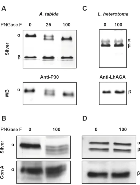
reported for eachα- and β-subunit of the human AGA that are N-glycosylated (N38 and N308) [38,39]. Sequences of AtAGA and LhAGA contain three potential N-glycosylation sites but only the two sites on theα-subunit (N52 and N153) of AtAGA and one site on the β-sub-unit (N326) of LhAGA were predicted as glycosylated by software analysis (Fig 1). N-glycosyla-tion of AtAGAα-subunit was previously demonstrated [9]. Accordingly, treatment ofA. Fig 4. Analysis of the native conformation of AtAGA and LhAGA. A. Non-reducing/reducing
electrophoretic migration of L. heterotoma venom. After venom separation on a 12.5% non-reducing gel, the full lane was excised and run under reducing conditions (gel on the left). The only band that showed a migration shift (boxed) was excised, run under reducing conditions, and silver stained (Gel) or probed with the anti-LhAGA (WB) (lanes on the right). B. Glutaraldehyde cross-linking analysis of the oligomerization state of FPLC purified native AtAGA and LhAGA (% of glutaraldehyde on top of the lane). First lane: molecular weight markers.
tabida venom with PNGase F to remove N-glycosylations induced a clear mobility shift of
AtAGAα-subunit from 25–27 kDa to approximately 24 kDa (Fig 6A), suggesting either an incomplete deglycosylation or occurrence of other post-translational modification. Besides, among various lectins tested on western blots ofA. tabida venom, the Con A lectin strongly
reacted with a band at the size of theα-subunit (27 kDa) (Fig 6B). It also reacted with the α-subunit from the FPLC-purified fraction. Although the intensity of the signal was reduced, Con A labeling was still observed on the deglycosylatedα-subunit, suggesting occurrence of PGNase F-resistant glycosylation sites (Fig 6B).
The migration of the LhAGAβ-subunit was not affected by the PNGase F treatment, even at high amounts and using a more resolutive 15% gel to control for the absence of molecular weight shift (Fig 6C). To ascertain whether the LhAGAβ-subunit is indeed glycosylated, we tested different lectins on western blot ofL. heterotoma venom. Again, a strong reaction was
observed with Con A only, at 20 kDa (Fig 6D), as well as with theβ-subunit of the LhAGA FPLC purified fraction, that was not affected by PNGase F treatment (Fig 6D). Yet, a more intense signal was sometimes observed for the 43 kDa band with the anti-LhAGA, suggesting partial unmasking (or better accessibility) of the epitope on the (αβ) heterodimer (or the pro-α-β-chain).
Mature AGA was reported to be produced through autocleavage/activation of a precursor form [34,37,40] and the immunoreactive bands at 43 kDa we observed in venom may
Fig 5. FPLC purification profiles of AtAGA and LhAGA. A and D. FPLC profiles of A. tabida (A) and L.
heterotoma (D) venom extracts at 280 nm. B and E. 12.5% SDS-PAGE analysis of each FPLC fraction for A. tabida
(B) and L. heterotoma (E). Lane T, total venom extract (3 venom apparatus/well). Aspartylglucosaminidase activity measured with 20μl of each FPLC fraction is overlay on gel pictures for AtAGA (B) and LhAGA (E). C and F. Detection of AGA on western-blots of 10μl of each FPLC fraction of A. tabida (anti-P30 antibody, C) and L.
heterotoma (anti-LhAGA, F). Only AGA positive fractions are shown.
correspond to the pro-α-β-chain. We thus tested whether parasitoid AGAs could undergo autocleavage /activation following venom collection. No change was observed after five days of venom incubation in the proportion ofA. tabida or L. heterotoma α- and β-subunits on
SDS-PAGE gels or in the aspartylglucosaminidase activity (S2 Fig), suggesting that all the AGA secreted in venom is already fully mature.
AGAs enzymatic activities
The venom of both species hydrolyzed AspAMC, the substrate of a sensitive aspartylglucosa-minidase assay [31]. Since only AGA immunoreactive FPLC fractions (Fig 5) hydrolyzed AspAMC, AGAs are likely responsible for all the activity detected in venom. We then defined the optimum pH of AGA activity using venom extracts (Fig 7):A. tabida AGA activity
in-creased sharply at pH 5, then more slowly, reaching a broad optimum between pH 6 and 8
Fig 6. Glycosylation profile of AtAGA and LhAGA subunits. Total venom collected in PBS was
heat-denatured in presence of SDS andβ-mercaptoethanol and incubated overnight at 37˚C in absence or presence of different quantities of PNGase F (in units). Silver stained gels (12.5% or 15%, 1/2 venom apparatus/well) and western-blots incubated with anti-P30 (A. tabida: A) or anti-LhAGA (L. heterotoma: C), or labeled with the Con A lectin (A. tabida: B; L. heterotoma: D) are shown.
after which the activity dropped, whereasL. heterotoma AGA had a sharper activity peak
cen-tered on pH 8.
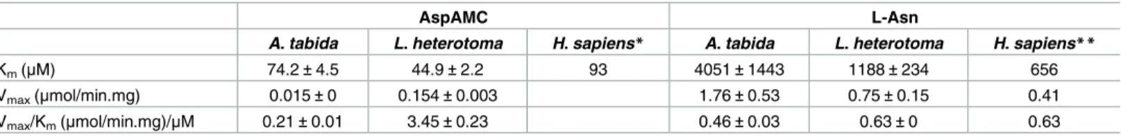
We further analyzed the catalytic properties using the FPLC purified enzymes at pH 8, opti-mum for the activity of both enzymes. Similar Kmvalues were obtained for AtAGA and
LhAGA, but LhAGA may have a better catalytic efficiency for AspAMC, with a 10-fold higher Vmax(Table 1).
As the recombinant human AGA also catalyzes the hydrolysis of the amino acid L-aspara-gine, which competitively inhibits the hydrolysis of aspartylglucosamine [21], we tested whetherA. tabida and L. heterotoma aspartylglucosaminidase activity was inhibited by
L-asparagine. We observed that AspAMC hydrolysis by venom decreased with increasing
Fig 7. Effect of pH on AGA activity in the venom. AGA activity was measured with the AspAMC substrate
in 50 mM Tris buffer at different pH. Each data point represents the mean of two separate assays with error bars showing standard deviation (SD). AFU (Arbitrary Fluorescence Units) values are indicated on the left for AtAGA and on the right for LhAGA.
https://doi.org/10.1371/journal.pone.0181940.g007
Table 1. Kinetic constants of AtAGA and LhAGA for AspAMC and L-asparagine.
AspAMC L-Asn
A. tabida L. heterotoma H. sapiens* A. tabida L. heterotoma H. sapiens**
Km(μM) 74.2±4.5 44.9±2.2 93 4051±1443 1188±234 656
Vmax(μmol/min.mg) 0.015±0 0.154±0.003 1.76±0.53 0.75±0.15 0.41
Vmax/Km(μmol/min.mg)/μM 0.21±0.01 3.45±0.23 0.46±0.03 0.63±0 0.63
*(data from [31]; Vmaxnot available) **(data from [21])
concentrations of L-asparagine, the dose-dependent inhibition reaching a maximum of about 40–45% inhibition with 2 mM of the free amino acid. In contrast, addition of 5 mM L-gluta-mine had no effect in these conditions, suggesting a lower affinity for this amino acid.
Finally, we confirmed that AtAGA and LhAGA have an asparaginase activity using FPLC purified enzymes. Kinetic parameters of the asparaginase activity were estimated using differ-ent L-asparagine concdiffer-entrations, other conditions being unchanged. The close Kmand Vmax
values for AtAGA and LhAGA suggest a similar catalytic efficiency for L-asparagine which is close also to the hAGA published one (Table 1).
Discussion
Here, we have analyzed and compared the biochemical features and enzymatic activities of secreted AGAs purified from the venom of two parasitoid wasps that belong to distant super-families:A. tabida (Ichneumonoidea) and L. heterotoma (Cynipoidea). AGA is the major
pro-tein in the venom of these species, representing 27% of the total propro-tein content inA. tabida
and 16% inL. heterotoma, based on SDS-PAGE estimation.
Structure and secretion
In eukaryotes, AGA is described as a soluble lysosomal amidase whose genetic deficiencies in human causes aspartylglucosaminuria [36,41]. Human and other mammalian AGAs have thus been purified and characterized from different tissues and cells [42,43], as were other verte-brate AGAs [44]. Yet, to our knowledge, AGA had only been described in one insect species (Sf9 cells derived fromSpodoptera frugiperda; Sf9AGA) [20]. All described AGAs share the same basic structure of two subunits of 19–25 kDa and 16–19 kDa, joined by non-covalent forces.
Human AGA is synthesized as an inactive single chain preproprotein precursor of about 45 kDa [35] which is translocated into the lumen of the endoplasmic reticulum (ER) [45]. After removal of the N-terminal signal peptide in the ER, two proprotein precursor chains dimerize and are glycosylated before an autocatalytic processing of each chain occurs, leading to forma-tion of an active heterotetramer composed of two pro-α- and two pro-β-subunits [46,47] and the exposition of the threonine T206 critical for the amidase activity at the N-terminus of the β-subunit. This activation mechanism is conserved among the N-terminal nucleophile hydro-lases (Ntn hydrohydro-lases) from bacteria to human [34,35,40,48]. The complex is further modified in the Golgi apparatus and then transported to the lysosomes whereα- and β-subunits are car-boxy-terminal proteolytically trimmed, reaching a final size of 19–25 kDa and 16–19 kDa, respectively (depending upon the studies) [49].
Sequence comparison of AtAGA, LhAGA and hAGA indicates that parasitoid venom pro-teins have a similar organization as lysosomal AGAs (signal peptide and glycosylasparaginase domain) and retained the threonine residue essential for enzyme autocatalytic activation and catalytic activity [34] and the triad (equivalent to hAGA W34, R234 and D237) essential for substrate binding, although they differ in their geometry [35,36]. AtAGA and LhAGA have similar predictedα- and β-subunits C-terminal disulfide bonds, which are known to be crucial in the early folding and activation of hAGA [47]. The essential hAGA stabilizing N-terminal disulfide bond [47] is also predicted for AtAGA (C66-C73) and LhAGA (C63-C70). The (αβ)2
structure of “native” secreted venom AGAs, suggested by the very good fit of the three-dimen-sional predicted structures with the hAGA solved structure, was confirmed by the 70–80 kDa molecular weight observed from gel filtration FPLC and by crosslinking experiments. Interest-ingly, LhAGA subunits did not dissociate on SDS-PAGE under non-reducing conditions, in contrast to AtAGA subunits. A similar resistance to SDS denaturation was previously reported
for hAGA [42] and the rat AGA [50]. The reason why it is not observed for AtAGA remains unclear.
Occurrence of high amount of secreted AGA in venom is puzzling. Indeed, human lyso-somal AGA is normally produced in low amounts [51,52] and AGA secretion in a specialized body fluid, albeit in low quantity, has only been reported once in mammals [53]. This suggests that specific mechanisms concur to the AGA high secretion level in parasitoid venom glands and may explain its non-lysosomal targeting.
AtAGA and LhAGA transcripts are abundant in venom tissues [9,10] compared to the residual wasp body, a pattern already observed for several potential virulence factors secreted in parasitoid venom [6]. These factors generally resemble “classical” intracellular proteins, sup-porting the hypothesis that the encoding genes evolved by duplication of genes involved in normal cellular processes. This mechanism may also explain the origin and evolution of venom proteins in other organisms [54,55].A. tabida and L. heterotoma venom AGAs have
thus likely evolved from an ancestral lysosomal AGA through duplication and convergent recruitment, to acquire specific features allowing overexpression and secretion. Interestingly,
in vitro cellular overexpression of hAGA was shown to increase the proportion of AGA
secreted in medium, as a prelysosomal form [38,56]. A similar process may occur for parasit-oid secreted AGA: once treated with PNGase F, AtAGA and LhAGAα- and β-subunits have an estimated molecular weight close to the one predictedin silico, suggesting they are not
trimmed at the carboxyterminal end (the predicted C-term peptide of LhAGAα-subunits was indeed obtained by mass spectrometry;S1 Fig), a process that occurs during the transport of hAGA to lysosomes.
Bothα- and β-subunits of hAGA contain a single N-glycosylated site holding high-man-nose-type oligosaccharides [38]. Yet, in agreement with software prediction, only AtAGA α-and LhAGAβ-subunits appear N-glycosylated, according to lectin binding. Interestingly, the Sf9 AGA also has a single potential glycosylation site located on theα-subunit [20]. After PNGase F treatment, AtAGAα-subunit, but not LhAGA β-subunit, showed a clear shift in mobility, but both enzymes retained the Con A labeling. PNGase F usually cleaves all aspara-gine-linked oligosaccharides, whether complex, hybrid, or high mannose, unless the core con-tains anα(1!3)-fucose. As arthropods do not produce complex glycans [57], AtAGA and LhAGA glycosylation could be either high mannose or pauci-mannose (man-3) oligosaccha-rides. Thus, although we cannot exclude that one of the two potential N-glycosylation sites of AtAGAα-subunit is occupied by a modified glycan with an α(1!3)-fucose, it may also be O-glycosylated. The presence of a modified glycan or an O-glycosylation could also explain the resistance of LhAGAβ-subunit to PNGase F on its sole glycosylation site. The role of hAGA glycosylations / phosphorylations on folding, activation, targeting and secretion has been assessed byin vitro mutagenesis and transient expression of mutant polypeptides in COS cells
[38]. Results showed that glycosylation of only one subunit was sufficient for lysosomal trans-port and normal processing, unglycosylated enzymes remaining trapped in the ER and being secreted in the medium. Glycosylation of theβ-subunit was more important than that of the α-subunit for folding, stability and transport. Thus, although the absence of glycosylation on one of the subunits may help for secretion of AtAGA and LhAGA, it is likely insufficient to explain their high secretion level.
In hAGA, three lysine residues (K177, K183 and K214) and one tyrosine residue (Y178) are necessary for proper phosphorylation of the oligosaccharides, and thus the binding to man-nose 6-phosphate receptors (MPRs) [58] which is responsible for acid hydrolases lysosomal targeting in mammals [59,60]. Interestingly, the non-conservative substitution K214A in hAGA decreases the lysosomal targeting efficiency by 70%, and it is correlated with enhanced secretion [58]. In insects, the existence of a MPR-dependent pathway is unclear. Notably, the
single MPR orthologue identified inD. melanogaster, named lysosomal enzyme receptor
pro-tein (LERP) [61], lacks the residues critical for mannose 6-phosphate binding and it contrib-utes only moderately to lysosomal enzyme targeting [62]. This may explain why two of the key lysine residues (K183 and K214), and the tyrosine one, are not conserved in AtAGA and LhAGA. Besides, the K214 lysine and the tyrosine residues do not seem conserved either in other invertebrates [9]. Understanding how AtAGA and LhAGA escape the lysosomal path-way thus requires further investigations. The identification of the parasitoids’ “lysosomal” AGA encoding gene(s) for instance could allow identifying the precise molecular features and mechanisms sustaining AGA overexpression in venom and their secretion.
Enzymatic activity
In addition to the expected aspartylglucosaminidase activity, both AtAGA and LhAGA exhib-ited asparaginase activity, similarly to the human andS. frugiperda AGAs. Although the
opti-mum pH range for aspartylglucosaminidase activity was broader for AtAGA than for LhAGA, both enzymes had a maximum activity around neutrality.S. frugiperda AGA also has a broad
optimum pH range (5 to 9) [20], like mammalian AGAs [34,42,63] except rat and mouse enzymes whose optimum pH is 7 to 9 [42]. Thus, although AtAGA and to a lesser extent LhAGA could be active in the lysosome acidic environment, they will have their maximum activity in the neutralDrosophila hemolymph environment where they are injected by the
female parasitoid. We found that LhAGA has a 10-fold higher activity for AspAMC than AtAGA, which may partly be explained by the AtAGA T257S substitution [34,37] and/or by the AtAGA reduced stability. In contrast, the two purified enzymes have a similar affinity for AspAMC, in the same range as the purified human AGA (hAGA Km= 93μM at pH 7.5;
simi-lar to the Kmfor the natural substrate GlcNAc-Asn) [31,64].
Human recombinant AGA has less affinity for L-asparagine and a lower conversion activity compared to aspartylglucosamine [21,31]. AtAGA and LhAGA also have less affinity for L-asparagine than AspAMC, similarly to Sf9AGA (Km= 3.0 mM for L-asparagine compared to
Km= 0.9 mM for GlcNAc-Asn) [20]. Yet, insect AGAs affinity for L-asparagine is similar to
that of the human asparaginase (hASNase-3; Km= 2 mM at pH 7.5) [65], and they have the
same catalytic efficiency as human AGA. Finally, we also estimated that AtAGA and LhAGA are 3- to 4-fold more active asparaginases than theE. coli asparaginase (used to calibrate the
assay).
Potential role of AtAGA and LhAGA in parasitism
Although the presence of N-aspartylglucosamine inDrosophila hemolymph seems unlikely, a
concentration of 2 mM of L-asparagine was reported [66] suggesting that once injected in the host, AtAGA and LhAGA can mainly act as asparaginases to transform circulating L-aspara-gine in L-aspartate. L-aspartate has an excitatory role similar to that of glutamate accumulation and it is a neurotransmitter in some synapses [67]. It can act as a signaling molecule or a com-petitor of L-glutamate either in the brain or at the neuromuscular junctions. Its increase in hemolymph might thus play a role in the transient paralysis of host larvae induced by oviposi-tion of bothA. tabida and L. heterotoma [14–16], but also in blocking sensory class IV neurons essential for the cellular immune response to parasitoid infestation [68]. Alternatively, depriv-ingDrosophila larvae from L-asparagine may induce cell division arrest and/or apoptosis and
thus slow down development [69], which may help protect the parasitoid larvae and synchro-nize the host development with its own [70]. Indeed bacterial secreted asparaginases are known to mediate virulence by inhibiting the proliferation of immune cells through mecha-nisms that involve asparagine starvation, a property largely used to treat acute lymphoblastic
leukemia [71–72]. The precise role of venom AGA in these suggested physiological mecha-nisms following from either aspartate production or asparagine depletion needs know to be elucidated.
In addition to their putative role in the success of wasp parasitism, an interest of venomous AGAs lies in the study of the mechanisms that led to their evolution from intracellular AGAs. Finally, understanding the structural features allowing a high secretion of active AGAs in the venom could potentially help improve the production of active AGA for pharmaceutical pur-poses [19,27,72].
Supporting information
S1 Fig. MS/MS identification of the AGA-immunoreactive 20- and 22-kDa bands resolved by SDS-PAGE ofL. heterotoma venom. Identification by mass spectrometry was performed
on 1D bands excised from SDS-PAGE that corresponded to the AGA-immunoreactive bands on western blots. Bands were treated with trypsin and peptides were extracted for MS/MS. Peptide identification was performed with the Mascot software (http://www.matrixscience. com) using the LhAGA sequence. Mascot analysis was performed with a fragment ion mass tolerance of 0.30 Da and a parent ion tolerance of 0.30 Da. Carbamidomethyl of cysteine was specified in Mascot as a fixed modification, and oxidation of methionine as a variable modifi-cation. The maximum miscleavage allowed was set to 2. Peptides identified with a p<0.05 are indicated in red.
(TIFF)
S2 Fig. Following of AGA auto-cleavage and auto-activation venom collection. PooledA. tabida and L. heterotoma venom extracts were incubated at room temperature and aliquots
were analyzed by SDS-PAGE and western blots at different times (seematerials and methods). No change in quantity of theα- and β-subunits (arrows) were observed by silver staining and forα-AtAGA and β-LhAGA by western blots (anti-P30 and anti-LhAGA, respectively). For each time, measured aspartylglucosaminidase activity is indicated (AspAMC in AFU/h). MW in kDa.
(TIFF)
Acknowledgments
We thank Dr M. Belghazi (CRN2M UMR 7286, Marseille) for mass spectrometry analysis, Christian Rebuf for technical help, and the PFIE unit (INRA Nouzilly) for antibody production.
Author Contributions
Conceptualization: Marylène Poirie´, Jean-Luc Gatti.
Funding acquisition: Geneviève Pre´vost, Marylène Poirie´.
Investigation: Quentin Coulette, Se´verine Lemauf, Dominique Colinet, Caroline Anselme,
Jean-Luc Gatti.
Methodology: Quentin Coulette, Se´verine Lemauf, Dominique Colinet, Caroline Anselme,
Jean-Luc Gatti.
Project administration: Marylène Poirie´, Jean-Luc Gatti.
Supervision: Geneviève Pre´vost, Caroline Anselme, Marylène Poirie´.
Writing – original draft: Quentin Coulette, Dominique Colinet, Caroline Anselme, Jean-Luc
Gatti.
Writing – review & editing: Dominique Colinet, Geneviève Pre´vost, Caroline Anselme, Mar-ylène Poirie´, Jean-Luc Gatti.
References
1. Grissell EE. Hymenopteran biodiversity: some alien notions. Am Entomol. 1999; 45: 235–244.
2. Godfray HCJ. Parasitoids: Behavioural and evolutionary ecology. Princeton: Princeton University Press; 1994.
3. Quicke DL. Parasitic wasps. Chapman & Hall, Springer Netherlands; 1997.
4. Asgari S, Rivers DB. Venom proteins from endoparasitoid wasps and their role in host-parasite interac-tions. Annu Rev Entomol. 2011; 56: 313–335.https://doi.org/10.1146/annurev-ento-120709-144849 PMID:20822448
5. Colinet D, Mathe´-Hubert H, Allemand R, Gatti JL, Poirie´ M. Variability of venom components in immune suppressive parasitoid wasps: From a phylogenetic to a population approach. J Insect Physiol. 2013; 59: 205–212.https://doi.org/10.1016/j.jinsphys.2012.10.013PMID:23103980
6. Poirie´ M, Colinet D, Gatti JL. Insights into function and evolution of parasitoid wasp venoms. Curr Opin Insect Sci. 2014; 6: 52–60.
7. Strand M. R. and Burke G. R. Polydnaviruses: From discovery to current insights. Virology 2015; 479, 393–402.https://doi.org/10.1016/j.virol.2015.01.018PMID:25670535
8. Moreau SJM, Cherqui A, Doury G, Dubois F, Fourdrain Y, Sabatier L, et al. Identification of an aspartyl-glucosaminidase-like protein in the venom of the parasitic wasp Asobara tabida (Hymenoptera: Braco-nidae). Insect Biochem Mol Biol. 2004; 34: 485–492.https://doi.org/10.1016/j.ibmb.2004.03.001PMID: 15110870
9. Vinchon S, Moreau SJM, Drezen JM, Pre´vost G, Cherqui A. Molecular and biochemical analysis of an aspartylglucosaminidase from the venom of the parasitoid wasp Asobara tabida (Hymenoptera: Braco-nidae). Insect Biochem Mol Biol. 2010; 40: 38–48.https://doi.org/10.1016/j.ibmb.2009.12.007PMID: 20036741
10. Colinet D, Deleury E, Anselme C, Cazes D, Poulain J, Azema-Dossat C, et al. Extensive inter- and intra-specific venom variation in closely related parasites targeting the same host: The case of Leptopilina parasitoids of Drosophila. Insect Biochem Mol Biol. 2013; 43: 601–611.https://doi.org/10.1016/j.ibmb. 2013.03.010PMID:23557852
11. Goecks J, Mortimer NT, Mobley JA, Bowersock GJ, Taylor J, Schlenke TA. Integrative approach reveals composition of endoparasitoid wasp venoms. PLoS One. 2013; 8: e64125.https://doi.org/10. 1371/journal.pone.0064125PMID:23717546
12. Monconduit H, Pre´vost G. Avoidance of encapsulation by Asobara tabida, a larval parasitoid of Dro-sophila species. Nor J Agric Sci. 1994; 16: 301–309.
13. Poirie´ M, Carton Y, Dubuffet A. Virulence strategies in parasitoid Hymenoptera as an example of adap-tive diversity. C R Biol. 2009; 332: 311–320.https://doi.org/10.1016/j.crvi.2008.09.004PMID: 19281961
14. Mabiala-Moundoungou ADN, Doury G, Eslin P, Cherqui A, Pre´vost G. Deadly venom of Asobara
japon-ica parasitoid needs ovarian antidote to regulate host physiology. J Insect Physiol. 2010; 56: 35–41.
https://doi.org/10.1016/j.jinsphys.2009.09.001PMID:19769980
15. Moreau SJM, Dingremont A, Doury G, Giordanengo P. Effects of parasitism by Asobara tabida (Hyme-noptera: Braconidae) on the development, survival and activity of Drosophila melanogaster larvae. J Insect Physiol. 2002; 48: 337–347. PMID:12770108
16. Van Lenteren JC, Bakker K, Samson-Boshuizen M, Van Lenteren JC, Bakker K. Success of parasitiza-tion of Pseudeucoila Bochei Weld (Hym., Cynip.): a matter of experience. Netherlands J Zool. 1973; 24: 67–85.
17. Enomaa N, Heiskanen T, Halila R, Sormunen R, Seppala R, Vihinen M et al. Human aspartylglucosami-nidase. Biochem J. 1992; 286: 613–618. PMID:1530592
18. Makino M, Kojima T, Ohgushi T, Yamashina I. Studies on enzymes acting on glycopeptides. J Biochem. 1968; 63: 186–192. PMID:5669921
19. Arvio M, Mononen I. Aspartylglycosaminuria: a review. Orphanet J Rare Dis. 2016; 24: 1–10.
20. Liu Y, Dunn G S, Aronson NN. Purification, biochemistry and molecular cloning of an insect glycosylas-paraginase from Spodoptera frugiperda. Glycobiology 1996; 6: 527–36. PMID:8877373
21. Noronkoski T, Stoineva IB, Petkov DD, Mononen I. Recombinant human glycosylasparaginase cata-lyzes hydrolysis of L-asparagine. FEBS Lett. 1997; 412: 149–152. PMID:9257709
22. Tanaka M, Kohno M, Yamashina I. Specificity Studies of 4-L-Aspartylglycosylamine amido hydrolase. J Biochem. 1973; 73: 1285–1289. PMID:4724304
23. Tarentino AL, Plummer TH. The first demonstration of a procaryotic glycosylasparaginase. Biochem Biophys Res Commun. 1993; 197: 179–86. PMID:8250923
24. Katoh K, Standley DM. MAFFT multiple sequence alignment software version 7: improvements in per-formance and usability. Mol Biol Evol. 2013; 30: 772–780.https://doi.org/10.1093/molbev/mst010 PMID:23329690
25. Kelley LA, Mezulis S, Yates CM, Wass MN, Sternberg MJE. The Phyre2 web portal for protein model-ing, prediction and analysis. Nat Protoc. 2015; 10: 845–858.https://doi.org/10.1038/nprot.2015.053 PMID:25950237
26. Benkert P, Biasini M, Schwede T. Toward the estimation of the absolute quality of individual protein structure models. Bioinformatics 2011; 27: 343–350.https://doi.org/10.1093/bioinformatics/btq662 PMID:21134891
27. Oinonen C, Tikkanen R, Rouvinen J, Peltonen L. Three-dimensional structure of human lysosomal aspartylglucosaminidase. Nat Struct Biol.1995; 2: 1102–1108. PMID:8846222
28. Laemmli UK. Cleavage of structural proteins during the assembly of the head of bacteriophage T4. Nature 1970; 227: 680–685. PMID:5432063
29. Morrissey JH. Silver stain for proteins in polyacrylamide gels: a modified procedure with enhanced uni-form sensitivity. Anal Biochem. 1981; 117: 307–310. PMID:6172996
30. Towbin H, Staehelin T, Gordon J. Electrophoretic transfer of proteins from polyacrylamide gels to nitro-cellulose sheets: procedure and some applications. Proc Natl Acad Sci USA 1979; 76: 4350–4354. PMID:388439
31. Mononen IT, Kaartinen VM, Williams JC. A fluorometric assay for glycosylasparaginase activity and detection of aspartylglycosaminuria. Anal Biochem. 1993; 208: 372–374.https://doi.org/10.1006/abio. 1993.1063PMID:8452235
32. Fernandez CA, Cai X, Elozory A, Liu C, Panetta JC, Jeha S, et al. High-throughput asparaginase activ-ity assay in serum of children with leukemia. Int J Clin Exp Med. 2013; 6: 478–487. PMID:23936585
33. Kemmer G, Keller S. Nonlinear least-squares data fitting in Excel spreadsheets. Nat Protoc. 2010; 5: 267–281.https://doi.org/10.1038/nprot.2009.182PMID:20134427
34. Tikkanen R, Riikonen A, Oinonen C, Rouvinen R, Peltonen L. Functional analyses of active site resi-dues of human lysosomal aspartylglucosaminidase: implications for catalytic mechanism and autocata-lytic activation. EMBO J. 1996; 15: 2954–60. PMID:8670796
35. Saarela J. Laine M, Tikkanen R, Oinonen C, Jalanko A, Rouvinen J. et al. Activation and oligomerization of aspartylglucosaminidase. J Biol Chem. 1998; 273: 25320–25328. PMID:9737998
36. Saarela J, Laine M, Oinonen C, von Schantz C, Jalanko A, Rouvinen J. et al. Molecular pathogenesis of a disease: structural consequences of aspartylglucosaminuria mutations. Hum Mol Genet. 2001; 10: 983–995. PMID:11309371
37. Saarela J, Oinonen C, Jalanko A, Rouvinen J, Peltonen L. Autoproteolytic activation of human aspartyl-glucosaminidase. Biochem J. 2004; 378: 363–71.https://doi.org/10.1042/BJ20031496PMID: 14616088
38. Tikkanen R, Enomaa N, Riikonen A, Ikonen E, Peltonen L. Intracellular sorting of aspartylglucosamini-dase: the role of N-linked oligosaccharides and evidence of Man-6-P-independent lysosomal targeting. DNA Cell Biol. 1995; 14: 305–312.https://doi.org/10.1089/dna.1995.14.305PMID:7710687
39. Rip JW, Coulter-Mackie MB, Rupar CA, Gordon BA. Purification and structure of human liver aspartyl-glucosaminidase. Biochem J. 1992; 288: 1005–1010. PMID:1281977
40. Guan C, Cui T, Rao V, Liao W, Benner J, Lin CL et al. Activation of glycosylasparaginase. J Biol Chem. 1996; 271: 1732–1737. PMID:8576176
41. Saito S, Ohno K, Sugawara K, Suzuki T, Togawa T, Sakuraba H. Structural basis of aspartylglucosami-nuria. Biochem Biophys Res Commun. 2008; 377: 1168–1172.https://doi.org/10.1016/j.bbrc.2008.10. 142PMID:18992224
42. Tollersrud OK, Aronson NN. Comparison of liver glycosylasparaginases from six vertebrates. Biochem J. 1992; 282: 891–897. PMID:1554372
43. Saarela J. Characterization of aspartylglucosaminidase activation and aspartylglucosaminuria muta-tions. Ph.D. Thesis, University of Helsinki. 2004.
44. Tarentino AL, Maley F. The purification and properties of aβ-aspartyl N-acetylglucosylamine amidohy-drolase from hen oviduct. Arch Biochem Biophys. 1969; 130: 295–303. PMID:5778645
45. Saarela J, von Schantz C, Peltonen L, Jalanko A. A novel aspartylglucosaminuria mutation affects translocation of aspartylglucosaminidase. Hum Mutat. 2004; 243: 350–351.
46. Ikonen E, Julkunen I, Tollersrud OK, Kalkkinen N, Peltonen L. Lysosomal aspartylglucosaminidase is processed to the active subunit complex in the endoplasmic reticulum. EMBO J. 1993; 12: 295–302. PMID:8428587
47. Riikonen A, Rouvinen J, Tikkanen R, Julkunen I, Peltonen L, Jalanko A. Primary folding of aspartylglu-cosaminidase. Significance of disulfide bridges and evidence of early multimerization. J. Biol. Chem. 1996; 271: 21340–21344. PMID:8702913
48. Oinonen C, Rouvinen J. Structural comparison of Ntn-hydrolases. Protein Sci. 2000; 9: 2329–2337. https://doi.org/10.1110/ps.9.12.2329PMID:11206054
49. Dunder U. The application of enzyme replacement therapy in vitro and in a mouse model in aspartylgly-cosaminuria. Ph.D. Thesis, University of Eastern Finland. 2010.
50. Tollersrud OK, Aronson NN. Purification and characterization of rat liver glycosylasparaginase. Bio-chem J. 1989; 260: 101–108. PMID:2775174
51. Arvio P, Arvio M, Kero M, Pirinen S, Lukinmaa PL. Overgrowth of oral mucosa and facial skin, a novel feature of aspartylglucosaminuria. J Med Genet. 1999; 36: 398–404. PMID:10353787
52. Halila R, Baumann M, Ikonen E, Enomaa N, Peltonen L. Human leucocyte aspartylglucosaminidase. Evidence for two different subunits in a more complex native structure. Biochem J. 1991; 276: 251–256. PMID:2039475
53. Moura AA, Souza CE, Stanley BA, Chapman DA, Killian GJ. Proteomics of cauda epididymal fluid from mature Holstein bulls. J Proteomics 2010; 73: 2006–2020.https://doi.org/10.1016/j.jprot.2010.06.005 PMID:20601273
54. Casewell NR, Wu¨ster W, Vonk FJ, Harrison RA, Fry BG. Complex cocktails: the evolutionary novelty of venoms. Trends Ecol Evol. 2013; 28: 219–229.https://doi.org/10.1016/j.tree.2012.10.020PMID: 23219381
55. Fry BG, Roelants K, Champagne DE, Scheib H, Tyndall JDA, King GF, et al. The toxicogenomic multi-verse: convergent recruitment of proteins into animal venoms. Annu Rev Genomics Hum Genet. 2009; 10: 483–511.https://doi.org/10.1146/annurev.genom.9.081307.164356PMID:19640225
56. Peltola M, Tikkanen R, Peltonen L, Jalanko A. Ser72Pro active-site disease mutation in human lyso-somal aspartylglucosaminidase: abnormal intracellular processing and evidence for extracellular activa-tion. Hum Mol Genet. 1996; 5: 737–743. PMID:8776587
57. Rendic D, Wilson IBH, Paschinger K. The glycosylation capacity of insect cells. Croat Chem Acta 2008; 81: 7–21.
58. Tikkanen R. Peltola M. Oinonen C. Rouvinen J, Peltonen L. Several cooperating binding sites mediate the interaction of a lysosomal enzyme with phosphotransferase. EMBO J. 1997; 16: 6684–6693.https:// doi.org/10.1093/emboj/16.22.6684PMID:9362483
59. Braulke T, Bonifacino JS. Sorting of lysosomal proteins. Biochim Biophys Acta 2009; 1793: 605–614. https://doi.org/10.1016/j.bbamcr.2008.10.016PMID:19046998
60. Hasanagic M, Waheed A, Eissenberg JC. Different Pathways to the lysosome: sorting out alternatives. Int J Biochem Cell Biol. 2015; 320: 75–101.
61. Dennes A, Cromme C, Suresh K, Kumar NS, Eble JA, Hahnenkamp A et al. The novel Drosophila lyso-somal enzyme receptor protein mediates lysolyso-somal sorting in mammalian cells and binds mammalian and Drosophila GGA adaptors. J Biol Chem. 2005; 280: 12849–12857.https://doi.org/10.1074/jbc. M410626200PMID:15664992
62. Hasanagic M, van Meel E, Luan S, Aurora R, Kornfeld S, Eissenberg JC. The lysosomal enzyme recep-tor protein (LERP) is not essential, but is implicated in lysosomal function in Drosophila melanogaster. Biol Open 2015; 4: 1316–1325.https://doi.org/10.1242/bio.013334PMID:26405051
63. Baumann M, Peltonen L, Aula P, Kalkkinen N. Isolation of a human hepatic 60 kDa aspartylglucosamini-dase consisting of three non-identical polypeptides. Biochem J. 1989; 262: 189–94. PMID:2818562
64. Kaartinen V, Williams JC, Tomich J, Yates, J, Hood LE, Mononen I. Glycosaparaginase from humanleu-kocytes. J Biol Chem 1991; 266: 5860–5869. PMID:2005122
65. Nomme J, Su Y, Lavie A. Elucidation of the specific function of the conserved threonine triad responsi-ble for human l-Asparaginase autocleavage and substrate hydrolysis. J Mol Biol. 2014; 426: 2471– 2485.https://doi.org/10.1016/j.jmb.2014.04.016PMID:24768817
66. Ragan TJ, Bailey AP, Gould AP, Driscoll PC. Volume determination with two standards allows absolute quantification and improved chemometric analysis of metabolites by NMR from submicroliter samples. Anal Chem. 2013; 85: 12046–12054.https://doi.org/10.1021/ac403111sPMID:24251761
67. Meldrum BS. Glutamate as a neurotransmitter in the brain: review of physiology and pathology. J Nutr. 2000; 130: 1007S–1015S. PMID:10736372
68. Hwang RY, Zhong L, Xu Y, Johnson T, Zhang F, Deisseroth K, Tracey D. Nociceptive Neurons Protect Drosophila Larvae from Parasitoid Wasps. Curr Biol. 2007; 17(24): 2105–2116.https://doi.org/10.1016/ j.cub.2007.11.029PMID:18060782
69. Kelo E, Noronkoski T, Mononen I. Depletion of L-asparagine supply and apoptosis of leukemia cells induced by human glycosylasparaginase. Leukemia 2009; 23: 1167–1171.https://doi.org/10.1038/leu. 2008.387PMID:19158835
70. Lawrence PO. Host-parasite hormonal interactions: an overview. J Insect Physiol. 1986; 32: 295–298.
71. Panosyan EH, Wang Y, Xia P, Lee WNP, Pak Y, Laks DR et al. Asparagine depletion potentiates the cytotoxic effect of chemotherapy against brain tumors. Mol Cancer Res. 2014; 12: 694–702.https://doi. org/10.1158/1541-7786.MCR-13-0576PMID:24505127
72. Tikkanen R, Rouvinen J, To¨rro¨nen A, Kalkkinen N, Peltonen L. Large-scale purification and preliminary x-ray diffraction studies of human aspartylglucosaminidase. Proteins 1996; 24: 253–8.https://doi.org/ 10.1002/(SICI)1097-0134(199602)24:2<253::AID-PROT12>3.0.CO;2-MPMID:8984501