FEBS Letters 386 (1996) 156-160 FEBS 17036
Molecular characterization o f a new urea transporter
in the h u m a n kidney
Bernadette Oliv+s% Sonia Martial b, Marie-Genevieve Mattei c, Giorgio Matassi a,
Germain Rousselet b, Pierre Ripoche b, Jean-Pierre Cartron a'*, Pascal Bailly ~
~INSERM U76, GIP-lnstitut National de la Transfusion Sanguine, 6 rue Alexandre Cabanel, 75015 Paris, France bService de Biologie Cellulaire, C.E.A./Saclay, 91191 Gif-sur-Yvette, France
° I N S E R M U242, Htpital de la Timone, 13385 Marseille, France Received 14 March 1996; revised version received 11 April 1996
Abstract A eDNA clone (HUT2) sharing 61.1% and 89.9% sequence identity with the human erythroid ( H U T l l ) and the rabbit (UT2) urea transporters, respectively, was isolated by homology cloning from a human kidney library. HUT2 transcripts were restricted to the kidney and the HUT2 polypeptide was not immunoprecipitated with blood group Kidd-related antibodies (anti-Jk3) in coupled transcription- translation assays. Functional expression studies in
Xenopus
oocytes demonstrated that HUT2-mediated urea transport was not inhibited by p-chloromercuribenzene sulfonate (pCMBS) which, however, inhibited the urea flux mediated by H U T l l . These findings demonstrate that at least two distinct urea transporters are present in human tissues. By in situ hybridiza- tion, the gene encoding HUT2 has been assigned to chromosome 18q12.l-q21-1, as found previously for the Kidd/urea transporter HUT11, suggesting that both genes evolved from duplication of a common ancestor.K e y words: Urea transport; Human kidney; Tissue expression; Transcription-translation assay; In situ hybridization
1. Introduction
In mammalian cells, urea is the chief end-product of nitro- gen catabolism and plays an important role in the urinary concentration mechanism [1,2]. Thus, the plasma membrane of human erythrocytes [3,4] and some renal epithelial cells [5] exhibit an elevated urea permeability which is mediated by highly selective urea transporters. At least two forms of urea transporters have recently been identified: (i) the presum- ably vasopressin-sensitive rabbit and rat transporters UT2 [6,7] and (ii) the constitutive human transporter H U T l l [8].
As expected for a vasopressin-regulated transporter, UT2 is localized in the inner medullary collecting ducts and probably in the descending thin limbs of Henle's loop [9], where it plays a central role in the establishment of a corticomedullary os- motic gradient. Two transcripts (4.0 and 2.9 kb) of a rat urea transporter (rUT2) were identified in the kidney [7]. They have a spatially distinct distribution and their expression is modu- lated by different pathways. Indeed, the 4.0 kb transcript is confined to the inner medulla and is responsive to changes in the protein diet, whereas the 2.9 kb transcript is predomi- nantly expressed in the inner stripe of the outer medulla and is responsive to the hydration state [7]. Thus, the two rUT2 messages are independently regulated by as yet unknown fac-
*Corresponding author. Fax: (33) (1) 47 34 74 31.
0014-5793/96/$12.00 © 1996 Federation of European Biochemical Societies. PH S00 1 4 - 5 7 9 3 ( 9 6 ) 0 0 4 2 5 - 5
tors, critical for the physiological and functional status of the kidney.
The human urea transporter ( H U T l l clone) has been cloned by cross-hybridization from a human bone marrow library [8]. The predicted translation product is a protein of 391 amino acids that displays 62.4% amino acid identity and a similar membrane topology to the rabbit transporter. HUT11 is responsible for the facilitated urea transport in human red cells and the m R N A is present in hematopoietic cell lines and in human (tumoral) kidney. It has been demonstrated that H U T l l is the product of the Kidd blood group locus [10] and that the rare J k ( a - b - ) red cells lack the Kidd/urea trans- port protein showing a selective defect of the urea transport capacity [10,11] whereas water permeability and aquaporin- associated Colton blood group antigens are normally ex- pressed [10]. Recently, immunofluorescence analysis of human kidney sections demonstrated that the Kidd/urea transporter protein HUT11 is constitutively expressed by endothelial cells of vasa recta in the inner and outer medulla kidney, but not in renal tubules [12]. This is in agreement with data indicating that the descending vasa recta contains a urea transporter [13,14].
Other urea transporters are likely to exist in tissues that use facilitated urea transport to manage the nitrogen catabolism [15]. Here we isolated the human homologue of the vasopres- sin-regulated rabbit UT2 clone by homology cloning from a human kidney library. The functional activity, immunochem- ical specificity, expression pattern and chromosome localiza- tion of the human transporters have been compared.
2. Materials and methods 2.1. Library screening
The UT2 eDNA encoding the rabbit urea transporter [6] was used as a random primed [32P]dCTP-labeled probe to screen about 1.7 X 10⁶ bacteriophage clones of a human kidney eDNA library 5'- stretch purchased from Clontech laboratories (Palo Alto, CA). Posi- tive clones were isolated and enzymatically amplified (annealing tem- perature 60°C; 30 cycles) between ~gtll primers. To prevent PCR errors, we employed the Pwo DNA polymerase (Boehringer, Mann- heim, Germany), which exhibits proofreading activity, and we se- quenced 3 independent clones. All PCR products were ligated into the Sinai-digested pUC vector and nucleotide sequences were deter- mined on both strands by the dideoxy chain termination method (Sanger) using the Sequenase enzyme (US Biochemical Corp). 2.2. In situ hybridization
The 730 bp NcoI fragment from the HUT2 eDNA clone was sub- cloned into pUC18 and 3H-labeled by nick-translation to a specific activity 8X l0 T dpm/gg. Hybridization to metaphase chromosome, Giemsa staining after R-banding and autoradiography were per- formed as previously described [16].
B. Olivbs et al./FEBS Letters 386 (1996) 156-160 157
2.3. Northern blot analysis
Poly(A +) RNAs (2 p.g, Clontech) from adults and fetuses (pool of different gestational ages) per lane were size separated on a denaturing formaldehyde 1.2% agarose gel, transferred to nylon membrane and fixed by UV light. Absence of degradation was checked by staining with ethidium bromide. The Northern blots were hybridized at 42°C in the presence of 50% formamide with the a2P-labeled probes corre- sponding to the NcoI fragment (730 bp) of the coding region of HUT2 and to the full length H U T l l cDNA clone. Stringent washes were performed (0.1 × SSC, 0.1% SDS at 42°C) according to the guidelines of the manufacturer.
2.4. Transcription-translation and immunoprecipitation assays
The HUT2 and HUT11 cDNAs were subcloned into the EcoRV-
digested pT7TS plasmid (kindly provided by P. Krieg, Austin, TX) under the control of the T7 promoter. The related proteins were synthesized in vitro using the transcription-translation coupled reticu- locyte lysate kit from Promega (Madison, WI) in the presence of L-[35S]methionine (Amersham, Bucks, UK) according to the manufac- turer's protocol. These reaction mixtures were used in immunopreci- pitation assay with the human anti-Jk 3 antiserum as previously de- scribed [10]. The different products obtained were analyzed by SDS- PAGE (15% separating gel) on a discontinuous buffer system [17] and followed by autoradiography.
2.5. Oocyte urea flux measurements
After linearization of the pT7TS-HUTll and -HUT2 construct vectors with SmaI and EcoRI restriction enzymes, respectively, capped sense RNAs were synthesized using T7 RNA polymerase from the mCAP mRNA capping kit (Stratagene, La Jolla, CA). Ex- pression studies were carried out by microinjection of cRNAs (10 ng/ oocyte) in collagenase-treated Xenopus laevis oocytes [18] and func- tional tests were realized 2 or 3 days after injection as described pre- viously [8].
P~UT2 1 ~ S - SE I K V E T ~ T T W I Q S SMI ~ U U ~ G X R ~ SYI T G 7 2 ~ E C G Z ~ K S p V F Q F L D 0C13UT2 i 14ED S-SEIKV~ T R ~ R T S W I Q S S yI ~ C A E G L - ~ D K S P % T ~ L D RUTII i ~ E D SPTMVRVD Sp TMVRCZNQVS P C Q G R R C T P K A L G Y V T ~ M K K L A N Q L K D K P V V L Q F ID
u . . .** *.**.**
conlen. • **.* . ..*.. * **** * ** *
± _2
HUT2 60 W V I , R G T S Q ~ M B ~ N P L SGI L I ILGLF i ~ P R R A I S G C L G T I ~ T L T A L I L S ~ RNUT2 60 WVLRGTSQV~a~;NNP L SG I L I V L G L F V ~ R W A I S G C L G T I ~ T L T A L I LSQ4)KSAIAA~ OCUUT2 60 W V L R G T S Q ~ i ~ B ' ~ L SG I L IVI G L F V ~ R R A I A G C L G T V M S T L T A L Z I ~ R S A I A S G HUT11 61 WI L R G Z S Q V V Y V N N P V S G I L I L V ~ L L V ~ N P W R ~ L T G W L G T V V S ~ Q D R S L IASG conlenlul *.*** * * * . * * * * * . * * * * " . . * * .******..* ***..*** **.****-* **.* EUT2 120 F B G Y N G V L V G L L M A V F $ D K ~ Y'ZRWLLLPVI IMS~SCP Z LS S A L G T I F S K W D L P V r TLPF RNUT2 120 L R G Y N G ~ L V G L L M ~ V r S D K G Z ~ Y ~ L L P ~ Z L S S A L S T V F S K w D L P V r TLPF O C U U T 2 120 LHG~'NGVLV~LL ZAVF S D K G ~ Y I ~ L L L P V I V M S ~ L ~ C P Z L S S A L G T IF S K ~ L P V r TLPF hUT11 121 L Y G Y N A T L V G V L M A V F S D K G ~ Y F W W L L L P ~ T C P I F S S A L N S ~ S K W D L P V r TLPF HUT2 180 N Z T V T L Y L A & T G H Y N L F F P T T L L ~ i~T~a , ~ , S E V Q V P L L L R A I P V G IG- -QVYGCDN RNUT2 180 N IAVT L Y L A A T G H Y N LFFP T I ~ ~ S D V ~ LLI2RAIPVG ZG- -Q~YC, C D N C~DUT2 180 N I A V T L Y L A A T G ~ Y N L F F P T T L L ~ V S SE ZQ~'P L L L R A Z P V G IG- -QVYGCDN HUTII 181 HI,EL S M Y L S A T G R YNP FFP A ~ L V I P SDLS]tLELLKS I P V ~ I Y G C D N
u
¢ o n s m n * • * ... **.****** ***. *. * . * .**.. * * . . * * * * . * *.*****
HUT2 238 p W T G G IFLIALF I $SPL ZCLHAAIGSTMC.MLAALT IATP F D S I Y ~ G L , , ~ , , , Y.AZG RNUT2 238 P W T G G Z F L V A L F V S S P L I C L H A ~ I G S T IGMLA~LS L%TP F D SZ Y ~ G L IAIG OCI~UT2 238 p W T G G IFLIALF I SSPL Z C L R A A I G S ~ T IATP F D S I Y F G L C IAVG BUTII 241 p W T G ~ IFLGAILLS S P L M ~ L B A & I G S L L ~ Z A A G L S L S A P F E D Z Y ~ G L
c o n a a n * u s ******** * . . * * * * . * * * * * * * * .*. * *....**. ***** . **.* HUT2 298 ~ V I T ~ A A ~ ' ~ n l s T - ~ L S V ~ G L P P C T ~ C L S A L T ~ L L L T ' F ~ A RNUT2 298 ~ Y V I T ~ A A ~ S V ~ G L P P C T W ~ F C L S A L T F L L L T T N N P G C ~ U U T 2 298 ~ Y V I T W ~ T g L L A V ~ C A L F U ~ E ~ A L T N V L S V F ~ L P T C T W P F C I SAL I F L L L T T ~ 4 P A R~TII 301 ~ M ~ L T ~ G C A L F T A ~ A C T W P F ~ LATLLFLI~I'T~NSN c o n a e n m u l *** .********. ****.**.* ..* .. *** * * * * * * . . . * **..** *
HUT2 358 I YKLP LSKVTYP ~ Y Y L S ~ T K y Q A Y D V S RNUT2 358 Z Y K L P L S ~ E A N R I ~ L S T K Y Q A Y D V S 0CEUT2 358 I YKLP L S K V T Y P E A N R T Y Y L T K Y Q A Y D V S HUTII 361 1 Y K ~ L S K V T Y P E E ~ R I F Y L Q A E X R M V E SPL ...
Fig. 1. Multiple sequence alignment of urea transporters. Amino acid sequence alignment (CLUSTAL W program [19]) of HUT2 with the rat (RNUT2), rabbit (OCUUT2) and human Kidd/urea transporter protein H U T l l . Conserved residues and conservative types are indicated as stars and as dots, respectively. Gaps appear as dashes. The 10 predicted membrane-spanning regions of rat UT2 protein are numbered and indicated by a solid line. Potential N-gly- cosylation and phosphorylation sites are boxed. HUT2 nucleotide sequence deposited to the EMBL data base (accession number X96969).
1 8
0 0 0 0 0 0 0 0 0 0 0 0 0 0 0 0 0 0 0 0 0 0 0 0 0 0 0 0 0 0 0 0 0 0 0 0 0 0 0 0 0 0 • 0 0Fig. 2. Chromosome localization of HUT2. Histogram of the hu- man G-banded chromosome 18, illustrating the distribution of la- beled sites with the HUT2 probe. In the 100 metaphase cells exam- ined, 204 silver grains associated with chromosomes, among which 23.5% were located on the long arm of chromosome 18. Over 79.2% mapped to the 18q12.1-q21.1 region.
3. Results and discussion
3.1. Cloning o f a new human kidney urea transporter
The H U T 2 clone was isolated and characterized from the h u m a n kidney library by h o m o l o g y cloning with the rabbit U T 2 c D N A probe. A l i g n m e n t of the deduced a m i n o acid sequence with those of the other known urea transporters is shown in Fig. 1.
The H U T 2 clone encoded a polypeptide chain of 397 a m i n o acids that exhibited 92.3% and 89.9% similarity with the rat and rabbit U T 2 proteins, respectively, but only 61.1% simi- larity with the urea transporter o f h u m a n erythrocytes H U T l l . C o m p a r i s o n of U T 2 and H U T 2 indicated that two potential phosphorylation sites (for protein kinase A), which are lacking in H U T 1 1 , the two N-linked glycosylation sites and the 10 cysteines were conserved at the same positions (Fig. 1). However, the potential protein kinase C phospho- rylation site conserved in the rat and rabbit U T 2 , is lacking in H U T 2 .
A n o t h e r difference between the H U T 2 , rat and rabbit trans- porters on the one hand and the H U T l l transporter on the other hand, concerned the base composition o f their nucleo- tide coding sequences. The G + C content at synonymous vari- able c o d o n positions was m u c h higher in the first group o f genes (69%, 71% and 74% respectively) than in the second (56%). In mammals, the G + C content at silent sites (codon usage) correlates with the G C level o f the genomic region (isochore) surrounding each gene (for reviews see [20,21]). Therefore, weakly constrained sites (i.e. synonymous sites) in coding sequences m a y have evolved under the same muta- tional pressure affecting the region where the gene was em- bedded [22]. This evolutionary model seems to apply to the two types o f h u m a n urea transporters analyzed in this work. G e n e duplication m a y have been followed by divergence taking place in two different genomic environments. Thus, a m i n o acid sequence and base composition comparisons sug- gest that H U T 2 is the h u m a n h o m o l o g o f the rabbit or rat U T 2 transporters.
3.2. Chromosomal localization
158 B. Olivbs et aL/FEBS Letters 386 (1996) 156-160
HUT11
kb 9.5- 7.5- 4.4- 2.4- ;~.~,~t.~>~,~ ~ ~ .~ ~" ~ . x ,,,~" ~'~ o ~ .~ .~%~';~p ~,~',,,o"~,~'~3" ~ NN1
~N
9.5 7.5HUT2
4.4 2.4Fetal Tissues Adult Tissues
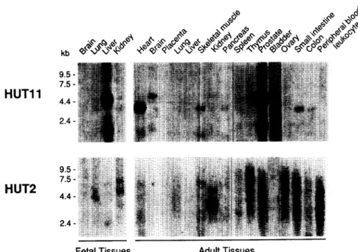
Fig. 3. Northern analysis of HUT11 and HUT2 expression in human tissues. Poly(A +) RNA (2 lag) from indicated human tissues (adult and fetal) were hybridized with 32p-labeled HUT1 1 and HUT2 eDNA probes as described in Section 2. Size markers (kb) are indicated on the left. somes by in situ hybridization with the NcoI eDNA fragment.
This probe mapped to the q l 2. l-q21.1 region of chromosome 18 (Fig. 2), a position identical to the H U T l l / K i d d blood group locus previously identified [10,23]. These results further suggest that both genes evolved from duplication, after that the internal repeats (found within H U T l l and UT2 se- quences), most likely resulting from another duplication, has occurred [24].
3.3. Tissue distribution o f H U T l l and H U T 2 m R N A s
Northern analysis revealed that HUT11 was predominantly expressed in human fetal liver, adult prostate and bladder as transcripts of 4.5 and 2.0 kb (Fig. 3). A similar pattern was previously detected in spleen erythroblasts [8]. HUT11 tran- scripts were also detected in other organs with distinct pat- terns. A weak signal at 4.5 kb was detected in thymus and brain. In heart, skeletal muscle, colon and small intestine, a predominant signal was detected at 3.6 kb with a weak band at 2.0 kb. A similar pattern was also observed in pancreas. In the adult kidney, there was only a very faint signal, consistent with the immunochemical localization of this transporter in endothelial cells of the vasa recta, but not in renal tubules [12]. This is also consistent with the detection of the tran- scripts in hydronephrotic rat kidneys [25].
When total RNAs from adult human kidney were analysed with the HUT2 eDNA, a faint signal was found at 4.5 kb and a strong signal at 3.6 kb. Preliminary studies by in situ hy- bridization on kidney sections revealed that HUT2 was selec- tively expressed by inner medulla collecting ducts, a nephron segment known to be under the regulatory control of vaso- pressin (E. Rondeau, P. Bailly and J.P. Cartron, unpublished data). In the fetal kidney only very weak hybridization signals (possibly cross-hybridization) were detected on Northern blots (Fig. 3), suggesting up-regulation during development. No clear signal could be detected in the other tissues examined (Fig. 3). Interestingly, the HUT2 transcript was not (or only very weakly) detected in the human colon (Fig. 3), at the
opposite of the UT2 transcript which was present in the rabbit colon [6]. Recently, it was reported that the rat UT2 transcript was undetectable in the colon and it was suggested that this may represent an adaptation towards the dietary protein con- sumed between herbivore (rabbit) and omnivore (rat) [7].
Taken together, our results indicate that HUT11 is present in several tissues whereas HUT2 is restricted to the kidney. HUT11 might be constitutively expressed and involved in the transport of urea in many organs whereas the HUT2 trans- porter might be vasopressin-inducible and restricted to the kidney to participate in the urinary concentration mechanism.
Direct a n a l y s i s of translation I m m u n o p r e c i p i t a t i o n p r o d u c t s with anti-Jk 3 1 2 3 4 5 6 7 8 40...-~ 36--D,.- M i c r o s o m e s - "t- - "4" - I I I I I I I I
HUT11 HUT2 control HUT11 HUT2 control
Fig. 4. Expression of urea transporters in transcription-translation assays. Left, autoradiogram of L-[35S]methionine-labeled proteins analyzed by SDS-PAGE (15% gels). Lanes 1 and 2: pT7TS-HUTll with and without canine pancreatic microsomal membranes; lane 3 and 4:pT7TS-HUT2 with and without microsomes; lane 5: control luciferase plasmid. Right: autoradiogram of ass-labeled proteins im- munoprecipitated with a human anti-Jk 3 and separated by SDS- PAGE. Lanes 6 and 7: immunoprecipitates from pT7TS-HUTll and pT7TS-HUT2, respectively; lane 8: luciferase control. Arrows on the left refer to product size (kDa).
R Olivbs et al./FEBS Letters 386 (1996) 15~160 Table 1
Urea permeabilities of water, HUT11 and HUT2 injected oocytes
159
Tissue localization Expression Purea (10 -6 cm s -1)
untreated + 0.5 mM pCMBS + 0.5 mM phloretin
Water 1.03+0.18 (n= 10) 0.74+0.12 (n=8) 1.09+0.16 (n=8)
HUT11 Red cell/kidney Constitutive 14.70 _+ 1.09*(n = 13) 4.02 + 0.67 (n = 4) 1.85 _+ 0.22 (n = 8) HUT2 Kidney ADH regulated? 45.50 + 2.38'(n = 12) 52.14 + 5.63 (n -- 4) 17.06 + 3.39 (n = 4) Oocytes injected with HUT2 cRNAs were incubated individually in 200 tal of Barth's medium containing 8 [.tCi (0.145 mM) [14C]urea (Amersham, UK) and 5/.tCi/ml [3H]raffinose (Dupont NEN, Germany) as a control of the oocyte integrity. Oocytes injected with HUT11 and water were used as positive and negative controls. Incubation time was 90 s at 18°C under gentle shaking. After washing and solubilization, the samples were counted by liquid scintillation in a Tri-Carb 4000 scintillation counter (Packard Inst., Rungis, France). For inhibition experiments, 0.5 mM phloretin (Sigma) and 0.5 mM para-chloromercuribenzene sulfonate (pCMBS) (Sigma) were added, 20 min and 10 min, respectively, before the assay and maintained during the urea uptake. Urea permeability was calculated from the oocyte-associated amount of [14C]urea and the oocyte surface area was calculated from the optically determined apparent diameter. Data are presented as mean + S.E. (n), where n indicates the number of oocytes for each experimental condition.
*P<0.001 vs. water-injected oocytes; P<0.001 vs. untreated oocytes.
3.4. Coupled transcription-translation o f H U T l l and HUT2 cDNAs
In the transcription translation coupled reticulocyte lysate system, b o t h plasmids p T 7 T S - H U T l l and - H U T 2 directed the synthesis o f 36 and 40 k D a protein bands when carried out with and without canine pancreatic microsomal mem- branes, respectively (Fig. 4, lanes 1-4). The translated product from H U T 1 1 , but not f r o m H U T 2 , could be immunoprecipi- tated with the anti-Jk 3 antibody, produced by immunized J k ( a - b - ) individuals (Fig. 4, lanes 6-8). Thus, the H U T 2 polypeptide does n o t carry the Jk a epitope o f K i d d (Jk) blood groups.
3.5. Transport studies
U r e a uptake o f H U T 1 1 and H U T 2 c R N A - i n j e c t e d Xenopus oocytes was 15-50 times faster than in water-injected oocytes depending u p o n individual experiments, as measured f r o m the initial rate (90 s). U r e a permeabilities (P~rea) o f H U T 1 1 - and H U T 2 - i n j e c t e d oocytes were respectively 1 4 . 7 + 1 . 0 9 x 1 0 6 cm/s (n=13) and 4 5 . 5 + 2 . 3 x 1 0 -6 cm/s (n=12) versus 1.03_+0.18x10 -6 cm/s (n=10) for water-injected oocytes (P<0.001) (Table 1). These values suggest that b o t h H U T 2 functions as an efficient urea transporter. It is u n k n o w n whether the higher values o f permeability for H U T 2 c R N A - injected oocytes could be due to a higher n u m b e r o f carrier copies inserted in the m e m b r a n e rather than to a different activities o f each transporter.
The urea flux mediated by H U T 2 was inhibited by 0.5 m M phloretin (65% inhibition), as expected for a urea transporter protein (Table 1). Because p C M B S inhibits water and urea transport by reacting with sulfhydryl groups of the transport- ers [26,27], the effect o f this reagent on H U T 2 - m e d i a t e d urea flux was also examined. Surprisingly, we found no inhibition o f urea uptake by 0.5 m M p C M B S (Table 1), although this reagent inhibited the erythroid H U T l l - m e d i a t e d urea flux (74% inhibition). These results are most likely related to the primary structure o f H U T 11 and H U T 2 proteins, particularly regarding the cysteine residues. H U T l l and H U T 2 have 10 cysteine residues, a m o n g which 7 are conserved and aligned at the same positions. C-25, -30 and -151 are present in H U T l l but n o t in H U T 2 , whereas C-45, -97 and -288 are present in H U T 2 but not in H U T l l (Fig. 1). Based on the predicted m e m b r a n e topology [6,8], C-25 and C-30 are m o s t likely in- tracellular, whereas C-151 is located in the 4th t r a n s m e m b r a n e d o m a i n and therefore m a y represent a likely candidate for p C M B S sensitivity.
In conclusion, we have shown that at least two distinct urea transporters present in h u m a n tissues can be distinguished by their functional properties and distribution. The correspond- ing genes colocalized on c h r o m o s o m e 18q12-q21 and might define a new gene family that evolved by duplication of a c o m m o n ancestor.
References
[1] Marsh, D.J. and Knepper, M.A. (1992) Renal handling of urea. In: Renal Physiology (Windhager, E.E., Ed.) pp. 1317-1347. Oxford University Press, Oxford.
[2] Chou, C.-L., Sands, J.M., Nonoguchi, H. and Knepper, M.A. (1990) Am. J. Physiol. 258, 486494.
[3] Galluci, E., Micelli, S. and Lippe, C. (1975) Nature 255, 722-723. [4] Macey, R.I. (1984) Am. J. Physiol. 246, C195 C203.
[5] Knepper, M.A. and Star, R.A. (1990) J. Physiol. 259, F393- F401.
[6] You, G., Smith, C.P., Kanai, Y., Lee, W.S., Stelzner, M. and Hediger, M.A. (1993) Nature 365, 844~847.
[7] Smith, C.P., Lee, W.-S., Martial, S., Knepper, M.A., You, G., Sands, J.M. and Hediger, M.H. (1995) J. Clin. Invest. 96, 1556- 1563.
[8] Olives, B., Neau, P., Bailly, P., Hediger, M.A., Rousselet, G., Cartron, J.P. and Ripoche, P. (1994) J. Biol. Chem. 269, 31649-31652.
[9] Smith, C., Shavakul, C., You, G., Martial, S., Lee, W-S., Mack- ensie, H.S., Sands, J.M., Knepper, M.A. and Hediger, M.A. (1995) American Society of Nephrology, 28th Annual Meeting, November 5 8, San Diego, CA (abstract, p. 329).
[10] Oliv6s, B., Mattei, M.-G., Huet, M., Neau, P., Martial, S., Car- tron, J.-P. and Bailly, P. (1995) J. Biol. Chem. 270, 15607 15610. [11] Fr6hlich, O., Macey, R.I., Edwards-Moulds, J., Gargus, J.J. and
Gunn, R.B. (1991) Am. J. Physiol. 260, C778-C783.
[12] Xu, Y., Olives, B., Bailly, P., Ripoche, P., Ronco, P., Cartron, P.-P. and Rondeau, E. (1995) American Society of Nephrology, 28th Annual Meeting, November 5 8, San Diego, CA (abstract, p. 331).
[13] Pallone, T.L., Work, J., Mayers, R.L. and Jamison, R.L. (1994) J. Clin. Invest. 93, 212-222.
[14] Pallone, T.L. (1994) Am. J. Physiol. 267, R260-R267.
[15] Gillin, A.G. and Sands, J.M. (1993) Semin. Nephrol. 13, 146- 154.
[16] Mattei, M.G., Philip, N., Passage, E., Moison, J.P., Mandel, J.P. and Mattei, J.F. (1985) Hum. Genet. 69, 268-271.
[17] Laemmli, U.K. (1970) Nature 227, 680-685.
[18] Martial, S., Ripoche, P. and Ibarra, C. (1991) Biochim. Biophys. Acta 1090, 86-90.
[19] Thompson, J.D., Higgins, D.G. and Gibson, T.J. (1994) Nucleic Acids Res. 22, 46734680.
[20] Sharp, P.M. and Matassi, G. (1994) Curr. Opin. Genet. Dev. 4, 851-860.
160
[22] Ellsworth, D.L., Hewett-Emmett, D. and Li, W.-H. (1994) Mol. Biol. Evol. 11, 875-885.
[23] Geitvik, G.A., Hoyheim, B., Gedde-Dahl, T., Grzeschilk, K.H., Lothe, R. Tomter, H. and Olaisen, B. (1987) Hum. Genet. 77, 205-209.
[24] Rousselet, G., Bailly, P., and Ripoche, P. (1996) Tandem repeats in urea transporters: identification of an urea transporter signa- ture sequence. Am. J. Physiol. 270, F554-F555.
B. Olivks et al./FEBS Letters 386 (1996) 156-160
[25] Promeneur, D., Rousselet, G., Bankir, L., Bailly, P., Cartron, J.P., Ripoche, P., Trinh-Trang and Tran, M.M. (1996) J. Am. Soc. Nephrol. (in press).
[26] Macey, R.I. and Farmer, R.E.L. (1970) Biochim. Biophys. Acta 211, 104-106.