V.A.C.
®
Abdominal Dressing System
A Temporary Closure for Open Abdomen
Ludwig Labler
1, Jörn Zwingmann
1, Dieter Mayer
2, Reto Stocker
1, Otmar Trentz
1, Marius Keel
1488
European Journal of Trauma 2005 · No. 5 © Urban & Vogel
Ab stract
Background and Purpose: The study reports experience
with the recently commercially available V.A.C.® Ab-dominal Dressing System, a system designed for a tem-porary closure of an open abdomen situation under negative pressure. The method allows a late primary fascial closure after laparotomy in case of damage con-trol, abdominal compartment syndrome or severe intra- abdominal spesis and facilitates delayed reconstruction of a large ventral hernia.
Patients and Methods: 18 patients with an open
abdo-men after laparotomy were managed between Febru-ary 2002 and September 2004.
Results: Twelve patients after primary, one patient after
secondary fascial closure and one patient with partially primary closure and resorbable mesh for abdominal wall reconstruction were free of wound infection or dehis-cence of the abdominal wall. Evisceration or enteric fis-tulas were not observed. Five patients died in conse-quence of severe injury, a multiple organ failure or septic complications.
Conclusion: V.A.C.® Abdominal Dressing System is an
effective temporary closure technique for open abdo-men in critically ill patients which makes a late primary fascial closure up to 2 months after initial laparotomy possible either in trauma patients or in case of severe intraabdominal infection. The technique is simple and easily mastered.
Key Words
Vacuum-assisted closure · V.A.C. · Open abdomen · Abdominal compartment syndrome · Damage control · Fascial closure
Eur J Trau ma 2005;31:488–94
DOI 10.1007/s00068-005-2031-y
Introduction
The management of abdominal compartment syndrome (ACS), with its effect on pulmonary, cardiac and renal functions, or the concept of damage-control laparotomy are techniques that proved to be an important advance of life-saving in trauma care [1–3]. A massive early re-suscitation of trauma patients is often accompanied by marked visceral edema, retroperitoneal hematoma or a packing of the peritoneal cavity and results in an open abdomen situation. The same applies to reexploration of visceral viability after damage-control procedures or to intraabdominal contamination [4–6]. Also ischemia and necrosis of the abdominal fascia, caused either by the tension after a forced abdominal wall closure or by an intraabdominal infection, may lead to a dehiscence of the abdominal wall up to an abdominal rupture and to an open abdomen situation [7]. A primary fascial clo-sure in all the above cases is not feasible [8] and a tem-porary abdominal closure (TAC) is indicated. Among diverse TAC techniques [5, 8, 9], vacuum-assisted clo-sure (VAC), the efficient dressing technique for the management of problematic wounds [10–16], was suc-cessfully used also for treatment of open abdomen situ-ations in the course of recent few years [17–23]. The V.A.C.® Abdominal Dressing System (KCI Vacuum
Assisted Closure, San Antonio, TX, USA), a modifica-tion of the VAC technique designed specifically as a temporary closure of open abdomen, appeared on the market recently. This study reports our experience with the device named.
1 Division of Trauma Surgery, Department of Surgery, University
Hospital Zurich, Switzerland,
2 Division of Cardiovascular Surgery,
University Hospital Zurich, Switzerland.
Received: March 18, 2005; accepted: June 12, 2005.
EJT05_0031_Labler.indd 488488
489
European Journal of Trauma 2005 · No. 5 © Urban & Vogel
Patients and Methods Patients
18 patients with open abdomen after laparotomy were treated with the commercial V.A.C.® Abdominal
Dress-ing System between February 2002 and September 2004. 15 patients were severely injured and three patients had other indications. The trauma patients were managed according to Advanced Trauma Life Support guidelines [24]. All hemodynamically stable patients with severe intraabdominal trauma or infection were evaluated by CT scan before laparotomy. The hemodynamically un-stable patients were submitted to a damage control and a packing in the operating room (OR) and transferred afterwards to the intensive care unit (ICU). The clinical records (Table 1) include patients’ data, Abbreviated Injury Scale (AIS), Injury Severity Score (ISS) [25], the ICU and hospital stay.
Technique
The commercially available V.A.C.® Abdominal
Dress-ing System (KCI Vacuum Assisted Closure) consists of a thin black polyurethane foam encapsulated in the center of a perforated polyethylene sheet, a separate black poly-urethane foam, a suction drain and of adhesive drapes. The sheet is cut to an appropriate dimension, placed over the viscera and tucked under the wound edges (Figure 1). A second black polyurethane foam, cut to fit the wound, is placed over the embedded plastic sheet with the thin foam. If necessary, the foam is fixed to the skin edges with staples (Figure 2). The surrounding skin is cleaned with benzine, and adhesive dressing drapes are trimmed as a patchwork to seal the wound. A hole of 2 cm diameter is cut out in the drape, the TRAC-PAD® (KCI Vacuum
Assisted Closure) is positioned on this occlusive seal (Figure 3) and connected with a vacuum pump (KCI
Table 1. Clinical characteristics. AIS: Abbreviated Injury Scale; F: female; ICU: intensive care unit; ISS: Injury Severity Score [25]; M: male. Patient Gender Age Cause for laparotomy AIS ISS Hospital ICU # (years) Traffic Work Gunshot Varia Head Face Thorax Abdomen Pelvis/ Integument stay stay accident accident wound extremities (days) (days)
1 M 24 + 3 4 4 3 2 41 71 25 2 M 69 + 41 26 3 M 58 + 4 4 4 2 48 46 46 4 F 24 + 4 3 5 2 50 103 69 5 M 13 + 5 4 2 45 89 57 6 M 47 + 14 14 7 M 24 + 5 1 26 27 27 8 M 62 + 2 2 4 3 1 29 60 60 9 M 33 + 5 1 26 93 37 10 M 48 + 99 99 11 M 58 + 3 4 25 30 7 12 M 17 + 3 4 3 3 34 27 26 13 M 21 + 2 5 5 52 60 35 14 F 21 + 3 5 2 38 50 13 15 M 43 + 3 4 1 26 67 2 16 M 15 + 6 4 3 61 2 2 17 M 37 + 4 2 5 4 2 3 57 23 23 18 M 18 + 3 5 5 59 7 7
Figures 1a to 1c. a) Open abdomen. b) Perforated polyethylene sheet with a thin polyurethane sponge encapsulated in the center. c) The sheet is
tucked under the wound edges to prevent adhesion of the viscera to the peritoneum.
a b c
EJT05_0031_Labler.indd 488489
490
European Journal of Trauma 2005 · No. 5 © Urban & Vogel Vacuum Assisted Closure) via a container. The pump isadjusted to a constant negative pressure of 75 mmHg and set in action. The layers collapse and exert a uniform pressure upon both the wound and the abdominal fascia, and the suction draws the wound edges slowly together (Figure 4). At the same time, the wound fluid is continu-ously removed through the drain and transferred into the container. Technical problems with the VAC system are rare. Occasional leak is repaired by an additional piece of the adhesive drape at the bedside. The dressing is re-placed by a new one generally in the OR, or if necessary, the procedure may also be carried out at the bedside in the ICU. As the V.A.C.® Abdominal Dressing System is
stable enough as a TAC, the patients may be extubated and mobilized in an armchair for a better pulmonary re-habilitation on the ICU. As soon as the edema resolves, no necrosis is present on the fascia layers and granulation tissue formation of subcutaneous tissue takes place, the fascia may be stepwise closed by suturing [12, 13, 26]. When the patients are already in a stable condition but the primary fascia closure still is not feasible, a conven-tional VAC dressing (KCI Vacuum Assisted Closure) is applied until a healthy granulation tissue covers the vis-cera and the wound edges. When the wound bed is free of infection, the granulated open wound is skin-grafted or an absorbable mesh is implanted.
Results
Indication for laparotomy and the management of open abdomen are summarized in Table 2. Laparotomy was performed for abdominal damage control in eight pa-tients (# 5, 9, 11–14, 16, 18). A gunshot injury (patient # 14) was managed by nephrectomy and segmental co-lon resection. The laparotomy wound developed an in-fection resulting in a disrupture of the abdominal wall caused supposedly by retroperitoneal abscess formation on the right side. In two patients (# 16, 18), the damage control was combined with a tamponade of retroperito-neal hematoma of pelvic ring injuries. Three patients developed ACS in the course of the therapy. Two (# 6, 10) were under treatment for necrotizing fasciitis with severe septic course and one patient (# 7) had severe head injury. Complications in open abdomen situation emerged after early total care in three patients. In one patient (# 3) the wound disrupted with intraabdominal peritonitis or developed an intraabdominal abscess (# 15) after splenectomy. In both cases the abdomen was left open for second-look interventions. Relaparot-omy had to be carried out also in patient # 4 2 weeks after early total care with splenectomy because of ini-tially overlooked small pancreas rupture with severe peritonitis. Further therapy required open abdomen treatment. One patient (# 2) was treated for pyoderma
gangraenosum of lower extremities. After 11 days of therapy, laparotomy was indicated because of spontane-ous ascending and descending colon perforation with severe peritonitis. Right hemicolectomy and Hartmann procedure were carried out, and the abdomen was left open for sec-ond-look operations. Because of clinical signs of acute abdomen on the 3rd day after trauma (# 1), lapa-rotomy was performed with
intraop-Figure 3. The
surround-ing skin is cleaned with benzine, and adhesive dressing drapes are placed as a patchwork. A hole is cut out, and the TRAC-PAD® is posi-tioned on the occlusive seal.s
Figure 2. A second
poly-urethane sponge shaped to the size of the wound is placed over the em-bedded plastic sheet and fixed to the skin edges with staples.
Figures 4a and 4b. a) V.A.C.® Abdominal Dressing System before suction is started. b) Suction is
started and the foam collapses.
a b
EJT05_0031_Labler.indd 488490
491
European Journal of Trauma 2005 · No. 5 © Urban & Vogel
erative diagnosis of posttraumatic pancreatitis. Because of intraabdominal finding and distended bowels, the ab-domen was left open for further surgical revisions and to prevent ACS development. One patient (# 8) developed ischemic small bowel perforation in the course of his trauma therapy. 10 days after resection and anastomo-sis, a leakage appeared with severe peritonitis. Relapa-rotomy with repeated lavage resulted in an open abdo-men situation. Shocking gastric bleeding took place in patient # 17 after 17 days of trauma therapy. Gastroto-my and surgical control of bleeding were carried out. 4 days later the patient sustained abdominal wall disrup-tion which, regarding his severe thoracic trauma, was treated by open abdomen therapy to prevent ACS. Dur-ing open abdomen procedure two patients (# 1, 9) de-veloped intraabdominal abscesses between small bowel loops, and in two patients (# 13, 14) a retroperitoneal abscess formation was observed during therapy which was surgically treated by irrigation and drainage. Four patients died during the treatment of open abdomen and before fascial closure. Two patients died because of severe head injury (# 7, 16), associated in patient # 16 with initially survived atlantooccipital dissociation (AOD). Patients # 17 and 18 died owing to a sepsis and multiorgan failure after severe trauma. One patient (# 8) died after performed fascial closure of the open abdomen due to multiorgan failure.
The VAC dressing was changed eight times on aver-age (range, one to 30 changes). The dressing remained in place according to the circumstances for an average of 2.5 days (range, 1–5 days), except patient # 2 because of severe pulmonary complications. In this case the dressing was left in place up to 7 days.
A primary fascial closure was achieved in twelve pa-tients. In the case of a penetrating trauma (patient # 9), the primary fascial closure failed because of persistent edema of the viscera. The open abdomen was then mesh-grafted and a secondary fascial closure was per-formed 22 days later when the patient was in stable con-dition (Figure 5). In one patient (# 15), a primary fascial closure was achieved in the medial laparotomy only, whereas the transverse accessory laparotomy had to be closed by an absorbable mesh.
All 13 patients who survived were available to fol-low-up examination in the time interval of 5–33 months. None developed a wound infection or a dehiscence of the abdominal wall.
Discussion
Open abdomen management of seriously injured or ill patients has been a challenge for the surgeon. Laparoto-my after damage control with a tamponade, occurrence of ACS, abdominal wall defects and severe intraabdomi-nal infections requiring repetitive exploration of the
ab-Table 2. Laparotomy and vacuum-assisted closure (VAC) dressing.
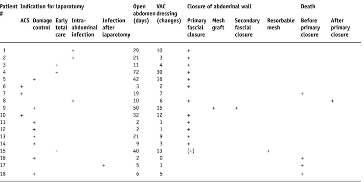
Patient Indication for laparotomy Open VAC Closure of abdominal wall Death
# abdomen dressing
ACS Damage Early Intra- Infection (days) (changes) Primary Mesh Secondary Resorbable Before After control total abdominal after fascial graft fascial mesh primary primary care infection laparotomy closure closure closure closure
1 + 29 10 + 2 + 21 3 + 3 + 11 4 + 4 + 72 30 + 5 + 42 16 + 6 + 3 2 + 7 + 19 7 + 8 + 10 6 + + 9 + 50 15 + + 10 + 32 12 + 11 + 2 1 + 12 + 2 1 + 13 + 21 9 + 14 + 9 3 + 15 + 40 13 (+) + 16 + 2 0 + 17 + 5 1 + 18 + 6 5 + EJT05_0031_Labler.indd 488491 EJT05_0031_Labler.indd 488491 30.09.2005 10:41:3430.09.2005 10:41:34
492
European Journal of Trauma 2005 · No. 5 © Urban & Vogel dominal cavity are situations where a closure of theab-dominal wall is impracticable and TAC is indicated. Also the systemic inflammatory response syndrome (SIRS) with its hypermetabolic state which leads to a capillary leakage and a consecutive swelling of the soft tissue en-counters the same problem. TAC prevents a contamina-tion of the peritoneal cavity, a bowel desiccacontamina-tion, an evis-ceration and a mechanical injury of the viscera. Further, it should be easily applied and managed. Several TAC techniques for management of open abdomen [5, 8, 9] and associated complications and problems [27–30] were reported. The negative pressure therapy with its wide range of indications was introduced into the clinical practice during the last decade. As TAC of open abdo-men, VAC for the first time was applied quite early [17]. Nevertheless, 5 years elapsed before further reports ap-peared in the literature [18–23]. The negative pressure as the open abdomen management reported in the litera-ture includes three systems: the vacuum-pack technique (A), VAC technique (B), and the commercial V.A.C.®
Abdominal Dressing System (C), which differ in techni-cal details. Unlike the customary VAC technique used for different sorts of wounds, all three open abdomen systems mentioned apply a perforated polyethylene sheet placed between the abdominal viscera and the an-terior peritoneum. The sheet prevents adherence of the viscera to the peritoneum, allows the abdominal wall to
glide over the viscera and facilitates an easy TAC removal at repeated ab-dominal entries. The sheet perfora-tion allows the wound fluid to be drained out of the abdominal cavity. The sheet delivered with the com-mercial device (C) is equipped with a thin foam encapsulated in its center helping minimize dressing shift with-in the abdomen. The polyethylene sheet is covered either with a moist sterile surgical towel (A) [17, 18, 20] or with a black polyurethane foam (B, C) [19, 21–23]. This layer fills up the open laparotomy wound, distrib-utes a uniform negative pressure over the abdomen [19], and filters the re-moved abdominal fluid thus prevent-ing a blockage of the drain. The skin is sutured (B) [19, 21–23] tightly [19, 21] or loosely [23] over the foam to prevent retraction of the fascial edge or is let free in vacuum-pack (A). In our study, the foam was let free or, if necessary, fixed to the skin edges with staples. In our opinion, suturing over the foam might cause necrosis of the wound edges and the underlying bowels.
Two suction drains are installed over the towel dressing (A) and connected via a Y-adapter with the suction source [17, 18]. Alternatively, one suction drain only is introduced into the foam (B) [19, 21, 22], and then the dressings (A, B) are covered with an adhesive drape placed over the entire wound to get an airtight seal. Continuous suction of 100–150 mmHg [17, 18, 23] or 175 mmHg [21, 22] is started. The system (C) seals the dressing with an adhesive drape first, then a hole is cut into the drape and, finally, a TRAC-PAD® is installed
and connected with a container and a vacuum pump. Unlike the published data, a negative pressure of 75 mmHg only was applied in our study. The lower pres-sure suffices entirely for an adequate fluid removal from the open abdomen and thus sufficiently prevents retrac-tion of the wound edges. The urinary output in patients was lower than expected. The cause supposedly was the large wound fluid volume, up to 3 l (1,640 ± 1,334 ml) in some cases, removed by the VAC system during the first dressing in accordance with the data (2–5 l) report-ed [23]. The removreport-ed volume then decreasreport-ed to 825 ± 119 ml before the open abdomen could be closed.
Figures 5a to 5d. a) Open abdomen after penetrating gunshot injury. b) Management of open
abdomen after penetrating gunshot injury with V.A.C.® Abdominal Dressing System. c) The primary fascial closure failed because of persistent edema of the viscera and mesh grafting of the granulation tissue over the bowels was performed. d) Reconstruction of the abdominal wall was without complications.
a b
c d
EJT05_0031_Labler.indd 488492
493
European Journal of Trauma 2005 · No. 5 © Urban & Vogel
In some of our patients (# 4, 5, 9, 15) the abdomen was left open for a rather long time of 51 days on aver-age (range, 40–72 days). In this group of patients the mean AIS for abdomen was 4.7 (range, 4–5) and the mean ISS was 37 (range, 26–50). Two of these patients suffered from severe intraabdominal septic complica-tions after early total care procedure (# 4, 15). Patient # 5 was treated for traumatic hemipelvectomy, and pa-tient # 9 had a gunshot injury to the liver with persistent edema of the viscera. On the other hand, this demon-strates that even such extremely long open abdomen situations may be practicable and, regarding the fact that all four patients survived, successful as well.
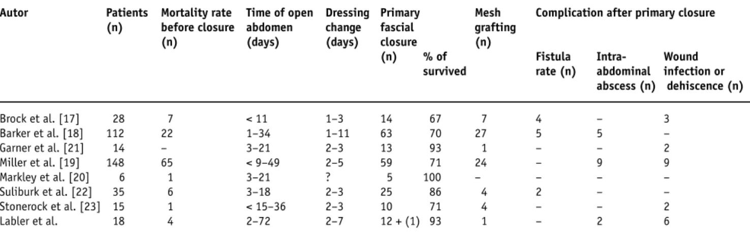
The results achieved by VAC therapy of open abdo-men published in the literature and our own results are summarized in Table 3. Some of the authors (Table 3) failed to close the fascia primarily and had to apply mesh grafting over the granulation tissue in order to close the wound as also was the case in our patient # 9 with a pen-etrating trauma. Some of the patients had to be operat-ed later for a second time in order to reconstruct the abdominal wall [18, 22]. Our five patients died in conse-quence of severity of their injuries, a multiple organ fail-ure and/or septic complications, which correlates to other studies in Table 3.
Complication reported in the literature and connect-ed with the use of VAC for open abdomen are ACS de-veloped after installation of the vacuum pack technique [18] and fistula formations [17, 18, 22]. It was, however, not quite clear whether the fistulas were directly related to the dressing technique or associated with the primary injury or the surgical treatment. The bleeding, a well-known complication of VAC treatment of open wounds, has not yet been observed. Complications
relat-ed to the abdominal wall reconstruction were anastomot-ic disruption and wound infection [17, 19, 21, 23]. Poor quality of the fascia after the use of VAC therapy leading to ventral hernia was also described [23]. In our study, in accordance with similar observations [18], we did not ob-serve any deep necrosis of the fascia after debridement and could perform the fascial closure without hernia for-mation or a dehiscence in 14 of our 18 patients. Fistula formation or bleeding was not observed. A formation of two intraabdominal abscesses between adherent bowels was a complication which may occur in a septic open ab-domen. Its occurrence was observed not only in VAC but also after application of other TAC techniques and prob-ably is not related to a particular dressing technique.
Conclusion
The VAC system, as well as its modification, the V.A.C.®
Abdominal Dressing System, is a useful alternative tech-nique for the management of open abdomen. The advan-tage of modifications B and C is the more uniform topic negative pressure to the wound edges and the fascia of the open abdomen. The encapsulated foam in the center of the polyethylene sheet of the commercial available V.A.C.® Abdominal Dressing System prevents the
in-traabdominal shift of the sheet. This modification keeps the device in position and secures the functionality of the system. In most cases, it allows a primary closure of the fascia thus preventing a formation of large ventral hernias and diminishes additional risks during subsequent recon-structive surgery of the abdominal wall. It seems that the traction force of the negative pressure on the wall edges of the open abdomen does not increase intraabdominal pres-sure. The technique is easy to handle, is performed in short time and can be mastered without difficulties.
Table 3. Vacuum-assisted closure (VAC) therapy of open abdomen.
Autor Patients Mortality rate Time of open Dressing Primary Mesh Complication after primary closure (n) before closure abdomen change fascial grafting
(n) (days) (days) closure (n)
(n) % of Fistula Intra- Wound survived rate (n) abdominal infection or
abscess (n) dehiscence (n) Brock et al. [17] 28 7 < 11 1–3 14 67 7 4 – 3 Barker et al. [18] 112 22 1–34 1–11 63 70 27 5 5 – Garner et al. [21] 14 – 3–21 2–3 13 93 1 – – 2 Miller et al. [19] 148 65 < 9–49 2–5 59 71 24 – 9 9 Markley et al. [20] 6 1 3–21 ? 5 100 – – – – Suliburk et al. [22] 35 6 3–18 2–3 25 86 4 2 – – Stonerock et al. [23] 15 1 < 15–36 2–3 10 71 4 – – 2 Labler et al. 18 4 2–72 2–7 12 + (1) 93 1 – 2 6 EJT05_0031_Labler.indd 488493 EJT05_0031_Labler.indd 488493 30.09.2005 10:41:3530.09.2005 10:41:35
494
European Journal of Trauma 2005 · No. 5 © Urban & VogelReferences
1. Wittmann DH, Iskander GA. The compartment syndrome of the abdominal cavity: a state of the art review. J Intensive Care Med 2000;15:201–20.
2. Shapiro MB, Jenkins DH, Schwab CW, et al. Damage control: col-lective review. J Trauma 2000;49:969–78.
3. Ertel W, Oberholzer A, Platz A, et al. Incidence and clinical pattern of the abdominal compartment syndrome after “damage-con-trol” laparotomy in 311 patients with severe abdominal and/or pelvic trauma. Crit Care Med 2000;28:1747–53.
4. Morris JA Jr, Eddy VA, Blinman TA, et al. The staged celiotomy for trauma. Issues in unpacking and reconstruction. Ann Surg 1993;217:576–84.
5. Aprahamian C, Wittmann DH, Bergstein JM, et al. Temporary ab-dominal closure (TAC) for planned relaparotomy (etappenlavage) in trauma. J Trauma 1990;30:719–23.
6. Garcia-Sabrido JL, Tallado JM, Christou NV, et al. Treatment of se-vere intra-abdominal sepsis and/or necrotic foci by an “open-ab-domen” approach. Zipper and zipper-mesh techniques. Arch Surg 1988;123:152–6.
7. Graham DJ, Stevenson JT, McHenry CR. The association of in-tra-abdominal infection and abdominal wound dehiscence. Am Surg 1998;64:660–5.
8. Howdieshell TR, Yeh KA, Hawkins ML, et al. Temporary abdominal wall closure in trauma patients: indications, technique, and re-sults. World J Surg 1995;19:154–8, discussion 158.
9. Schein M, Saadia R, Jamieson JR, et al. The “sandwich technique” in the management of the open abdomen. Br J Surg 1986;73: 369–70.
10. Fleischmann W, Strecker W, Bombelli M, et al. Vacuum sealing as treatment of soft tissue damage in open fractures. Unfallchirurg 1993;96:488–92.
11. Morykwas MJ, Argenta LC, Shelton-Brown EI, et al. Vacuum-assist-ed closure: a new method for wound control and treatment: ani-mal studies and basic foundation. Ann Plast Surg 1997;38:553–62. 12. Argenta LC, Morykwas MJ. Vacuum-assisted closure: a new
meth-od for wound control and treatment: clinical experience. Ann Plast Surg 1997;38:563–76.
13. Morykwas MJ, Argenta LC. Nonsurgical modalities to enhance healing and care of soft tissue wounds. J South Orthop Assoc 1997;6:279–88.
14. Fabian TS, Kaufman HJ, Lett ED, et al. The evaluation of subatmo-spheric presssure and hyperbaric oxygen in ischemic full-thick-ness wound healing. Am Surg 2000;66:1136–43.
15. Philbeck TE Jr, Whittington KT, Millsap MH, et al. The clinical and cost effectiveness of externally applied negative pressure wound therapy in the treatment of wounds in home healthcare Medi-care patients. Ostomy Wound Manage 1999;45:41–50.
16. Labler L, Oehy K. Vacuum sealing of problem wounds. Swiss Surg 2002;8:266–72.
17. Brock WB, Barker DE, Burns RP. Temporary closure of open abdom-inal wounds: the vacuum pack. Am Surg 1995;61:30–5.
18. Barker DE, Kaufman HJ, Smith LA, et al. Vacuum pack technique of temporary abdominal closure: a 7-year experience with 112 pa-tients. J Trauma 2000;48:201–6.
19. Miller PR, Thompson JT, Faler BJ, et al. Late fascial closure in lieu of ventral hernia: the next step in open abdomen management. J Trauma 2002;53:843–9.
20. Markley MA, Mantor PC, Letton RW, et al. Pediatric vacuum pack-ing wound closure for damage-control laparotomy. J Pediatr Surg 2002;37:512–4.
21. Garner GB, Ware DN, Cocanour CS, et al. Vacuum-assisted wound closure provides early fascial reapproximation in trauma patients with open abdomens. Am J Surg 2001;182:630–8.
22. Suliburk JW, Ware DN, Balogh Z, et al. Vacuum-assisted wound closure achieves early fascial closure of open abdomens after se-vere trauma. J Trauma 2003;55:1155–60.
23. Stonerock CE, Bynoe RP, Yost MJ, et al. Use of a vacuum-assisted device to facilitate abdominal closure. Am Surg 2003;69:1030–4. 24. Collicott PE, Hughes I. Training in advanced trauma support.
JAMA 1980;293:1156–9.
25. Baker SP, O’Neill B, Haddon W Jr, et al. The Injury Severity Score: a method for describing patients with multiple injuries and evalu-ating emergency care. J Trauma 1974;14:187–96.
26. Greer SE, Longaker MT, Margiotta M, et al. The use of subatmo-spheric pressure dressing for the coverage of radial forearm free flap donor-site exposed tendon complications. Ann Plast Surg 1999;43:551–4.
27. Sherck J, Seiver A, Shatney C, et al. Covering the “open abdomen”: a better technique. Am Surg 1998;64:854–7.
28. Nagy KK, Fildes JJ, Mahr C, et al. Experience with three prosthetic materials in temporary abdominal wall closure. Am Surg 1996; 62:331–5.
29. Wouters DB, Krom RAF, Slooff MJH, et al. The use of Marlex mesh in patients with generalized peritonitis and multiple organ sys-tem failure. Surg Gynecol Obstet 1983;156:609–14.
30. Fry DE, Osler T. Abdominal wall considerations and complications in reoperative surgery. Surg Clin North Am 1991;7:1–11.
Address for Correspondence
Ludwig Labler
Division of Trauma Surgery Department of Surgery University Hospital Zurich Rämistraße 100 8091 Zürich Switzerland Phone (+41/1) 255-1111, Fax -4406 e-mail: ludwig.labler@usz.ch EJT05_0031_Labler.indd 488494 EJT05_0031_Labler.indd 488494 30.09.2005 10:41:3530.09.2005 10:41:35
![Table 1. Clinical characteristics. AIS: Abbreviated Injury Scale; F: female; ICU: intensive care unit; ISS: Injury Severity Score [25]; M: male.](https://thumb-eu.123doks.com/thumbv2/123doknet/14875684.641979/2.971.115.849.201.534/table-clinical-characteristics-abbreviated-injury-intensive-injury-severity.webp)