HAL Id: hal-02303874
https://hal.sorbonne-universite.fr/hal-02303874
Submitted on 2 Oct 2019HAL is a multi-disciplinary open access archive for the deposit and dissemination of sci-entific research documents, whether they are pub-lished or not. The documents may come from teaching and research institutions in France or abroad, or from public or private research centers.
L’archive ouverte pluridisciplinaire HAL, est destinée au dépôt et à la diffusion de documents scientifiques de niveau recherche, publiés ou non, émanant des établissements d’enseignement et de recherche français ou étrangers, des laboratoires publics ou privés.
Serotonin neuron development: shaping molecular and
structural identities
Evan Deneris, Patricia Gaspar
To cite this version:
Evan Deneris, Patricia Gaspar. Serotonin neuron development: shaping molecular and structural identities. Wiley Interdisciplinary Reviews: Developmental Biology, Wiley, 2018, 7 (1), pp.e301. �10.1002/wdev.301�. �hal-02303874�
OVERVIEW
Serotonin neuron development:
shaping molecular and structural identities
Evan Deneris1 and Patricia Gaspar2,3,4
1
Case Western Reserve University School of Medicine Department of Neurosciences, Cleveland OH, 44106, USA
esd@case.edu
2
Institut du Fer à Moulin, Institut National de la Santé et de la Recherche Médicale (INSERM), UMR-S839, Paris, France, 3Université Pierre et Marie Curie, Paris, France,
4
Institut du Fer à Moulin, Paris, France patricia.gaspar@inserm.fr
The continuing fascination with serotonin (5-HT) as a nervous system chemical messenger began with its discovery in the brains of mammals in 1953. Among the many reasons for this decades-long interest is that the small numbers of neurons that make 5-HT influence the excitability of neural circuits in nearly every region of the brain and spinal cord. A further reason is that 5-HT dysfunction has been linked to a range of psychiatric and neurological disorders many of which have a neurodevelopmental component. This has led to intense interest in understanding 5-HT neuron development with the aim of determining whether early alterations in their generation lead to brain disease susceptibility. Here, we present an overview of the neuroanatomical
organization of vertebrate 5-HT neurons, their neurogenesis, and prodigious axonal architectures, which enables the expansive reach of 5-HT neuromodulation in the CNS. We review recent findings that have revealed the molecular basis for the tremendous diversity of 5-HT neuron subtypes, the impact of environmental factors on 5-HT neuron development, and how 5-HT axons are topographically organized through disparate signaling pathways. We summarize studies of the gene regulatory networks that control the differentiation, maturation and maintenance of 5-HT neurons. These studies show that the regulatory factors controlling acquisition of 5-HT-type transmitter identity continue to play critical roles in the functional maturation and the maintenance of 5-HT neurons. New insights are presented into how continuously expressed 5-HT regulatory factors control 5-HT neurons at different stages of life and how the regulatory networks themselves are maintained.
Introduction
In all species harboring a nervous system, from invertebrates to mammals, a small percentage of brainstem neurons become specialized to use serotonin (5-hydroxytryptamine, 5-HT) as a neurotransmitter (1-4). Unlike glutamate or Gaba, the main excitatory and inhibitory neurotransmitters of the brain, the action of 5-HT is that of a neuromodulator, adjusting neuronal excitability, to increase or decrease cell
excitability according to the 5-HT receptors engaged (5, 6). Moreover, 5-HT has long-term effects on cell function such as trophic effects on growth (7, 8). Although the pool of 5-HT that is made in the brain accounts for a small percentage of total body 5-HT synthesis, brain 5-HT has been implicated in a multitude of functions from basic homeostatic processes such as thermoregulation and breathing (9, 10), to more elaborate functions such as affect and mood control (11-16), memory (17) and reward (18). 5-HT also plays a key role in several complex social behaviors such as male dominance, aggressive behavior, and maternal care (19-27).
In mammals, all brain 5-HT neurons are generated in a part of the developing hindbrain called the rhombencephalon, which gives rise to the pons and medulla (Figure 1) but later extend also into the mesencephalon following developmental
reorganizations such as migration and folding of the neural tube (28, 29). 5-HT neurons are organized in cell clusters mainly, but not exclusively, along the midline (30-32). The raphe nuclei refer to the midline cell clusters. From there, 5-HT axons radiate broadly to innervate the entire central nervous system from the olfactory bulb to the spinal cord (30). Together with their highly divergent anatomical organization, hindbrain 5-HT neurons show anatomical and physiological specializations: specific neuronal subsets
deliver 5-HT to different brain regions, and have different molecular and physiological identities (4). This compartmentalized regional organization of hindbrain 5-HT neurons into distinctive neural circuits forms the basis of their diverse brain functions.
Understanding the development of 5-HT brainstem neurons implies the dissection of developmental mechanisms that operate in parallel but also with
interaction: a) mechanisms that specify 5-HT molecular identity and b) mechanisms that specify structural/regional identity and network wiring. These processes are controlled by intrinsic genetic programming and are also modulated by environmental factors. Thus, variants of developmental genes or environmental insults might impair the sequential developmental programs that instruct brain 5-HT neuron networks. These developmental variations could in turn underlie different susceptibility to neurological and psychiatric disorders. They may also underlie different responsiveness to
pharmaceuticals targeting the 5-HT system (33-35).
In this review, we present a general overview of the neuroanatomical organization and the molecular diversity of vertebrate 5-HT neurons from a developmental perspective. We describe recent findings revealing the regulatory networks implicated in the differentiation, maturation, and maintenance of 5-HT neurons. We highlight some of the mechanisms through which 5-HT regulatory
networks are thought to shape 5-HT neuron identity and how these networks are stably sustained into adulthood to preserve 5-HT function. Finally, we describe the signaling pathways that have been implicated in guiding 5-HT axons to their appropriate
innervation zones and the potential of environmental factors to alter 5-HT neurotransmission through effects on 5-HT neuron morphology and migration.
Nomenclature and anatomical organization of the brain 5-HT neuron nuclei
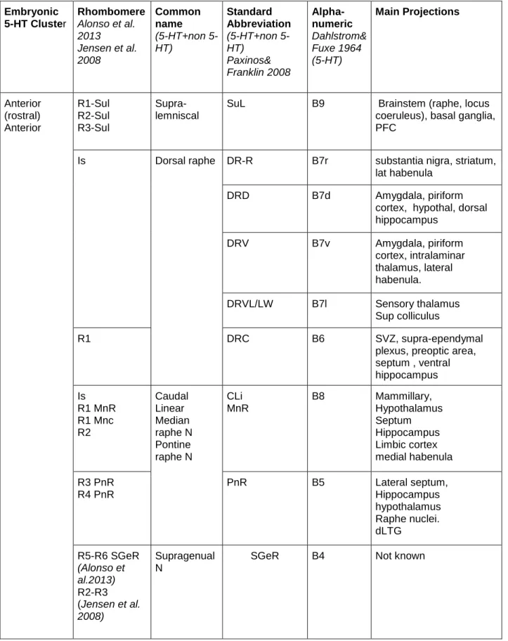
From the early discovery of indolamine containing neurons in the rodent and human brain (31, 32) different 5-HT neuron clusters were identified in the hindbrain. The precise neuroanatomical boundaries of these cell clusters and their denominations have varied according to studies (summarized in Table 1). Some nomenclatures refer only to the 5-HT containing neurons (the B1-B9 cell groups, named from caudal B1 to rostral B9), whereas other nomenclatures refer to cytoarchitectonic structures that contain 5-HT neurons, such as the dorsal raphe (DRN) and median raphe (MRN) nuclei.
Equivalences between these terms are valid to some extent but one must keep in mind that raphe nuclei contain a variable mix of 5-HT and non-5-HT neurons. For instance, the proportion of 5-HT neurons ranges from 50% in the DRN (36) to 21 % in the MRN (37) and less than 10 % in the paramedian raphe and the supra-lemniscal nuclei (37, 38).
Importantly, from a developmental point of view, 5-HT neurons can be separated into two main clusters, anterior (rostral) and posterior (caudal), with opposite polarity of their axonal projections. The anterior cluster neurons are the main source of 5-HT inputs to the forebrain and midbrain with additionally important projections in the
hindbrain, including the cerebellum and the raphe neurons themselves (26, 39, 40). The posterior cluster is an important source of innervation of major brainstem nuclei and is the exclusive source of 5-HT innervation to the spinal cord. This primary division is best seen during early embryonic stages, (E11-E14 in rodents) (Figure 1) when 5-HT
Embryonic and fate mapping studies indicated that these primary anterior and posterior clusters are further segmented along the rostrocaudal axis according to rhombomeric (r) divisions (44). Rhombomeres are longitudinal compartments of the rhombencephalon from rostral (r1) to caudal (r8) where specific transcriptional codes (secreted morphogens and homeodomain-containing proteins) confer positional values. In this scheme, the rostral 5-HT cluster is generated from neural precursors in the isthmus, r1-r3, and the caudal cluster is generated from neural precursors of r5-r8 (44). An intervening gap between these two clusters corresponds to r4, where 5-HT identity is repressed and viscero-motoneurons are formed instead (45).
The final migration of 5-HT cell bodies and general morphogenetic remodeling of the hindbrain causes the 5-HT neurons of the rostral group to split into the B4 –B9 cell groups, and the caudal group to split into the B1-B3 cell groups (32, 42, 43). For
instance, fate mapping of 5-HT progenitors generated in the isthmus and r1 showed that they gave rise to 5-HT neurons populating the DRN (B6, B7), B4, and parts of the MRN (B5, B8, B9) (44) (Table 1).5-HT neurons from these cell groups then establish
regionalized connections to different parts of the forebrain (39, 40) as summarized in Table 1. As can be seen from this table, different rhombomeres can contribute to a given raphe nucleus. In addition to the established divergent polarity of axons arising from the rostral and caudal 5-HT neuron clusters, there is a distinct topographic
organization within the ascending forebrain projections as 5-HT axons originating from the DRN and MRN occupy complementary terminal territories in the forebrain (40, 46, 47). The DRN and MRN receive in turn a wide range of afferent inputs from the
terminalis, nucleus accumbens, preoptic area, hypothalamus, zona incerta and lateral habenula (48). As recently evaluated with transynaptic labeling strategy, these diverse brain areas establish monosynaptic glutamatergic, gabaergic or peptidergic inputs directly on the 5-HT (and gabaergic) raphe neurons (49, 50).
A rostral to caudal gradient of neurogenesis
Studies on the embryonic development of the raphe neurons have shown that 5-HT neurons are among the earliest generated neurons. They are produced at mid-gestation in rodents (embryonic day (E) 9.5-E12 in mice; E10.5-E13 in rats) and from the 5th to 7th week of gestation in humans (32, 42, 43, 51, 52). Immediately upon differentiation raphe neurons start producing 5-HT, facilitating their developmental analysis (Figure 2A, B). However, brain 5-HT synthesis does not begin to contribute to forebrain 5-HT levels until E16.5 and is instead supplied by the placenta prior to this stage (53).
Differences in date of birth, together with axial position, could contribute to the specification of raphe neurons and account for the large transcriptional differences between the anterior and posterior cell groups reported in transcriptome analyses (54). 5-HT-labeled perikarya appear one day before in the anterior raphe cluster than in the posterior cluster (41-43) (Figure 2B). Other neurogenic gradients might also exist within the anterior and posterior raphe clusters. For instance, 5-HT progenitors originating from different sources have been described within the anterior raphe cluster. Part of the 5-HT progenitors are generated in the isthmus and secondarily migrate into r1 (28, 55) while the other r1-r3 progenitors originate from a ventral hindbrain progenitor pool called
the p3 domain. Birthdating experiments in embryos showed that neurogenesis in r1 ceased approximately one day before r3 (56). This appears to match thymidine birthdating experiments in the midbrain suggesting that the peak of
neurogenesis occurs earlier in the DRN than in the MRN (57). Maturation gradients were also noted within the posterior raphe cluster. A lateral group of 5-HT neurons was noted to emerge 2 days after the midline cell groups (41, 43) (Figure 2B), although it is unclear whether this reflects a late wave of 5-HT neurogenesis or a secondary migration of earlier born 5-HT neurons.
More recent high throughput molecular analyses showed that the rostrocaudal position of the 5-HT raphe neurons, as defined by rhombomeric origin, was the major component of the genetic variability amongst the 5-HT raphe neurons. This evidence was obtained using unbiased comparison of RNA-sequencing analyses from isolated 5-HT neurons sorted by position and by genetic fate-mapping of rhombomeres (58).
5-HT neuron molecular identity
5-HT neuron function derives from 5-HT neuron molecular identity. The identity of a 5-HT neuron is defined by its transcriptome: the collective of transcription products in a 5-HT neuron at a particular stage in life that encodes the stably expressed terminal effector proteins needed to build and maintain rich 5-HT connectivity, enable 5-HT neurotransmission and provide for their complex synaptic responsivity to environmental stimuli. It is now well established that 5-HT neurons do not possess a unitary
transcriptome (54, 58-61). Indeed, intersectional and single cell analyses have identified tremendous diversity of 5-HT neuron transcriptomes with evidence in support of
functional subtypes (26, 58-60, 62, 63). Moreover, unique 5-HT molecular identities may dictate highly selective axonal connectivity patterns of small 5-HT neuron
subpopulations within specific raphe nuclei (64). Nevertheless, all 5-HT neurons do share a set of continuously expressed common characteristics with the most
conspicuous being the capacity to biosynthetically make 5-HT (Figure 3A). The enzymes, tryptophan hydroxylase 2 (TPH2) and aromatic amino-acid decarboxylase (AADC), are the two terminal effectors directly catalyzing brain 5-HT synthesis. In addition, there are several enzymes needed for synthesis and recycling of BH4: the tetrahydrobiopterin cofactor obligatory for TPH2 enzyme catalysis. De novo BH4 synthesis depends on several enzymatic steps catalyzed by terminal effectors GTP cyclohydrolase I, (GTPCH, Gch1), 6-pyruvoyl-tetrahydropterin synthase (PTPS, Pts), and sepiapterin reductase (SR, Spr). Although it is an essential defining common feature of 5-HT neurons, some 5-HT neurons in the MRN express low levels of TPH2 (Figure 3B) (58).
In addition to 5-HT synthesis, 5-HT neurotransmission requires reuptake and vesicular transport activities. These functions are provided by expression of the high-affinity 5-HT transporter (SERT, Slc6a4) for 5-HT reuptake and the vesicular
monoamine transporter 2, (VMAT2, Slc18a2) for loading of vesicles. Together, the common terminal effectors for 5-HT synthesis, reuptake and vesicular transport constitute a set of 5-HT pathway proteins whose activation confers 5-HT-type
transmitter identity (Figure 3A). None of these terminal effectors is restricted to 5-HT neurons; even TPH2 is expressed in non-neuronal cells (65, 66). It is their
5-HT neuron-type identity. The 5-5-HT1a autoreceptor (Htr1a) was thought to be a common feature of 5-HT neurons in order to enable auto-inhibitory feedback on 5-HT neuron firing but recent studies presented evidence that some 5-HT neurons lack expression of this autoreceptor (67).
Subtype-specific identity features
As alluded to above, 5-HT neurons are heterogeneous (4) and are diversified by the restricted expression of a large number of different terminal effector and
transcription factor genes (58-60). For example, a rich assortment of neuropeptides are expressed in subsets of human, rat, monkey, and cat 5-HT neurons, which is a well-documented example of how subtype-specific features diversify 5-HT neuron molecular identities (61, 63, 68, 69) (Figure 3A).
Peptide expression has been suggested to show species-specific differences based on an extensive Immunohistochemical study in the adult mouse raphe that failed to demonstrated peptide coexpression in DRN 5-HT neurons (70). However, a recent study of flow sorted neonatal DRN/MRN neurons indicated strong transcription of several peptide genes as evidenced by abundant RNAseq reads (71). The most abundantly transcribed genes in decreasing order were Sst (somatostatin), Penk, (proenkephalin), Pdyn (prodynorphin), Gal (galanin), and Nts (neurotensin). Single cell RNA-seq analyses of more mature 5-HT neurons revealed low reads for most peptide genes, however presence of galanin, and preprotachykinin transcripts helped define DRN/MRN subtype identities (58, 59). The discrepancies might be explained by lower peptide expression in mouse DRN 5-HT neurons, which escaped immunohistochemical
detection. Another possibility is that peptides are more efficiently transported out of mouse 5-HT cell bodies to axon terminals or distal cell compartments. It will be interesting to determine whether peptidergic transcription in mouse 5-HT neurons is developmentally regulated and might vary in different physiological circumstances.
The vesicular glutamate transporter 3, (VGLUT3, Slc17a8) is another subtype specific feature through which 5-HT neurons in the DRN and MRN are diversified (72-74) (Figure 3A). As for peptides, estimates of VGLUT3/5-HT colocalization vary across the raphe nuclei, and shows species (rat/mice) differences (59, 75, 76). For example, in the ventromedial portion of the DRN and in the MRN of rats roughly 80% of 5-HT
neurons are VGLUT3+ and a similar percentage of VGLUT3+ neurons are TPH2+. However, in the lateral regions of DRN (wings) the percentage of Tph2+ neurons that express VGLUT3 is about 5% (74). There is also a cluster of VGLUT3+/TPH2- neurons in the dorsomedial portion of the DRN (dmDRN, shell region) (74). The raphe
distribution of VGLUT3+ neurons is similar in mice but with overall lower %
colocalization with TPH2 (59). Optogenetically stimulated 5-HT neurons provides evidence that 5-HT and glutamate are co-released and that VGLUT3 is required for release of glutamate (77-79). One possibility is that a portion of DRN 5-HT neurons may be glutamatergic and co-release glutamate for fast synaptic transmission, which was first suggested in cell culture studies (80). In addition, VGLUT3 might facilitate filling of vesicles with HT to increase HT release and therefore it may function to enhance 5-HT transmission (81). VGLUT3+/5-HT+ neurons may be a specialized 5-HT subtype as these cells were found to have specific electrophysiological signatures in patch clamp analyses, with lower activation thresholds, suggesting that they may be recruited only
by strong stimuli to boost serotonergic/glutamatergic signals (59). Further, in vivo recordings indicated that VGLUT3+/5-HT-, VGLUT3-/5-HT+, and VGLUT3+/5-HT+ neurons in the MRN possess significantly different firing rates, responses to sensory stimulation, and phase coupling to forebrain oscillations (82).
Maturation of 5-HT neurons
Newborn 5-HT neurons are in an immature state as they have yet to migrate to their adult raphe locations, develop axonal and dendritic structures, acquire adult firing characteristics and make connections with neuronal targets and afferents (83). The stage of HT neuron maturation in rodents begins concomitant with the initiation of 5-HT synthesis and extends up to at least the third week of life (84-86). RNA-sequencing of flow sorted 5-HT neurons followed by hierarchical clustering revealed complex ascending and descending gene expression trajectories during the fetal to early postnatal phase of maturation (71). For example, expression of peptide genes, Sst,
Penk, Pdyn and Nts was low at E11.5 when 5-HT-type transmitter identity is acquired
but subsequently increased 2-3 orders of magnitude through the remaining fetal stage and into early postnatal life. Similarly, expression of the noradrenergic receptor gene,
Adra1b; GABA receptor subunit genes, Gabra1, Gabra3, Gabra4, Gabra5; NMDA
receptor subunit genes, Grin1, Grin2b and sodium channel genes, Scn1a, Scn1b, was not detected or was very low at the time 5-HT pathway genes are activated but
exhibited progressive increases during the maturation stage (71). The up-regulation of these genes precedes the arrival of excitatory and inhibitory synaptic inputs to the DRN neurons, and the maturation of the electrophysiological firing properties of raphe
neurons (85, 86). Expression of other genes, for example Slc17a8 and Gria4 encoding the GluR4 AMPA receptor subunit, was already robust at the time of 5-HT pathway gene activation and continued without significant change; other genes displayed modest increases or decreases. Groups of genes with descending expression trajectories were associated with gene ontology terms suggestive of earlier progenitor stage functions. These disparate expression trajectories suggest that maturation of 5-HT neurons results from the induction of specific genes, subsequent to acquisition of 5-HT-type transmitter identity, turning off of earlier stage genes, and adjusting expression levels of others.
Migration of 5-HT neurons shapes adult raphe nuclei and commences soon after they acquire 5HT-type transmitter identity. 5-HT neurons are born in the ventricular zone, close to the floor plate and then rapidly migrate out by a saltatory migration toward the pial surface. Videomicroscopic analyses at E10 and E11 showed that this migration results from soma translocation within a leading process (87). This study also provided evidence for an additional longitudinal migration along the rostral-caudal pial surface and suggested that the later born 5-HT neurons pile up behind the earlier born ones (87) although this would need to be confirmed by analyses over longer time periods.
Later migration events have been demonstrated by sequential developmental studies on fixed brain samples from different embryonic stages. This consists in a lateral to medial migration of 5-HT raphe neurons resulting in their progressive fusion along the midline. These movements coincide with the disappearance of the floor plate, and are preceded by 5-HT process ingrowth into the midline. The fusion follows a clear rostral to caudal temporal sequence in accordance with the time of generation of the 5-HT cell
clusters (41, 43) (Figure 2A). Fusion is only completed at postnatal day 3 (P3) in the anterior cluster and by P6 in the posterior cluster (88). Overall these different cellular morphogenetic movements progressively shape the adult-like 5-HT raphe cell nuclei and are terminated by the end of the first postnatal week in rodents.
As soon as they are born 5-HT neurons are polarized and burgeon axons that contain HT at particularly high concentrations in the growth cones suggesting that 5-HT can be released while neurons are still growing. 5-5-HT neurons initially form
distinctive bundles of straight fibers. Their day-to-day progression has been well
described in rat embryos (42, 43, 84). Ascending fiber tracts are visible at E12, running on both sides of the midline in the marginal zone; they reach the mesencephalic flexure by E14, the diencephalon by E15- E16, and the cerebral cortex by E17 (Figures 2B, 4). At that stage part of the 5-HT axons leave the main tract of the medial forebrain bundle (mfb) and start following other fascicles toward the habenula (fasciculus retroflexus), the hypothalamus (mamillotegmental tract), the amygdala (external capsule), the cerebral cortex and hippocampus (supracallosal stria and fornix) (Figure 4). In all these routes, raphe axons follow pre-existing fiber tracts rather than pioneering new pathways (42). 5-HT axons reach all the forebrain areas at the time of birth, but the terminal innervation takes another 3 full weeks to become established in different brain areas (Figure 4). Extensive axon terminal branching continues over a long postnatal period, with
variations in the timing of invasion (42). For instance, the medial thalamus, septum and amygdala are among the earliest innervated areas, while substantia nigra, caudate nucleus and suprachiasmatic nucleus lag behind, as most of the terminal innervation occurs during the 2nd and 3rd postnatal weeks (Figure 4). These regional differences in
timing of innervation, recently confirmed with precise quantitative analyses in mice (89), are not related to the time of arrival of axons but rather, seem to be correlated with the maturation of targets, suggesting that target derived factors promote fiber ingrowth (84, 90).
Regional 5-HT axonal targeting appears to be selective from the outset with no evidence of exuberance or pruning. Although some early studies of 5-HT axon
development had reported the existence of exuberant 5-HT raphe innervation in the primary somatosensory cortex (barrel cortex) and the visual thalamic nuclei (dLGN), these transient 5-HT innervation patterns were later found to be due to uptake of 5-HT by thalamic and retinal axons respectively and not to raphe 5-HT innervation; as both sensory thalamic neurons and retinal ganglion cells transiently express the 5-HT (Slc6a4) and vesicular monoamine (Slc18a2) transporters during an early postnatal period (7, 91).
A remarkable, feature of the 5-HT axons, which sets them apart from most other neuronal cell types in the adult central nervous system, is their capacity to regenerate after injury, and to show collateral sprouting in response to different types of neural damage. One of the most striking examples is the sprouting of 5-HT axons in response to lesions of the dopaminergic neurons in rodent or primate models of Parkinson’s disease (92-94). The other striking example is the growth of the descending 5-HT axons after a spinal cord injury. 5-HT axons are among the rare axons that are able to cross or at least penetrate a traumatic or ischemic scar (95, 96). This unusual response of
serotonergic neurons after CNS injury could be due to a lack of axonal dieback of the axotomized 5-HT neurons and the fact that their growth is not inhibited by the
chondroitin sulfate proteoglycans present in glial scars (97). However recent in vivo imaging data showed that cut 5-HT axons could also undergo an initial regression process followed by a regrowth (98).
Gene regulatory networks controlling 5-HT neuron identity
Early patterning events, reviewed in depth elsewhere (99-101), generate
proliferating progenitors expressing the homeobox transcription factor (TF) gene,
Nkx2-2. These Nkx2-2+ progenitors are tripotent and have competence to generate cranial
motor neurons (MNs), as well as later born 5-HT neurons and oligodendrocyte
precursors (OLPs). In young Nkx2-2+ progenitors expression of Phox2b ensures MN production while suppressing 5-HT neuron generation (45). Tgf-beta signaling is then activated in Nkx2-2+ progenitors and serves as a switching signal to shut down Phox2b
expression, restricting Nkx2-2+ progenitor potency and permitting the sequential
generation of 5-HT neurons and OLPs (102).
Nkx2-2+ progenitors also transiently express Ascl1 and Foxa2 constituting a 5-HT
progenitor stage gene regulatory network (GRN). This primary GRN activates a secondary GRN in postmitotic precursors that performs three critical functions: first, it directly selects terminal effector genes for coexpression in HT neurons such as the 5-HT pathway genes. Second, 5-5-HT GRNs set the levels of transcription of these genes and therefore the level of 5-HT signaling. Third, they provide the regulatory apparatus to enable external stimuli to make adjustments to default transcriptomes according to the physiological demands placed on the animal (103).
While it is now evident (see below) that postmitotic 5-HT GRNs are
heterogeneous, there is a set of core TFs, GATA2, GATA3, INSM1, LMX1B, PET1, that control co-expression of 5-HT synthesis, reuptake and vesicular transport effector genes (25, 104-108). Together, these TFs constitute a postmitotic stage master GRN that governs acquisition of 5-HT neuron identity in postmitotic 5-HT neurons from E9.5-E12.5 (Figure 5). Like the common 5-HT pathway genes, none of the TFs in 5-HT GRNs is expressed exclusively in 5-HT neuronal lineage and so it is their co-expression and presumed combinatorial function in the small number of cells fated to be 5-HT neurons that enables co-expression of 5-HT neuron identity genes.
Gata2 and Insm1 sit atop the regulatory hierarchy in postmitotic precursors to
control induction of Gata3, Pet1 and Lmx1b (104, 106, 109) (Figure 5). Insm1 expression in 5-HT precursors is almost completely lost in Ascl-/- embryos (104)
Chromatin immunoprecipitation (ChIP) analyses suggest ASCL1 directly induces Insm1 in 5-HT neuron precursors in cooperation with a presently unknown BRN-type POU TF (104, 110). A candidate POU factor is POU3F2 as presumed hypomorphic POU3F2 alleles led to deficits in the number of DRN TPH2+ cells (111). RNA-seq revealed POU3F2 expression in fetal 5-HT neurons but it is not yet known if it is coexpressed with ASCL1 at the progenitor/precursor stage. Insm-1 seems to be transiently
expressed and is required in a subset of 5-HT neurons. GATA2 appears to directly control induction of Pet1 in 5-HT precursors as GATA2 binds near conserved GATA binding sites in the Pet1 upstream enhancer region and mouse transgenic studies indicate these sites are required for 5-HT neuron specific activity of the enhancer (112).
Subsequently, Gata2 expression is dramatically downregulated as 5-HT neurons enter the maturation stage (28, 113).
Several groups have investigated the role of Gata3 in acquisition of 5-HT neuron-type identity (108, 109, 113, 114). In Gata3 null embryos there is an increasing
requirement for Gata3 along the rostrocaudal hindbrain axis with a substantial depletion of 5-HT+ neurons in r8 and sparing of most 5-HT+ neurons in r1 (108, 114). Similarly, conditional targeting of Gata3 beginning at around E12.5 in newborn 5-HT neurons resulted in moderate deficiencies in 5-HT+ neurons in all raphe nuclei (113). However, a recent study of Gata3 in which earlier conditional targeting was achieved with an
En1-cre transgene led to severe deficiencies of r1-derived TPH2+ and SERT+ neurons (109).
The basis for the discrepancy between null vs. En1-cre targeting of Gata3 in r1-derived 5-HT neurons is presently unknown.
Lmx1b deficiency obtained through either germ line or 5-HT neuron-directed
conditional targeting resulted in a nearly complete elimination of TPH2 and SERT expression and a near complete absence of 5-HT in the mouse brain and spinal cord (107, 115) (Figure 5). Although 5-HT cell bodies were reported missing in 5-HT
conditionally targeted Lmx1b mice, the stage at which this occurs is not known (115). Expression of secretogranin II (SCGII) and calcitonin receptor (Ctr) was also eliminated in 5-HT conditionally targeted Lmx1b mice indicating that Lmx1b function is not limited to 5-HT pathway genes (115).
The ETS domain transcription factor gene, Pet1, (human ortholog, FEV) has a rostral to caudal temporal pattern of expression, similar to the rostral to caudal temporal sequence of 5-HT neurogenesis noted above. Pet1 expression precedes the
appearance of 5-HT immunoreactivity in both cell groups by 6-12 hours (55, 56, 116).
Nkx2-2+ progenitors in the ventral hindbrain generate Pet1+ postmitotic precursors
through control of Pet1 by Gata2 (106, 109, 112). Similar to Lmx1b, Pet1 is required for acquisition of 5-HT transmitter identity in nearly all 5-HT precursors as SERT is strongly reduced in all raphe nuclei of Pet1 deficient mice (25, 113, 117). SERT radio-ligand binding is also reduced by 90% in Pet1-/- brain (118); the residual binding may reflect
SERT expression on the remaining 5-HT neurons whose axons are concentrated in very specific brain areas (amygdala, paraventricular nucleus, midline thalamic nuclei). Moreover, expression of Slc22a3 (encoding the low affinity, high capacity 5-HT
transporter, Oct3) and Htr1a is nearly eliminated in Pet1-/- 5-HT neurons further
supporting the essential function of Pet1 in 5-HT neuron development (117). However, 5-HT synthesis is intact, albeit reduced, in about 25% of Pet1-deficient 5-HT neurons because of residual TPH2, VMAT2, and AADC expression (118). BAC transgenic rescue lines in which FEV is expressed at different levels in the Pet1-/- background led to corresponding graded levels of rescue of 5-HT identity (i.e., TPH2 and 5-HT)
supporting the idea that the level of 5-HT GRN function determines the level of 5-HT signaling (23).
Pet1 and engrailed function in 5-HT neuron maturation
Pet1 expression continues through the maturation stage suggesting that in
addition to controlling induction of the 5-HT pathway genes as 5-HT neurons are born it may directly control gene expression trajectories supporting 5-HT neuron maturation. In support of this idea, whole cell recordings in slices taken from P21 and adult animals
indicated that passive and active membrane properties of Pet1-/- DRN 5-HT neurons
were significantly different from those in wild type 5-HT neurons and in general resembled the immature properties of neonatal 5-HT neurons (71). Moreover, excitability was increased while EPSC amplitudes, noradrenergic input, 5-HT
autoreceptor function, and G-protein signaling were significantly reduced in Pet1-/- 5-HT
neurons; collectively these mutant properties were indicative of an immature state (86). RNA-seq of E15.5 Pet1-/- 5-HT neurons revealed significantly altered expression
of hundreds of other genes in addition to the common 5-HT pathway genes, hence revealing Pet1’s broad role in shaping the maturation of 5-HT neuron transcriptomes
(71). For example, expression of several neurotransmitter-type GPCR and ligand-gated ion channel genes were decreased in Pet1-/- neurons including Adra1b, Htr1a, Lpar1,
and Gria4 (Figure 5). Multielectrode array and whole cell recordings revealed severely reduced responses of mutant 5-HT neurons to agonists selective for each of the GPCRs (71). Therefore, Pet1 coordinates expression of different surface receptors required for the diverse neuromodulatory synaptic input to 5-HT neurons.
The engrailed homeodomain paralogs, En1 and En2, play important early roles in 5-HT neuron development although it is not clear whether this early role is in patterning of the mid/hindbrain territory or in 5-HT progenitor specification or both (119-121). En1 and En2 are also expressed in postmitotic DRN 5-HT neurons that are generated from progenitors in r1 but not in 5-HT neurons that develop at more posterior rhombomeres (54, 58, 122, 123). These expression patterns indicate that EN1 and EN2 are subtype-specific TFs of r1-derived 5-HT neurons (Figure 5). En1 persists into adulthood while
Conditional targeting of En1/2 in postmitotic 5-HT neurons revealed a
requirement for these TF genes in the positioning of 5-HT neurons in the DRN and thus maturation of DRN cytoarchitecture (122). These studies further indicated that En1/2 were required in the perinatal stage for maintenance of TPH2 expression in the DRN with En1 being the predominant functional paralog (Figure 5). Following this loss of identity, a large proportion of the En1/2-deficient DRN 5-HT neurons did not survive (122), which could result from a poor integration in functional neural circuits and lack of trophic interactions.
Regulation of 5-HT subtype identity
Insight into the development of VGLUT3+ neurons in the raphe has been gained from a number of recent studies. Genetic fate mapping has recently revealed previously unrecognized complexity in the cell lineages derived from ventral r1 Nkx2-2+ progenitors (109). While confirming that these progenitors give rise to Pet1+ precursors, which in
turn differentiate into 5-HT neurons that populate the DRN it was reported that subsets of Pet1+ precursors also generate neurons with dual VGLUT3/5-HT identity and some
with just VGLUT3 identity. However, quantitative estimates of the number of Pet1+ precursors that give to VGLUT3+/5-HT- neurons in the DRN were not reported and would require combined triple labeling studies of the three markers. VGLUT3+/TPH2- neurons clustered in the dmDRN may not derive from Nkx2-2/Pet1 lineage as
expression of Pet1 in these cells is very low or absent (109).
Pet1 also controls Slc17a8 (VGLUT3) and peptide gene expression in the DRN
targeting of Pet1 in adult 5-HT neurons, which resulted in a partial loss of Slc17a8 expression (113). More recently ISH and RNA-seq studies of flow-sorted fetal Pet1-/-
5-HT neurons revealed decreased Slc17a8 expression relative to wild type 5-5-HT neurons (4, 71). Similarly, several peptide genes were significantly decreased in flow-sorted fetal
Pet1-/- 5-HT neurons suggesting Pet1 controls the complex patterns of peptidergic traits
in 5-HT neurons (71). Pet1’s control of Slc17a8 and peptide genes indicates that it is not
only required for expression of terminal identity features shared by all 5-HT neurons, but also for expression of subtype-specific features. Lmx1b has also been implicated in regulating the expression of several peptides in these neurons (124). Gata2 but not
Gata3 is required for VGLUT3 expression in r1-derived DRN and MRN 5-HT neurons
(109). Thus, a regulatory program comprising Nkx2-2, Gata2 and Pet1 controls
Slc17a8/VGLUT3 expression in r1-derived 5-HT neurons.
How is Slc17a8 and peptide gene expression restricted to a subset of 5-HT neurons? One possibility is that Pet1 function is repressed in some 5-HT neurons by repressor TFs, which is a recently established mechanism through which neuronal subtype diversification is controlled in C. elegans (125). Alternatively, distinct Pet1 cofactor combinations may be involved that select subtype-specific traits in some 5-HT neurons, hence contributing to subtype-specific diversification. Although distinct Pet1 cofactor combinations have not yet been identified a large number of regulatory genes are differentially expressed in Pet1+ neurons across the various raphe nuclei including
the MRN (54, 58).
In some Pet1+ cells of the DRN and MRN Tph2 expression could not be detected
was detected in the MRN B8 and B9 groups where it reached 26% whereas in the B6/B7 DRN the proportion was only about 1% (126). Thus, the vast majority of DRN
Pet1+ neurons possess overt 5-HT neuron-type identity some of which express
Slc17a8/VGLUT3 (Figure 3B). The presence of Pet1+/Tph2- neurons in the MRN was
interpreted as evidence that some Pet1+ neurons are not 5-HT neurons (37, 126).
However, recent single neuron RNAseq and two-color single molecule FISH
demonstrated that virtually all Pet1+ neurons in the r2-derived MRN transcribe Tph2
although in some single Pet1+ neurons the level of Tph2 expression is much lower than
in other Pet1+ neurons (58). These findings identify two 5-HT subtypes based on Tph2
and Slc17a8 expression patterns: high Tph2/low Slc17a8 expression and low Tph2/high
Slc17a8 expression (Figure 3B). Gch1 and Slc6a4 expression also varied with Tph2 in
r2-derived MRN raising a question about the 5-HT synthesis and reuptake capacity of some Pet1+ 5-HT MRN neurons. It is possible that this represents a
reserve quiescent /dormant subset of 5-HT neurons, in which 5-HT identity features are normally repressed. An interesting idea yet to be investigated is that Pet1+ neurons are
poised to switch between high Tph2/low Slc17a8 expressing and low Tph2/high
Slc17a8 expressing identities in response to appropriate types of stimuli as has been
described for sensory stimulus switching of dopamine (DA) vs. somatostatin identities of neurons in the hypothalamus (127).
Maintenance of 5-HT neurons
It is becoming increasingly evident that transcriptional programs actively maintain the identity and function of adult neurons (128). These programs often comprise
continuously expressed TFs that in many instances play crucial early roles in controlling the acquisition of identity of the very neurons they work to maintain later in life. Further, there is now growing attention to the possibility that transcriptional destabilization of adult neuron-type identity may predispose neurons to degeneration. For example, the continuously expressed TF, NURR1, is necessary for the acquisition and maintenance of DA neuron identity. Conditional targeting of Nurr1 in adult DA neurons led to a
phenotype with features of early stage Parkinson’s disease (PD) (129, 130). In addition,
Nurr1 is also required to maintain DA neuroprotective pathways through its control of
the glial-cell line derived neurotrophic factor (GDNF) receptor gene, Ret (131). Recent findings suggest that alpha-synuclein toxicity may lead to PD by interfering with Nurr1 regulatory programs that function to maintain GDNF/Ret trophic signaling in adult DA neurons (132).
Integrity of transcriptional maintenance programs may also be crucial for
preserving the health of brain HT neurons. Age-related progressive degeneration of 5-HT axonal morphology is evident as early as 12 months in rats and is followed over the ensuing several months by a progressive loss of forebrain 5-HT axonal density (133). Large deficits in the number of 5-HT neurons in the DRN and MRN are consistently observed in Alzheimer’s disease and some genetic mouse models of AD (134). 5-HT cell body deficits appear to follow the 5-HT axonal degeneration that is evident at earlier stages of AD (133, 134). These findings raise the potential importance of regulatory mechanisms that might act continuously to maintain 5-HT neuronal identity and the ability to appropriately respond to neurotoxic injury.
Tamoxifen inducible targeting approaches were used to investigate requirements for Lmx1b and Pet1 in adult 5-HT neurons (113, 135). The findings obtained made it clear that both TFs are needed in adulthood to maintain 5-HT neuron identity and 5-HT levels. Further, mice in which Pet1 was targeted in adult 5-HT neurons exhibited a robust anxiety-like behavioral phenotype suggesting that maintenance of 5-HT neuron identity in adulthood is required for normal emotional behaviors (113). While TPH2 and SERT expression was reduced after targeting of either Lmx1b or Pet1, VMAT2 was reduced only after Lmx1b targeting, suggesting distinct roles for the two TFs in
maintenance of 5-HT neuron identity (Figure 5). Although these studies have revealed continued functions for Pet1 and Lmx1b in adult 5-HT neurons it remains far from clear to what extent they are required to regulate 5-HT gene expression in adulthood and preserve the health of 5-HT neurons.
Role of neural activity in specification of 5-HT neurons
Several environmental factors affecting the specification of 5-HT neurons have been suggested to converge on activity-dependent mechanisms. In Xenopus,
expression of the TF LMX1B was controlled by neuronal activity during the period of specification of 5-HT neurons in the hindbrain (103). Increased activity inhibited Lmx1b and downstream Tph expression, while decreased activity increased Lmx1b expression, with no alterations of Nkx2-2+ progenitors. These activity-dependent mechanisms occur in a context of spontaneous neural activity observed in both chick and mouse embryos. Calcium imaging showed that in the embryonic mouse hindbrain spontaneous waves of activity are initiated in the midline, by E11-E13, and propagate rostrocaudally and
mediolaterally (136, 137). The driver population of this activity appears to be the serotonergic neurons of the nascent raphe at the level of r2 (138). In addition, in the raphe region, more sustained calcium increases that can last minutes have been noted, that could act as important modulators of gene expression (139). Whether such activity-dependent mechanisms regulate the genesis of 5-HT neurons in mammals would be an important point to address in the future.
Terminal selector mechanisms in 5-HT gene regulatory networks
Pet1 possesses many of the defining properties of terminal selector-type
transcription factors (140). Terminal selectors were originally described in C. elegans as TFs that are continuously expressed in postmitotic neurons and function to initiate and maintain coexpression of neuron-type identity genes through direct binding to common conserved motifs in cis-regulatory regions. Thus, terminal selector TFs “select” genes for coexpression to establish and sustain the differentiated state of postmitotic neurons (140-142). Some invertebrate terminal selectors have been shown to maintain their own expression via direct positive autoregulation and in some cases are sufficient for
generating terminal neuron-type identities when ectopically expressed (143-147).
Pet1 is continuously expressed in postmitotic 5-HT neurons and continuously
controls 5-HT neuron-type identity through common PET1/FEV ETS binding sites. Pet1 also appears to maintain its own expression through direct autoregulation. Lmx1b and
Gata3 are also continuously expressed in 5-HT neurons and likely function as terminal
selectors. Neither Pet1 nor Lmx1b appears to be sufficient for directing 5-HT neuron-type identity. However, together they are sufficient when coexpressed in ventral but not
dorsal spinal cord (105). Recent findings described below have provided insight into how the Pet1 terminal selector controls 5-HT neuron-type identity.
Pet1 controls a postmitotic regulatory network
Studies in C. elegans have shown that terminal selectors are not regulatory endpoints in that they not only directly control terminal effector genes but also other TFs (143). These secondary TFs act in regulatory subroutines to control specific terminal identity features of a neuron (142). Similarly, Pet1 is not a regulatory terminus in postmitotic 5-HT neurons and part of the mechanism through which it regulates maturation and maintenance of 5-HT neurons is by controlling the expression of a downstream TF network (71). Although this postmitotic TF network is just beginning to be understood recent studies have established a regulatory link between Pet1 and the
En genes.
RNA-seq and ISH showed that Pet1 is required to maintain expression of En1 in r1-derived 5-HT neurons (71). In support of direct control of En1 by PET1, ChIP-seq studies revealed PET1 occupancy near several high affinity PET1/FEV binding motifs upstream and downstream of En1. Together, these findings support a 5-HT neuron subtype-specific transcriptional subroutine in which Pet1 directly maintains En1 in r1-derived postmitotic 5-HT neurons to enable expression of an En1-dependent program needed for maturation of the DRN, maintenance of 5-HT identity in the DRN and survival of some DRN 5-HT neurons (Figure 5).
Autoregulation
ChIP experiments have indicated that PET1 binding is enriched in the immediate 2kb upstream region of Pet1 near conserved high affinity PET1 DNA binding motifs (71, 113). This upstream region, which functions as an enhancer, is sufficient to direct 5-HT neuron-specific transgene expression in the mouse brain (112, 148, 149). Tamoxifen-inducible targeting of Pet1 in adult 5-HT neurons led to a loss of enhancer activity suggesting that Pet1 was required to maintain its own expression through direct binding to its upstream enhancer region (113). With Pet1 expression locked in by
autoregulation, the expression and function of other continuously expressed TFs can be maintained through direct control by Pet1, for example En1, thereby preserving 5-HT identity (Figure 5). However, Pet1 autoregulation appears to be critical only later in life, as Lmx1b is necessary to maintain Pet1 at fetal stages but not in adult 5-HT neurons (105, 115, 135). Maintenance of Lmx1b expression is also not dependent on Pet1 in adult 5-HT neurons and whether Lmx1b autoregulates to maintain expression has not been investigated. Neither Pet1 nor Lmx1b are required to control the induction of each other. Interestingly, the loss of expression of some 5-HT neuron identity genes seems to worsen as Pet1-/- mice age suggesting that deficiency of a single TF does not simply
result in a static unitary mutant phenotype (71). These findings suggest a continuing decay of 5-HT neuron cell state with mutant cells taking on phenotypes progressively less like that of a 5-HT neuron. Perhaps, the progressive destabilization of terminal identity in Pet1-/- neurons is caused by a loss of Pet1 autoregulation and a continuing collapse in reinforcing terminal selector and other TF interactions governed by PET1. Later changes in Pet1-/- could also be due to indirect consequences of a lack of proper
integration in a cellular network, with the resultant changes in evoked activity and trophic interactions.
Repression helps to shape 5-HT neuron identity
RNA-seq studies of flow sorted Pet1 deficient 5-HT neurons revealed more upregulated genes than downregulated ones (71). Upregulated were numerous peptide, ion channel, GPCR, and TF genes. ChIP-seq revealed PET1 occupancy near
PET1/FEV high affinity binding sites located within 5kb upstream of transcriptional start sites or downstream of transcriptional termination sites of numerous upregulated genes in Pet1 deficient neurons. The potential for Pet1 to repress transcription is not
unexpected as cell line reporter assays published many years ago indicated that PET1 and FEV can repress through high affinity ETS binding sites (150, 151). Further, the alanine-rich carboxyl-terminal region of FEV is a strong transcriptional repressor domain that acts independently of the ETS DNA binding domain (151).
One such gene whose expression was significantly increased in Pet1 deficient 5-HT neurons was the hypocretin receptor 1 (Hcrtr1) gene (71) (Figure 5). Hcrtr1 is a GPCR gene that is normally expressed in many but not all DRN 5-HT neurons and is required on 5-HT neuron plasma membranes for modulation of their activity by direct orexigenic synaptic input (152, 153). Loss of Pet1 appeared to derepress Hcrtr1 in the DRN so that more 5-HT neurons expressed the GPCR or the levels of Hcrtr1 were increased in 5-HT neurons that already expressed it (71). Similar effects on Hcrtr1 expression were observed after conditional deletion of Pet1 in the early postnatal period (71). These findings suggest ongoing Pet1-mediated repression may determine the
number of 5-HT neurons that express subtype-specific characteristics and in the case of
Hcrtr1 set the level of orexinergic input to the 5-HT system. It is not yet known how Pet1
terminal selector function is modified to enable transactivation of some genes and repression of others in the same neuron-type.
Terminal selector target switching
Another intriguing aspect of Pet1 terminal selector function revealed through temporal conditional targeting is that the dependency of transcriptional targets on Pet1 appear to shift as 5-HT neurons mature (71). In constitutive Pet1-/- mice, expression levels of 5-HT synthesis genes, Tph2, Gch1, Gchfr are severely reduced to about 10-15% normal levels. However, when Pet1 is targeted in the early postnatal period with AAV-Cre (Pet-1floxed/-;AAV-Cre), expression of these genes became nearly insensitive to
Pet1 deficiency. Instead, a different group of target genes (GPCRs: Adra1b, Htr1a) that
function to control 5-HT neuron excitability and whose expression is upregulated around birth were highly sensitive to postnatal loss of Pet1. These findings suggest an intriguing regulatory strategy for a continuously expressed neuronal terminal selector: as
postmitotic development proceeds there is a switch in Pet1 targets from those required in newborn 5-HT neurons for initiation of 5-HT synthesis to those required postnatally for maturation of extrinsically controlled 5-HT neuron excitability.
Some genes such as Slc22a3, which encodes a low affinity, high capacity 5-HT transporter, remains completely dependent on Pet1 at all stages of life. In contrast,
Htr1a requires Pet1 for its induction and also its maintenance but only up to the early
independent of Pet1 at later stages (Figure 6). Thus, there is a postmitotic sensitive period for Pet1 regulation of Htr1a (71). Transcriptional dependencies of some target genes to particular terminal selector TFs may actually be dynamic throughout life. For example, Tph2 appears to regain sensitivity to loss of Pet1 in adulthood (113).
Moreover, although Slc18a2 loses dependence on Pet1 it remains highly sensitive to
Lmx1b loss in adult 5-HT neurons (113, 135). These findings suggest that terminal
effector genes possess distinct and dynamic temporal dependencies on specific
terminal selectors at different stages of postmitotic life (Figure 6). A similar type of target switching has also been uncovered for the bHLH TF, HAND2, during peripheral
sympathetic neuron maturation suggesting switching may be a common regulatory strategy of continuously expressed TFs in postmitotic neurons (154). Understanding the importance of terminal selector target switching and the mechanism though which switching is controlled should provide further insight into stage specific control of postmitotic neuronal gene expression by terminal selectors.
Genetic and environmental perturbations of 5-HT neuron migration
Although the precise mechanisms controlling raphe neuron migration are still not elucidated, several genetic and environmental factors have been found to play a role. As discussed above, En1 and En2 are required in a cell-autonomous manner for midline fusion of raphe 5-HT neurons (122). Selective deletion of En1/En2 in maturing 5-HT neurons prevented their normal migration resulting in many mis-positioned neurons laterally on either side of the midline (122).
Another gene involved in the maturation and migration of 5-HT raphe neurons is
Necdin (Ndn), whose action is most conspicuous for the maturation of the posterior
raphe cluster. Ndn is one of the genes involved in Prader-Willi syndrome that associates an autistic syndrome and breathing deficits. Ndn is expressed in the early born
medullary 5-HT neurons and controls their growth and positioning (155). Ndn knock out mice show alterations in the maturation of medullary raphe neurons, with perturbed serotoninergic transmission that in turn causes breathing defects (155).
Mouse models of Zellweger syndrome were also reported to show an altered maturation of 5-HT raphe neurons. These mice are deficient in brain peroxisome biogenesis through Nestin-cre targeting of Pex13, which encodes a protein component (PEX13) of the peroxisome protein import machinery (156). Pex13 deficient mice exhibit numerous dystrophic serotonergic axons, reduced numbers and abnormal distribution of 5-HT neurons (157). However these alterations likely reflect secondary neurodegeneration rather than primary developmental defects.
In addition to genetic factors, environmental exposure to toxics such as ethanol, valproate, and thalidomide at the time of early serotonergic maturation modifies the migration of 5-HT neurons and reduces their numbers. After an ethanol diet during gestation (E8-E10), 5-HT raphe neurons of the exposed embryos were clustered along the midline as though unable to migrate out of the neurogenic zone (158). Similar observations were made after exposure to valproate or thalidomide at mid-gestation (159, 160). Further studies found that these chemicals reduce sonic-hedgehog expression in the hindbrain, which could explain the developmental alterations. The consequences on the final number of 5-HT neurons were estimated differently
according to species. In zebrafish, VPA exposure resulted in an important failure of 5-HT neurons specification whereas no difference in the final number of 5-5-HT neurons generated was observed in VPA-treated rodents (161), suggesting compensatory mechanisms such as increases of Gata3 expression (162).
Overall, it is likely that diverse genetic and environmental factors acting during early stages of raphe neuron development -mid embryonic life in rodents, first trimester of gestation in humans - could influence the morphogenesis of 5-HT neurons. This could then impact 5-HT neurotransmission and hence have a role in the
pathophysiology of brain disorders, such as autism or sudden infant death syndrome (51).
Molecular control of 5-HT axon growth
Surprisingly, given the extensive influence of 5-HT on CNS circuitry, and the remarkable capacity of 5-HT axons to regrow in response to injury, very few studies have analyzed the molecular control of raphe axon growth and guidance. This is likely due to a prevailing view of 5-HT neurons as a diffuse, highly collateralized system with limited specificity. This view has changed in recent years in particular with increasing availability of mouse genetic models and pan-genomic analyses showing that
developing 5-HT neurons express a wide array of receptors for guidance molecules. These analyses further indicated that expression of guidance molecules varied between the different raphe neurons (54, 58) and could therefore be key to construct their
Several broadly expressed growth factors such as BDNF, S100and GAP43 were found to promote the growth and branching of 5-HT axons (163-168). These effects were particularly marked for GAP43, a phosphoprotein that is associated with the membrane skeleton and is particularly enriched in growing and regenerating axons (169). Suppressing GAP43 expression has widespread adverse effects on axon
outgrowth. In particular, the 5-HT raphe axons of Gap43-/- mice failed to invade the cerebral cortex, with axon stalling in subcortical brain areas and increased 5-HT innervation of the brainstem and thalamus (164) (Figure 7). GAP43, together with integrin beta, are molecules with continued high expression in adult 5-HT neurons that could be involved in their remarkable regenerative properties (97, 98).
An autocrine trophic effect of 5-HT has recently been clearly demonstrated. As mentioned earlier, 5-HT is concentrated in growth cones and likely released by the growing axons. While lack of 5-HT does not impact early raphe development it plays a role during the later period of target invasion in postnatal life. In Tph2-/- mice and
Slc18a2-/- mice that are unable to synthesize or store 5-HT, the raphe neurons develop
normally and the main axon tracts are unaffected (170, 171). However alterations in the distribution of 5-HT terminal innervation was found at later developmental stages. This was made possible with Tph2-/- mice in which a green fluorescent (GFP) reporter is knocked into the 5-HT locus, allowing unambiguous labeling of the 5-HT raphe neurons despite their lack of 5-HT. In these mutants, terminal raphe innervation was decreased in selected areas, such as the suprachiasmatic nucleus and the thalamic paraventricular nucleus, and increased in the hippocampus (90) (Figure 7). Similar effects were
observed after adult conditional depletion of Tph2 in this same model (172), indicating that 5-HT terminal innervation is highly plastic.
Besides these general trophic factors, a handful of more specific axon guidance molecules have been shown to act in the selective orientation and targeting of the 5-HT raphe axons. WNT signaling has a major role in the initial polarity and orientation along the anterior to posterior axis (173). WNT is a secreted morphogen that controls neuron specification and axon growth. Different WNT sources are present in the brainstem close to the site of 5-HT neuron precursors. WNT5 is highly expressed in the isthmus (at the mid-hindbrain boundary) and caudally in the spinal cord. Gain and loss of function experiments indicated that WNTs act as chemoattractants for both the ascending and the descending 5-HT raphe neurons. Embryonic (E11, E12) raphe neurons express FRIZZLED3, CELSR3 and VANGL2, which are WNT receptors of the planar cell polarity complex. Mice deficient for one or another of these receptors showed abnormal orientations of raphe 5-HT axons. The rostrally-oriented axons were
misrouted laterally and caudally. Conversely 5-HT axons arising from the posterior cluster showed aberrant rostral orientation of their axons. As a result, abnormal ingrowth of 5-HT was observed in r4, which is normally devoid of 5-HT axons during early embryonic development (173).
5-HT axon tract organization is in part controlled by SLIT/ROBO signaling (174). The repellent molecules, SLITs, are highly expressed along the midline and ROBO1/2 receptors are expressed in the embryonic raphe 5-HT neurons. In Slit1-/-/Slit2-/- mice, a
large fraction of the raphe axons are directed away from the mfb toward the hypothalamus, whereas such midline crossing is more limited in WT mice.
Invasion of 5-HT axons in their targets is controlled by protocadherins that play an important role in the tiling of 5-HT axons in brain targets (175, 176). Protocadherins are a large family of genes whose combinatorial expression is thought to play a role in conferring specific neuronal identities. Katori et al. (2009) showed that the protocaherin alpha genes are strongly expressed in the developing rostral 5-HT raphe groups. Mouse mutants that lack the common cytoplasmic region of protocaherin alpha, have normal early axon growth until P7, but thereafter abnormal clustering of axons is visible in the periphery of target structures such as the globus pallidus, substantia nigra or
hippocampus, resulting in an abnormal distribution of 5-HT terminal innervation (176) (Figure 7). This abnormal innervation might explain the anxiety and learning deficits observed in these mutants (177). Recently, these observations were extended by the use of 5-HT-specific deletion of the 3 main protocadherin families, which demonstrated a cell-autonomous role of protocadherin alpha for 5-HT axon tiling in target structures, while protocadherin beta and gamma are dispensable (175). Repulsive interactions between the different 5-HT ingrowing axons are thought to mediate these effects (175).
Other guidance molecules such as ephrins appear to be involved in the selective targeting of brain areas by different 5-HT raphe nuclei. Ephrin signaling plays a major role in the establishment of a variety of topographic sensory maps (178). Recent evidence shows that it is involved in the selective targeting of dorsal (DRN) versus median raphe (MRN) nuclei. Expression of the EphA5 receptor gene is unequally distributed across the raphe nuclei with a rostral to caudal gradient: DRN 5-HT neurons have high EphA5 expression and growth cones from these axons are repelled by ephrin A in vitro and in vivo (179). This correlates with the fact that DRN axons avoid
innervating brain areas expressing high levels of ephrinA5. Conversely, MRN 5-HT neurons with low EphA5 expression innervate areas with high ephrinA expression. This pattern was particularly clear in the olfactory bulb, and in several hypothalamic nuclei. In ephrinA5 knock out mice (efna5-/-), DRN 5-HT axons are mistargeted to these structures (Figure 7). These results indicated that ephrinA5 signaling contributes to the selective targeting of 5-HT axons by repelling the ingrowth of 5-HT axons originating from the DRN, while being permissive to 5-HT axons originating from the MRN (179). These findings could explain increased hypothalamic 5-HT innervation and altered aggressive behavior observed in EphA5-/- mice (180).
Concluding remarks and future directions
This review has highlighted the impressive progress that has been made in understanding the specification, differentiation, and maturation of the small number of brain neurons that make and use 5-HT as a neurotransmitter. The continuing
application of classical histological approaches together with more recently established molecular genetic approaches has further refined our understanding of the
neuroanatomical development of 5-HT neurons and the topographic organization of their axonal projections. These approaches have also uncovered an astonishing diversity of 5-HT neuron molecular subtypes, which suggest that no two 5-HT neurons in the brain are likely to share an identical transcriptome. Key TFs that compose 5-HT GRNs have been identified and their respective functions in controlling acquisition of 5-HT neuron transmitter identity are understood in some depth. Focus is beginning to shift
to the study of how other common and subtype-specific characteristics of these neurons are controlled, and how they contribute to specific circuits and functions.
New knowledge of developmental 5-HT GRNs and the transcriptomes they create have revealed genetic loci whose allelic variants could in theory produce a spectrum of 5-HT signaling capacities. Two open and important questions are which variants cause alterations in 5-HT signaling and what is the extent to which they increase vulnerability for behavioral disorders. Knowledge of 5-HT GRNs and 5-HT transcriptomes has been applied to create strategies for efficient production of neurons with 5-HT identities from human pluripotent stem cells and human fibroblasts (181-183). These strategies could be used for examining the effects of allelic variants of human 5-HT neuron expressed genes on 5-5-HT signaling capacity and to screen for potential new therapeutic agents that target the 5-HT system.
Still many questions remain. First, an important challenge is to better understand the molecular and functional diversity of 5-HT neurons. Second, it is not well understood whether activity and environmental factors such as acute and chronic stress alter 5-HT GRNs and produce functionally consequential changes in 5-HT neuron identities. Third, a reasonable estimate is that for each 5-HT neuron there are roughly 200,000 non-5-HT neurons in the human brain (184-186). Yet, despite their miniscule numbers 5-HT
neurons are able to reach and influence nearly all CNS circuitry through an impressive axonal architecture. Although GAP43, Ephs/ephrins, and protocadherins have been implicated in the patterning of 5-HT axons, the intrinsic regulatory mechanisms that enable the remarkable growth and connectivity of 5-HT axonal architecture throughout the brain and spinal cord are poorly understood. Finally 5-HT neurons have become a
good model with which to investigate how continuously expressed vertebrate terminal selector-type transcription factors function. Further study of Pet1 and Lmx1b in 5-HT neurons is likely to reveal how neurons control patterns of gene expression at different stages of life.
Table 1: Nomenclature and main projections of serotonin midbrain neurons Embryonic 5-HT Cluster Rhombomere Alonso et al. 2013 Jensen et al. 2008 Common name (HT+non 5-HT) Standard Abbreviation (HT+non 5-HT) Paxinos& Franklin 2008 Alpha-numeric Dahlstrom& Fuxe 1964 (5-HT) Main Projections Anterior (rostral) Anterior R1-Sul R2-Sul R3-Sul Supra-lemniscal
SuL B9 Brainstem (raphe, locus coeruleus), basal ganglia, PFC
Is Dorsal raphe DR-R B7r substantia nigra, striatum, lat habenula
DRD B7d Amygdala, piriform cortex, hypothal, dorsal hippocampus DRV B7v Amygdala, piriform cortex, intralaminar thalamus, lateral habenula. DRVL/LW B7l Sensory thalamus Sup colliculus R1 DRC B6 SVZ, supra-ependymal
plexus, preoptic area, septum , ventral hippocampus Is R1 MnR R1 Mnc R2 Caudal Linear Median raphe N Pontine raphe N CLi MnR B8 Mammillary, Hypothalamus Septum Hippocampus Limbic cortex medial habenula R3 PnR R4 PnR PnR B5 Lateral septum, Hippocampus hypothalamus Raphe nuclei. dLTG R5-R6 SGeR (Alonso et al.2013) R2-R3 (Jensen et al. 2008) Supragenual N
Posterior (caudal) R5 R6 Raphe Magnus RMg B3 All Brainstem-viscero-motor and chemosensitive nuclei (NTS, X, XI) R7 Raphe Pallidus
RPa B2 Spinal cord, sympathetic ¶sympathetic preganglionic neurons R8 Raphe obscurus ROb B1 Brainstem Spinal cord
Figure Legends
Figure 1. Anatomy of developing 5-HT neurons in the mouse brain.
3-D imaging of whole E12.5 mouse embryos. Tissue was fixed, immunostained in toto with 5-HT antisera and imaged with light sheet microscopy after tissue clarification. A) Dorsal view of the embryo, showing the anterior (B4-B9) and posterior (B1-B3) cell clusters. At this stage 5-HT neurons form two continuous parasagittal bands on either side of the midline. B) Higher magnification of the same embryo shows the 3D organization of cell bodies and the 5-HT efferent fiber tracts (arrowheads) that are directed both rostrally or caudally. The tentative position of earlier rhombomeric
divisions (r1-r7) is indicated with different colors. Note the absence of 5-HT-labeled cells and axons in the brainstem segment corresponding to r4 at this embryonic stage.
Figure 2. Maturation gradient of serotonin neurons.
A) Progressive midline fusion of the newborn 5-HT neurons in rat embryonic brains. The scheme indicates the different anteroposterior levels of sectioning in the hindbrain shown as red lines (1,2,3,4) that are illustrated in the micrographs. The position of the 5-HT neurons is indicated as green dots. Micrographs on the right show 5-5-HT
immunostained sections at E13, E15 and E18 at these different rostral to caudal levels. Note that 5-HT cell groups are initially entirely separated by floor plate cells at E13. The floor plate starts to be invaded by outgrowing neurites at E15 at rostral levels (1, 2), midline fusion of the anterior cluster neurons begins at E18, while the fusion of the cells in the posterior cluster (3,4) is achieved postnatally. Pictures were taken from