Cell-free fetal DNA in the maternal circulation does not stem from the transplacental passage of fetal erythroblasts
7
0
0
Texte intégral
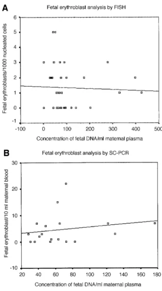
(2) Fetal DNA and fetal cells in maternal blood pre-eclampsia (Lo et al., 1999b; Leung et al., 2001; Zhong et al., 2001b,c, 2002). In addition, we have observed a very high number of fetal erythroblasts and high concentrations of cell-free fetal DNA in a polyhydramnic pregnancy (Zhong et al., 2000c). Of further interest is that increments in these two fetal parameters have also been observed in pregnancies with certain aneuploid fetuses, in particular those with trisomy 21 (Bianchi et al., 1997; Lo et al., 1999a; Zhong et al., 2000a). These varied independent results have suggested that some form of inter-relationship may exist between the presence of fetal erythroblasts, or fetal cell trafficking in general, and the release of cell-free fetal DNA into the maternal circulation (Bianchi and Lo, 2001). This hypothesis is further supported by data indicating that a significant proportion of fetal erythroblasts exhibit an apoptotic phenotype in the form of fragmented nuclear DNA (Sekizawa et al., 2000; Hristoskova et al., 2001) and that apoptotic fetal cell remnants are detectable in the plasma of pregnant women (van Wijk et al., 2000). We have now addressed this hypothesis by examining the levels of fetal erythroblasts and cell-free DNA levels in the same maternal blood samples from different pregnancy conditions.. Materials and methods Ethical approval for this study was obtained from our institutional review committee. Written informed consent was obtained in all instances. The gestational age was confirmed by first trimester ultrasound in all cases. As we quantified the number of male cells or amount of cell-free fetal DNA via the Y chromosome, we have only studied women pregnant with singleton male fetuses. Several parts of this study have relied on the re-analysis of data regarding the number of fetal erythroblasts in relation to cell-free DNA levels in the same sample. Although these data have been obtained from previous unrelated studies, they have not been viewed in this context before. These parts have been annotated accordingly. Four groups of pregnant women were examined in an independent manner.. Groups of pregnant women Normal pregnant women in the first or second trimester of pregnancy All blood samples (15 ml on average) were drawn immediately prior to an invasive procedure. The samples were processed within 24 h. The average gestational age at the time of blood sampling was 14 ⫹ 4 weeks. In this study we examined 36 normal maternal blood samples by a combination of XY FISH on enriched fetal cells and real-time PCR on the plasma DNA. We have previously examined fetal erythroblasts by SC-PCR for the RhD gene and the Y chromosome-specific SRY locus in maternal blood samples from 19 RhD pregnant women (Troeger et al., 1999). In a parallel study, we had examined the accuracy of a real-time PCR assay in determining these two fetal loci from the cell-free fetal DNA (Zhong et al., 2001a). We have now examined whether a quantitative relationship exists between these two parameters. Pregnant women with manifest pre-eclampsia Pre-eclampsia was defined by a blood pressure of 140/90 mmHg in two determinations 4 h apart or by a diastolic blood pressure of 110 mmHg and an associated proteinuria of 300 mg/24 h after 20 weeks gestation (Holzgreve et al., 1998). In this study, we examined blood samples from seven pregnant women with manifest pre-eclampsia and from 17 matched controls. The mean gestational ages of the subjects were 30 ⫹ 1 weeks (X.Y.Zhong, W.Holzgreve and S.Hahn, unpublished data). Pregnant women at risk for pre-eclampsia This part of our examination was again performed by re-analysing data regarding fetal erythroblast numbers and cell-free DNA concentrations that had been obtained previously. Details of this study cohort have been published previously (Holzgreve et al., 2001; Zhong et al., 2001c, 2002). Pregnant women at risk for preterm labour Preterm contractions were defined as four or more contractions/20 min according to the Canadian preterm labour investigators’ group (Goldenberg et al., 2001). In this study, 31 pregnant women between 20 ⫹ 0 and 33 ⫹ 6 weeks of gestation with preterm contractions were examined. Our study. included 13 gestationally matched controls who all delivered normally at term (Hoesli, Holzgreve and Hahn, unpublished data; Zhong, Holzgreve and Hahn, unpublished data).. Quantification of fetal erythroblasts Erythroblasts were enriched according to our established protocol using miniMacs magnetic separation (Miltenyi Biotec, Bergisch Gladbach, Germany) (Hahn et al., 1999; Troeger et al., 1999). Positively enriched cells were analysed by XY chromosome-specific FISH analysis (Hahn et al., 1999) or by SC-PCR for the presence of the Y chromosome and/or fetal RhD (Troeger et al., 1999; Zhong et al., 2000c). We have previously shown that a significant proportion (up to 50%) of the erythroblasts in the maternal circulation are of fetal origin, both by the use of FISH and SC-PCR (Holzgreve et al., 1998; Hahn et al., 1999; Troeger et al., 1999; Zhong et al., 2000c).. Quantitative analysis of cell-free fetal DNA Plasma was separated by centrifugation and stored at –80°C as described previously (Hahn et al., 2000; Zhong et al., 2000a,b,c). Quantitation of the concentration of male fetal DNA was performed using a well established Y chromosome-specific TaqMan real-time PCR. (Zhong et al., 2000a,b,c). The concentrations are expressed as copies per ml maternal plasma.. Data analysis The data were analysed by Excel software (Microsoft, Redmond, WA, USA) and are represented by a scatter diagram wherein the number of enriched fetal cells have been plotted against the quantity of fetal DNA determined in the same samples. As the data are not parametric, the significance of the correlation between the two parameters was analysed using Spearman Rank analysis (SPSS Statistical Software Package for Windows).. Results Normal pregnancies Our initial intention was to compare the number of male fetal erythroblasts with the concentration of male cell-free fetal DNA in the maternal circulation of normal healthy pregnancies. In the first part of this examination, we used FISH for the X and Y chromosomes to quantify the number of fetal erythroblasts following enrichment from 36 pregnancies with male singleton fetuses; XY-positive cells could be detected in 22 of 36 samples, corresponding to a sensitivity of 61.1%. Our analysis of cell-free fetal DNA by use of real-time TaqMan PCR indicated that male fetal DNA could be detected and quantified in the plasma samples of 34 out of 36 women bearing male fetuses (sensitivity ⫽ 94.4%). These values correspond well with the results obtained in previous large scale studies (Hahn et al., 1999; Zhong et al., 2001a). Due to the extreme scarcity of fetal erythroblasts in the maternal circulation, being in the order of 1 in 106–107 maternal nucleated cells, their frequency is low even after enrichment (Holzgreve and Hahn, 2001). Consequently, we were only able to detect between one and five XY-positive cells amongst the 1000 nucleated cells scored by FISH analysis following enrichment. On the other hand, in those instances where circulatory fetal DNA was detectable, the concentration of cell-free fetal DNA ranged between 28.8 and 424.7 copies per ml maternal plasma, as determined using a Y chromosomespecific real-time PCR assay. With the determination of these two parameters we could now address our original question and examine whether any correlation exists between the number of enumerated fetal erythroblasts and the level of cell-free fetal DNA. To our surprise, this analysis indicated that no correlation exists between these two parameters (Table I; Figure 1). As we had previously observed that the analysis of fetal erythroblasts was potentially more efficient by SC-PCR than by FISH. 865.
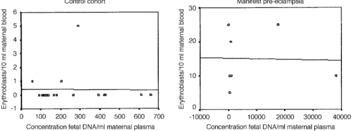
(3) X.Y.Zhong, W.Holzgreve and S.Hahn. Table I. Lack of correlation between fetal erythroblast numbers and cell-free fetal DNA concentrations in normal 1st and 2nd trimester pregnancies Study. Fetal erythroblasts. Concentration of fetal DNA/ml maternal plasma. Correlation. P-value. FISH analysis SC-PCR analysis. 1 (0–5) 3 (0–22). 70.12 (28.7–424.7) 58.93 (28.3–176.7). –0.22 0.28. 0.20 0.27. SC-PCR ⫽ single cell PCR. Fetal erythroblasts were either identified by FISH and enumerated per 1000 nucleated cells, or by SC-PCR and enumerated per 10 ml maternal blood. Concentration of fetal DNA is in copies/ml maternal plasma. Values are indicated as median with range in brackets. Correlation was determined using Spearman Rank analysis, which was considered significant at P ⬍ 0.05.. et al., 2001a). Even though we were able to detect almost twice as many fetal erythroblasts in these samples, no correlation was found to exist with the corresponding concentration of cell-free DNA (Table I; Figure 1B). In this regard it is worth noting that even though we are able to detect more fetal erythroblasts by SC-PCR than by FISH, a very good concordance between these two techniques is achieved in cell recycling studies whereby fetal cells are first analysed by FISH and then subsequently by PCR (Sekizawa et al., 1999).. Pregnancies affected by pre-eclampsia As we and others have previously observed significant increases in fetal cell trafficking as well as release of cell-free fetal DNA in pregnancies affected by pre-eclampsia (Holzgreve et al., 1998; Lo et al., 1999b; Zhong et al., 2001c), we examined these two parameters under these conditions. For this analysis we made use of a recently performed study in which the numbers of fetal erythroblasts were determined by SC-PCR (Zhong, Holzgreve and Hahn, unpublished data); we were again able to confirm that pre-eclampsia is associated with a significant increase in the trafficking of fetal erythroblasts into the maternal periphery compared with that in matched controls (median of 10 versus 0 per ml maternal blood). The simultaneous assessment of the fetal DNA concentrations in these samples indicated that the levels of cell-free fetal DNA were also elevated in those pregnancies affected by pre-eclampsia compared with matched control pregnancies (median of 933.9 versus 179.3 copies per ml maternal plasma) (Zhong, Holzgreve and Hahn, unpublished data). In our assessment of the possible relationship between these two parameters we could, however, determine no correlation between fetal erythroblasts numbers and cell-free fetal DNA levels in either the pre-eclamptic or normotensive pregnancies (Figure 2).. Pregnancies at risk for pre-eclampsia. Figure 1. Lack of correlation between circulating fetal DNA concentrations and erythroblast numbers in the maternal circulation of normal pregnancies. (A) Fetal erythroblasts studied by FISH; (B) Fetal erythroblasts analysed by single cell PCR. There was no significant correlation between these two parameters in either instance (r ⫽ –0.22, P ⫽ 0.2 and r ⫽ 0.28, P ⫽ 0.27 respectively).. (Garvin et al., 1998; Troeger et al. 1999), we next examined the quantitative relationship between the number of fetal erythroblasts identified by SC-PCR with the comparable quantity of cell-free fetal DNA. This analysis was performed on data we had previously obtained regarding the examination of individual micro-manipulated fetal erythroblasts by SC-PCR for the RhD and SRY loci (Troeger et al., 1999), and the parallel evaluation of a real-time PCR assay in determining these fetal genetic traits on cell-free fetal DNA (Zhong. 866. We and others have previously observed that fetal cell trafficking (Al-Mufti et al., 2000; Holzgreve et al., 2001) and cell-free fetal DNA (Leung et al., 2001; Zhong et al., 2001c, 2002) in maternal blood are already elevated at 20 weeks of pregnancy in those pregnancies which later develop pre-eclampsia. These results imply that these parameters may be able to serve as novel markers to predict the disorder (Holzgreve and Hahn, 1999). We have now examined whether the release of cell-free fetal DNA into maternal blood is associated with the observations we had previously made regarding increased levels in fetal cell trafficking (Holzgreve et al., 2001) and elevated fetal DNA concentrations (Zhong et al., 2001c, 2002). Although our previous observations indicated an increase in erythroblast numbers (~6-fold) and cell-free DNA (~3-fold) in those pregnancies which subsequently developed pre-eclampsia (Holzgreve et al., 2001; Zhong et al., 2001c, 2002), our re-assessment of their corresponding quantities indicated that no correlation exists between these two parameters under such conditions (Figure 3)..
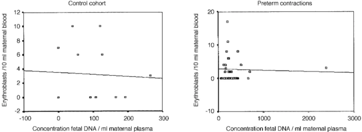
(4) Fetal DNA and fetal cells in maternal blood. Figure 2. Lack of correlation between circulating fetal DNA concentrations and erythroblast numbers in the maternal circulation of pregnancies with manifest pre-eclampsia (n ⫽ 7, r ⫽ 0.02, P ⫽ 0.97) or matched controls (n ⫽ 17, r ⫽ –0.01, P ⫽ 0.67). Erythroblasts were identified morphologically and subsequently characterized by single cell PCR. Values are indicative of total erythroblast numbers per 10 ml maternal blood. Concentration of fetal DNA is in copies/ml maternal plasma. Correlation was determined using Spearman Rank analysis, which was considered significant at P ⬍ 0.05.. Figure 3. Lack of correlation between circulating fetal DNA concentrations and erythroblast numbers in the maternal circulation of pregnancies which subsequently developed pre-eclampsia (r ⫽ 0.23, P ⫽ 0.55) or in those pregnancies with normal outcome (r ⫽ 0.02, P ⫽ 0.89). Erythroblasts were identified morphologically and enumerated per 10 ml maternal blood. Concentration of fetal DNA is in copies/ml maternal plasma. Correlation was determined using Spearman Rank analysis, which was considered significant at P ⬍ 0.05.. Pregnancies affected by preterm labour. Discussion. It has recently been reported that preterm labour is associated with an increased release of cell-free fetal DNA into the maternal circulation (Leung et al., 1998). For this purpose we have recently examined fetal erythroblast (Hoesli et al., unpublished data) and cell-free fetal DNA levels (Zhong, Holzgreve and Hahn, unpublished data) under these conditions. We have now determined whether a correlation exists between these two parameters under these conditions. A striking feature of this study was that we determined that there was no increment in erythroblast numbers in pregnancies with preterm contractions when compared with the control group (median ⫽ 0 erythroblasts per ml maternal blood in both instances; P ⫽ 0.8) (Hoesli et al., unpublished data). Single cell PCR analysis of the few erythroblasts we were able to retrieve indicated that 48% were of fetal origin. On the other hand, we observed significant elevations in cell-free DNA concentrations in pregnancies with preterm labour when compared with the control group (median concentration of 251.5 versus 91.6 copies per ml maternal plasma; P ⫽ 0.003) (Zhong, Holzgreve and Hahn, unpublished data). This result therefore indicates that cell-free fetal DNA levels can be elevated in the absence of elevations in the traffic of fetal erythroblasts, a feature which is graphically represented in Figure 4.. The novel discoveries of cell-free fetal DNA (Lo et al., 1997), fetal cell remnants (van Wijk et al., 2000) and fetal erythroblasts with apoptotic character (Sekizawa et al., 2000; Hristoskova et al., 2001) in the maternal circulation have led authors to question the relationship between these molecular and cellular entities (Bianchi and Lo, 2001). In this regard, it has been proposed that cell-free fetal DNA may be derived from the interaction of fetal cells with maternal immune effector cells (Sekizawa et al., 2000). This proposal is based upon an observation that numerous erythroblasts in the maternal circulation exhibit an apoptotic phenotype in the form of nuclear fragmentation as measured by the TUNEL assay (Sekizawa et al., 2000). In a separate study, we have shown that numerous fetal erythroblasts already display such characteristics while still in the fetal circulation (Hristoskova et al., 2001), a feature which is consistent with other observations that erythroblast terminal differentiation and enucleation involves apoptosis-like processes (Zermati et al., 2001). Since erythroblast enucleation has been observed in the periphery in the absence of accessory cells (Marmont, 1998), it is possible that this end stage differentiation step may be a key contributor to the presence of cellfree fetal DNA in the maternal circulation. This hypothesis would be reinforced by observations that elevations in both fetal erythroblasts. 867.
(5) X.Y.Zhong, W.Holzgreve and S.Hahn. Figure 4. Lack of correlation between circulating fetal DNA concentrations and erythroblasts numbers in the maternal circulation of pregnancies affected by onset of preterm contractions. The study included 31 cases with preterm contractions (r ⫽ –0.44, P ⫽ 0.82) and 13 matched controls (r ⫽ –0.06, P ⫽ 0.84). Erythroblasts were identified morphologically and subsequently characterized by single cell PCR. Values are indicative of total erythroblast numbers per 10 ml maternal blood. Concentration of fetal DNA is in copies/ml maternal plasma. Correlation was determined using Spearman Rank analysis, which was considered significant at P ⬍ 0.05.. (Holzgreve et al., 1998, 2001) and cell-free fetal DNA (Lo et al., 1999b; Leung et al., 2001; Zhong et al., 2001b,c, 2002) occur in several pregnancy-related pathologies, such as pre-eclampsia. In this study, we have now examined this issue in greater detail by determining the relationship between fetal erythroblast numbers and cell-free fetal DNA levels in the same maternal blood samples under different pregnancy conditions. In our analysis, we have used different technologies to enumerate the numbers of fetal erythroblasts, namely FISH and SC-PCR, and real-time PCR to quantify the concentration of cell-free DNA. The reason for this technical diversity is that currently no reliable method exists for the quantification of fetal erythroblasts by real-time PCR. Therefore, some small deviation in our results may be attributable to subtle differences in these technologies. Because we have observed very comparable results for either of the techniques used in numerous blinded and prospective studies, we feel confident that the error margin is very small. Our present study has shown that no correlation exists between fetal erythroblast numbers and cell-free fetal DNA levels in normal pregnancies, or under a variety of pregnancy-related pathologies associated with increased fetal cell traffic or release of cell-free fetal DNA. The disparity between fetal erythroblast numbers and cell-free DNA concentration was most notable in pregnancies with preterm contractions, where we observed significant increments in cell-free fetal DNA levels without any discernible increase in fetal erythroblast numbers. Therefore, our data strongly suggest that another cell type is responsible for the release of cell-free fetal DNA into the maternal circulation. Our data further imply, provided that fetal erythroblasts can be used as a marker for fetal cell traffic across the placenta in general, that the cell type responsible for the release of cell-free fetal DNA is affected by conditions which do not alter fetal cell traffic. In this manner our results may help elucidate fundamental differences between various pregnancy-related pathologies such as pre-eclampsia and preterm labour. Although we have not determined the source of cell-free fetal DNA and it is possible that the source may vary depending on the type of pregnancy related disorder, our data do, however, favour hypotheses which propose that cell-free fetal DNA is derived from the placenta and is not the result of fetal cell trafficking (Lo, 2000). Observations which support a placental component include the very rapid disappearance of cell-free DNA from the maternal circulation following delivery (Lo et al., 1999c), conditions under which fetal erythroblasts are still very readily detectable for several. 868. days post-delivery (Ganshirt et al., 1994b). Indeed, it was the similarity between the placenta and tumours (Lo, 2000) that first led to the original investigations of whether cell-free DNA existed in the maternal circulation, in that: (i) the placenta and tumours are large tissues in direct contact with the peripheral circulation; (ii) both have a very high degree of cell division and cell turnover; (iii) both express high levels of certain proto-oncogenes; and (iv) tumours are known to release cell-free DNA into the circulation of cancer patients. Other observations favouring placental origin are that cell-free fetal DNA levels are in general several orders of magnitude greater than fetal cell levels (Lo, 2000). Furthermore, cell-free fetal DNA concentrations are not increased by processing conditions which would lead to the demise of fetal cells present in the maternal blood sample being processed, for instance by the preparation of serum which involves blood clotting (Chui et al., 2001; Hahn et al., 2001). These experiments have also demonstrated that most of the cell-free fetal DNA in maternal plasma exists in a distinct acellular form and very little is present in the form of apoptotic cell remnants (Chui et al., 2001; Hahn et al., 2001). Furthermore, there is no significant increase in the amount of cell-free fetal DNA in serum samples, conditions under which the fetal cells present would be expected to die and release their DNA during the clotting process. This is in strong contrast to the total cell-free DNA concentrations, which are significantly increased in serum samples. Although it is unclear by which means cell-free DNA is released from the placenta, one attractive hypothesis is that it is mediated by the shedding of cell fragments and excess nuclei from the syncytiotrophoblast as it is constantly replenished by the underlying differentiating cytotrophoblast (Huppertz et al., 2001). It has been estimated that the amount of cellular material released by this process into the maternal circulation is in the order of several grams per day (Huppertz et al., 2001). Since the half life of cell-free fetal DNA in the maternal circulation has been estimated to be in the order of 15 min (Lo et al., 1999c), it becomes clear that prodigious quantities of input material would be required to facilitate a high level steadystate concentration (Lo, 2000). It is likely that the true source of cell-free DNA in the maternal circulation will remain a scientifically contentious issue (Bianchi and Lo, 2001), which will require many more studies to be resolved. The best approach will probably be to examine cell-free fetal DNA levels with fetal genes which are epigenetically modified in different fetal tissues (Poon et al., 2002)..
(6) Fetal DNA and fetal cells in maternal blood Our data do, however, indicate that fetal erythroblasts are not involved to a significant degree in the generation of this material. Furthermore, our study provides new evidence of discrete differences between pre-eclampsia and preterm labour, perhaps at a placental level, in that preterm contractions are only associated with release of cell-free fetal DNA whereas both fetal cell traffic and cell-free DNA release are perturbed in pre-eclampsia. Therefore, further exploration of these phenomena may aid in our understanding of these intriguing disorders.. Acknowledgements This work was supported in part by Swiss National Science Foundation Grant Number:3200–059275.99/1 and 3200–055614.98, NIH (USA) Contract Number: N01-HD-4–3202.. References Al-Mufti, R., Hambley, H., Albaiges, G., Lees, C. and Nicolaides, K.H. (2000) Increased fetal erythroblasts in women who subsequently develop preeclampsia. Hum. Reprod., 15, 1624–1628. Bianchi, D.W. and Lo, Y.M. (2001) Fetomaternal cellular and plasma DNA trafficking: the Yin and the Yang. Ann. NY Acad. Sci., 945, 119–131. Bianchi, D.W., Mahr, A., Zickwolf, G.K., Houseal, T.W., Flint, A.F. and Klinger, K.W. (1992) Detection of fetal cells with, 47, XY, ⫹21 karyotype in maternal peripheral blood. Hum. Genet., 90, 368–370. Bianchi, D.W., Williams, J.M., Sullivan, L.M., Hanson, F.W., Klinger, K.W. and Shuber, A.P. (1997) PCR quantitation of fetal cells in maternal blood in normal and aneuploid pregnancies. Am. J. Hum. Genet., 61, 822–829. Bianchi, D.W., Simpson, J.L., Jackson, L.G., Evans, M.I., Elias, S., Holzgreve, W., Sullivan, L.M. and de la Cruz, F. (1999) Fetal cells in maternal blood: NIFTY clinical trial interim analysis. DM-STAT. NICHD fetal cell study (NIFTY) group. Prenat. Diagn., 19, 994–995. Chen, X.Q., Stroun, M., Magnenat, J.L., Nicod, L.P., Kurt, A.M., Lyantey, J., Lederberg, G. and Anker, P. (1996) Microsatellite alterations in plasma DNA of small cell lung cancer patients. Nature Med., 2, 1033–1035. Cheung, M.C., Goldberg, J.D. and Kan, Y.W. (1996) Prenatal diagnosis of sickle cell anaemia and thalassaemia by analysis of fetal cells in maternal blood. Nature Genet., 14, 264–268. Chui, R.W.K., Poon, L.L.M., Lau. T.K., Leung, T.N., Wong, E.M.C. and Lo, Y.M.D. (2001) Effects of blood processing protocols on fetal and total DNA quantification in maternal plasma. Clin. Chem., 47, 1607–1613. de la Cruz, F., Shifrin, H., Elias, S., Bianchi, D.W., Jackson, L., Evans, M.I., Simpson, J.L., Holzgreve, W. and Klinger, K. (1998) Low false-positive rate of aneuploidy detection using fetal cells isolated from maternal blood. Fetal Diagn. Ther., 13, 380. Elias, S., Price, J., Dockter, M., Wachtel, S., Tharapel, A., Simpson, J.L. and Klinger, K. (1992) First trimester prenatal diagnosis of trisomy 21 in fetal cells from maternal blood. Lancet, 340, 1033. Ganshirt, D., Borjesson-Stoll, R., Burschyk, M., Garritsen, H.S., Neusser, M., Smeets, F.W., Velasco, M. and Walde, C. (1994a) Successful prenatal diagnosis from maternal blood with magnetic-activated cell sorting. Ann. NY Acad. Sci., 731, 103–114. Ganshirt, D., Garritsen, H., Miny, P. and Holzgreve, W. (1994b) Fetal cells in maternal circulation throughout gestation. Lancet, 343, 1038–1039. Garvin, A.M., Holzgreve, W. and Hahn, S. (1998) Highly accurate analysis of heterozygous loci bysingle cell PCR. Nucleic Acids Res., 26, 3468–3472. Goldenberg, R.L., Iams, J.D., Mercer, B.M., Meis, P.J., Moawad, A., Das, A., Miodovanik, M., van Dorster, P.J., Caritis, S.N., Thurman, G. et al. (2001) The Preterm Prediction Study: toward a multiple-marker test for spontaneous preterm birth. Am. J. Obstet. Gynecol., 185, 643–651. Hahn, S., Kiefer, V., Brombacher, V., Troeger, C. and Holzgreve, W. (1999) Fetal cells in maternal blood. An update from Basel. Eur. J. Obstet. Gynecol. Reprod. Biol., 85, 101–104. Hahn, S., Zhong, X.Y., Burk, M.R., Troeger, C. and Holzgreve, W. (2000) Multiplex and real-time quantitative PCR on fetal DNA in maternal plasma. A comparison with fetal cells isolated from maternal blood. Ann. NY Acad. Sci., 906, 148–152. Hahn, S., Zhong, X.Y. and Holzgreve, W. (2001) Quantification of circulating DNA: in the preparation lies the rub. Clin. Chem., 47, 1577–1578. Holzgreve, W. and Hahn, S. (1999) Novel molecular biological approaches for the diagnosis of pre-eclampsia. Clin. Chem., 45, 451–452.. Holzgreve, W. and Hahn, S. (2001) Prenatal diagnosis using fetal cells and free fetal DNA in maternal blood. Clin. Perinatol., 28, 353–365. Holzgreve, W., Garritsen, H.S. and Ganshirt-Ahlert, D. (1992) Fetal cells in the maternal circulation. J. Reprod. Med., 37, 410–418. Holzgreve, W., Ghezzi, F., Di Naro, E., Ganshirt, D., Maymon, E. and Hahn, S. (1998) Disturbed feto-maternal cell traffic in pre-eclampsia. Obstet. Gynecol., 91, 669–672. Holzgreve, W., Li, J.C., Steinborn, A., Kulz, T., Sohn, C., Hodel, M. and Hahn, S. (2001) Elevation in erythroblast count in maternal blood before the onset of pre-eclampsia. Am. J. Obstet. Gynecol., 184, 165–168. Hristoskova, S., Holzgreve, W. and Hahn, S. (2001) More than one-half of the erythroblasts in the fetal circulation and cord blood are TUNEL positive. Clin. Chem., 47, 1870–1871. Huppertz, B., Tews, D.S. and Kaufmann, P. (2001) Apoptosis and syncytial fusion in human placental trophoblast and skeletal muscle. Int. Rev. Cytol., 205, 215–253. Leung, T.N., Zhang, J., Lau, T.K., Hjelm, N.M. and Lo, Y.M. (1998) Maternal plasma fetal DNA as a marker for preterm labour. Lancet, 352, 1904–1905. Leung, T.N., Zhang, J., Lau, T.K., Chan, L.Y. and Lo, Y.M. (2001) Increased maternal plasma fetal DNA concentrations in women who eventually develop pre-eclampsia. Clin. Chem., 47, 137–139. Lo, Y.M. (2000) Fetal DNA in maternal plasma: biology and diagnostic applications. Clin. Chem., 46, 1903–1906. Lo, Y.M., Corbetta, N., Chamberlain, P.F., Rai, V., Sargent, I.L., Redman, C.W. and Wainscoat, J.S. (1997) Presence of fetal DNA in maternal plasma and serum. Lancet, 350, 485–487. Lo, Y.M., Tein, M.S., Lau, T.K., Haines, C.J., Leung, T.N., Poon, P.M., Wainscoat, J.S., Johnson, P.J., Chang, A.M. and Hjelm, N.M. (1998) Quantitative analysis of fetal DNA in maternal plasma and serum: implications for noninvasive prenatal diagnosis. Am. J. Hum. Genet., 62, 768–775. Lo, Y.M., Lau, T.K., Zhang, J., Leung, T.N., Chang, A.M., Hjelm, N.M., Elmes, R.S. and Bianchi, D.W. (1999a) Increased fetal DNA concentrations in the plasma of pregnant women carrying fetuses with trisomy 21. Clin. Chem., 45, 1747–1751. Lo, Y.M., Leung, T.N., Tein, M.S., Sargent, I.L., Zhang, J., Lau, T.K., Haines, C.J. and Redman, C.W. (1999b) Quantitative abnormalities of fetal DNA in maternal serum in pre-eclampsia. Clin. Chem., 45, 184–188. Lo, Y.M., Zhang, J., Leung, T.N., Lau, T.K., Chang, A.M. and Hjelm, N.M. (1999c) Rapid clearance of fetal DNA from maternal plasma. Am. J. Hum. Genet., 64, 218–224. Marmont, A.M. (1998) Nuclear extrusion by erythroblasts in the peripheral blood of a splenectomized patient. Haematologica, 83, 943. Poon, L.L., Leung, T.N., Lau, T.K., Chow, K.C. and Lo, Y.M. (2002) Differential DNA methylation between fetus and mother as a strategy for detecting fetal DNA in maternal plasma. Clin. Chem., 48, 35–41. Sekizawa, A., Samura, O., Zhen, D.K., A. and Bianchi, D.W. (1999) Fetal cell recycling: diagnosis of gender and RhD genotype in the same fetal cell retrieved from maternal blood. Am. J. Obstet. Gynecol., 181, 1237–1242. Sekizawa, A., Samura, O., Zhen, D.K., Falco, V., Farina, A. and Bianchi, D.W. (2000) Apoptosis in fetal nucleated erythrocytes circulating in maternal blood. Prenat. Diagn., 20, 886–889. Sekizawa, A., Sugito, Y., Iwasaki, M., Watanabe, A., Jimbo, M., Hoshi, S., Saito, H. and Ohai, T. (2001) Cell-free fetal DNA is increased in plasma of women with hyperemesis gravidarum. Clin. Chem., 47, 2164–2165. Troeger, C., Zhong, X.Y., Burgemeister, R., Minderer, S., Tercanli, S., Holzgreve, W. and Hahn, S. (1999) Approximately half of the erythroblasts in maternal blood are of fetal origin. Mol. Hum. Reprod., 5, 1162–1165. van Wijk, I.J., de Hoon, A.C., Jurhawan, R., Tjoa, M.L., Griffioen, S., Mulders, M.A., van Vugt, J.M. and Oudejaans, C.B. (2000) Detection of apoptotic fetal cells in plasma of pregnant women. Clin. Chem., 46, 729–731. Zermati, Y., Garrido, C., Amsellem, S., Fishelson, S., Bouscary, D., Valensi, F., Varet, B., Solary, E. and Hermine, O. (2001) Caspase activation is required for terminal erythroid differentiation. J. Exp. Med., 193, 247–254. Zhong, X.Y., Burk, M.R., Troeger, C., Jackson, L.R., Holzgreve, W. and Hahn, S. (2000a) Fetal DNA in maternal plasma is elevated in pregnancies with aneuploid fetuses. Prenat. Diagn., 20, 795–798. Zhong, X.Y., Holzgreve, W. and Hahn, S. (2000b) Detection of fetal Rhesus D and sex using fetal DNA from maternal plasma by multiplex polymerase chain reaction. Br. J. Obstet. Gynaecol., 107, 766–769. Zhong, X.Y., Holzgreve, W., Li, J.C., Aydinli, K. and Hahn, S. (2000c) High levels of fetal erythroblasts and fetal extracellular DNA in the peripheral blood of a pregnant woman with idiopathic polyhydramnios: case report. Prenat. Diagn., 20, 838–841.. 869.
(7) X.Y.Zhong, W.Holzgreve and S.Hahn Zhong, X.Y., Hahn, S. and Holzgreve, W. (2001a) Prenatal identification of fetal genetic traits. Lancet, 357, 310–311. Zhong, X.Y., Laivuori, H., Livingston, J.C., Ylikorkala, O., Sibai, B.M., Holzgreve, W. and Hahn, S. (2001b) Elevation of both maternal and fetal extracellular circulating deoxyribonucleic acid concentrations in the plasma of pregnant women with pre-eclampsia. Am. J. Obstet. Gynecol., 184, 414–419.. 870. Zhong, X.Y., Holzgreve, W. and Hahn, S. (2001c) Circulatory fetal and maternal DNA in pregnancies at risk and those affected by pre-eclampsia. Ann. NY Acad. Sci., 945, 138–140. Zhong, X.Y., Holzgreve, W. and Hahn, S. (2002) The levels of circulatory fetal DNA in maternal plasma are elevated prior to onset of pre-eclampsia. Hypertens. Preg., 21, 77–83. Submitted on April 26, 2002; accepted on June 19, 2002.
(8)
Figure
Documents relatifs