CELLULAR RESPONSES TO PLATINUM-BASED ANTICANCER DRUGS
by
KEVIN J. YAREMA
B. S., Walla Walla College, 1988
SUBMITTED IN PARTIAL FULFILLMENT OF THE REQUIREMENTS FOR THE DEGREE OF
DOCTOR OF PHILOSOPHY
at the
MASSACHUSETTS INSTITUTE OF TECHNOLOGY
February 1994
© 1994 Massachusetts Institute of Technology All rights reserved
Signature of Author Certified by -Accepted by Department of Chemistry 3 February 1994
Prof. John M. Essigmann Thesis Supervisor
/
Prof. Glen A. Berchtold Chairprson, Committee on Graduate Students
MASSACU.JLc 3 NS;Twu
-' n,
ny
MAR 21
1994
ciru
lt
,, ! ,~~~~~~~~~~~~ ,_N;T4
I v . . _,COMMITTEE PAGE
This doctoral thesis has been examined by a Committe of the Department of Chemistry as follows:
I
Professor Steven R. Tannenbaum I/
-Chairman
Professor John M. Essigmann
:-
\
Thesis SupervisorI
Professor Stephen J. Lippard
Professor William H. Orme-Johnson_
Professor Joanne Stubbe
t-~j7l4t
(72>
- -
j
-.
v>
CELLULAR RESPONSES TO PLATINUM-BASED ANTICANCER DRUGS
byKevin Jon Yarema
Submitted to the Department of Chemistry on 3 February 1994 in partial fulfillment of the requirements for the Degree of Doctor of Philosophy
in Biological Chemistry
ABSTRACT
This dissertation describes the genetic effects of platinum DNA adducts in Escherichia coli from two perspectives. First, the
genotoxicities and mutagenicities of the three major DNA adducts formed by cis-diamminedichloroplatinum(II) (cis-DDP), cis-[Pt(NH3)2{d(GpG)}]
(G*G*), cis- [Pt(NH3)2{d(ApG) }] (A*G*), and cis- [Pt (NH3)2{d(GpNpG) } ]
(G*NG)*, were investigated. These studies were conducted by using viral genomes in which specific nucleotides in the natural sequence were replaced with the aforementioned DNA adducts. The results of the site specific evaluation of these adducts suggested principles applicable to the design and evaluation of novel anticancer platinum-based therapeutic agents. Accordingly, the second aspect of this work was the genotoxic and mutagenic evaluation of
cis-ammine(cylcohexylamine)dichloroplatinum(II) (ACDP), a metabolite of the platinum(IV) compound, cis, trans,
cis-ammine(cyclohexylamine)dibutyrato-dichloroplatinum(IV) (ACDDP), now undergoing clinical trials to determine its efficacy as an anticancer drug.
The first aspect of this work, the site specific evaluation of the cis-DDP G*G*, A*G*, and G*TG* adducts, was done by incorporating
oligodeoxynucleotide 24-mers, containing these lesions, into M13mp7L2 derived ss genomes. Both the platinum modified and unmodified, ss genomes were transfected into E. coli DL7 cells that had been, or had not been, irradiated with ultraviolet light in order to induce the SOS response. The genotoxicities of the adducts were determined by using the M13 plaque forming assay. Genomes containing the cis-DDP G*G*, A*G*, and G*TG* adducts had survival levels of 5.2 + 1.2%, 21.6 + 2.6%,
13.5 ± 2.5%, respectively, compared to unmodified control genomes. Upon SOS induction of the host E. coli cells, the survival of genomes
containing the cis-DDP G*G* and A*G* adducts rose significantly to 30.8
+ 5.4% and 32.4 + 4.9%, respectively. By contrast, the survival of genomes containing the cis-DDP G*TG* adduct did not increase upon SOS
induction, remaining at 14.4 + 3.7%.
Mutations attributable to cis-DDP were observed only for modified genomes replicated in SOS induced E. coli DL7 cells. The cis-DDP G*G* and A*G* adducts produced highly targeted mutations at the 5' modified base, with the predominant mutation consisting of G T and A - T
transversions for the two adducts, respectively. A G transitions also arose from the cis-DDP A*G* adduct as well as a low level of tandem mutations from both adducts affecting the 5' modified base as well as
the adjacent 5' base. The cis-DDP A*G* adduct was considerably more mutagenic than the G*G* adduct, with a mutation frequency of 6.0%
compared to 1.4% for the latter adduct. In accordance with the lack of an SOS-dependent increase in survival, the cis-DDP G*TG* adduct also was not mutagenic.
A drawback of current platinum based chemotherapy is the potential carcinogenicity of cis-DDP. The site specific results suggest a
strategy for reducing the mutagenicity, and hence the potential
carcinogenicity, of platinum compounds. The high mutagenicity of the A*G* adduct suggests that, if a platinum compound directed against the
formation of this highly mutagenic DNA lesion, its overall mutagenicity would be decreased. ACDP is a compound that meets this criterion; its cyclohexyl ring causes an orientational isomerism which, upon binding to DNA, reduces the number of A*G* adducts threefold as compared with cis-DDP DNA binding. ACDP, therefore, was predicted to be less mutagenic than cis-DDP. This hypothesis was tested by comparing the
genotoxicities and mutagenicities of cis-DDP and ACDP DNA adducts in E.
coli. Both compounds displayed similar levels of genotoxicity in a
bacteriophage M13mp18 plaque-forming assay, with survival for genomes platinated by either drug increasing by threefold in cells pretreated with uv irradiation to induce the SOS functions of the host. The mutagenicity of ACDP was lower than that of cis-DDP in the lacZ'
a-complementation forward mutational assay and was also SOS-dependent. The mutational spectra for both drugs were similar; G T transversions at d(GpG) sites were the most common mutations while G A transitions and A T transversions, many at d(ApG), d(GpNpG), and d(GpG) sites, were also well represented. Adduct mapping experiments revealed
excellent correlation between the location of DNA lesions and the sites of mutations, confirming that the induced mutations were a consequence of the platinum adducts. Analysis of the distribution of mutations suggested that there were no sequence-dependent mutation hotspots; mutagenesis was random throughout the lacZ' region of the M13mp18 bacteriophage genome.
Thesis Supervisor: Dr. John M. Essigmann
ACKNOWLEDGEMENTS
The primary, and perhaps only, reason that I came to MIT was to obtain an education. Accordingly, I would like to thank all the people who contributed to my learning process. First and foremost, I am
grateful for my faculty advisor, John Essigmann who provided me with the research project described in this dissertation, financial support, and an enjoyable, stimulating, yet highly professional, work environment. During my time at MIT, I've probably learned more from other members of the Essigmann laboratory than from anyone else. Amongst this group of people, the greatest contributor to the practical aspects of my
education was my mentor, the relatively benevolent Lisa Naser Bradley, who taught me everything there was to know about working in a
laboratory. Other laboratory members, especially Ashis Basu, Mike Wood, Tim Connors, Dan Treiber, and Lisa Bailey have also provided invaluable technical advice during numerous helpful discussions. Additionally, I would like to thank our collaborator, Stephen Lippard, and his
laboratory, for important intellectual contributions and expert
technical assistance. Finally I would like to express my appreciation for the faculty members I've had the privilege of interacting with, and learning from, especially the members of my thesis committee, Steven Tannenbaum, Bill Orme-Johnson, and Joanne Stubbe.
As I've found out, the graduate school experience involves more practical work than actual theoretical learning. At times, laboratory
work can seem tedious and apparently unrewarding. Therefore, I
gratefully acknowledge the friendship and encouragement from my family and labmates that helped me to persevere and ultimately experience the massive emotional, physical, intellectual, and potential financial, rewards of successful completion of this dissertation. From a practical perspective, the excellent technical assistance received from Wendy Rowell and Jennifer Wilson greatly facilitated my work and helped me graduate with (relatively) great alacrity. A special thanks is due Brian Donahue and Jill Mello who valiantly played crucial roles in battling the heartless MIT parking bureaucracy by helping me to obtain, collectively, four parking permits. Finally, a most special thanks to Marjie Solomon for critical reading of this manuscript, thereby enabling it to become what it is today.
TABLE OF CONTENTS COMMITTEE PAGE . . . 2 ABSTRACT . . . 3 ACKNOWLEDGEMENTS ... ... . 5 TABLE OF CONTENTS ... ... 6 LIST OF FIGURES ... . ... 10 LIST OF TABLES ... . ... 11 LIST OF ABBREVIATIONS . ... 12 I. INTRODUCTION ... . ... 14
II. LITERATURE SURVEY . . . .. 21
A. Platinum-DNA Interactions . . . .22
1. Mode of Cytotoxicity Induced by cis-DDP Modified DNA .. . . . 23
2. Platinum-DNA Adducts. .25 3. Structural Effects of Platinum-DNA Adducts . . . 28
a. Macroscopic effects on the structure of platinum modified DNA . . . 28
b. Structural effects of single platinum DNA adducts . . . 29
c. Platinum induced perturbation of duplex DNA . 30 4. Replication Blockage Effects of Platinum DNA Adducts . 34 B. Cellular Effects of Platinum Anticancer compounds . . .. 39
1. Genotoxicity of Platinum-DNA Adducts . . . 39
2. Mutagenesis of cis-DDP in E. coli . . . 40
a. Mutagenesis in E. coli ... 40
b. Mutational specificity of cis-DDP in E. coli. . . . . . . . . . . . . . . . . . 40
3. Mechanism of cis-DDP Mutagenesis in Prokaryotes .. 41
a. SOS dependence of mutagenesis . . . 41
b. The role of UmuDC and RecA in SOS mutagenesis . . . 43
c. The role of UvrA and UvrB . . . 46
4. umuDC-like Mutagenesis in Other Species . . . . . 48
5. Mutagenicity and Genotoxity of cis-DDP in Eukaryotic Cells ... . 50
C. Platinum (IV) Anticancer Drugs . . . 52
1. The Need for New Platinum Drugs . . . 52
2. Platinum(IV) Ammine/amine Dicarboxylates . . . . 53
3. cis-DDP Resistance in Tumor Cells . . . 54
a. Intracellular sequestration and conjugation of cis-DDP .56 b. cis-DDP Resistance mediated by enhanced DNA repair ... 57
4. Circumvention of cis-DDP Resistance by Platinum(IV) Drugs . . . . .. 58
5. ACDP, a Cellular Metabolite of ACDDP, Forms DNA Adducts . . . 60
a. DNA binding properties of platinum(IV) drugs . . . 60 b. Mutagenic implications of the orientational
adducts ... 62 D. Background of Experimental Systems Used in this Work . 66
1. Site Specifically Situated versus Randomly
Distributed DNA Adducts . . . 66 a. Site specific modification of
oligonucleotides . . . 67
b. Methods for constructing singly modified
genomes ... . 68
2. M13 Bacteriophage . . ... 69
a. Life cycle and replication of M13 . . . 70
b. Strand bias effects . . . . 71
3. The M13 lacZ' Forward Mutational Assay . . . 72 a. Forward mutational assays . . . .. 72 b. Molecular basis of lacZ' mutational assay .. 72
III. MATERIALS AND METHODS . . . 83
A. Materials ... 84
B. SOS-Induction of E. coli DL6 and DL7 Cells . . . . . 85 1. cis-DDP Modification of M13mp18 Bacteriophage
Genomes . . . .. 85 2. Transformation of E. coli with cis-DDP Modified
M13mp18 Genomes ... . 86 3. UV Irradiation of E. coli DL7 Cells . . . . . . 87 C. Site Specific Comparison of cis-DDP DNA Adducts . . . 88
1. Preparation of cis-DDP Modified
Oligodeoxynucleotides . . . 88 a. Design of oligodeoxynucleotide sequence . .. 88 b. Synthesis of Platinated
Oligodeoxynucleotides . . 89
2. Preparation of cis-DDP Modified Genomes . . . 92 a. Scaffold oligodeoxynucleotides . . . 93 b. Preparation of linearized M13mp7L2 ss
genomes . . . 93 c. Phosphorylation of oligodeoxynucleotide
24-mers ... 94
d. Annealing and ligation reactions . . . 94 e. Characterization of genetically engineered
genomes . . . 96
3. Genotoxicities of cis-DDP Modified Genomes . . . . 96 4. Mutagenicity of Site-Specifically Modified
Genomes . . . ... .. 97
a. RF DNA preparations from progeny phage . .. 98 b. Restriction digestion of M13 bacteriophage
RF DNA . . . 99 c. Determination of mutation frequency . . . 99 d. Determination of mutational specificity .. 100 D. Comparison of the Mutagenicities, Genotoxicities, and
Adduct Distributions of cis-DDP and ACDP DNA Adducts 107 1. Random Platination of Bacteriophage M13mp18 RF
Genomes . . ... . . 107
2. Transfection of Platinated M13mp18 RF Genomes .. 107
3. Assay of -Galactosidase Activity . . . 108
4. Identification and Sequencing of Mutants . . .. 108 5. Replication Mapping of cis-DDP and ACDP DNA
Adducts ... . 108
IV. RESULTS ... 113
A. SOS Induction of E. coli DL7 cells ... 114 1. Transformation of E. coli with cis-DDP modified
M13mp18 RF Genomes . . 114
B. Comparison of the Genotoxicities and Mutagenicities of
the cis-DDP G*G*, A*G*, and G*TG* DNA Adducts .. 122 1. Preparation of cis-DDP Modified
Oligodeoxynucleotides . . . 122 2. Preparation of Singly cis-DDP Modified M13 SS
Genomes . . . .. . . 126
3. Genotoxicities of Singly cis-DDP Modified M13 SS
Genomes . . . 128 4. Mutagenicities of Singly cis-DDP Modified M13 SS
Genomes . . . 129 C. Comparison of the Mutagenicities, Genotoxicities and
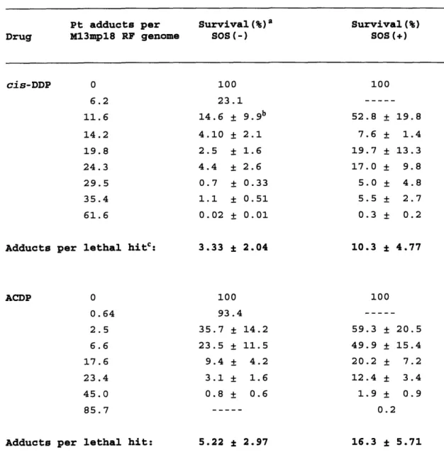
Adduct Distributions of cis-DDP and ACDP DNA Adducts 144 1. Survival of cis-DDP and ACDP Modified M13mp18 RF
Genomes . . . 144
2. Identification of Mutants . . . 144 3. Determination of Mutation Frequency . . . 145 4. lacZ' Mutational Spectra of cis-DDP and ACDP
Modified DNA . . . 146
5. Replication Mapping of DNA Adducts . . . 147
V. DISCUSSION ... 167
A. Genotoxicities of the G*G*, A*G*, and G*TG* Adducts . 168
1. Comparison to Previous Results . . . 168
2. Comparative Genotoxities of cis-DDP adducts . .. 169
a. Differential repair . . . 170
b. Translesion synthesis. ... 172 3. Mode of SOS-Dependent Survival Increase . . . 173
a. Enhanced survival is not due to repair by UvrABC .174
b. Alternate mechanisms for repair of cis-DDP
adducts . 176
c. SOS-dependent bypass of cis-DDP adducts . 177 B. Mutagenicities of the G*G*, A*G*, and G*TG* Adducts . 178 1. SOS-Dependence of Mutagenesis . . . 178 2. Mutational Specificity of cis-DDP Adducts . . . 179
a. Comparison to previous results . . . 179
b. The cis-DDP G*TG* adduct is not mutagenic . 180 c. Mutational frequencies of the cis-DDP G*G*
and A*G* adducts. . . . 181 d. Structural determinants of mutagenicity . . 183 C. Comparison of the Genotoxicities and Mutagenicities of
cis-DDP and ACDP DNA Adducts . . . . . 189
1. Survival . . . .. . . 189 2. Comparative mutation frequencies of cis-DDP and
ACDP ... 190
a. Comparison to other mutational assays . .. 190 b. Mechanistic rationale for differences in the
mutation frequencies of cis-DDP and ACDP 191 c. Comparison of mutation frequencies
determined from the lacZ' and the site
specific studies . . . 194 d. The absolute mutation frequency differs
between singly and randomly cis-DDP
modified genomes . . . 195 3. Features of the LacZ' Forward Mutational Assay 197 4. Dark Blue plaques ... .198
5. Mutational Spectra of cis-DDP and ACDP Modified
DNA
... ... .
199
6. Correlation of Sites of Adduct Formation with
7. Sequence Dependence of Mutagenesis . . . 206 a. Sensitivity to mutation is related to the
local function of the lacZ' sequence .. 206 b. Distribution of missense mutations . . .. 207 c. Distribution of nonsense mutations . . .. 208 d. Mutations are correlated to a position
within a codon . . . 209 e. The lack of mutational hotspots reinforces
the validity of the site specific results 210 8. Technical Features of the Adduct Mapping
Experiments . .. . . .. . 211 D. Application of Results to Drug Design and Evaluation 220
1. Relevance results from prokaryotes in the analysis
of human drugs . . . 220 2. Implications of replication blockage, mutagenic
and genotoxic studies for platinum-based drug
design . . . ... 221
VI. SUGGESTIONS FOR FUTURE RESEARCH . . . 223
A. Site Specific Experiments . . . 224
1. cis- and trans-DDP G*NG* Adducts . . 224
2. Interstrand Crosslinks . . 225
3. Site Specific ACDP Adducts . . 226
B. Genetic Experiments ... . 227
C. Drug Design ... . 228
LIST OF FIGURES
Figure 1. 2. 3. 4. 5. 6. 7. Figure 8. Figure 9. Figure 10. Figure 11. 12. 13. 14. Figure 15. Figure 16. 17. 18. 19. 20. Figure 21. Figure 22.Structures of platinum compounds discussed in this dissertation . . . .
cis-DDP G*G*, A*G*, and G*TG* adducts
Orientational isomerism of DNA adducts formed by ACDP General methods used to construct site specifically modified M13 based bacteriophage genomes .
Outline of the life cycle of the M13 bacteriophage . Molecular basis of the lacZ' mutational assay . Functional regions of the lacZ' DNA sequence of the M13mp18 genome .
Method used to construct singly cis-DDP modified M13 ss genomes ...
Method of mutational analysis.
Method for quantitation of stop sites used in the replication mapping of cis-DDP and ACDP DNA adducts Genotoxicity of cis-DDP modified DNA replicated in host E. coli with various levels of repair
proficiency . .
Survival of E. coli DL7 cells after uv irradiation Mutation frequency as a function of uv dose . Survival of singly cis-DDP modified M13 ss genomes in non-SOS induced E. coli DL7 cells . .
Survival of singly cis-DDP modified M13 ss genomes in SOS induced, or non induced, E. coli DL7 cells Survival of cis-DDP and ACDP modified M13mp18 RF genomes in E. coli DL7 cells
Mutation frequency as a function of adduct level Mutation and DNA binding spectra for the lacZ' region
Spontaneous mutational spectra . . . . Molecular basis for the targeting of mutations to
the 5' base of a bifunctional cis-DDP adduct . .
Speculative model to account for enhanced P-galactosidase activity of phenotypically "dark blue" plaques .
Comparison of the predicted and actual adduct
distributions . Figure Figure Figure Figure Figure Figure Figure Figure Figure 19 37 64 75 77 79 81 103 105 110 116 118 120 140 142 158 160 162 165 187 216 218 Figure Figure Figure Figure
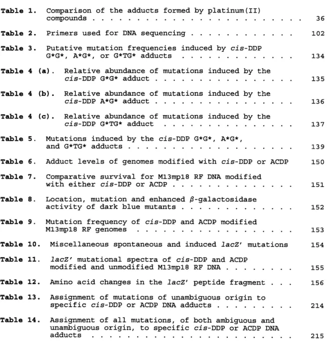
LIST OF TABLES
Table 1. Comparison of the adducts formed by platinum(II)
compounds . . . 36
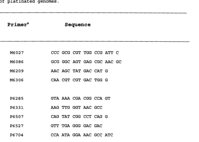
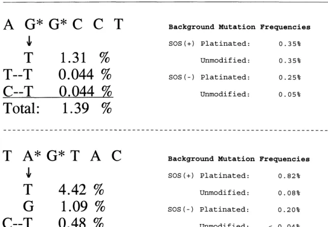
Table 2. Primers used for DNA sequencing . . 102 Table 3. Putative mutation frequencies induced by cis-DDP
G*G*, A*G*, or G*TG* adducts . . 134
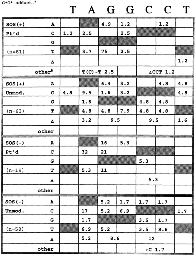
Table 4 (a). Relative abundance of mutations induced by the
cis-DDP G*G* adduct ... 135
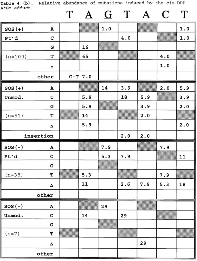
Table 4 (b). Relative abundance of mutations induced by the
cis-DDP A*G* adduct ... 136
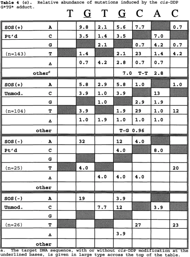
Table 4 (c). Relative abundance of mutations induced by the
cis-DDP G*TG* adduct . . 137
Table 5. Mutations induced by the cis-DDP G*G*, A*G*,
and G*TG* adducts ... . 139
Table 6. Adduct levels of genomes modified with cis-DDP or ACDP 150 Table 7. Comparative survival for M13mp18 RF DNA modified
with either cis-DDP or ACDP . . 151
Table 8. Location, mutation and enhanced -galactosidase
activity of dark blue mutants . . 152
Table 9. Mutation frequency of cis-DDP and ACDP modified
M13mp18 RF genomes ... 153
Table 10. Miscellaneous spontaneous and induced lacZ' mutations 154
Table 11. lacZ' mutational spectra of cis-DDP and ACDP
modified and unmodified M13mp18 RF DNA . . . 155
Table 12. Amino acid changes in the lacZ' peptide fragment . .. 156
Table 13. Assignment of mutations of unambiguous origin to
specific cis-DDP or ACDP DNA adducts . . 214
Table 14. Assignment of all mutations, of both ambiguous and
unambiguous origin, to specific cis-DDP or ACDP DNA
LIST OF ABBREVIATIONS
AAF ACDDP ACDP A-DNA AF A*G* ANLL AP ApaL Pt ApaL 24 ApaL 64 B-DNA bp BSA CAP carboplatin CHO Ci CIP dA or A cis-DDP dC or C dG or G Do DNA ds dT or T E. coli EDTA EthBr FA form I form Io form II form III G* G*G* G*NG* G*TG* HMG HPLC hUBF iproplatin IPTG J/m2 kV LB MF mRNA MT nt PAGE PEG Phage acetylaminofluorenecis, trans,
cis-ammine(cyclohexylamine)dibutyrato-dichloroplatinum(IV)
cis-ammine(cyclohexylamine)dichloroplatinum(II) DNA in the A conformation
aminofluorene
cis- [Pt (NH3)2{d(ApG) -N7 (1) , -N7 (2)}]
acute non-lymphocytic leukemia apurinic/apyrimidinic
cis-DDP modified ApaL 24 oligodeoxynucleotide
24-mer oligodeoxynucleotide containing the ApaL I site 64-mer oligodeoxynucleotide used as a scaffold in the construction of the G*TG* containing genome
DNA in its biologically predominant B conformation base pair
bovine serum albumin
catabolite activator protein
diammine(1,1-cyclobutanedicarboxylato)platinum(II) Chinese hamster ovary
Curie
calf intestinal phosphatase deoxyadenosine
cis-diamminedichloroplatinum(II) deoxycytosine
deoxyguanosine
dose required for 50% mortality deoxyribonucleic acid
double stranded thymidine
Escherichia coli
disodium salt of ethylenediaminetetraacetic acid ethidium bromide
Fanconi's anemia supercoiled DNA
covalently closed circular ds DNA nicked circular ds DNA
linear ds DNA platinum monoadduct cis- [Pt (NH3)2{d(GpG) -N7 (1) , -N7 (2) }] cis-[Pt(NH3)2{d(GpNpG)-N7(1),-N7(3)}] where N = A, C or T cis- [Pt (NH3)2{d(GpTpG) -N7 (1) , -N7 (3)}]
high mobility group
high pressure liquid chromatography human upstream binding factor
cis, trans,
cis-dichlorobisdihydroxy(isopropyl-amine)platinum(IV)
isopropylthio--D-galactoside
joules per meter squared kilovolt Luria broth mutation frequency messenger RNA metallothionein nucleotide
Polyacrylamide gel electrophoresis polyethylene glycol, average MW = 8000 M13 bacteriophage
[Pt (dach) C12] [Pt(dien)Cl] ]+ [Pt (en) Cl2] RF RNA rRNA S. cerevisiae S. typhimurium Sca Pt Sca 24 Sca 64
ss
Stu Pt Stu 24 Stu 64 TAE TBE TE Tris-HC1 trans-DDP tRNA uv X-gal XP YT Z-DNA cis-diaminocyclohexanedichloroplatinum(II) cis-chlorodiethylenetriamineplatinum(II) dichloroethylenediamineplatinum(II) replicative form ribonucleic acid ribosomal RNA Saccharomyces cerevisiae Salmonella typhimuriumcis-DDP modified Sca 24 oligodeoxynucleotide
24-mer oligodeoxynucleotide containing the Sca I site 64-mer oligodeoxynucleotide used as a scaffold in the construction of the A*G* containing genome
single stranded
cis-DDP modified Stu 24 oligodeoxynucleotide
24-mer oligodeoxynucleotide containing the Stu I site 64-mer oligodeoxynucleotide used as a scaffold in the construction of the G*G* containing genome
200 mM Tris, 0.57% glacial acetic acid, 0.2 mM Na2EDTA
89 mM Tris, 89 mM boric acid, 0.2 mM Na2EDTA 10 mM Tris-HC1 (pH 8.0), 1 mM EDTA tris(hydroxymethyl)aminomethane hydrochloride trans-diamminedichloroplatinum(II) transfer RNA ultraviolet 5-bromo-4-chloro-3-indolyl-3-D-galactoside xeroderma pigmentosum yeast-tryptone media DNA in the Z conformation
The genotoxic properties of chemicals that bind to DNA and
selectively kill rapidly dividing cells by inhibition of replication or transcription are exploited in cancer chemotherapy. One of the most effective anticancer drugs believed to act by this mechanism is cis-diamminedichloroplatinum(II) (cis-DDP, Figure 1). cis-DDP cures over 95% of testicular cancer cases and is also used in the treatment of cancers of the head, neck, lung, stomach, esophagus, and urogenital tissues. Despite being one of the most widely used chemotherapeutic agents of the last decade, cis-DDP has several significant liabilities. Dose-limiting toxicity, acquired and intrinsic resistance, and
cumbersome intravenous administration decrease the clinical efficacy of
cis-DDP. In recent years intense effort has been devoted to the design
and development of platinum-based drugs that have diminished toxicity, are effective against cis-DDP resistant tumors and can be administered orally. Conversely, a drawback of platinum-based chemotherapy that has not yet been addressed adequately is the suspected human carcinogenicity of cis-DDP. Accordingly, this thesis explores what attributes of
platinum compounds contribute to their mutagenicity, and implied carcinogenicity, with the goal of elucidating principles that will
facilitate the design and evaluation of less mutagenic, and therefore potentially less carcinogenic, drugs.
cis-DDP is a proven carcinogen in the mouse and rat (Leopold et
al., 1979). As has been shown for many electrophilic carcinogens,
cis-DDP is also a mutagen in bacterial (Beck & Brubaker, 1975; Benedict
et al., 1977) and in mammalian (Johnson, N. P. et al., 1980) cell systems. Mutation is believed to be one of the multiple steps that results in neoplastic transformation of cells (Weinberg, 1989). Therefore, the appearance of second malignancies in patients treated with cis-DDP is not surprising (Johnson, D. C. et al, 1980; Stewart & Wilkinson, 1981; Meadet et al., 1983; Redman et al., 1984; Redman et
al., 1985; Bassett and Weiss, 1986), fueling speculation that these
cancers may have resulted from cis-DDP treatment. The incidence of second cancers is highly specific. Acute non-lymophocytic leukemia
(ANLL) occurred in 95% of cis-DDP treated patients suffering a second cancer, at a 50 fold higher rate of incidence than expected (Ratain et
al., 1987; Nichols et al., 1990). It should be noted that, because
cis-DDP typically is administered in conjunction with a battery of other chemotherapeutic agents, some of which are also suspected carcinogens, its carcinogenity in humans is implied but not firmly established
(Greene, 1992).
Several key questions arise as to how cis-DDP induces the mutations that presumably engender the cancer phenotype. Are all of its multiple DNA adducts mutagenic or is the mutagenic activity of the drug
attributable only to one, or to a subset, of its adducts? Are the lesions that kill cells different from those that cause mutations and, if so, could this observation be exploited in the development of a safer therapeutic regimen? In order to address these questions, methodologies have been developed that enable one to situate a single DNA lesion at a specific site within a viral or plasmid genome (Green et al., 1984). The resulting singly modified genomes can be used to determine the mutation frequency and specificity of each adduct, and to what extent the adduct compromises the viability of the genome. By using these techniques to determine the relative contributions of each cis-DDP adduct to the total mutagenicity and genotoxicity of the compound, it may be possible to identify those lesions that are principally genotoxic and those that are primarily premutagenic. This information could then be used for the design or evaluation of new platinum based drugs;
ideally a new drug would maximize the number of genotoxic lesions and minimize the proportion of premutagenic lesions (Bradley, 1991).
This dissertation reports studies of the effects on survival and mutagenesis of the three major cis-DDP DNA adducts in Escherichia coli. For this purpose, M13mp7L2 derived ss viral genomes, each containing a single cis-DDP A*G*, G*G*, or G*TG* adduct at a unique site, were constructed. This work affords significant insights into the
biochemical and molecular processing of platinum DNA adducts. In more general terms, the extensive use of the lacZ' mutational assay also elucidates the molecular mechanisms eliciting mutagenicity in E. coli.
As mentioned, a practical application of this work would be the design or evaluation of new platinum based drugs. Therefore, in
conjunction with the site specific studies, the comparative DNA binding, genotoxic, and mutagenic effects of cis-DDP and cis-ammine(cyclohexyl-amine)dichloroplatinum(II) (ACDP, Figure 1) were determined by
replicating drug-modified M13mp18 RF genomes in E. coli.
ACDP is the major metabolite of cis,trans,cis-ammine(cyclo-hexylamine)dibutryratodichloroplatinum(IV), (ACDDP, Figure 1) a promising new platinum-based anticancer drug now in clinical trials. ACDDP is an orally active platinum (IV) compound undergoing testing as a
less toxic analogue of cis-DDP that is chemotherapeutically active against resistant tumors. Its inclusion in this study is motivated by the prediction that this drug will be less mutagenic than cis-DDP. This prediction is based on the DNA-binding spectrum of ACDP combined with
the results of previous site-specific studies on platinum DNA adducts. Hartwig and Lippard (1992) showed that ACDP binds to DNA in a manner similar to cis-DDP but with the notable difference that ACDP formed 3
fold fewer intrastrand crosslinks at d(ApG) sites. Considering that the cis-DDP A*G* adduct was determined to be 4-5 fold more mutagenic than either the G*G* or G*TG* adducts, ACDP is predicted to have an overall mutagenicity lower than that of cis-DDP. This hypothesis was
experimentally tested and confirmed by comparing the two drugs in the
lacZ' -galactosidase a-complementation forward mutational assay in E. coli. Simultaneously, the genotoxicity of the various platinum DNA
adducts was studied to ensure that a diminution of mutagenicity was achieved while retaining a level of lethality necessary to retain the chemotherapeutic effectiveness of these compounds.
Figure 1. Structures of platinum compounds discussed in this
H3N\ /CI Pt H3N/ \CI
cis-DDP
H3N\ /CIl Ptr_
NH2'CI
ACDP
0
H3N,I .C l Pt NH I 'Cl0
ACDDP
H3N\ /CI Pt CI NH3trans-DDP
NH2 CI Pt NH2 Cl[Pt(dach)C1
2]
I-K
OH NH2 I lkCI PtNHI
Cl
.Y OHiproplatin
NH2 NH 2 N Pt NH2 CI [Pt(dien)C1]+ NH2 /CI Pt NH2 CI [Pt(en)C12] H3N\ / Pt H3N 0carboplatin
This section' briefly reviews what is known about the formation and structural effects of platinum DNA adducts and the subsequent mutagenic and genotoxic effects of these adducts in a prokaryotic system.
Limitations of current platinum-based chemotherapy are outlined, demonstrating the need for new drugs. Platinum(IV) ammine/amine
dicarboxylates, a class of promising new drugs, are described, followed by a discussion of ACDP, the biologically active metabolite of ACDDP, a platinum(IV) drug now undergoing evaulation in clinical trials.
A. Platinum-DNA Interactions
DNA is considered to be the principal target of cis-DDP in vivo. When mammalian cells are treated with cis-DDP, inhibition of replication occurs preferentially over inhibition of transcription and translation
(Harder & Rosenberg, 1970; Howle & Gale, 1970). Pascoe and Roberts (1974) have shown that pharmacologically relevant doses of cis-DDP given to HeLa cells result in more platinum bound per molecule of DNA than RNA or protein. At the 37% survival level (i.e. the survival that
corresponds to one lethal event per cell), the DNA contained 22 adducts, whereas only 1 in 8 mRNA, 1 in 30 rRNA, and 1 in 1500 tRNA or protein molecules were modified. Similarly, Akaboshi et al. (1992) reported platinum adduct levels of 1 per 3-10,000 protein molecules, and 1 per 10 - 1,000 RNA molecules, while each DNA molecule had more than 9 adducts present for D values of 50% in HeLa S-3 cells. Considering that a large excess of unmodified RNA and protein molecules remain in cells undergoing cis-DDP induced mortality, it is reasonable that this drug effects cytotoxicity through it interactions with DNA.
'Portions of the literature survey are adapted (with permission) from Bradley (1991).
1. Mode of Cytotoxicity Induced by cis-DDP Modified DNA
The type and distribution of cis-DDP adducts in mammalian cells (Plooy et al., 1985a; Akaboshi et al., 1992; Kusumoto et al., 1993) and in cancer patients receiving platinum therapy (Fichtinger-Schepman et
al., 1987; Fichtinger-Schepman et al., 1990) is similar to that
determined in vitro. The large size and complexity of the mammalian genome, however, has hindered the study of the mechanisms of
cytotoxicity, as well as the mutagenicity and repair, of cis-DDP adducts in these systems. Nevertheless, a variety of models has been proposed to account for the chemotherapeutic effectiveness of cis-DDP. cis-DDP has been proposed to kill cells by apoptosis, with sensitivity to this drug only occurring at certain points in the cell cycle (Evans & Dive, 1993; Krishnaswawy & Dewey, 1993).
Many early studies focused on the levels of platinum interstrand crosslinking of DNA as the factor responsible for the cytotoxicity of cis-DDP (Connors et al., 1979; Zwelling & Kohn, 1979). Interstrand crosslinks, however, are a minor component of the adduct spectrum of
cis-DDP, comprising <5% of the total binding in vitro and in vivo.
Moreover, there have been conflicting reports about the correlation between the levels of interstrand crosslinks and biological response
(see Pinto & Lippard, 1985a, for a review). A significant piece of evidence refuting the importance of interstrand crosslinks is the chemotherapeutic ineffectiveness of trans-DDP, despite the fact that this geometric isomer of cis-DDP forms twice as many interstrand
crosslinks (Brabec & Leng, 1993; Decoville et al., 1993). Intrastrand crosslinks, however, that are formed by cis-DDP and related compounds are strongly correlated with the effectiveness of certain platinum drugs.
Of note are the 1,2 intrastrand crosslinks formed by cis-DDP at adjacent nucleotides that, due to steric constraints, are not formed by
the trans-DDP isomer, suggesting that 1,2 adducts may be responsible for the chemotherapeutic effectiveness of the former compound. The
predominant 1,2 intrastrand crosslinks formed by cis-DDP are the G*G* and A*G* adducts. A class of HMG-box containing proteins has been reported that bind specifically to these two adducts (Toney et al.,
1989; Donahue et al., 1990; Pil & Lippard, 1992; Brown et al., 1993). A potential consequence of the binding of these proteins to cis-DDP
adducts is that bound adducts are shielded from repair thereby allowing them to persist in the genome to mediate their cytotoxicity. Indeed, the cis-DDP G*G* adduct is not repaired in human cell extracts, implying that it may be important in mediating the chemotherapeutic effects of this drug (Szymkowski et al., 1992). Alternatively, these proteins, which include important cellular regulatory proteins such as the rRNA transcription factor hUBF, may bind to cis-DDP G*G* and A*G* adducts. Once bound, the proteins are prevented from performing their normal cellular functions ultimately leading to cell death (X. Zhai & D. Treiber, personal communication). The importance of this group of proteins in mediating the chemotherapeutic effects of cis-DDP remains unclear, however, as a recent report shows no correlation between the levels of such proteins and the in vitro sensitivity of tumor cells to cis-DDP (Bissett et al., 1993). Notwithstanding the current lack of mechanistic insight into how cis-DDP DNA adducts mediate their cytotoxic effects, evidence remains strong that cis-DDP DNA adducts are in some way responsible for the therapeutic activity of this drug. Accordingly, platinum-DNA adducts will be discussed in detail below. An underlying
knowledge of platinum-DNA interactions is necessary to understand the genotoxic and mutagenic effects of cis-DDP reported in this
2. Platinum-DNA Adducts.
cis-DDP is a bifunctional electrophilic compound that reacts with DNA to form a variety of intra- and interstrand crosslinks (reviewed in Sherman & Lippard, 1987). The principal adducts are the
cis- [Pt (NH3)2{d(GpG) }] and cis- [Pt (NH3)2{d(ApG) }] intrastrand crosslinks
(referred to as G*G* and A*G*, respectively, Figure 2); minor adducts include cis-[Pt(NH3)2{d(GpNpG)}] intrastrand crosslinks (G*NG*, where N
is any intervening nucleotide), interstrand crosslinks, and monoadducts (G*). In each of these adducts, the N7 atoms of the purine bases have replaced the chloride ligands in the cis-DDP square plane. The G*G* intrastrand crosslink constitutes 50-65% of the cis-DDP adducts formed in DNA in vitro, with the next most abundant adduct, A*G*, comprising approximately 25% of the total (Eastman, 1983; Fichtinger-Schepman et
al., 1985b).
trans-Diamminedichloroplatinum(II) (trans-DDP) is the geometric isomer of cis-DDP. It also binds to DNA to produce intra- and
interstrand crosslinks at the N7 positions of purine bases, although these adducts have not been as well characterized as those of cis-DDP. Binding studies with oligodeoxynucleotides have shown trans-DDP to form principally 1,3-intrastrand adducts (Eastman et al., 1988). Because this compound is clinically inactive, the formation and removal of its DNA adducts have often been compared to those of cis-DDP with the goal of explaining the potent biological activity of the latter agent.
Studies of structure-activity relationships among various platinum compounds have presented a compelling case that the cis geometry of the chloride ligands is a key feature for biological activity. For example, the monofunctional platinum compound
the N7 position of guanine (Johnson, N. P. et al., 1982), does not exhibit antitumor activity. Clinically active platinum compounds include cis-dichloroethylenediamine-platinum (II) ([Pt (en) Cl2]),
cis-diaminocyclohexanedichloroplatinum(II) ([Pt (dach) Cl2] ) , and
diammine (1,1-cyclobutanedicarboxylato)platinum(II) (carboplatin) (Figure 1). It is noteworthy that these drugs, like cis-DDP, have ligands bound in a cis geometry. At least one of these compounds, [Pt(en)C12], forms
an adduct spectrum that is very similar to that of cis-DDP (Eastman, 1983; Eastman, 1986). Due to the apparent requirement of
bifunctionality and a cis geometry for clinical activity, and the fact that for stereochemical reasons trans-DDP can form neither the G*G* nor A*G* adducts in which adjacent bases in the same strand are crosslinked
(Table 1), it is believed that the 1,2-crosslinks are responsible for the unique biological activities of cis-DDP and the other clinically active drugs (Pinto & Lippard, 1985a). The therapeutic ineffectiveness of trans-DDP has been used to justify the contention that 1,3 intra- and interstrand crosslinks are not important in the cytotoxicity mediated by
cis-DDP. Recent results, however, indicate that 1,3 intra- and
interstrand crosslinks formed by cis-DDP have significantly different structural characteristics than those formed by trans-DDP.t The possibility remains, therefore, that the cis-DDP 1,3 intra- or
interstrand crosslinks may be therapeutically relevant lesions.
1
Both the cis- and trans-DDP interstrand crosslinks predominately form at d(GpC)/d(GpC) sites. The cis-DDP adduct forms between the guanines offset by 1 base on the adjacent strands whereas the
interstrand crosslink formed by trans-DDP is between a guanine residue and the cytosine directly opposite (Brabec & Leng, 1993; Decoville et
al., 1993). Considering the 1,3 intrastrand crosslinks, the trans-DDP
G*NG* 1,3 adduct unwinds the DNA duplex by 9-10 ° and imposes a flexible hinge joint bending on the helix whereas the comparable cis-DDP lesion unwinds the DNA by 230 and produces a directed bend (toward the major groove) in the DNA helix (Bellon et al., 1991; Keck & Lippard, 1992; Zou
et al., 1993). These structural differences allow differential
processing of 1,3 cis- and trans-DDP DNA lesions by various polymerases (Corda et al., 1993). In conclusion, care should be exercised in
concluding that cis-DDP 1,3 intra- and interstrand crosslinks are
As mentioned above, cis-DDP binds to DNA at the N7 position of the purine bases. In general, other N7 purine adducts (as formed by
aflatoxin B or by alkylating agents) destablize the glycosidic bond, resulting in the release of the adducted base. Apurinic (AP) sites are generated in DNA by this process. AP sites are SOS dependent
premutagenic lesions, and it has been postulated that they serve as a common intermediate in the pathway leading to mutagenesis by many chemical and physical agents (Schaaper & Loeb, 1981). The facile generation of an AP site, leading to a mutation, is unlikely to be the cause of cis-DDP induced mutations. cis-DDP DNA adducts actually
stabilize the glycosidic bond and are very resistant to treatments that induce depurination (Royer-Pokora et al., 1981; Forsti et al., 1986). They can, however, be removed from DNA in vitro by cyanide or thiourea treatment, which reverses most adducts by breaking the coordinate bonds between the platinum atom and the bases (Bauer et al., 1978; Lippard & Hoeschele, 1979; Filipski et al., 1979). One cannot rule out the possibility that some powerful intracellular nucleophile might act similarly in vivo, although there is no evidence for such a "repair" mechanism at present.
Mapping experiments utilizing exonuclease III digestion of (Tullius & Lippard, 1981; Royer-Pokora et al., 1981) and replication blockage by
(Pinto & Lippard, 1985b; Hemminiki & Thilly, 1988; Villani et al., 1988; Bubley et al., 1991; Murray et al., 1992) platinum modified DNA
demonstrate that for cis-DDP, enzyme blockage occurs at guanine bases and at oligo (dG) sequences. trans-DDP treated DNA also poses a block to replication and exonuclease III digestion, but exhibits less of a sequence specificity with some indication of blockage at d(GpNpG) sequences. The ability of these adducts to interfere with normal enzymatic activity suggests that there is some aspect of the structure of these adducts that may account for their biological activity.
3. Structural Effects of Platinum-DNA Adducts
a. Macroscopic effects on the structure of platinum modified DNA
Many studies have focused on defining the detailed structural and physical properties of platinum DNA adducts. The binding of both cis-and trans-DDP to DNA in vitro shortens cis-and unwinds the helix (Cohen et
al., 1979). In addition, spectroscopic studies have shown that the
binding of platinum compounds to DNA disrupts normal base stacking, and results in a decreased melting temperature of the DNA (reviewed by Sherman & Lippard, 1987). Studies with enzymatic probes sensitive to the structure of DNA have also indicated that platinated DNA has an abnormal structure. Single-strand specific nucleases can digest duplex DNA platinated with either cis-DDP or trans-DDP (Mong et al., 1981;
Scovell & Capponi, 1982; Scovell & Capponi, 1984). Restriction
endonuclease digestion experiments with globally platinated DNA suggest that the presence of a cis-DDP adduct within three base-pairs of a
recognition sequence can inhibit recognition and digestion by the enzyme (Cohen et al., 1980; Ushay et al., 1981).
Antibodies that recognize each of the four DNA nucleosides have been used as probes to define the extent of cis-DDP perturbation of DNA structure (Sundquist et al., 1986). The antibodies recognize denatured DNA, but do not bind to native duplex DNA. DNA treated with increasing amounts of cis-DDP bind increasingly more antibodies to dC, and to a lesser extent dA, and dT, but not those elicited against dG. DNA
treated with very low levels of the trans compound was able to bind all four antibodies, whereas [Pt(dien)Cl]+ treated DNA was not recognized by the antibodies. These results suggest that there is disruption of base pairing upon binding of bifunctional platinum drugs, and that this disruption is greater for trans-DDP than cis-DDP.
An altered and destabilized DNA structure that may still
accommodate some form of hydrogen bonding between modified nucleotides and their complements is consistent with the physical and biochemical data presented above. It has been suggested that the high levels of platination (1 platinum adduct in 5-10 nucleotides) required for single strand nuclease digestion may lead to cooperative destabilization of the helix, resulting in single stranded regions that would not ordinarily be present in platinated DNA (Scovell & Capponi, 1982; den Hartog et al., 1985a). This cooperativity could also explain the nonlinear increase in anti nucleoside-antibody binding to DNA treated with increasing amounts of cis-DDP (Sundquist et al., 1986); alternatively, while the antibodies cannot bind to native duplex DNA, they may be able to bind to a
distorted duplex DNA, and thus binding may be an indication either of distorted or single stranded regions of DNA.
As mentioned, many of the early biochemical and physical characterization studies of cis-DDP modified DNA were performed at levels of drug of up to 1 platinum adduct per 5-10 nucleotides.
Evidence is accumulating that the chemotherapeutic response to cis-DDP is elicited from much lower levels of drug bound to DNA. Reed et al.
(1993) report that patients successfully treated with cis-DDP typically accrue 1 adduct per 10-20,000 nucleotides of their genome. This finding suggests that single adducts, and not the cooperative effects of two or more closely spaced lesions, are the biologically relevent species in eliciting the cytotoxicity of cis-DDP. Accordingly, the structural effects of single cis-DDP adducts is addressed below.
b. Structural effects of single platinum DNA adducts
X-ray diffraction studies of the single stranded dinucleotide
guanine bases, resulting in a dihedral angle between the ring planes of 76-87° (Sherman et al., 1985). In the crystal form, the 3' deoxyribose is in the conformationally more flexible C(2')-endo configuration that is typical of the B-DNA architecture; the deoxyribose of the 5'-linked nucleotide, by contrast, is in the more rigid C(3')-endo geometry
characteristic of A-DNA (Sherman et al., 1985).
The crystal structure of a single stranded dinucleotide is not necessarily reflective of the structure of such lesions in larger DNA
contexts. NMR analysis of the cis-DDP modified d(GpG) dinucleotide is consistent with the crystal structure result depicting a severe kink
(den Hartog et al., 1982). The G*G* adduct in the d(CpGpG)
trinucleotide, however, appears to be less disruptive, with the bases able to adopt a stacking conformation consistent with B-DNA despite the distortions imposed by platinum binding (den Hartog et al., 1985b).
Indeed, the structural repercussions of the G*G* adduct become
ameliorated as the lesion is incorporated into oligodexoynucleotides of increasing length. For example, small oligodeoxynucleotides up to six bases in length containing a single G*G* crosslink will not form duplex structures (Caradonna et al., 1982; Sherman & Lippard, 1987). Singly modified decamer sequences containing the G*G* crosslink will form duplexes, however, indicating that the structural distortion of a G*G* crosslink is fairly well localized (den Hartog et al., 1984; van
Hemelryck et al., 1984; den Hartog et al., 1985b). NMR studies on these decamer sequences do not conclusively demonstrate the presence of
hydrogen bonding occurring at the platinum modified guanines, however, it cannot be ruled out at present. Each study suggests that the NMR data is consistent with the presence of a kink or bend induced in the oligodeoxynucleotide by the adduct.
The structural perturbation of DNA upon cis-DDP binding has been evaluated in the context of native duplex DNA. Gel electrophoretic mobility assays have been used to determine cis-DDP induced bend angles
in DNA. By using duplex oligomers containing G*G*, A*G*, or G*NG* adducts, it was shown that each adduct imparts a directed bend in the double helix, with bend angles calculated to be 32-35° for all three adducts (Bellon & Lippard, 1990). The direction of the bend of the G*G* adduct is toward the major groove (Rice et al., 1988); it is expected that the other adducts also have bends directed toward the major groove. By contrast, 1,3 intrastrand crosslinks formed by trans-DDP consist of flexible hinge joints that allow the DNA to bend alternately toward either the major or minor grooves (Bellon et at., 1991). Unwinding of the DNA helix also occurs upon cis-DDP binding. The cis-DDP G*G* and A*G* adducts both unwind the helix by 130 while the G*NG* adduct unwinds the helix by 230; trans-DDP unwinds the helix by 90 (Bellon et al., 1991; Keck & Lippard, 1992). The unwinding of the DNA duplex appears to be more important than drug-induced bending in determining the biological responses to platinum-modified DNA (Bellon et al., 1991).
The chemical reactivity of DNA bases to compounds that are
sensitive to the structure of DNA has provided enhanced resolution to the picture of the local distortion induced by single cis-DDP-DNA
adducts (Marrot & Leng, 1989; Schwartz et al., 1989; Anin & Leng, 1990). These data suggest that the helix is distorted to a greater extent on the 5' side of A*G* and G*G* adducts than on the 3' side, but that this asymmetrical distortion does not result in local denaturation in the area of the lesion. These distortions differ for the two adducts, however, as demonstrated by their different patterns of chemical reactivity, with the A*G* adduct showing evidence of more distortion than the G*G* adduct.
In contrast to the cis-DDP 1,2 intrastrand crosslinks, where, despite some distortion, the modified nucleotides retain base stacking
(den Hartog et al., 1985b) and base pairing abilities (van Hemelryck et
al., 1984), 1,3 crosslinks are more severely distorted. The structure
of the cis-DDP G*CG* adduct has been studied by NMR (den Hartog et al., 1983; Marcelis et al., 1983). This lesion does not exhibit
characteristics consistent with normal base stacking, instead the intervening cytosine is destacked, in effect "bulged out" or "turned away" from the chelated guanines. The 1,3 intrastrand crosslinks at d(GpTpG) and d(GpApG) sequences' appear to adopt similar conformations. The disruption of hydrogen bonding occurring upon the destacking of the intervening nucleotide is reflected in the thermal stability of the DNA duplex. The melting temperatures of ds decamers containing any of the cis-DDP 1,3 adducts (G*AG*, G*CG*, or G*TG*) are decreased by 300C. This result suggests that these adducts are structurally similar (Urata
et al., 1992). Interestingly, the presence of a T opposite the destacked central nucleotide stabilized the duplex form of DNA containing any of the 1,3 adducts (Urata et al., 1992).
Destacking of the central nucleotide, similar to that observed for the cis-DDP G*CG* adduct, occurs for trans-DDP adducts. The structures of the G*TG* (van der Veer et al., 1986a), G*CG* (Gibson & Lippard,
1986), and G*AG* (Lepre et al., 1990) adducts have all revealed
destacking. The similarities in destacking exhibited by cis- and trans-DDP 1,3 intrastand crosslinks, however, is insufficient to explain the biological effects of these adducts. To give two examples, trans-DDP adducts are easily bypassed by DNA polymerases (Comess et al., 1992;
'The G*GG* adduct is not expected to form. The N7 of the central guanine is the most nucleophilic site in the d(GpGpG) trinucleotide
sequence and reacts first with cis-DDP to form a monoadduct. The monoadduct then closes to a 1,2 intrastrand crosslink, preferentially binding, at least in the sequence context studied, to the 3' guanine residue (Yohannes et al., 1993).
Corda et al., 1993) while cis-DDP adducts are not (Comess et al., 1992). Additionally, 1,3 adducts formed by cis acting platinum compounds are good substrates for UvrABC excision repair (Page et al., 1990) while the
comparable trans lesions do not appear to be repaired by this enzyme (Popoff et al., 1987). As mentioned, cis- and trans-DDP 1,3 intrastrand crosslinks bend and unwind the DNA duplex differently, likely accounting
for their different biological activities.
In summary, the cis-DDP intrastrand crosslinks induce bends in duplex DNA toward the major groove of approximately 32-35°. Unwinding of the helix is more severe, at 230, for 1,3 cis-DDP intrastrand
crosslinks than for either the A*G* or G*G* adducts, both of which unwind the DNA duplex by 130. Base-pairing probably remains intact for the A*G* and G*G* adducts, albeit in a distorted manner, and this
distortion is greater on the 5' side of the adduct than on the 3' side. 1,3 Intrastrand crosslinks disrupt the DNA duplex more severely, with the central base destacking from the helix.
In addition to the consequences of localized structural
perturbation of the DNA architecture that results from single platinum adducts, cis-DDP's chemotherapeutic effectiveness also has been proposed to result from a larger disruption of the DNA duplex. These models are based on the targeted binding of cis-DDP at certain nucleotide sequences
(Hemminki & Thilly, 1988; Jones et al., 1991). Such binding might result in multiple adducts being formed in close proximity to each other, allowing the cooperative distortion of DNA by multiple adducts. Regions thought to be affected in this manner are alternating d(GpT), and d(GpC), stretches (Johnson et al., 1992). These sequences are
almost always located in the 5' promotor region of "house-keeping genes" and often in tissue specific gene promoters. cis-DDP modification
putatively inhibiting transcription of these essential genes ultimately leading to cell death (Johnson et al., 1992). Another model is based on the demonstration that cis-DDP modification disrupts the ordered
structure of oriented DNA fibers in vitro. This result was interpreted to imply that cis-DDP could disrupt the orderly packing of DNA in the chromosome with dire ramifications for the survival of a cell subjected to such an insult (Rampino & Johnson, 1991; Rampino, 1992). A final model is based on the ability of the G*G* cis-DDP adduct to form and stabilize cruciform and hairpin structures. These structures are substrates for the aforementioned HMG-box proteins that bind specifically to cis-DDP modified DNA and may play a role in the chemotherapeutic response to this drug (Yohannes et al., 1993).
4. Replication Blockage Effects of Platinum DNA Adducts
The therapeutic effects of cis-DDP are attributed, at least in part, to the ability of platinum DNA adducts to block DNA replication and transcription. cis-DDP treated DNA substrates have been shown to inhibit human DNA polymerases in vitro (Harder et al., 1976), and the sites of termination of eukaryotic DNA polymerase a have been mapped to oligo d(G), sequences in the cis-DDP treated template (Villani et al., 1988). More recently, an in vitro translesion synthesis assay
demonstrated that cis-DDP adducts were effective blocks to DNA
replication. The G*G* adduct was the most inhibitory (with as little as 2% replication bypass) and the G*NG* adduct was the least inhibitory (as much as 25% replication bypass) (Comess et al., 1992). Similarly,
transcription elongation of DNA containing either A*G* or G*G* cis-DDP adducts was blocked at the site of the lesion (Corda et al., 1991).
The ability of cis-DDP DNA adducts to block polymerases is derived from the structural distortion these lesions impose on the DNA
architecture rather than from the presence of the platinum moiety itself. Monofunctional platinum adducts do not bend or unwind the DNA helix to the extent that bifunctional crosslinks do, neither do they provide a block to polymerases. In contrast, the cis-DDP G*G* and A*G* adducts inhibit a variety of polymerases, allowing translesion synthesis at a frequency less than 10% (Comess et al., 1992). Interestingly, the thymine-thymine cyclobutane dimer, a lesion with structural
characteristics similar to the cis-DDP G*G* and A*G* adducts, is also a potent inhibitor of DNA replication (LeClerc et al., 1991). Facile
bypass of trans-DDP 1,3 intrastrand crosslinks (Comess et al., 1992; Corda et al., 1992) is attributable to one of two factors. First,
trans-DDP 1,3 intrastrand crosslinks can interconvert with the 1,4 form
(Comess et al., 1990), presumably by means of a monofunctional
intermediate. A polymerase stalled at such a lesion could reinitiate replication while the adduct is in the monofunctional form and not effective at blocking DNA synthesis. Alternately, the flexible hinge joint of a trans-DDP adduct could allow the lesion to adopt a bypassable conformation allowing a stalled polymerase to resume DNA replication.
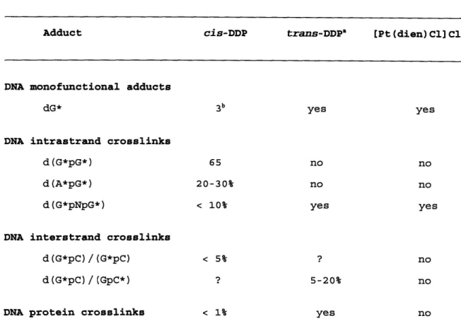
Table 1. Comparison of the adducts formed by platinum(II) compounds.
Adduct cis-DDP trans-DDP" [Pt (dien) C1] Cla
DNA monofunctional adducts
dG* 3b yes yes
DNA intrastrand crosslinks
d(G*pG*) 65 no no
d(A*pG*) 20-30% no no
d(G*pNpG*) < 10% yes yes
DNA interstrand crosslinks
d(G*pC)/(G*pC) < 5% ? no
d(G*pC)/(GpC*) ? 5-20% no
DNA protein crosslinks < 1% yes no
a. The majority of DNA adducts formed by trans-DDP and [Pt(dien)Cl]C1 has not been quantitated.
b. Numbers indicate the percentage of each adduct compared to the total binding spectrum. In cases where accurate numbers are not available,
Figure 2. cis-DDP G*G*, A*G*, and G*TG* adducts. Only the chemical composition of the cis-DDP DNA adducts are depicted. Structural
conformations are discussed at length in the text and are diagrammed in the literature referenced therein.
I I
I
=z
z
I ha
a
v
a
m ed:
U
o
h r3a
v
a
4h"
r3
CC) 3(·Z
v oa
U
.$o
ha
r3v
a
(u 3e-v
o
a
o
I
0
I
0
C.0
I
0
IB. Cellular Effects of Platinum Anticancer compounds
1. Genotoxicity of Platinum-DNA Adducts
The genotoxicity of platinum DNA adducts is well established in a variety of biological systems and is expected based on the in vitro experiments that demonstrated cis-DDP adducts to be potent blocks to DNA polymerases. Early work by Rosenburg and colleagues identified
inhibition of DNA synthesis as the mechanism by which cis-DDP reduced the growth of E. coli (Rosenberg et al., 1967; Harder & Rosenburg, 1970). DNA adducts formed by [Pt(dach)C12], a chemotherapeutically active cis-DDP analogue, are genotoxic in a plasmid-based assay (Husain
et al., 1985). The level of genotoxicity of these adducts, 5.5 adducts
per lethal hit, is comparable to that of DNA lesions formed by
benzo(a)pyrene-7,8-dihydrodiol-9,10-oxide (BPDE) or uv photoproducts (Mizusawa et al., 1981; Strike & Roberts, 1981). Different platinum DNA adducts exhibit variable levels of genotoxicity. In an SV40-based in
vitro replication system utilizing HeLa cell extracts, bifunctional
cis-DDP adducts inhibited DNA replication 30 fold more effectively than the monofunctional DNA adducts formed by the therapeutically ineffective compound [Pt(dien)Cl]+ (Heiger-Bernays et al., 1990). A
site-specifically located G*G* cis-DDP adduct in M13mp18 DNA reduced DNA replication by 78% in E. coli (Bradley et al., 1993). In addition to inhibition of DNA synthesis, cis-DDP exhibits other genotoxic effects including chromosomal abnormalities (Kliesch & Adler, 1987) and sister chromatid exchanges (Vogel et al., 1991).1
'It should be noted, however, that gross chromosomal abnormalities and sister chromatid exchanges may be a result of the transformed phenotype of carcinoma cells and not a direct consequence of cis-DDP treatment.
2. Mutagenesis of cis-DDP in E. coli
a. Mutagenesis in E. coli.
cis-DDP induced mutagenesis was first demonstrated in E. coli by Beck & Brubaker (1975). cis-DDP is a base-pair substitution mutagen by virtue of its ability to revert 2-aminopurine and N-methyl-N'-nitro-N-nitrosoguanidine induced mutations and, conversely, by the ability of these mutagens to revert cis-DDP induced mutations. As expected, the frameshift mutagen ICR-191 is unable to revert any cis-DDP mutants.
cis-DDP mutagenesis is also dependent upon LexA (Venturini &
Monti-Bragadin, 1978) and RecA activities (Konishi et al., 1981), in particular the recombinational capabilities of RecA (Jarosik & Beck, 1984). Mutagenic dependence on LexA activity, and the ability of the mutagenesis enhancing plasmid pKM101 to increase the level of cis-DDP induced mutants in various cell types (Venturini & Monti-Bragadin, 1978; Jarosik & Beck, 1984) indicates that SOS processing of cis-DDP lesions is required for mutagenesis. Direct evidence for SOS involvement in mutagenic processing has been provided by Fram et al. (1985), who demonstrated that cis-DDP mutagenesis is abolished in a umuD,C background.
b. Mutational specificity of cis-DDP in E. coli.
The exact nature of the mutations of cis-DDP in E. coli has been studied in several forward mutational assays but with conflicting results. By using the endogenous E. coli lacI gene as the genetic target, Brouwer et al. (1981) found that the mutations arising from cis-DDP treatment to cells were primarily at d(GpApG) and d(GpCpG)
sequences. No mutations were detected with the trans compound. In this work, no specific treatment was used to induce SOS other than the drug
treatment itself. By contrast, Burnouf et al. (1987) found that the majority (>90%) of mutations occurred at d(ApG) and d(GpG) sequences in the tetR gene of cis-DDP modified pBR322 that was replicated in SOS induced E. coli cells. The differences between the results of these two studies could be due to differences in the mode of DNA damage (in the former study intact cells were treated, whereas in the latter DNA was modified and then introduced into the host for mutation fixation), to differences in the range of mutations detectable in the respective genetic systems, or to the state of SOS induction of the cells in which the mutations were fixed. More recently, the mutagenicity of cis-DDP G*G* (Bradley et al., 1993) and A*G* (Burnouf et al., 1990) adducts has been determined site specifically. The results of these studies are analyzed in detail in comparison with the site specific analysis of the cis-DDP G*G*, A*G*, and G*TG* adducts done in the present work (see Discussion).
3. Mechanism of cis-DDP Mutagenesis in Prokaryotes
a. SOS dependence of mutagenesis
A wide range of DNA damaging agents can induce the SOS response in
E. coli, including uv radiation, methylmethanesulfonate,
4-nitroquinolone-l-oxide, and aflatoxin B. cis-DDP also induces the
SOS response, as evidenced by its ability to stimulate the filamentous growth of bacteria (Rosenberg et al., 1967), to induce prophage from
lysogenic bacteria (Reslova, 1971), and to induce increased cellular levels of RecA protein (Salles & Lesca, 1982). As with many of the treatments mentioned, cis-DDP is also an SOS-dependent mutagen; this property will be described in detail in the next section. Thus the
state of SOS induction is important to any mutagenesis studies of cis-DDP in E. coli. The SOS response of E. coli, therefore, will be
examined in detail below.
The SOS response to DNA damage is characterized by enhanced DNA repair activities, enhanced mutagenesis, inhibition of cell division, and prophage induction.' This diverse array of physiological responses result from the expression of an inducible set of genes that are under the control of the RecA and LexA proteins. Most of the literature on the SOS response describes the UvrABC exinuclease complex, which
efficiently repairs a wide array of bulky lesions in DNA (Sancar & Rupp, 1983), including adducts formed by cis-DDP (Brouwer et al., 1988). This pathway has been extensively reviewed (Witkin, 1976; Little & Mount, 1982; Walker, 1984; Battista et al., 1990; Echols & Goodman, 1990; Lin & Sancar, 1992; Altshuler, 1993; Grossman & Thiagalingam, 1993; Sancar & Tang, 1993). The work done for this dissertation does not directly address the repair of platinum-DNA adducts.2 This discussion, therefore, will emphasize those aspects of the SOS response that engender the mutagenic processing of damaged DNA.
Under physiologically normal cellular conditions, a repressor protein, LexA, is bound to the operator regions of the at least 17-20 genes thought to comprise the SOS regulon. The SOS network is activated when a cell is exposed to a DNA damaging agent. The inducing signal is
'The SOS response has been most extensively studied in E. coli and, unless otherwise stated, this discussion describes the SOS response in
E. coli. It should be noted that related gram-negative bacteria mount
very similar responses to DNA damage. In addition, gram-positive bacteria, exemplified by Bacillus subtilis also exhibit SOS-like
responses when confronted with DNA damage (Cheo et al., 1993) and it is postulated that eukaryotes also have similar responses to DNA damage (Ho
et al., 1993).
2The possible role of DNA repair by UvrABC in mediating the
observed SOS-dependent enhanced survival of platinum drug modified M13 bacteriophage genomes replicated in E. coli is briefly addressed in the dissertation in the experiments comparing the survival of cis-DDP
modified DNA transfected into either E. coli DL7 (wt) or DL6 (uvrA) cells (see Figure 11).