CHARACTERIZATION OF THE AGGREGATION PATHWAY COMPETING WITH PRODUCTIVE PROTEIN FOLDING
BY
MARGARET A. SPEED BSE Chemical Engineering Princeton University, 1991
Submitted to the Department of Chemical Engineering in partial fulfillment of the requirements for the degree of
DOCTOR OF PHILOSOPHY in
BIOCHEMICAL ENGINEERING at
MASSACHUSETTS INSTITUTE OF TECHNOLOGY June, 1996
@ Margaret A. Speed. All rights reserved.
The author hereby grants to MIT permission to reproduce and to distribute publicly paper and electronic copies of this thesis document in whole or in part.
Signature of Author
Certified by Certified by. Accepted
Mfgaret A. Speed, Ibepartment of Chemical Engineering
April 4, 1996
fessor Daniel I.C. Wang, Thesis cosupervisor
'7Thesis cosupervisor Professor Robert Cohen, Chairman; Departmental Committee for Graduate Students
,ASSACHUSGEITTS INS IU
OF TECHNOLOQGY
JUN 2 71996 Science
CHARACTERIZATION OF THE AGGREGATION PATHWAY COMPETING WITH PRODUCTIVE PROTEIN FOLDING
BY
MARGARET A. SPEED
Submitted to the Department of Chemical Engineering Massachusetts Institute of Technology
on April 4, 1996
in partial fulfillment of the requirements for the degree of Doctor of Philosophy in Biochemical Engineering ABSTRACT
The formation of insoluble aggregates due to protein misfolding is a phenomenon
associated with in vivo inclusion body formation, in vitro aggregation, and various human diseases. Analysis of the polymerization mechanism of the aggregation reaction requires study of the polypeptide chain association pathway and characterization of the multimeric intermediates.
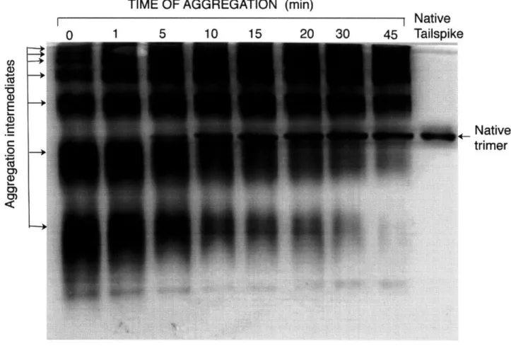
The model system that was chosen for these folding and aggregation studies was the phage P22 tailspike protein, which has been investigated previously both in vivo and in vitro. Nondenaturing polyacrylamide gel electrophoresis (PAGE) was used to isolate early intermediates in the in vitro pathway to the aggregated state for the tailspike protein. A sequential ladder of multimers of a partially folded species was present in the initial stages of the aggregation reaction. Discrete species formed during the polymerization process involving the noncovalent association of monomeric folding intermediates. The
aggregation intermediates were nonnative multimers that were sensitive to SDS and protease unlike the thermostable native trimer. Using a nondenaturing Western blot protocol, reactivity with monoclonal antibodies against native and nonnative tailspike was screened. Aggregation intermediates displayed nonnative epitopes in common with
productive folding intermediates. The nonnative epitopes on the intermediates were located on the partially folded N-terminus in a region nonessential to folding, and this had
structural implications as to the mechanism of folding and aggregation.
Quantitative modeling of the aggregation reaction and two-dimensional PAGE data indicated that tailspike aggregation occurred by multimeric cluster-cluster
polymerization, in which two multimers of any size could associate to form a larger aggregate. The specificity of aggregation was tested for a mixture of proteins refolding in vitro, and the results indicated that folding intermediates of different proteins did not aggregate heterogeneously with each other but only self-associated.
This methodology of isolating early multimers along the aggregation pathway has been shown to be applicable to other proteins, such as the P22 coat protein and carbonic anhydrase II, and may potentially be used to monitor the extent of aggregation in the production of therapeutic proteins, identify inclusion body intermediates, or isolate early multimeric intermediates in amyloid fibril formation.
Thesis supervisors: Dr. Daniel I.C. Wang, Institute Professor Dr. Jonathan King, Professor of Biology
ACKNOWLEDGMENTS
This project was made possible by an interdepartmental collaboration between the laboratories of Professor Wang and Professor King within the Biotechnology Process Engineering Center of MIT. My coadvisors have been instrumental at bringing together the Chemical Engineering, Biology, and Chemistry Departments at MIT in the area of
biotechnology and biomedical research. Financial support for this research project came from the National Science Foundation's Engineering Research Center Initiative under the Cooperative Agreement CDR-8803014 and the NIH Biotechnology Training Grant (NIGMS-2-T32-GM08334).
Professor Wang has been a source of inspiration for me and probably for everyone else in biotechnology. When I first began this research project, I was working in the shadow of my immediate predecessor, Dr. Jeffrey Cleland, who set the bar for thesis research in protein folding and aggregation. Professor Wang was supportive throughout the course of my project and gave me encouragement when I needed it. He also had a great ability to refocus my project in my periods of frustration and guided my research to address problems relevant to biotechnology.
Professor King welcomed me into his lab with open arms and adopted me as a card carrying biology graduate student. Without his guidance, this thesis project would not have gotten off the ground as smoothly as it did. His understanding of the scientific details of these experiments and ability to interpret results with an "alternative hypothesis" in mind have turned troubleshooting into enjoyable philosophical discussions. Jonathan King has taught me a lot about scientific methodology, and I admire him for his scientific, social, and political convictions.
I want to thank both past and present members of the King laboratory for their guidance throughout this project. Special thanks goes to Anna Mitraki who served as my mentor in teaching protein biochemistry to a chemical engineer. Anne Robinson has been a great labmate and friend who understands me. Cammie Haase-Pettingell should receive a medal of honor for attempting to teach microbiology to an engineer. The non-tailspike members of the lab, Carolyn Teschke, Peter Privilege, and Barrie Greene, have also provided endless hours of scientific discussion and amusement.
Thanks also go to my parents for bringing me into this world and to my brother for being himself. Hats off to my Princeton friends for making the last 5 years endurable and to my housemates for putting up with my annoying sense of humor.
Nothing is rich but the inexhaustible wealth of nature.
She shows us only surfaces, but she is a million fathoms deep. -Ralph Waldo Emerson
TABLE OF CONTENTS
page
List of figures and tables ... 11
Abbreviations... ... ... 14
I. Introduction ... 15
II. Literature review ... 21
A. In vitro refolding and aggregation ... 21
B. In vivo folding and inclusion body formation ... 30
C. Bacteriophage P22 tailspike protein ... . . ... . 34
D. Polymerization mechanisms ... . 50
E. Methods to monitor folding and aggregation .. ... 54
F. Strategies to address the protein folding problem ... 58
III. Multimeric intermediates in the pathway to the aggregated inclusion body state 63 A. Materials and methods .... .. . ... .. .. 66
i. Protein .... . . . ... . . . .. . . .. 66
ii. In vitro refolding and aggregation ... 66
iii. Gel electrophoresis ... 67
iv. Proteolysis . . . 68
B. Results ... ... ... 68
i. Identification of aggregation intermediates ... 69
ii. Chain composition of the aggregation intermediates ... 73
iii. Intermolecular bonding ... 75
iv. Conformation of aggregation intermediates ... 79
v. Extension of methodology to other proteins ... 83
C. Discussion ... ... 83
i. Characterization of aggregation intermediates ... 84
ii. Determination of aggregation pathway ... 88
iii. The mechanism of polymerization ... ... .88
II
A. Materials and methods ... ... . . . . 93
i. Materials . ... ... 93
ii. In vitro refolding ... 93
iii. Gel electrophoresis . . . . 93
iv. Western blotting ... 94
v. Refolding in presence of antibodies . ... . . . . . 94
vi. Immunoprecipitation . ... 95
B. Results ... ... .... 96
i. Probing the structure of aggregation intermediates . ... 96
ii. Characterizing the nonnative epitopes . ... . . 98
iii. Location of the nonnative epitopes . ... 104
iv. Nonnative epitopes on productive folding intermediates . . . . 108
C. Discussion . . . . . .. . . . . . . 121
i. Domains nonessential for folding . ... 121
ii. Structural implications ... 123
V. Polymerization mechanism of aggregation . . . . 125
A. Materials and methods . . .. . . .. . .... . . . 126
i. In vitro aggregation. .... . . . ... . 126
ii. Classical light scattering . . .. .... . . .. . . 127
iii. Gel electrophoresis. ... . ... . 127
iv. Computer simulations . . . . .. . . . . . 129
B. Results ... ... ... .. ... 129
i. Classical light scattering .... ... . 130
ii. Gel electrophoresis . . . . . . . . 133
iii. Mechanism of aggregation determined by 2D-PAGE . . . . . 139
C. Discussion ... ... .... 145
i. Polymerization mechanism ... . ... 145
ii. Aggregation of folding intermediates . ... . .. 147
iii. Irreversibility of aggregation . ... . . . . 148
VI. Specific aggregation of partially folded polypeptide chains . ... . . . 151
A. Materials and methods .. .. . . . ... 155
i. Materials .... ... .... 155
ii. In vitro refolding and aggregation . .. ... . 156
iv. Western blotting ... 157
B. Results . . . 157
i. Characterization of folding and aggregation intermediates . ... 158
ii. Mixed aggregation studies . . . 161
iii. Effects of polypeptide chain concentration ... 170
C. Discussion ... 174
i. Mechanism of aggregation ... 176
ii. Oligomeric proteins ... .. 177
iii. Kinetic factors ... 178
iv. Specificity of macromolecular interactions ... 179
VII. Summary and conclusions. .. ... 181
i. Mechanism of aggregation ... 181
ii. Structural implications on folding and aggregation ... 182
iii. General refolding strategies ... 188
VIII. References . . . . . . . . . . 191
IX. Appendices ... 207
A. Temperature sensitive folding mutations in P22 tailspike ... 208
B. Classical light scattering data analysis ... ... . 209
i. Multimeric polymerization model ... 209
ii. Sequential polymerization model ... 210
C. Matlab programs to model aggregation ... 211
i. Differential equations for multimeric polymerization (CLS data) . 211 ii. Differential equations for sequential polymerization (CLS data) . . 212
iii. Differential equations for multimeric polymerization & folding . . 213
LIST OF FIGURES AND TABLES 1.1 Folding and aggregation of therapeutic proteins
1.2 Human diseases caused by protein misfolding, aggregation, & improper trafficking 2.1 Electron micrographs of inclusion bodies in E. coli
2.2 'Ribbon diagram of the structure of native tailspike trimer 2.3 In vivo folding and aggregation pathway
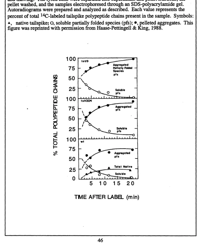
2.4 Location of temperature-sensitive folding mutations in P22 tailspike protein 2.5 Kinetics of intracellular aggregation for temperature-sensitive folding mutants vs.
wild-type tailspike protein
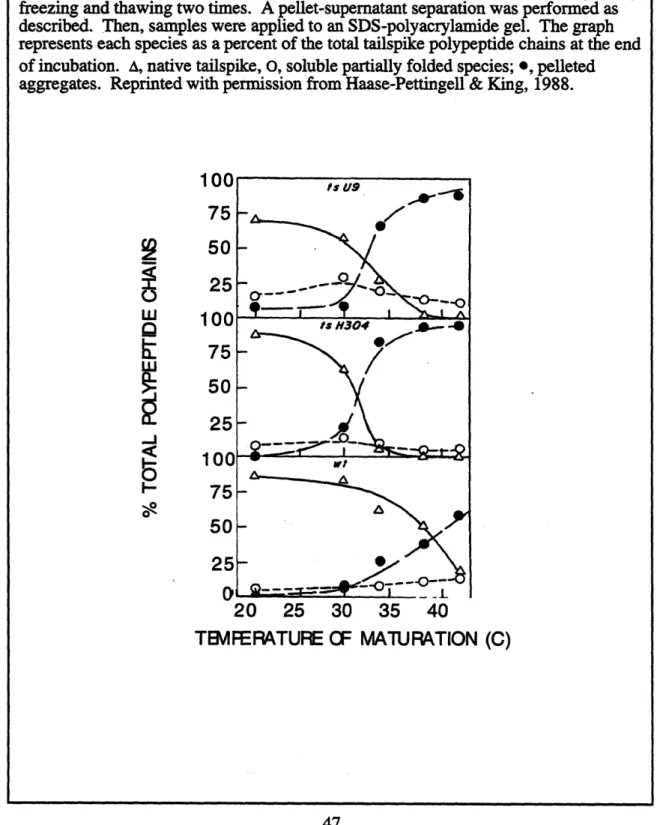
2.6 Temperature dependence of in vivo folding and aggregation for temperature-sensitive folding mutants vs. wild-type tailspike protein
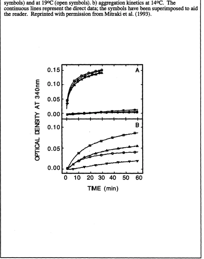
2.7 Kinetics of in vitro aggregation of wild-type tailspike protein, temperature-sensitive folding mutants, and suppressor mutants
3.1 In vitro refolding and aggregation pathway
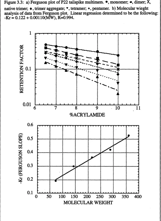
3.2 Isolation of in vitro aggregation intermediates by nondenaturing gel electrophoresis 3.3 Ferguson plot of P22 tailspike multimers
3.4 Protocol for determining intermolecular bonding of aggregates 3.5 2D-nonreducing SDS gel of aggregation intermediates
3.6 Protocol for proteolysis of tailspike folding and aggregation intermediates 3.7 Proteolysis of tailspike folding and aggregation intermediates
3.8 Extension of methodology to isolate multimers of carbonic anhydrase and P22 coat protein
4.1 Monoclonal antibody reactivity with native trimer and refolding tailspike 11
4.2 Screening of individual monoclonal antibody reactivity with native trimer and refolding tailspike
4.3 Model of anti-nonnative tailspike monoclonals' reactivity with folding and aggregation intermediates
4.4 Monoclonal antibody reactivity with denatured polypeptide chains
4.5 Monoclonal antibody reactivity with peptide fragments and truncated tailspike 4.6 Model of anti-nonnative tailspike monoclonals' reactivity with productive folding
intermediates
4.7 Protocol for refolding tailspike in the presence of monoclonal antibodies 4.8 Refolding tailspike in the presence of monoclonal antibodies
4.9 Protocol for immunoprecipitation of aggregation intermediates
4.10 Immunoprecipitation of tailspike folding and aggregation intermediates
5.1 Classical light scattering of tailspike aggregation
5.2 Nondenaturing gel electrophoresis of folding and aggregation intermediates generated at various refolding temperatures
5.3 Quantitative densitometry and computer simulations depicting refolding and aggregation kinetics at various refolding temperatures
5.4 Multimeric polymerization: 2D-PAGE of aggregation intermediates (250C)
5.5 Irreversibility of aggregation: 2D-PAGE of aggregation intermediates (40C) 5.6 Models to depict the polymerization mechanism of aggregation
6.1 In vitro refolding and aggregation pathways for P22 tailspike and P22 coat polypeptide chains
6.2 Nondenaturing gel electrophoresis of native tailspike trimer, refolding and aggregation tailspike, refolded coat protein, and aggregating coat chains 6.3 Protocol for mixed refolding experiment
12
6.4 Western blot of nondenaturing gels from mixed refolding experiment probing with anti-tailspike monoclonal antibodies
6.5 Western blot of nondenaturing gels from mixed refolding experiment probing with anti-coat antibodies
6.6 Western blot of nondenaturing gels from mixed refolding experiment probing with anti-tailspike and anti-coat antibodies
6.7 Model of specificity of aggregation
7.1 Structural cartoon of tailspike aggregation intermediates 7.2 Intrachain vs. interchain P-helix
ABBREVIATIONS Ac Coat refolding and aggregation At Tailspike refolding and aggregation
At+c Mixed refolding of tailspike and coat chains Abc Anti-coat polyclonal antibodies
Abt Anti-tailspike monoclonal antibodies ANS Anilino-naphthalene sulfonate
anti-I Anti-intermediate tailspike monoclonal antibody anti-N Anti-native tailspike monoclonal antibody
bGH Bovine growth hormone
PME 3-mercaptoethanol
CAB Carbonic anhydrase B
CD Circular dichroism
CLS Classical light scattering
DTT Dithiothreitol
ECL Enhanced chemiluminescence EDTA Ethylene diamine tetraacetic acid ELISA Enzyme-linked immunosorbent assay FTIR Fourier transform infrared spectroscopy GuHCl Guanidine hydrochloride
HPLC High performance liquid chromatography I Partially folded intermediate
MAb Monoclonal antibody
MW Molecular weight
N Native protein
Nc Native coat monomer
Nt Native tailspike trimer
PAGE Polyacrylamide gel electrophoresis PDI Protein disulphide isomerase PPI Peptidyl-prolyl isomerase
PEG Polyethylene glycol
PM Powdered milk (blocking reagent) pt Protrimer folding intermediate QLS Quasi-elastic light scattering
SDS Sodium dodecyl sulfate
TBS Tris buffered saline
tsf Temperature-sensitive folding
TTR Transthyretin
U Unfolded polypeptide chains
UV Ultraviolet
CHAPTER 1: INTRODUCTION
The study of protein folding and aggregation has its historical roots in food science technology. The food and biotechnology industry arose from the practice of fermentation to produce beer and wine as early as 7000BC (Bailey & Ollis, 1986). With the
development of microbiology, food science has expanded to include the production of cheese, yogurt, soy, and whipped cream, which involve protein denaturation and aggregation of egg, milk, and soy proteins (Ferry, 1948). Rheology and texture are the issues in protein aggregation during food preparation rather than enzymatic activity, as in biotechnology. The principles of protein denaturation by heat treatment, shear stress, or acidic conditions are utilized in food processing. Gel formation depends on the
denaturation conditions of pH, salt, and protein concentration (Mulvihill & Kinsella, 1988). Biochemical studies have been performed on the aggregation of many food proteins, including lysozyme (Li-Chan & Nakai, 1991), ovalbumin (Painter & Koenig,
1976), Pf-lactoglobulin (Clark & Lee-Tuffnell, 1986; Langton & Hermansson, 1991),
ic-casein (Haque & Kinsella, 1988), whey (Hermansson, 1986), soy glycinin (Badley et al.,
1975), myosin (Kaminer & Bell, 1966), and peanut arachin (Kumar et al., 1980).
Experienced cooks know in making an egg and lemon sauce to add the hot chicken broth to the egg and lemon sauce in that order and not the reverse (Mitraki, personal cooking lesson). Aggregation in cooking can be avoided by ensuring proper heat transfer
and dilution of the acid-denatured egg protein. If the order of addition is reversed or the transfer is not done in stages with continuous mixing, the resulting concoction will resemble egg drop soup. Whether frying an egg, whipping cream, or making an egg and lemon sauce, years of experience in the kitchen have utilized the principles of denaturation and aggregation of egg and milk proteins and heat and mass transfer principles.
With the advent of biotechnology, protein folding became a recognized scientific problem (Marston, 1986). Before the 1980's, therapeutic proteins such as insulin and growth hormone were isolated from their natural source (Schein, 1989). The disadvantage of this practice is the large amount of material that must be processed to extract enough of the desired protein, the lack of availability of a reliable source, and the possibility of
spreading infectious agents to human recipients. Recombinant DNA technology offered the possibility of producing heterologous protein in a bacterial host (Cohen et al., 1973). This technique promised a continuous supply of any protein that could be cloned, expressed, and purified. However, it became evident that the proteins overexpressed in bacteria formed insoluble aggregates, or inclusion bodies (Prouty & Goldberg, 1972; Prouty et al.,
1975). In the aggregated state, the protein did not have any enzymatic activity or
therapeutic value. Therefore, the biotechnology industry has catalyzed an intense effort in solving the protein folding problem.
Seminal work in in vitro protein refolding was performed on RNase A by Anfinsen (Anfinsen, 1961). This research was the first documentation that a polypeptide chain could regain its enzymatic activity by refolding. The primary sequence of the protein was
sufficient to direct the refolding into the native conformation without the addition of
exogenous cofactors. In vivo, Prouty discovered that abnormal E. coli proteins aggregated in cells to form inclusion bodies (Prouty & Goldberg, 1972). Two decades later, the biotechnology industry is utilizing knowledge of in vivo and in vitro folding and aggregation to refold recombinant proteins from the aggregated inclusion body state (Marston, 1986; Cleland, 1993; Schein, 1989). Table 1.1 shows that most therapeutic proteins important in the biotechnology industry are recovered from the aggregated inclusion body state.
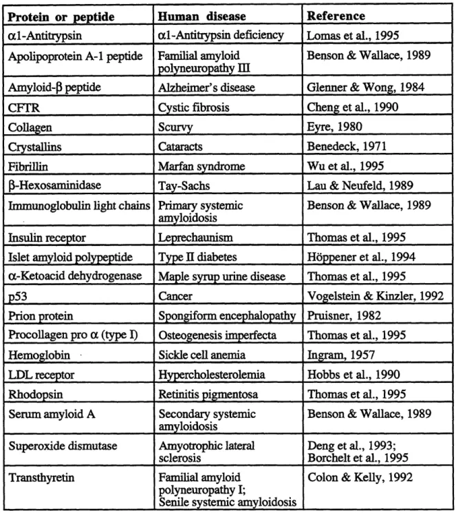
Protein misfolding also plays a role in human diseases (Benson & Wallace, 1989; Thomas et al., 1995). Table 1.2 lists some examples of diseases caused by protein
Table 1.1: Folding and aggregation of therapeutic proteins.
Protein Expression Conformation Reference
System State
Antibody OKT3 Hybridoma Native Cleland, 1993
al-Antitrypsin E. coli Nativet Courtney et al., 1984
rh DNase CHO Native Cleland, 1993
rh Erythropoietin CHO Native Lin et al., 1985 Factor VIII hamster kidney Native Wood et al., 1984
cells
Factor IX CHO Native Kaufman et al., 1986
basic-FGF / E. coli Native Squires et al., 1988
FMDV (foot&mouth E. coli Inclusion body Builder&Ogez, 1985;
disease viral coat) Olsen&Pai, 1985
h Fibroblast Interferon E. coli Inclusion bodyt Builder&Ogez, 1985;
Olsen&Pai, 1985
G-CSF E. coli Inclusion body Cleland, 1993
GM-CSF E. coli Inclusion body Cleland, 1993
hGH V E. coli Inclusion bodyt Builder&Ogez, 1985;
Olsen&Pai, 1985
IGF-1 / E. coli Inclusion body Chang&Swartz, 1993 IL-2 / E. coli Inclusion bodyt Koths et al., 1985 h Insulin E. coli Inclusion body Marston, 1986 a-IFN E. coli Inclusion bodyt Schein & Notebornm,
1988
1-IFN E. coli Inclusion body Konrad et al, 1984;
Hershenson, 1990
rh y-IFN E. coli Inclusion bodyt Schein & Noteborn,
1988
h TNF E. coli Native Pennica et al., 1984
rh tPA CHO Native Lubiniecki et al., 1988
tIndicates that the fraction of polypeptide chains in the aggregated inclusion body state was highly dependent on growth conditions
Table 1.2: Human diseases caused by protein misfolding, aggregation, and improper trafficking.
Protein or peptide Human disease Reference
al-Antitrypsin al-Antitrypsin deficiency Lomas et al., 1995 Apolipoprotein A-1 peptide Familial amyloid Benson & Wallace, 1989
polyneuropathy
Ill
Amyloid-f3 peptide Alzheimer's disease Glenner & Wong, 1984
CFTR Cystic fibrosis Cheng et al., 1990
Collagen Scurvy Eyre, 1980
Crystallins Cataracts Benedeck, 1971
Fibrillin Marfan syndrome Wu et al., 1995
j~-Hexosaminidase
Tay-Sachs Lau & Neufeld, 1989Immunoglobulin light chains Primary systemic Benson & Wallace, 1989
amyloidosis
Insulin receptor Leprechaunism Thomas et al., 1995 Islet amyloid polypeptide Type II diabetes H6ppener et al., 1994 a-Ketoacid dehydrogenase Maple syrup urine disease Thomas et al., 1995
p53 Cancer Vogelstein & Kinzler, 1992
Prion protein Spongiform encephalopathy Pruisner, 1982 Procollagen pro a (type I) Osteogenesis imperfecta Thomas et al., 1995 Hemoglobin Sickle cell anemia Ingram, 1957 LDL receptor Hypercholesterolemia Hobbs et al., 1990 Rhodopsin Retinitis pigmentosa Thomas et al., 1995
Serum amyloid A Secondary systemic Benson & Wallace, 1989
amyloidosis
Superoxide dismutase Amyotrophic lateral Deng et al., 1993;
sclerosis Borchelt et al., 1995
Transthyretin Familial amyloid Colon & Kelly, 1992 polyneuropathy I;
misfolding, aggregation, and improper trafficking. Amyloidosis is the most recognized disease involving protein aggregation (Kelly & Lansbury, 1994). The formation of insoluble fibrils either systemically or in certain organs, such as the brain, can cause polyneuropathy and neurodegenerative disorders. Examples of amyloidogenic proteins include 3-amyloid (Glenner & Wong, 1984), transthyretin (Colon & Kelly, 1992), prions (Pruisner, 1982), immunoglobulin light chains (Myatt et al., 1994; Hurle et al., 1994), and islet amyloid polypeptide protein (Hbppener et al., 1994). Other proteins that cause disease via aggregation include sickle cell hemoglobin (Ingram, 1957), crystallin proteins in
cataract formation (Benedek, 1971), and superoxide dismutase associated with
amyotrophic lateral sclerosis, or Lou Gehrig's disease (Deng et al., 1993; Borchelt et al., 1995). The molecular mechanism of these diseases may involve the absence of the active folded native protein, the improper trafficking of protein within the cell, or the presence of toxic aggregates (Thomas et al., 1995).
Despite of the link between protein aggregation and human diseases, protein aggregation is a natural process regulated by the body. For example, the aggregation of lysophospholipase is an essential step in blood clotting. Von Willebrand factor, or factor VIII, is required to mediate platelet aggregation in blood clotting, the absence of which causes hemophilia (Wood et al., 1984).
Due to the need of the the biotechnology industry, biochemical engineers have been recruited to deal with the protein folding problem. The chemical engineering of protein folding and aggregation includes thermodynamics, kinetics, and engineering principles in conjunction with biochemistry and molecular biology. Thermodynamic data and molecular models originally designed for the petroleum industry are now applied to protein folding and aggregation (Chen et al., 1995). Group contribution theory can be employed to estimate the AG of transferring amino acid side chains from a solvated state of the unfolded polypeptide chain to the protein core of the native species. In modeling the kinetics of
II 11
aggregation, principles of polymerization chemistry and irreversible reactions are incorporated into the kinetic analysis (Sonntag & Strenge, 1987). Fundamental reactor
engineering principles are also used in the design of novel refolding strategies. One such design involves immobilizing unfolded polypeptide chains onto a column matrix and passing the refolding buffer through the matrix (Sinna & Light, 1975). Other refolding
strategies attempt to keep the concentration of folding intermediates below the critical aggregation concentration by performing a series of step-wise dilutions of the denatured protein (Rudolph & Fischer, 1987).
To address the protein folding problem, this thesis research focuses on the competition between productive folding and aggregation. The point at which one can control the folding reaction is the critical folding intermediates at the junction between the productive folding pathway and the off-pathway aggregation reaction (Mitraki & King, 1989). An understanding of the folding pathway as well as the polymerization mechanism of aggregation is required to address the protein folding problem. Aggregation occurs by the association of folding intermediates via a defined polymerization mechanism. The resulting off-pathway intermediates are multimeric precursors to the aggregated inclusion body state (Speed et al., 1995). The objectives of this thesis research are: 1) identify the
multimeric aggregation intermediates of P22 tailspike polypeptide chains, 2) characterize the biophysical properties of the aggregation intermediates, 3) characterize the structural basis of aggregation, 4) determine the polymerization mechanism of aggregation, 5) determine the specificity of aggregation, and, 6) extend the methodologies to other protein systems.
CHAPTER 2: LITERATURE REVIEW
A major problem in biotechnology is the incorrect folding of newly synthesized polypeptide chains and formation of insoluble aggregates which are biologically inactive (Marston,
1986; Mitraki and King, 1989; DeBernardez-Clark and Georgiou, 1991; Wetzel, 1994). In vivo, this folding problem frequently arises with heterologous proteins overexpressed in E. coli, which form inclusion bodies, or amorphous aggregates within the cell. The
analogous aggregation problem occurs in vitro via a similar association mechanism
(Zettlmeissl et al., 1979; Colon & Kelly, 1992; Mitraki et al., 1991). In the cases where the competition between the refolding and aggregation reactions has been directly studied, the precursor to aggregation is a defined intermediate in the refolding pathway (Brems,
1988; Mitraki et al., 1993). For numerous proteins, this class of folding intermediates is the species that is recognized by the GroE chaperonin, which assists in protein folding by binding to folding intermediates and preventing improper association of the polypeptide chains (Goloubinoff et al., 1989). Non-native multimerization subsequently leads
irreversibly to the formation of large aggregates or inclusion bodies. Therefore, further study of the aggregation reaction is essential in designing effective refolding strategies. A. In vitro refolding and aggregation
In the early 1960's, Anfinsen performed the first experiments in protein refolding in which he successfully renatured bovine pancreatic ribonuclease in vitro without additional
cofactors (Anfinsen et al., 1961). The blueprint for folding was located in the primary amino acid sequence and was not dependent upon external templates (Anfinsen, 1973). Over the course of three decades of research in protein folding with significant advances, the protein folding problem has not been solved. However, there is now a greater understanding of the structural changes, thermodynamic forces, and kinetic competition associated with protein folding and aggregation.
.ll II
In the simplest model for protein folding, the unfolded polypeptide chain is in equilibrium with the native state. For most proteins, the folding pathway is more
complicated and involves either a two-step folding process with a single intermediate or a complicated multistage mechanism with several intermediates. Most globular proteins are hypothesized to refold through a molten globule intermediate, in which the folding
intermediate has an exposed hydrophobic surface that can lead to aggregation. Examples of proteins that have molten globule folding intermediates include carbonic anhydrase B, a-lactalbumin, p-lactamase, bovine growth hormone, phosphoglycerate kinase, and P-lactoglobulin (Ptitsyn et al., 1990).
The forces that are important in protein folding and aggregation are electrostatics, hydrogen bonding, van der Waals interactions, and hydrophobic interactions (Dill, 1990; Creighton, 1993). Electrostatics are long-range interactions with a strong dependence on ionic strength and pH. High salt concentrations can shield charges and reduce the
electrostatic effect. The pH determines the global charge of a protein, and aggregation is more likely to occur when the solution pH equals the isoelectric point of the protein due to the lack of charge repulsion for neutral species. Although each ion pair in the native protein
can provide 1-3 kcal/mol stabilization (Fersht, 1972), ion pairing generally is not the dominant force in protein folding, as determined by a weak dependence on ionic strength near the isoelectric point (Hermans & Sheraga, 1961) and lack of conservation of ion pairing in structural evolution (Barlow & Thorton, 1983). Hydrogen bonds and van der Waals interactions, which provide 2-10 kcal/mol stabilization (Pauling, 1960), are critical
in the formation of secondary structure, such as helix-coil transitions (Schellman, 1958) and P-sheet formation (Mattice & Scheraga, 1984). The main driving force of folding is generally hydrophobicity, or the "transfer of nonpolar solutes into an aqueous solution, characterized by the ordering of water molecules around nonpolar solutes" (Dill, 1990). The dominant force opposing protein folding is the loss in degrees of freedom of the polypeptide chain in folding into the native conformation. There is an unfavorable loss in
entropy upon ordering water molecules around the protein, an excluded volume effect, and steric constraints. Generally, the overall change in free energy associated with protein folding is 5-20 kcal/mol (Pace, 1975; Privalov, 1979).
Seminal work in identifying and characterizing intermediates on the folding pathway was first done on bovine growth hormone (bGH) by Brems and colleagues (Brems et al., 1986; Brems, 1988; Lehrman et al., 1991). Equilibrium experiments of bGH in various concentrations of guanidine hydrochloride (GuHC1) were performed using size-exclusion HPLC to measure the hydrodynamic radius or compactness, far-ultraviolet circular dichroism at 222nm (far-UV CD) to measure secondary structure, and ultraviolet absorbance at 290nm to measure exposure to solvent or extent of packing of the aromatic residues (Brems & Havel, 1989). The data indicate that the unfolding transition measured by the above three techniques did not occur at the same GuHC1 concentrations. This noncoincidence of signals indicated that the unfolding process was a multistate transition and not a simple two-step mechanism. The initial sign of denaturation was the disruption of the packed aromatic residues (tyrosine and tryptophan residues), followed by a loss in compactness and secondary structure at higher GuHCI concentrations. Tryptophan fluorescence quenching experiments were performed on bGH over a range of denaturant concentrations with various chemical quenchers (iodide, acrylamide, and trichloroethanol), measuring fluorescence at 350nm. The data indicated that the quenching was greater for the intermediate than native bGH but less than fully denatured protein, which suggested that the tertiary structure was disrupted during the unfolding process. From the other data comparing the properties of native, intermediate, and unfolded species, the intermediate was determined to be a compact globule that retained much of its a-helical structure (far-UV CD data) but had disrupted packing of the aromatic side chains ((far-UV absorption data).
The problem with aggregation arises when the monomeric folding intermediates associate to form off-pathway multimeric species. Brems used a two-step dilution method
II II
to investigate the folding intermediates and aggregation process during refolding of bovine growth hormone (Brems, 1988). In the first step, certain bovine growth hormone
conformers were populated by adjusting GuHCl and protein concentrations or by taking samples during kinetic refolding experiments. The second step was to dilute the sample to solvent conditions (low GuHCl concentration) to cause precipitation of only associated intermediates. To determine the amount of aggregate, the turbidity was measured at 450nm using a spectrophotometer. During refolding, the initial unfolding conditions affected the extent of aggregation and partially unfolded bGH led to aggregation. In addition, the mechanism was temperature dependent, with more aggregation occurring at higher temperatures.
Previous work by Brems (1986) suggested that aggregation was reduced by adding peptide fragments of bGH (residues 96-133) to prevent association of intermediates. This peptide fragment was thought to be an amphipathic helix, and perhaps it interacted with the hydrophobic surface of the intermediate and blocked self-aggregation. Experiments were performed to see the effects of adding various concentrations of peptide 96-133 in refolding assays (Brems, 1988). The peptide fragment effectively inhibited precipitation and
demonstrated half of its inhibition potential at a peptide concentration in excess of three fold over the bGH concentration. Further studies showed that peptide fragment 109-133 was just as effective in preventing precipitation as fragment 96-133, but peptide 96-112 was
not. Therefore, the C-terminal section of fragment 96-133 plays an important role in intermolecular self-association that leads to precipitation of folding intermediates. The aggregation mechanism appears to involve a specific hydrophobic patch or helix docking site on the surface of the protein folding intermediate. The free energy of the helix-helix docking reaction during dimer formation can be calculated by group contribution theory
Another globular protein that has been extensively studied is carbonic anhydrase B (CAB), which is an esterase that regulates the ratio of carbon dioxide to bicarbonate concentration in blood. Using quasi-elastic light scattering (QLS) and size exclusion chromatography (HPLC) to monitor the refolding and aggregation process, the folding pathway and association of folding intermediates were characterized (Cleland & Wang, 1991). The refolding pathway involved two folding intermediates and an aggregation pathway that is caused by association of the first folding intermediate. The conditions under which micron-sized aggregates immediately formed defined the aggregation regime, and the multimer formation-regime was characterized by observable dimer and trimer species that formed before micron-sized aggregation. The lower limit for refolding was the regime which allowed the formation of multimers that did not develop into micron-sized aggregates. Measuring the formation of multimeric species by QLS, the aggregation regime and lower limit of refolding was mapped with respect to protein and denaturant concentration. The protein refolded into the native structure or a stable intermediate that did not aggregate at denaturant concentrations between the lower refolding limit and 1M GuHC1. The association of the early monomeric intermediate II to form a dimer was a slow reaction at equilibrium with a Keq of 1.3mM-1 and association rate of 5.16x10⁻³min-1. After dimerization, trimerization proceeded rapidly (Keq of 0.42M-1 and association rate of 0.133min-1), which directly led to irreversible aggregation (Cleland & Wang, 1991).
In kinetic and equilibrium experiments, aggregation occurs by the association of folding intermediates with exposed hydrophobic patches. Certain cosolvents can inhibit aggregation by interacting with folding intermediates to block self-association. These small organic and inorganic substances include an assortment of cofactors, metal ions, specific ligands, surfactants, sugars, and other solvent additives (Schein, 1990). The simplest possible mechanism by which cosolvents act involves the solvent molecule (e.g. low concentration of PEG) binding directly to the protein to stabilize that conformer and prevent self association. If the formation of the binding site is the rate limiting step of protein
II II
folding, then specific binding of the cosolvent to stabilize the folding intermediate enhances the rate of refolding. An alternative mechanism is that the cosolvent alters the solution properties by preferential hydration or exclusion (Arakawa & Timasheff, 1985).
Preferential hydration occurs when cosolvents, such as sugars, increase the surface tension of an aqueous solution and have a higher affinity for water than the hydrophobic protein does. The resulting hydration shell surrounding the protein is conducive for the burial of the hydrophobic core during refolding. The hydration leads to less free surface area and a lower free energy for the protein, which is the driving force for forming a compact
structure such as the native species or aggregate. The effectiveness of cosolvent addition in protein folding or solubility of the native species is dependent on the specific protein, protein concentration, cosolvent type, and cosolvent molecular weight (Arakawa et al.,
1990).
The cosolvent polyethylene glycol (PEG) of molecular weight 1000 to 8000 Da inhibited CAB aggregation and increased the refolding yield (Cleland & Wang, 1990a). The molecular weight of PEG was critical because too low MW PEG (200-600) did not
effectively shield the exposed hydophobic patches on the folding intermediate, whereas high MW PEG (20,000 Da) actually increased aggregation by the PEG molecule binding to more than one polypeptide chain (Cleland, Hedgepeth, & Wang, 1992). In addition, the
solvent exclusion effect caused high molecular weight PEG (10-50 wt%) to exclude CAB sterically from the bulk solution and caused the protein to precipitate because of the higher effective CAB concentration (Cleland & Wang, 1990a). The mechanism by which PEG interacted with CAB involved the cosolvent molecule binding to the first folding
intermediate of CAB (Cleland & Randolph, 1992). Because the hydrophobic patches on the protein surface were the sites for aggregation interactions, PEG binding to the
aggregation sites induced changes in the surface characteristics and prevented dimerization. Using PEG of MW 3350, stoichiometric analysis indicated that 2 PEG molecules were bound to the hydrophobic patches of each CAB polypeptide chain (Cleland, Hedgepeth, &
Wang, 1992). PEG bound to the first folding intermediate and not the second intermediate and native CAB. The mechanism of PEG-assisted refolding involved preventing the first intermediate from dimerizing rather than increasing the folding rate to form the second intermediate (Cleland, Hedgepeth, & Wang, 1992). PEG effectively assisted refolding at a concentration of 3 to 30g/L to produce an active species without aggregation, and optimal refolding was observed for PEG concentrations of 0.15-0.20 g/L (MW 3350) at a
[PEG]/[CAB] molar ratio of 2-3 under the refolding conditions of 0.50 mg/mL CAB, 1M GdnHC1, 50mM Tris, pH 7.5 (Cleland, Hedgepeth, & Wang, 1992). The apparent refolding rate increased three fold by adding 3g/L PEG of molecular weight 3350Da, as measured by the recovery of enzymatic activity (Cleland & Wang, 1990a). The ability of a cosolvent to assist refolding allowed recovery of an enzymatically active protein at
conditions which normally resulted in aggregation. In this respect, the mechanism by which cosolvents assist refolding is similar to the role of molecular chaperones in in vivo
folding (Cleland, Hedgepeth, & Wang, 1992).
Several amyloidogenic proteins in the human body have a propensity to aggregate to form insoluble plaques which then lead to neurodegenerative disorders. One such species is the [3-amyloid peptide, which is associated with Alzheimer's disease (Glenner & Wong, 1984). The C-terminus of amyloid peptide (Al1-42) is critical in fibril formation (Halverson et al., 1990), and this sequence is homologous to the region of the
amyloidogenic prion protein 94-111 involved in scrapie (Jarrett & Lansbury, 1992). Aggregates of the [3-amyloid peptide are found in the brain as plaques with substantial neurofibrillary tangles. The dimension of the fibrils are >1000A in length and 70-100A in width (Kelly & Lansbury, 1994). FTIR data indicate that the fibrils contain 13-sheet secondary structure with the [3-sheet contacts parallel to the axis of the fibril. This motif is similar to the parallel [3-spiral of the tailspike protein, except the local [3-sheet contacts are antiparallel for amyloid. The cross D3-fibril structure has specific interstrand alignment determined by solid state NMR (Sun et al., 1995). There is also extreme sheet pleating
with the side chains packed together tightly without the presence of water molecules. Examination of the bond angles shows an unusual Gly-Gly structure in a cis conformation. Fragment Ar334-42, which is extremely insoluble, may initiate fibril formation by the hydrophobic cluster stabilizing the cis isomer. The kinetics of fibril polymerization follow nucleation growth kinetics, which will be discussed in a later section (Jarrett & Lansbury,
1992). Seeding of the aggregation reaction with a preformed fibril bypasses the slow nucleation step, characteristic of nucleation-growth reactions (Jarrett et al., 1993).
Prion diseases are neurodegenerative disorders related to 03-amyloid fibril
formation. Types of prion diseases or scrapie include bovine spongiform encephalopathy, Creutzfeldt-Jakob disease, Gerstmann-Straussler-Schienker syndrome, fatal familial insomnia. The term "prion" was coined by Pruisner to describe a PROteinaceous INfectious agent which did not require nucleic acids to pass along its genetic code
(Pruisner, 1982). Infectious scrapie prion protein was resistant to nucleic acid modifying agents but susceptible to protein modifiers (Prusiner, 1982; Prusiner, 1992). Caughey, Lansbury, and colleagues developed an in vitro cell-free conversion of PrPC to PrPSc to prove that prion protein is the infectious agent for scrapie (Kocisko et al., 1994). In mixing 35S-labeled PrPC and PrPse, PrPSc was found to convert PrPC to the scrapie form, and the transition was monitored by the protease-resistance characteristic of PrPse. The conversion was specific in that other types of amyloid could not seed the scrapie transition and certain strains of scrapie were not infectious due to slight conformational differences in the scrapie seed (Kocisko et al., 1995). Similar to
1-amyloid
fibril formation, the conversion of normal cellular prion protein (PrPC) to the scrapie form (PrPSC) was accompanied by an a-helix to P-sheet conformational transition, as measured by CD (Pan et al., 1993). In fact, all known inherited scrapie mutations except one,F198S, have a higher predicted propensity to adopt a n-sheet conformation (Baldwin et al., 1995). Structural models of the normal cellular prion protein predict a 4-helix bundle motif in an x-orientation (Baldwin et al., 1995). Evidence suggests that the first helix and
specifically the hydrophobic peptide 104-122 is involved in converting prion protein to the scrapie form with 3-sheet structure, as measured by CD and FTIR spectroscopy (Nguyen et al., 1995). As in other protein aggregation phenomena, scrapie infectivity displayed a concentration dependence on prion protein, with greater susceptibility to scrapie occurring at higher prion concentrations.
Transthyretin (TTR) is another amyloidogenic protein that has been extensively studied. TTR is a plasma protein involved in binding thyroxine and retinol binding protein, which then binds vitamin A (van Jaarsveld et al., 1973). The native state of TTR is a tetramer, with each monomer having a 3-sheet sandwich motif (Blake et al., 1974). The proposed model is that the tetramer dissociates under acidic conditions and the resulting monomer is the amyloidogenic intermediate, which may form in the acidic environment of the lysosomes (Colon & Kelly, 1992). Near UV CD spectroscopy data indicated that the tetramer was still intact at pH 5.1, although tertiary structure was disrupted. At pH 3, TTR was monomeric and in the acid denatured A-state. However, at an intermediate pH of 4, the plateau in fluorescence indicated an accumulation of a monomeric intermediate. This intermediate was a structured monomer and not analogous to a hydrophobic folding
intermediate, as determined by the lack of ANS binding. The familial mutations of TTR destabilized the tetramer and caused the transition to the amyloidogenic monomer to occur at a higher pH (McCutchen et al., 1993). Proteolysis experiments using endoproteinase Glu-C (commonly known as protease V-8) indicated that a loop between 2 P-sheet strands (C-D loop) rearranged in fibril formation and was critical in the polymerization reaction. Since the H-strand of the 3-sheet has a reduced affinity for self-association causing the TTR tetramer to dissociate under acidic conditions, the amyloidogenic monomers assemble in a head-to-tail orientation.
B. In vivo folding and inclusion body formation
The cellular environment in which the protein folds in vivo and interactions with molecular chaperonins that aid in folding are generally unique aspects of in vivo folding that are difficult to fully replicate in vitro. Although the in vivo and in vitro folding pathways consist of similar thermolabile intermediates, folding in a cellular environment differs from in vitro folding. The progressive exposure of a nascent polypeptide chain to the cellular environment is not identical to in vitro initiation of refolding by dilution of denaturant. At a standard translation rate of 40-50 nucleosides/s/ribosome (Spirin, 1986), it takes 40-50 sec for the complete tailspike polypeptide chain to be synthesized, and the N-terminus is exposed to the folding conditions within the cell while the incomplete chain is still tethered to the ribosome. Within the cell, the presence of ions, cofactors, chaperones, isomerases, proteases, and other cellular components which interact with nascent polypeptide chains can influence the folding process. The concentration of polypeptide chains is also relatively high in the cell (approximately 150 mg/mL), whereas standard protein concentrations for in vitro refolding are much more dilute (3-4 orders of magnitude lower, King et al., 1996). Generally, the cell is better equipped to fold polypeptide chains correctly than in vitro systems.
Although the amino acid sequence contains the information to determine the native tertiary structure, certain cofactors and auxiliary proteins are often required to ensure proper folding. Molecular chaperones are one type of auxiliary protein that aid in the folding process by associating with partially folded proteins to prevent incorrect interactions (Ellis,
1994). Chaperones help stabilize the folding intermediates of nascent polypeptide chains, translocating proteins, and proteins destabilized under stress. Chaperones recognize certain protein structural characteristics that are only exposed to the solvent in partially folded or stress-destabilized proteins. Like an enzyme, the molecular chaperone is not associated with the final folded protein substrate. It does not determine the final steric conformation
but only prevents inappropriate interactions. Most of the moleular chaperone proteins in E. coli are constitutively expressed, and their rate of synthesis is dramatically increased when the cell is exposed to stressful growing conditions, such as heat shock.
GroE proteins are heat-shock molecular chaperones found in prokaryotic cytoplasm and eukaryotic organelles. GroEL, or chaperonin 60, is an 812kD complex of fourteen 58kD subunits in the form of 2 heptameric rings. GroEL was originally discovered for its role in assembling T4 phage tails and bacteriophage lambda heads. Further studies with the photosynthetic enzyme ribulose bisphosphate carboxylase, known as Rubisco (Viitanen,
1990; Gatenby et al., 1990; Goloubinoff, 1989), bacteriophage P22 coat protein (Gordon et al., 1994), and numerous other proteins revealed that GroEL and other related molecular chaperones are important in protein folding and assembly of oligomeric proteins. Closely associated with GroEL is chaperonin 10 or the GroES protein, a ring of seven 10kD subunits. ATP hydrolysis and K+ cations are required to catalyze the release of the polypeptide chains. Through a series of binding and release reactions, the nascent
polypeptide chain folds within the GroEL-ES complex, which shields the chains from self-association.
Other heat shock proteins (hsp) play an essential role in protein folding within the cell. DnaK, a member of the hsp70 family in E. coli, binds to nascent polypeptide chains, keeping them in the unfolded state. Subsequent binding of the chaperone DnaJ to the DnaK-polypeptide complex stabilizes the partially folded intermediate. ATP hydrolysis and
the addition of chaperonin GrpE catalyzes the release of DnaK and DnaJ, and the folding intermediate then binds to GroEL. SecB is another cytoplasmic chaperone in E. coli that binds to folding intermediates and maintains the chains in a nonnative conformation to enable export into the periplasmic space (Diamond et al., 1995). The homologue of hsp70 in eukaryotes is the heavy chain binding protein BiP which aids the folding of polypeptide
chains in the endoplasmic reticulum. Heat shock cognate hsc70 is another related 31
chaperonin which is synthesized constitutively in eukaryotic cytoplasm. In addition, other heat shock proteins display proteolytic activity, such as ion and clpP proteases in E coli, and aid in the degradation of misfolded polypeptide chains. The analogous protein in eukaryotes is ubiquitin, which binds to polypeptide chains and presents the chains to proteolytic enzymes.
Frequently when overexpressing heterologous protein in bacteria, the polypeptide chains are found in the aggregated inclusion body state. Locally, the concentration of nascent polypeptide chains may be relatively high, resulting in aggregation. The predominant factor in inclusion body formation is growth temperature rather than
production rate or protein concentration, although the rate of protein synthesis is important for secreted proteins (Schein & Notebom, 1988). The host chaperonins and foldases may not recognize the foreign protein or may be present in insufficient amounts to aid the overexpressed polypeptide chains. Constitutive expression of human erythropoeitin, human granulocyte colony stimulating factor, and S. pombe acid phosphatase in yeast depleted the amount of free protein disulphide isomerase (PDI) and the chaperone BiP (Robinson & Wittrup, 1995). Cotranslation and overexpression of heterologous
chaperonins or amplification of native chaperonins can improve the yield of recombinant proteins. In yeast, overexpression of PDI improved the secretion of S. pombe acid
phosphatase by 4-fold and human platelet derived growth factor B by 10-fold (Robinson et al., 1994). Bacteria lack certain cellular machinery to produce certain native eukaryotic proteins. Since the bacterial cytosol is a reducing environment, disulfide bonds that may be required in the native recombinant protein are not generally formed. This problem can be circumvented by expression as a fusion peptide with a signal sequence to direct the protein to the periplasmic space. Bacteria also lack certain cellular compartments, the endoplasmic reticulum and Golgi apparatus, which are required for the secretion of glycosylated
thioredoxin, the recombinant polypeptide chains often accumulate folding intermediates that are susceptible to aggregating to form inclusion bodies.
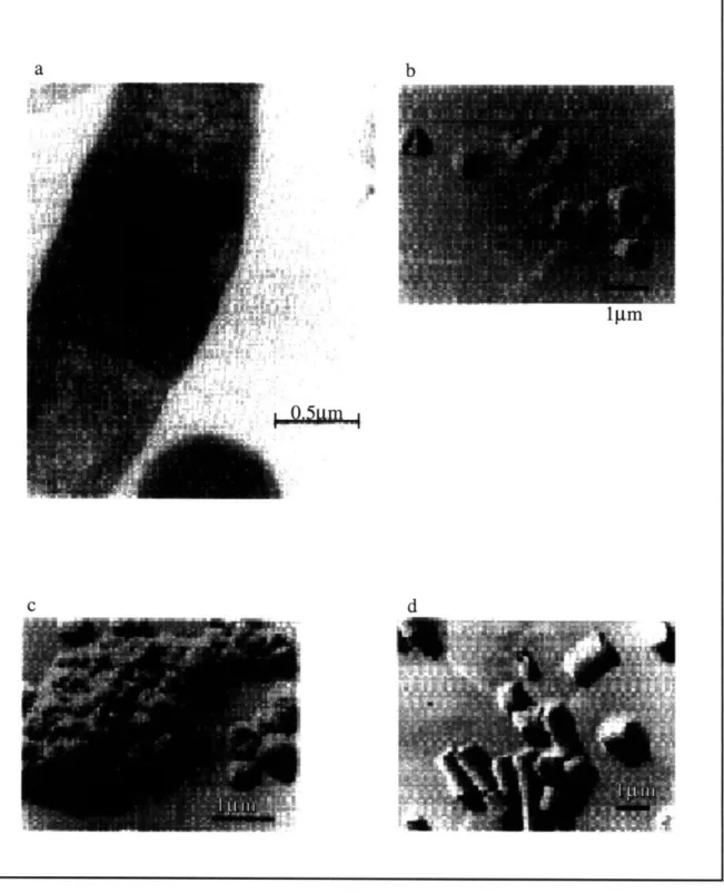
Scanning electron microscopy has been used to determine the morphology of inclusion bodies, also known as refractile bodies. Electron micrographs of cells show that inclusion bodies are large amorphous or spherical particles that occupy a substantial
fraction of the cell volume (Fig. 2.1). Generally, the size of inclusion bodies in E. coli are approximately 1 pm in diameter. In a statistical analysis of particle size, y-interferon and prochymosin inclusion bodies were 0.81pm (std deviation 0.17) and 1.28 pm (std
deviation 0.46), respectively (Taylor et al., 1986). Sedimentation studies by centrifugation determined the apparent density of the y-interferon and prochymosin inclusion bodies to be approximately 1.37 and 1.20 g/mL with a 70% and 80% voidage, respectively (Taylor et al., 1986). Theoretical correlations between the measured density and the particle size suggested that the y-interferon inclusion bodies were more ordered than the prochymosin aggregates. Although most inclusion bodies in E. coli are cytosolic, aggregated inclusion bodies can form in the periplasmic space by having a signal peptide sequence directing the polypeptide chain across the inner membrane. Since the periplasmic environment differs from the cytosol in terms of redox potential and cofactors, the inclusion bodies that form in the perisplasmic space have different biophysical characteristics, including the possibility of disulfide bond formation. 1-lactamase inclusion bodies located in E. coli cytoplasm were amorphous and spherical, whereas periplasmic inclusion bodies were more regular and cylindrical (Bowden et al., 1991).
Besides folding and aggregation, various other posttranslational modifications are in kinetic competition acting upon the nascent polypeptide chains. Proline bond
isomerization to the cis isoform may be a rate-limiting step of folding, and this reaction is catalyzed by peptidyl-prolyl isomerase (PPI). Sequence analysis has correlated empirically high proline content of proteins to the propensity to aggregate (Schein, 1989; Wood et al.,
1995). Other reactions are associated with proteins secreted into cellular compartments, such as the periplasmic space of Gram-negative bacteria or the eukaryotic endoplasmic reticulum, which has a redox potential, pH, and posttranslational enzymes different from the cytoplasm. Oxidation of cysteines and correct pairing in disulfide bond formation are reactions that often require export into the periplasmic space because disulfide bonds
generally do not form in the reducing environment of the cytoplasm with glutathione present (DeBernardez & Georgiou, 1991). In eukaryotic cells, disulfide bond
rearrangement is catalyzed by protein disulfide isomerase (PDI) which shuffles the bonding of cysteines to the correct pairing. Also, glycosylation of eukaryotic proteins in the
endoplasmic reticulum can increase protein solubility and refolding yield (Schein, 1990). Other chemical modifications, such as acylation, can occur for secreted proteins. Proteins must undergo all of the above posttranslational modifications correctly to produce the active native molecule. If a certain mammalian protein requires posttranslational modifications that the bacterial cell is not equipt to perform, then mammalian cell culture-must be used. C. Bacteriophage P22 tailspike protein
The folding and aggregation of P22 tailspike protein has been studied both in vitro and in vivo. The tailspike protein has a number of temperature-sensitive mutations that can be used to elucidate further information about the kinetics of the individual steps of the folding reaction and assembly of the native trimer species. At restrictive temperatures, the
refolding tailspike protein does not reach the protrimer conformation, and so temperature-sensitive mutants indicate which amino acids are critical for the folding process. Further investigation of the folding mechanism will identify the thermolabile folding intermediates and further characterize the species along the folding pathway of an oligomeric protein.
The tailspike endorhamnosidase is the organelle by which phage P22 attaches to the Salmonella cell. Infectious phage contain six tailspikes forming a small neck for cell attachment. The individual tailspike is a trimer consisting of identical 72kD subunits
Figure 2.1: Electron micrographs of inclusion bodies in E. coli. a) EM of cytosolic inclusion bodies of signal sequence deletion mutant of P-lactamase expressed in E. coli. Transmission electron micrographs of platinum replicas of purified inclusion bodies of: b) OmpA-p-lactamase (periplasmic), c) wild type f-lactamase
(periplasmic), and d) the signal sequence deletion mutant (cytosolic). Reprinted with permission of GA Bowden, AM Paredes, and G Georgiou (1991).
b
I jim
d
i,. 1'49·:
containing 666 residues encoded by phage gene 9. The function of the tailspike protein is to recognize phage by binding to the O-antigen receptor of Salmonella typhimurium. This
attachment process involves the tailspike endorhamnosidase hydrolyzing the rhamnosylgalactose linkage in the Salmonella O-antigen.
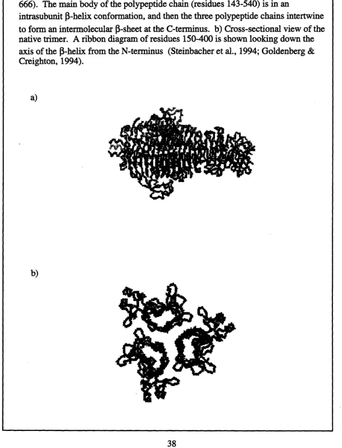
The structure of the tailspike has been solved by x-ray crystallography (Steinbacher et al., 1994). Figure 2.2 is a ribbon diagram depicting the structure of the tailspike
monomer and native trimer. The central region of each 72kD subunit of the native trimer has a right-handed parallel 5-helix conformation similar to pectate lyase, a monomeric protein (Yoder et al., 1993). Recently, a left-handed helix motif was discovered in solving the structure of acetylglucosamine, an enzyme required in the lipid A synthesis (Raetz & Roderick,1995). Similar to the tailspike protein, acetylglucosamine acetyltransferase (LpxA) is a trimer with each of the subunits in the 3-helix conformation. Unlike the tailspike protein and pectate lyase, LpxA has a hexapeptide repeat sequence in which alaphatic amino acid (isoleucine, leucine, or valine) is found every sixth residue, followed by a small amino acid (usually glycine). Two residues before the conserved aliphatic residue is a hydrophobic amino acid. The cross section of the three 3-sheets of the LpxA (-spiral resemble equilateral triangles, whereas the tailspike polypeptide chain has a dimpled configuration with two (-sheets forming a (-sandwich and a third sheet at a 900 angle. In (-spirals, the intrachain (-sheet contacts occur between each successive rung in the (3-spiral and not between the monomeric subunits.
For the tailspike protein, the main body of the polypeptide chain is in the (-helix conformation with 13 (-strands. The C-terminal regions interdigitate and form further (-sheet structures. For most of the tailspike molecule, the central cavity contains hydrophilic residues and several water molecules stabilizing the trimer. The conformation of the
Figure 2.2: a) Polypeptide backbone of the native tailspike trimer (residues 108-666). The main body of the polypeptide chain (residues 143-540) is in an
intrasubunit [3-helix conformation, and then the three polypeptide chains intertwine to form an intermolecular [3-sheet at the C-terminus. b) Cross-sectional view of the native trimer. A ribbon diagram of residues 150-400 is shown looking down the axis of the [-helix from the N-terminus (Steinbacher et al., 1994; Goldenberg & Creighton, 1994).
a)
The monomer depicted in Fig. 2.2 is the monomeric polypeptide chain within the native trimer and does not represent a proposed structure of the folding intermediate.
Structural data indicate that the 3-spiral motif of the tailspike protein may be similar to the local structure within amyloid fibrils composed of 1-amyloid peptide, prion protein, transthyretin, and immunoglobulin light chains. FTIR and solid state NMR data indicate the fibrils have a cross-3-structure, which is a repeating pattern of anti-parallel 1-sheets. Although the 1-spiral structure of the tailspike contains parallel 1-sheets, the structure has the similar property of the 1-strands being perpendicular to the fibril axis. The fibrils have a characteristic green birefringence upon binding the hydrophobic dye, Congo red (Jarrett & Lansbury, 1992). This phenomenon occurs when the Congo red molecules bound to the 1-sheets are aligned in a regular fashion.
Inclusion bodies, which form by the association of partially folded intermediates, may contain secondary structure that resembles the native state, including significant amounts of 0-sheet content. The aggregates may contain incorrect cross-3-sheet structures (either intermolecular or intramolecular) or associated hydrophobic sites involving 1-sheet interactions. These structural issues will be discussed further in Chapter 7, including an analysis of the structural data on the aggregation intermediates found in Chapter 4.
In order to obtain soluble tailspike protein instead of tailspike attached to the virus, a mutation in phage gene 5 blocks viral shell assembly by interfering with synthesis of the coat protein. This allows DNA to remain unpackaged and levels of tailspike protein to increase ten times the wild type concentrations. The final step in viral assembly is the tailspike binding to the heads to create infectious phage. Since this process occurs readily in vitro, the ability to produce infectious phage is a convenient assay to test tailspike activity.
The native tailspike is thermally stable with a melting temperature of 880C. Partially
folded or misfolded tailspikes are not heat resistant and can be distinguished from native tailspikes by checking activity at high temperatures (-800C). Besides thermal stability, the tailspike is also resistant to common denaturing conditions. The native tailspike does not denature in either protease or sodium dodecyl sulfate (SDS). Since partially folded species or misfolded aggregates are susceptible to protease and SDS, assays testing resistance to protease, SDS, and heat can distinguish native from non-native tailspikes.
Most in vivo tailspike folding experiments involve bacteriophage P22 infection of Salmonella host cells, which then synthesize the phage proteins. Recent work by A.S. Robinson has been in expressing tailspike off a plasmid in E. coli, which is analogous techniques utilized by the biotechnology industry. In a pulse-chase assay, a transient dose of radioactively labeled amino acids is added in order to monitor the amount of protein synthesized within a given time. In using phage-infected cells for these assays, the phage proteins can be easily recovered when the phage genes cause lysozyme production to break open the host cell. When expressing tailspike in E. coli, the addition of lysozyme and EDTA is necessary to lyse the cells.
Although most proteins have transient folding intermediates that cannot be isolated, the relatively large tailspike with significant secondary structure can be trapped after lysis.
Quickly chilling the lysed solutions of radiolabeled infected cells and running the samples on an acrylamide gel isolates certain folding intermediates. Since the tailspike protein is overexpressed in the host cells, the main radiolabeled species is the protein of interest.
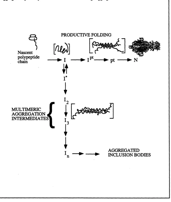
The in vivo folding pathway (Figure 2.3) involves an unfolded nascent polypeptide chain coming off the ribosome forming a partially folded species with significant structure. This single-chain intermediate (I) can associate to form a protrimer (pt) of partially folded chains (Goldenberg et al., 1983). Although the protrimer is a transient species, it is metastable enough to be trapped on an acrylamide gel at low temperatures. The final
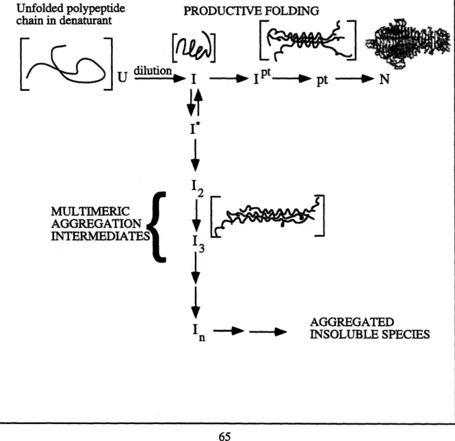
Figure 2.3: In vivo folding and aggregation pathway. Nascent polypeptide chain is released from the ribosome and forms a partially folded intermediate (I). This critical folding intermediate is at the junction between productive folding and aggregation. Along the productive folding pathway, the polypeptide chain folds into a protrimer intermediate (Ipt), an SDS-sensitive protrimer species (pt), and then the fully stable native trimer (N). The first off-pathway intermediate (I*) is in equilibrium with the productive folding intermediate (I). Higher ordered multimeric aggregation intermediates along the aggregation pathway lead to the formation of large aggregated inclusion bodies.
PRODUCTIVE FOLDING ~- -Fa Nascent polypeptide chain ---
,.
MULTIMERIC AGGREGATION INTERMEDIATES{ I123
4
I n -- -- AGGREGAT'i'EDINCLUSION BODIES
10 1
![[PDF] Essentiel Windows 2003 pdf](data:image/gif;base64,R0lGODlhAQABAIAAAP///wAAACH5BAEAAAAALAAAAAABAAEAAAICRAEAOw==)