HAL Id: hal-01584076
https://hal.sorbonne-universite.fr/hal-01584076
Submitted on 8 Sep 2017
HAL is a multi-disciplinary open access
archive for the deposit and dissemination of
sci-entific research documents, whether they are
pub-lished or not. The documents may come from
teaching and research institutions in France or
abroad, or from public or private research centers.
L’archive ouverte pluridisciplinaire HAL, est
destinée au dépôt et à la diffusion de documents
scientifiques de niveau recherche, publiés ou non,
émanant des établissements d’enseignement et de
recherche français ou étrangers, des laboratoires
publics ou privés.
Distributed under a Creative Commons Attribution| 4.0 International License
Mireille Centlivre, Nicolas Legrand, Sofieke Klamer, Ying Poi Liu, Karin
Jasmijn Von Eije, Martino Bohne, Esther Siteur-Van Rijnstra, Kees Weijer,
Bianca Blom, Carlijn Voermans, et al.
To cite this version:
Mireille Centlivre, Nicolas Legrand, Sofieke Klamer, Ying Poi Liu, Karin Jasmijn Von Eije, et al..
Preclinical In Vivo Evaluation of the Safety of a Multi-shRNA-based Gene Therapy Against
HIV-1. Molecular Therapy - Nucleic Acids, Nature Publishing Group, 2013, 2, �10.1038/mtna.2013.48�.
�hal-01584076�
Citation: Molecular Therapy—Nucleic Acids (2013) 2, e120; doi:10.1038/mtna.2013.48
© 2013 The American Society of Gene & Cell Therapy All rights reserved 2162-2531/12 www.nature.com/mtna
Introduction
HIV-1 and AIDS remains as a major public health problem with more than 34 millions infected individuals worldwide in 2011. The development of highly active antiretroviral therapy (HAART) has dramatically reduced both mortality and mor-bidity, and improved life expectancy and quality of life of the treated individuals. Despite its success, HAART does not fully suppress viral replication, especially in some critical sites (e.g., gut),1 and allows the persistence of a latent viral reservoir.2,3
The efficacy of HAART relies on daily adherence to drug regimens, but this life-long treatment is associated with side effects and emergence of drug-resistant viral variants, there-fore promoting the search for alternative therapeutic options.
Among these, gene therapy has emerged as new thera-peutic option. Ideally, such a gene therapy approach should target the CD34+ human hematopoietic progenitor cells
(hHPC), allowing the generation of a HIV-resistant multilin-eage immune system with constant supply of HIV-resistant cells that will colonize the blood and tissues. Such approaches were reinforced by the recent clinical success described for the “Berlin patient”. In early 2007, a HIV-positive individual with acute myeloid leukemia received a compatible CD34+
hHPC transplant from one of the rare individuals who are nat-urally resistant to HIV-1 (CCR5Δ32) in an effort to eradicate both diseases at once.4 The patient was declared
“function-ally cured” as no sign of viral replication was found even 3–4
years after cessation of HAART.5 Although successful, this
approach is impractical for widespread use due to the dif-ficulty of finding suitable CCR5-negative donors and the high risk of the required whole body irradiation.
To circumvent allogeneic transplantation, ex vivo modifi-cation of individuals own hHPC have been proposed, using new antiviral gene therapy approaches based on RNA inter-ference (RNAi).6–8 Gene silencing through RNAi can be
induced by expression of double-stranded RNA that will lead to sequence-specific degradation of the target RNA.9
Anti-HIV-1 short hairpin RNA (shRNA) expression in CD4+ T cells
strongly inhibits HIV-1 replication in vitro.10,11 The
shRNA-expressing gene cassettes are delivered to autologous hHPC via transduction with a lentiviral vector (LV), which stably inte-grates into the host genome. There is growing evidence from longitudinal studies in patients that LV can be used safely without signs of abnormal cell expansion induced by vector integration-mediated activation of proto-oncogenes.12
To study safety issues of gene therapy approaches, mouse models of the human immune system (HIS) have been used.11,13,14 Over the last two decades, several humanized
mouse models have been generated and optimized for the experimental study of HIS development and function.15–17 We
and others have transplanted CD34+CD38− hHPC into
immu-nodeficient newborn mice, such as BALB/c Rag2−/− IL-2Rγ c−/−
(BRG) or NOD.scid IL-2Rγc−/− (NSG) mice, resulting in the
development of a multilineage HIS in vivo.18–20 Since target
Received 8 March 2013; accepted 24 June 2013; advance online publication 3 September 2013. doi:10.1038/mtna.2013.48
2162-2531
e120
Molecular Therapy—Nucleic Acids
10.1038/mtna.2013.48
ORIGINAL ARTICLE
3September20132
8March2013
24June2013
2013
© 2013 The American Society of Gene & Cell Therapy Safe Anti-HIV-1 Multi-shRNA Gene Therapy Centlivre et al.
Highly active antiretroviral therapy (HAART) has significantly improved the quality of life and the life expectancy of HIV-infected
individuals. Still, drug-induced side effects and emergence of drug-resistant viral variants remain important issues that justify
the exploration of alternative therapeutic options. One strategy consists of a gene therapy based on RNA interference to
induce the sequence-specific degradation of the HIV-1 RNA genome. We have selected four potent short hairpin RNA (shRNA)
candidates targeting the viral capside, integrase, protease and tat/rev open-reading frames and screened the safety of them
during human hematopoietic cell development, both in vitro and in vivo. Although the four shRNA candidates appeared to be
safe in vitro, one shRNA candidate impaired the in vivo development of the human immune system in Balb/c Rag2
−/−IL-2Rγ
c−/−
(BRG) mice. The three remaining shRNA candidates were combined into one single lentiviral vector (LV), and safety of the
shRNA combination during human hematopoietic cell development was confirmed. Overall, we demonstrate here the preclinical
in vivo safety of a LV expressing three shRNAs against HIV-1, which is proposed for a future Phase I clinical trial.
Molecular Therapy—Nucleic Acids (2013)
2, e120; doi:
10.1038/mtna.2013.48
; published online 3 September 2013
Subject Category: siRNAs, shRNAs, and miRNAs
The first two authors contributed equally to the work.
1Department of Medical Microbiology, Laboratory of Experimental Virology, Center for Infection and Immunity Amsterdam (CINIMA), Academic Medical Center, University of Amsterdam, Amsterdam, the Netherlands; 2Department of Cell Biology & Histology, Center for Immunology of Amsterdam (CIA), Academic Medical Center, University of Amsterdam, Amsterdam, the Netherlands; 3Department of Hematopoiesis, Sanquin Research and Landsteiner Laboratory, Academic Medical Center, University of Amsterdam, Amsterdam, the Netherlands; 4HIS mouse facility, Academic Medical Center, University of Amsterdam, Amsterdam, the Netherlands; 5Tytgat Institute of Intestinal and Liver Research, Academic Medical Center, University of Amsterdam, Amsterdam, the Netherlands; 6Laboratory of Immunity and Infection, Institut National de la Santé et de la Recherche Médicale, INSERM UMR-S 945, and Université Pierre et Marie Curie, UPMC Univ Paris 06, 91 Bld de l’Hôpital, 75013 Paris, France; 7AXENIS, 28 rue du Docteur Roux, 75015 Paris, France. Correspondence: Ben Berkhout, Department of Medical Microbiology, Laboratory of Experimental Virology, Center for Infection and Immunity Amsterdam (CINIMA), Room K3-110, AMC-UvA, Meibergdreef 15, 1105 AZ Amsterdam, The Netherlands. E-mail: b.berkhout@amc.uva.nl
keywords: combinational RNA; gene therapy; HIV-1; humanized mouse model; RNA interference
Preclinical
In Vivo Evaluation of the Safety of a
Multi-shRNA-based Gene Therapy Against HIV-1
Mireille Centlivre1,6, Nicolas Legrand2,7, Sofieke klamer3, Ying Poi Liu1, karin Jasmijn von Eije1, Martino Bohne2,
Molecular Therapy—Nucleic Acids
cells for HIV-1 infection are present in the HIS mice, such animal models have become popular for assessing human pathogenesis21–23 and addressing new HIV therapeutic
strat-egies.24,25 Considering a future RNAi-based gene therapy
approach in the clinic, CD34+ hHPC would be exposed
ex vivo to LV and reinfused into the patient, where they would
home to the bone marrow and produce mature hematopoietic cells that are protected against HIV-1 infection. Thus, gen-erating HIS mice with genetically engineered hHPC for the expression of anti-HIV shRNA is likely to represent a sensi-tive safety screen for human hematopoiesis consistent with the envisioned clinical strategy.
In this study, we have selected four shRNA candidates tar-geting conserved regions of the viral genome for the devel-opment of the combinatorial shRNA-based gene therapy against HIV-1. These four shRNA were first tested individu-ally in HIS mice and the safe shRNAs were combined into a single LV that was evaluated for antiviral activity in vitro and for safety in HIS mice.
Results
Selection of effective anti-HIV-1 shRNAs targeting highly conserved HIV-1 regions
We have previously identified 21 shRNAs targeting eight highly conserved regions of the HIV-1 genome that exhibit potent inhibition of HIV-1 replication.10 Based on long-term
in vitro culture experiments, we selected the four most
effective shRNAs.26–28 shGag5, shPol1, shPol47, and shR/
T5, respectively targeting the viral capside, integrase, pro-tease, and tat/rev open-reading frames (Figure 1a). The respective four shRNA cassettes were cloned individually in a self-inactivating LV (Figure 1b).
No signs of toxicity of shRNA expression in a human colony-forming cell assay
We first evaluated the safety of the four shRNAs by perform-ing a human colony-formperform-ing cell assay (hCFC). This in vitro assay is commonly employed for determining the colony- and burst-forming capacity of hematopoietic progenitor cells and is widely used for measurement of drug toxicity on human hematopoietic progenitor cells (hHPC). We transduced CD34+CD38− hHPC with the different LVs expressing a single
shRNA candidate or the empty control vector JS1. Transduced GFP+ hHPC were isolated by fluorescence-activated cell
sort-ing and cultured for 2 weeks in the appropriate conditions driving development of colony-forming unit- macrophages (CFU-GM), colony-forming unit- granulocytes-erythroid-macrophages-megakaryocytes (CFU-GEMM) and burst-forming unit-erythroid (BFU-E) (Figure 2a).
CFU-GM, CFU-GEMM, and BFU-E were then counted in four independent experiments. For all the different LV conditions analyzed, transduced hHPC gave rise to the three types of col-ony/BFU. We counted between 13 and 54 CFU-GM, 5 and 16 BFU-E and 0 and 4 CFU-GEMM for all the conditions analyzed, with high variability between the experiments and between the LV conditions tested, which likely reflects the interdonor hHPC variability. Still, the LV-transduced hHPC – encoding a single or no shRNA candidate – generated comparable numbers of CFU-GM, BFU-E, and CFU-GEMM (Figure 2b–d), indicat-ing no obvious toxicity of shRNA expression on the CFU/BFU
capacity of CD34+ human hematopoietic progenitor cells in this
relatively short-term in vitro assay.
In vivo monitoring of the shRNA candidates reveal some
toxicity for shGag5 during human hematopoiesis
We next assessed the in vivo safety of single shRNA treatment in the humanized BRG-HIS mouse model.18,19 CD34+CD38−
hHPC were transduced with LVs expressing a single shRNA or the empty control LV prior to xenograft transplantation in immunodeficient BRG newborn mice.29 The BRG mice were
injected with the bulk of cultured hHPC, i.e., they received a mixture of transduced and nontransduced hHPC, providing for each animal an internal control for human immune system development and maturation. An aliquot of the transduced hHPC was kept in culture in vitro for 3–4 days to determine the transduction efficiency based on GFP expression.
BRG-HIS mice were killed between 10 and 13 weeks post-hHPC transplantation and we first analyzed the absolute human cell number in the blood and the lymphoid organs (bone marrow, thymus, spleen, and liver) of the animals. Simi-lar numbers of human cells were detected in all the analyzed organs of BRG-HIS mice transplanted with shRNA-transduced hHPC as compared with the control JS1 group (Table 1). This highlights that all four shRNA treatments do not have a nega-tive impact on human hematopoiesis at a global level.
We next monitored the relative frequency of shRNA-trans-duced hematopoietic cells (GFP+ hCD45+ cells). The in vivo
frequency of human GFP+ cells in the blood and lymphoid
organs of the animals was measured and compared with the original in vitro transduction efficiency (Figure 3a). An
in vivo/in vitro GFP+ ratio around 1.0 indicates no selection
bias with a good recovery of human GFP+ cells in the
ani-mals as compared with the initial transduction efficiency. We observed a similar efficient GFP+ recovery in the analyzed
organs of the BRG-HIS mice that received hHPC express-ing shPol1, shPol47, or shR/T5 (Figure 3b–f). In contrast, shGag5 expression appeared to be detrimental for the recovery of human GFP+ cells in vivo in all lymphoid organs,
Figure 1 Anti-HIV-1 shRNA target regions and cloning strategy.
(a) The shGag5, shPol1, shPol47, and shR/T5 target positions
within the HIV-1 genome are indicated. (b) The third generation
self-inactivating lentiviral vector JS1 expresses the green fluorescent protein (GFP) reporter. Single shRNA vectors express in addition a shRNA targeting the HIV-1 genome from the human polymerase III H1 promoter.
GAG5
5′LTR 3′LTR
gag vif nef
tat rev pol env vpu mcs vpr POL1 POL47 JS1 RSV R U5 RRE ψ shRNA H1 cPPT PGK GFP pre ∆U3 3′LTR R/T5
a
b
www.moleculartherapy.org/mtna
Safe Anti-HIV-1 Multi-shRNA Gene Therapy
Centlivre et al.
3
suggesting a negative impact of this specific shRNA on the development of the transduced human hematopoietic cells.
We further analyzed the GFP expression in several human leukocyte subsets of the spleen of BRG-HIS mice, namely human T cells, B cells, monocytes, and pDC (Figure 4a). Good GFP recovery with a ratio around 1.0 was observed for all human cell subsets in JS1 control BRG-HIS mice, but with high variation between the individual animals, especially in the T-cell compartment (Figure 4b). In contrast, all human GFP+
cell subsets were negatively affected in shGag5-expressing BRG-HIS mice (Figure 4c). The other shRNAs (shPol1, shPol47, and shR/T5) did not affect the recovery of human B cells, monocytes, and pDC (Figure 4d–f). For shPol1- and shR/T5-expressing animals, but not shPol47, a lower recov-ery of human GFP+ T cells was observed (Figure 4d–f).
Overall, these observations allowed us to formally exclude
shGag5 as a potential anti-HIV-1 shRNA candidate, due to its long-term, negative impact on human hematopoiesis in vivo.
Delayed seeding of HIS mice thymus by shPol47-expressing human progenitor cells
Reduced accumulation of shRNA-expressing T cells was noticed in 10–13-week old BRG-HIS mice as compared with control LV-transduced T cells, except for shPol47. Still, we questioned whether shPol47-expressing hHPC also showed hindered capacity for T-cell generation at earlier time points, i.e., during early events of BRG-HIS mouse thy-mus colonization and human T-cell differentiation. We trans-duced CD34+CD38− hHPC with JS1 or shPol47-encoding LV
prior to xenograft transplantation in BRG newborn mice. At 1-week post-hHPC transplantation, the site of hHPC injec-tion and major hematopoietic cell niche at this age, i.e. the liver of the BRG-HIS mice, was analyzed for the presence of human hematopoietic GFP+ cells. We did not observe any
difference in the absolute number of human cells nor in the
in vivo/in vitro ratio of human GFP+ cells between JS1
con-trol and shPol47-expressing BRG-HIS mice (Figure 5a,b). At 4 weeks post-hHPC transplantation, the bone marrow and the thymus of BRG-HIS mice are the major sites of human hematopoietic cell accumulation. No difference in the abso-lute number of human cells nor in the in vivo/in vitro ratio of human GFP+ cells was noted between the two groups
of mice in the bone marrow at this age (Figure 5a,b). By contrast, the seeding of the thymus by shPol47-transduced (GFP+) human progenitors was almost abolished whereas a
GFP+ ratio around 1.0 was observed in the thymus of control
animals (Figure 5b). A similar number of human CD45+
thy-mocytes was retrieved from the two BRG-HIS mouse groups, suggesting that seeding of the thymus by human progenitors
Figure 2 Impact of in vitro shRNA expression in early human hematopoietic progenitors. (a) Human fetal liver CD34+CD38− hHPC were transduced with JS1, shGag5, shPol1, shPol47, or shR/T5-expressing lentiviral vector. Transduced (GFP+) hHPC were sorted and a human colony-forming cell assay was performed. (b–d) Graphs show the relative colony counts of CFU-GM (b), BFU-E (c), and CFU-GEMM (d), as
compared with the control JS1 LV. The results (mean ± SD) are pooled from four independent experiments with duplicates for each experiment.
0.0 JS1 Gag5 Pol1 CFU-GM CFU-GEMM Pol47 R/T5 0.5 1.0 Relativ e colon y counts 1.5 2.0 0.0
JS1 Gag5 Pol1 Pol47 R/T5
0.5 1.0 Relativ e colon y counts 1.5 2.5 2.0 BFU-E 0.0
JS1 Gag5 Pol1 Pol47 R/T5
0.5 1.0 Relativ e colon y counts 1.5 Transduced hHPC GFP+ hHPC Cell sorting hCFC assay 2.5 2.0
a
c
b
d
Table 1 Absolute number of human CD45+ cells in the organs of
shRNA-expressing BRG-HIS mice
(×105)
Bone
marrow Thymus Spleen Liver
Blood (cells/ml) JS1 47.2 ± 31.4 14.7 ± 10.3 21.1 ± 18.6 5.2 ± 3.4 1.53 ± 1.86 shGag5 25.4 ± 21.6 10.1 ± 3.2 8.5 ± 6.0 4.8 ± 2,.5 0.91 ± 0.53 shPol1 72.4 ± 56.8 16.3 ± 10.9 39.0 ± 25.5 5.7 ± 2.6 8.22 ± 14.70 shPol47 34.9 ± 18.9 14.4 ± 10.9 16.5 ± 14.0 5.1 ± 2.9 0.71 ± 0.35 shR/T5 41.9 ± 38.5 9.1 ± 6.2 18.5 ± 15.0 4.8 ± 2.5 1.17 ± 1.40 BRG-HIS mice generated with LV-transduced hHPC for the expression of shGag5 (n = 5), shPol1 (n = 9), shPol47 (n = 17) or shR/T5 (n = 11) were analyzed 10–13 weeks post-hHPC transplantation for absolute number of hu-man CD45+ cells and compared with control animals (empty JS1-transduced
hHPC; n = 26). Bone marrow analysis was performed on two femurs per animal.
Molecular Therapy—Nucleic Acids
is exclusively impaired for the shPol47-expressing GFP+ cell
subpopulation. In the case of shPol47, the delay in thymus seeding by human shRNA-expressing progenitors was over-come at weeks 10–13 post-hHPC transplantation, with simi-lar human cell number and GFP recovery in the thymus and all the analyzed organs of JS1 and shPol47- treated animals (Table 1 and Figure 3).
Simultaneous expression of three nontoxic shRNAs is safe in vitro and in vivo
As for HAART, combination of multiple shRNAs into a single LV resulted in additive inhibition of HIV-1 in vitro.26 Even more
importantly, the combinatorial RNAi approach prevented the selection of RNAi-escape variants.26 We combined three
shRNAs (shPol1, shPol47, and shR/T5; R3) or four shRNAs candidates (shGag5, shPol1, shPol47, and shR/T5; R4) into the JS1 LV (Figure 6a) and evaluated the in vitro safety of these combinatorial LV in the hCFC assay (Figure 2a). The num-bers of CFU-GM, BFU-E, and CFU-GEMM were comparable between R3 LV and JS1 control LV (Figure 6b). The hHPC
transduced with the shGag5-encoding R4 LV gave rise to a reduced number of CFU-GM as compared with control hHPC.
We next assessed the in vivo safety of a combinatorial shRNA treatment in BRG-HIS mice. As for the single shRNA treatment experiments, similar numbers of human cells were detected in all the analyzed organs of BRG-HIS mice trans-planted with shRNA-transduced hHPC as compared with the control JS1 group (Supplementary Table S1, online).
When the in vivo frequency of human GFP+ cells in the blood
and lymphoid organs of the animals was measured and compared with the original in vitro transduction efficiency, we observed a severely reduced recovery of human GFP+
cells in vivo in the R4 animals (Figure 6c–g). This recovery was even lower than observed for the single shGag5 experi-mental group (Figure 3). The recovery of GFP+ cells from
R3-treated BRG-HIS mice was lower than in the JS1 control BRG-HIS mice, although it was significantly lower only for the spleen and blood. This reduced global recovery in the spleen was explained in the analysis of different human immune cell subsets, where we measured a reduced frequency of
Figure 3 Recovery of LV-transduced human hematopoietic cells in the lymphoid organs of BRG-HIS mice. BRG-HIS mice generated
with LV-transduced hHPC for the expression of shGag5 (grey diamonds, n = 5), shPol1 (black diamonds, n = 9), shPol47 (grey triangle, n = 17) or shR/T5 (black triangle, n = 11) were analyzed 10–13 weeks post-hHPC transplantation and compared with control animals (empty JS1-transduced hHPC; black circles, n = 26). (a) The flow cytometry dot plots show the transduction efficiency in vitro of hHPC transduced with LV
(left panel, SSC side scatter) and the frequency of GFP expression among human hCD45+ hematopoietic cells in vivo in the BRG-HIS mice (right panel). (b–f) The results are expressed as the GFP+ ratio between the frequency of human GFP+ cells measured in the animals (in vivo) and the frequency of GFP+ hHPC injected in the newborn mice (in vitro transduction efficiency). The GFP+ ratio is shown for the bone marrow (b), thymus (c), spleen (d), liver (e), and blood (f). The results (mean) are pooled from two to eight independent experiments, and each dot
represents an individual animal. *P < 0.05; **P < 0.01 (Kruskall–Wallis test).
Thymus Spleen
0.0
JS1 Gag5 Pol1 Pol47 R/T5
0.5 1.0 GFP + ratio 1.5 2.5 0 SSC 102 102 103 104 105 0 103 34.3 43.3 65.6 GFP 104 105 in vitro hHPC in vivo BRG-HIS mice 0 50 k 100 k150 k200 k250 k 0 102 103 104 105 * 2.0 0.0
JS1 Gag5 Pol1 Pol47 R/T5
0.5 1.0 GFP + ratio 1.5 2.5 2.0
c
Liver 0.0JS1 Gag5 Pol1 Pol47 R/T5
0.5 1.0 GFP + ratio 1.5 2.5 ** 2.0
e
Blood 0.0JS1 Gag5 Pol1 Pol47 R/T5
0.5 1.0 GFP + ratio 1.5 2.5 ** ** 2.0
f
d
Bone marrow 0.0JS1 Gag5 Pol1 Pol47 R/T5
0.5 1.0 GFP + ratio 1.5 2.5 2.0 *
b
a
hCD45www.moleculartherapy.org/mtna
Safe Anti-HIV-1 Multi-shRNA Gene Therapy
Centlivre et al.
5
GFP+ cells in the human B- and T-cell subsets (Figure 6h).
Still, R3-transduced hHPC gave rise to multilineage in vivo reconstitution of the human immune system, demonstrating the in vivo safety of the R3 construct. When compared with the shGag5 group, the R3-treated BRG-HIS mice exhibited higher recovery of GFP+ cells in the primary lymphoid organs
(bone marrow, thymus) and the liver, suggesting that the reconstitution kinetics by the R3-transduced cells might be delayed, as observed with shPol47.
Inhibition of HIV-1 replication on in vivo generated human CD4+ T cells expressing three shRNA against HIV-1
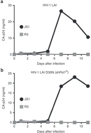
To evaluate the capacity of human shRNA+ cells to inhibit
HIV-1 replication, we isolated mature human CD4+ T cells
from NSG-HIS mice produced either with control JS1 or with R3-transduced hHPC. At sacrifice, splenocytes were harvested and GFP+ human CD4+ T cells were isolated by
fluorescence-activated cell sorting. The sorted CD4+ T cells
were challenged ex vivo by either a wild-type HIV-1 strain (LAI) or a shPol1-resistant HIV-1 virus (HIV-1 LAI-D30N) that was selected during mono-RNAi therapy.28 HIV-1 LAI
replication was inhibited in transduced GFP+ CD4+ T cells
from R3-expressing NSG-HIS mice but not in the control GFP+ CD4+ T cells from JS1-expressing NSG-HIS mice
(Figure 7a). When the sorted cells were challenged with the shPol1-resistant HIV-1 virus, viral inhibition was still observed for the transduced GFP+ CD4+ T cells from R3-expressing
NSG-HIS mice but not in the control GFP+ CD4+ T cells from
Figure 4 Differential shRNA expression in human cell subsets of BRG-HIS mice. GFP expression by several human immune cells
was measured in the spleen of BRG-HIS mice. (a) The analyzed subsets were human T cells (CD3+), B cells (BDCA2−CD19+), plasmacytoid dendritic cells (pDC; CD19−BDCA2+), and monocytes (Mono; CD19−BDCA2−CD14+). (b–g) GFP expression by these subsets was measured in BRG-HIS mice reconstituted with the following LV: control JS1 (b), shGag5 (c), shPol1 (d), shR/T5 (e), or shPol47 (f). The results are
expressed as the GFP+ ratio between the frequency of human GFP+ cells measured in the spleen of the animals (in vivo) and the frequency of GFP+ hHPC injected in the newborn mice (in vitro transduction efficiency). The results (mean) are pooled from two to eight independent experiments, and each dot represents an individual animal. *P < 0.05; **P < 0.01 (Friedman test).
0 102 102 103 104 105 0 103 CD19 104 105 0.0
T cells B cells Mono pDC
0.5 1.0 GFP + ratio 1.5 2.5 shPol47 2.0
f
0.0T cells B cells Mono pDC
0.5 1.0 GFP + ratio 1.5 2.5 shR/T5 * 2.0
e
0.0T cells B cells Mono pDC
0.5 1.0 GFP + ratio 1.5 2.5 shGag5 2.0
c
0.0T cells B cells Mono pDC
0.5 1.0 GFP + ratio 1.5 2.5 ** ** * shPol1 2.0
d
0.0T cells B cells Mono pDC
0.5 1.0 GFP + ratio 1.5 2.5 JS1 2.0
b
a
0 102 103 CD14 104 105 0 102 103 CD3 104 105 T cells B cells 58 pDC 35 5 7 BDCA2 32 Mono 102 103 104 105 0 hCD45 102 103 104 105 0 hCD45Molecular Therapy—Nucleic Acids
JS1-expressing NSG-HIS mice (Figure 7b). Taken together, these results demonstrate that in vivo generated human CD4+
T cells that express R3 are resistant to HIV-1 replication. Fur-thermore, as for HAART, pre-existing resistance to one of the inhibitors (shPol1) is not sufficient to confer escape from inhi-bition by the other two shRNAs (shPol47 andshR/T5).
Discussion
We have previously reported the proof-of-concept of testing the in vivo impact of expressing a shRNA directed against the HIV-1 nef gene in CD34+CD38− hHPC in BRG-HIS mice.11
The development of human hematopoiesis-derived cells was not affected in the shNef-hHPC engrafted mice and the shNef-transduced CD4+ T cells that develop in the animals
are resistant to HIV-1 challenge ex vivo. This shNef targets a nonconserved region of the HIV-1 genome and a nonessen-tial gene for HIV-1 replication. Consequently, this shRNA was not attractive for further development of an anti-HIV RNAi-based gene therapy. Here, we selected four anti-HIV-1 shRNA
candidates that target essential HIV-1 genes and highly con-served regions of the HIV-1 genome among all HIV-1 subtypes (>73% target conservation of the shRNA target sequence in all virus isolates present in the Los Alamos database), in par-ticular subtype B isolates (>80%).30 We evaluated the
preclini-cal safety profile in vivo in view of further development of this RNAi-based gene therapy towards clinical trial phase I.
Evaluation of the safety of shRNA expression by hHPC was initially performed using the classical human colony-forming cell assay. No apparent toxicity was scored in this assay for single shRNA expression. In contrast, when safety of shRNA expression by human hematopoietic cells was assessed in vivo in mice humanized for the immune sys-tem, a single shRNA (shGag5) exhibited a negative impact on the development of human hematopoietic cells. Only the shGag5+expressing cells were affected, as
develop-ment of nontransduced cells occurred normally in these animals to reach absolute number of human cells similar to control humanized mice. These results demonstrate that mice humanized for the human immune system constitute a valuable and sensitive in vivo preclinical model to evalu-ate the hematopoietic safety of gene therapy strevalu-ategies, for several reasons. First, the hHPCs that are engrafted in the immunodeficient newborn mice are similar to the ones that would be genetically engineered for ex vivo gene therapy of HIV-infected patients. Second, as the hHPCs transplanted in the mice consist of a mixture of transduced (GFP+) and
nontransduced hHPC (GFP−), the human immune system
in the animals is thus constituted of shRNA-expressing cells (GFP+) and nontransduced cells (GFP−). Besides evaluating
the control mice vs. the shRNA-treated mice, this approach provides a perfect internal control to test for adverse effects of shRNA expression on hematopoiesis by simply following the frequency of GFP+ cells over time, as compared with the
original hHPC inoculum. This approach turned out to be more sensitive than a simple screen of the total human cell popula-tion. We previously used such a strategy to develop a sensi-tive cell culture system to detect transgene toxicity.31 Finally,
HIS mice allow for the long-term evaluation of associated tox-icity, which could for instance not be observed in the relatively short-term colony-forming cell assay with shGag5.
We demonstrate in HIS mice that shPol1, shPol47, and shR/ T5 were safe, as they did not affect the in vivo development of the human immune system, both in terms of absolute human cell numbers and shRNA-expressing cell recovery. When com-bined into a single LV, simultaneous expression of these three shRNAs did not affect the in vivo generation of a multilineage human immune system, even though GFP recovery is less effective for R3 than the vectors that express a single shRNA from this combination. Still, GFP recovery with the R3 vector was better than with shGag5 alone, and similar or close to the JS1 control in the primary organs. Moreover, we provide proof-of-concept for the capacity of human shRNA+ CD4+ T cells
harvested from R3-treated HIS mice to resist HIV-1 infection. As for HAART, combinatorial shRNA treatment exhibits addi-tive HIV-1 inhibition and prevents the selection of escape virus variants. In fact, we demonstrated that R3-expressing cells remain nonsusceptible for an HIV-1 variant with an escape mutation in a single shRNA target sequence. These data should be extended in the future by long-term HIV infection
Figure 5 Impaired seeding of BRG-HIS mouse thymus by shPol47-expressing human progenitor cells. CD34+CD38− hHPC were transduced with JS1-control or shPol47-expressing LV and injected into newborn BRG mice. At 1 and 4 weeks post-hHPC transplantation, JS1 (white boxes; n = 4) and shPol47 (grey boxes; n = 5) BRG-HIS mice were analyzed in the major sites of hematopoiesis for the absolute number of human CD45+ cells (a) and for GFP+ ratio in human hematopoietic cells (b). The GFP+ ratio is the ratio between the frequency of human GFP+ cells measured in the animals (in vivo) and the frequency of GFP+ hHPC injected in the newborn mice (in vitro transduction efficiency). The results are pooled from four independent experiments, and each dot represents an individual animal. *P < 0.05 (Mann–Whitney test). BM, bone marrow; LIV, liver; THY, thymus.
0.0 LIV T + 1 week 0.5 1.0 GFP + ratio 1.5 2.5 2.0 b a 102 103 104
Absolute human cell number
105 106 107 JS1 shPol47 THY T + 10/13 weeks BM THY * T + 4 weeks LIV T + 1 week THY T + 10/13 weeks BM THY T + 4 weeks
www.moleculartherapy.org/mtna
Safe Anti-HIV-1 Multi-shRNA Gene Therapy
Centlivre et al.
7
experiments in the HIS mouse to analyze the potential emer-gence of HIV escape mutants over time.
Analysis of the early events of tissue colonization and cell differentiation taking place in the BRG-HIS mice engrafted with shRNA-hHPC revealed that seeding of the thymus by human bone marrow GFP+ progenitors was delayed, with
kinetics varying depending on the respective shRNA. This observation could reflect several nonmutually exclusive mechanisms affecting e.g., generation of thymus seeding progenitor cells,32–34 migration of thymus seeding progenitor
cells in the circulation, their entry into the thymus or the earli-est steps of T-cell lineage commitment. Consequently, such a delayed capacity of shRNA-transduced cells to initiate thy-mopoiesis and to accumulate in secondary lymphoid organs
may cause a reduced frequency of human GFP+ T cells in
the periphery of adult HIS mice.
One explanation for this phenomenon could be off-target effects. The shGag5 may target unrelated mRNAs that are involved in hematopoiesis. Transcriptional interference of the shRNA-cassette with GFP transcription from the PGK poly-merase II promoter seems unlikely as all single shRNA con-structs were driven from the same H1 polymerase III promoter, yet exhibited a different outcome with only shGag5 showing toxic effects. Similarly, as all tested shRNA constructs were designed in the same JS1 backbone, reduced LV integration can be ruled out. Alternatively, a more general mechanism such as saturation of the miRNA pathway can be consid-ered.11,35–39 The miRNA pathway is involved in hematopoiesis
Figure 6 In vitro and in vivo safety of combinatorial shRNA treatment. (a) Schematic representation of the R3 and R4 LV. The R3
vector expresses the three validated shRNAs (shR/T5, shPol1, and shPol47) and the R4 vector also contains shGag5. (b) Human fetal liver
CD34+CD38− hHPC were transduced with JS1, R3 or R4-expressing LV and analyzed as in Figure 2b. (c–g) JS1 (black circles, n = 10), R3 (grey squares, n = 7), R4 (black squares, n = 3) groups of BRG-HIS mice were analyzed 10–13 weeks after transplantation. The GFP+ ratio in human hematopoietic cells was determined in the bone marrow (c), thymus (d), spleen (e), liver (f), and blood (g). The GFP+ ratio is the ratio between the frequency of human GFP+ cells measured in the animals (in vivo) and the frequency of GFP+ hHPC injected in the newborn mice (in vitro transduction efficiency). (h) GFP expression by human immune cell subsets was measured in the spleen of BRG-HIS mice generated
with R3-expressing hHPC. The results are pooled from two independent experiments, and each dot represents an individual animal. *P < 0.05; **P < 0.01 (Kruskall–Wallis test for group comparison and Friedman test for cell subset comparison).
0.0
JS1 R3
Thymus Spleen Liver
** ** * * R4 0.5 1.0 GFP + ratio 1.5 2.5 2.0 0.0 JS1 R3 CFU-GM * ** R4 0.5 1.0 Relativ e colon y counts 1.5 2.0 0.0 JS1 R3 BFU-E U1 R/T5 R3 R4
Pol1 Pol47 R/T5 Pol1 Gag5 Pol47
U6 H1 U1 U6 7SK H1 R4 0.5 1.0 1.5 2.0 0.0 JS1 R3 CFU-GEMM R4 0.5 1.0 1.5 2.0 0.0 JS1 R3 Bone marrow R4 0.5 1.0 1.5 2.5 2.0
d
a
b
c
0.0 JS1 R3 R4 0.5 1.0 GFP + ratio GFP + ratio 1.5 2.5 2.0e
0.0 JS1 R3 R4 0.5 1.0 GFP + ratio 1.5 2.5 2.0f
Blood ** ** * * 0.0 JS1 R3 R4 0.5 1.0 GFP + ratio 1.5 2.5 2.0g
R3 0.0T cells B cells Mono pDC 0.5 1.0 GFP + ratio 1.5 2.5 2.0
h
Molecular Therapy—Nucleic Acids
and differentiation of hHPC into lineage-committed progeni-tors and the migration of hematopoietic cells from the bone marrow to the thymus and the periphery.40,41 One can imagine
that any disturbance in this network might result in impaired hematopoietic development. To explain the shGag5-specific effects, one could propose that it is ineffectively processed, thus occluding the RNAi machinery by saturation of one of the critical proteins. Saturation of the miRNA pathway may also occur if the shRNA is expressed at too high levels due to, for example, multiple integrations of the LV in the target cells. In the clinical setting one would therefore aim for a low number of integrations per cells, preferably only a single copy, as we tried to mimick in the HIS mouse experiments.
A reduction of the number of modified human T cells seems to be a common feature of diverse treatments in the humanized HIS mouse model, including an inducible LV,42 a
LV expressing a human/rhesus macaque TRIM5α isoform, a CCR5 shRNA and a TAR decoy,43 or a LV expressing two
shRNAs.44 Notably, a reduced frequency of
shRNA-express-ing T cells was also observed in another humanized mouse setting, using hHPC transduced with a LV encoding three anti-HIV genes (shRNA against Tat/Rev, CCR5-ribozyme, and TAR-decoy) and subsequently injected in human thymic grafts of the SCID-hu mice.14 Six weeks after hHPC
injec-tion in the mice, the frequency of GFP+ cells was particularly
reduced for the shRNA-expressing vector in human thymo-cytes. This combinatorial therapy was tested in a pilot clinical trial in four patients with AIDS-related non-Hodgkin’s lym-phoma where autologous CD34+ hHPC were transduced with
a LV encoding the three anti-HIV small RNA and infused back into the patients.45 This gene therapy was well tolerated with
no signs of toxicity and shRNA expression was detected in PBMCs. Interestingly, the relative number of transduced cells increased in two patients after a spike of HIV-1 viremia, sug-gesting selective expansion of the protected cells.46
What would be the significance of the temporary reduction of human shRNA-expressing T cells for a clinical application? Based on the latter clinical study and our data showing potent virus inhibition with the shRNA combination, it is likely that the developmental disadvantage of shRNA-expressing cells will be counterbalanced by the survival advantage upon HIV-1 infection. Even with maintenance of a high plasma viral load, such a selective survival advantage of treated human CD4+
T cells has already been observed in HIS mice treated with either a triple-combination anti-HIV LV or a dual shRNA LV and subsequently infected by HIV-1 (both R5- and X4-tropic strains).43,44 In this scenario, HIV-1 resistant
shRNA-express-ing cells are likely to be positively selected, as the unpro-tected cells will be removed by the immune system upon HIV-1 infection, leading to a clinical benefit for the patient. Still, this hypothesis remains to be formally proven, and alternative therapeutic strategies can also be envisioned, e.g., using clin-ical-grade cell sorting of modified cells – as already performed in HIS mice47 – or in vivo delivery of T cell-targeted shRNA.13 It
should be emphasized that human T-cell homeostasis is par-ticularly sub-optimal in BRG-HIS mice.15,18,19 Thus it remains
unclear whether a similar delayed seeding of the thymus and peripheral lymphoid organs will be seen in a human clinical setting. Overall, the R3 LV that encodes the shPol1, shPol47, and shR/T5 shRNAs – respectively targeting the HIV-1 open-reading frames for integrase, protease and Tat/Rev – might represent an attractive prototype for further clinical develop-ment, which requires further evaluation, especially concerning the potential delay in reconstitution by R3-expressing T cells.
Materials and methods
Constructs, LV preparation, virus titer determination and cell culture. LV – JS1, R/T5, Gag5, Pol1, Pol47, R3, and R4 –
were previously described.10,11 The JS1 LV carries the GFP
reporter gene that allows for distinction between transduced (GFP+) cells and nontransduced (GFP−) cells. LV48 were
pro-duced by cotransfection of vector constructs and packaging constructs pSYNGP, pRSV-rev, and pVSVg in 293T cells with lipofectamine-2000 (Invitrogen, Carlsbad, CA, USA), as pre-viously described.49 The virus stocks were concentrated on a
5% sucrose layer by ultracentrifugation (32 K, 90 minutes) and aliquots of 350 µl were stored at −80 °C. Virus production was determined by CA-p24 ELISA and virus titers were determined by serial dilution of virus and transduction on SupT1 cells. Human embryonic kidney 293T cells were grown at 37 °C and 5% CO2 in DMEM (Gibco BRL) and SupT1 cells were grown in
RPMI 1640 (Gibco BRL). Both media were supplemented with 10% fetal calf serum, minimal essential medium nonessential amino acids, penicillin (100 U/ml) and streptomycin (100 µg/ml).
Figure 7 R3+human CD4+T cells from NSG-HIS mice inhibit
HIV-1 replication ex vivo. Transduced (GFP+) human CD4+ T cells from spleen of JS1 and R3-treated NSG-HIS mice were sorted and infected either with HIV-1 LAI (a) or a HIV-1 LAI strain resistant
to shPol1 (HIV-1 LAI D30N) (b). Virus spread was monitored by
measuring CA-p24 production with ELISA. The HIV-1 inhibition experiment was repeated three-times and one representative experiment is shown. 0 5 10 15 JS1
HIV-1 LAI D30N (shPol1R)
R3 JS1 R3 CA-p24 (ng/ml) 20 25 0 2 4 6
Days after infection
8 10 0 HIV-1 LAI CA-p24 (ng/ml) 0 10 20 30 2 4 6
Days after infection
8 10
a
www.moleculartherapy.org/mtna
Safe Anti-HIV-1 Multi-shRNA Gene Therapy
Centlivre et al.
9
Isolation of human hematopoietic progenitor cells and trans-duction procedure. Human hematopoietic progenitor cells
(hHPC) were isolated from human fetal liver tissue samples obtained from elective abortions, with gestational age of 14–18 weeks. The use of fetal liver tissues was approved by the Medical Ethical Committee of the Academic Medical Center of the University of Amsterdam and was contingent on informed consent. Single-cell fetal liver suspensions were pre-pared and hHPC were isolated as previously described.29 In
brief, CD34+ cells were enriched by immunomagnetic sorting
(indirect CD34 human progenitor cell isolation kit, Miltenyi Bio-tech) and the CD34+CD38−CD3−CD19−CD56−BDCA2− (further
referred to as CD34+CD38−) hHPC were sorted using a
FAC-SAria (BD Biosciences, Franklin Lakes, NJ) to purity ≥99%. The sorted CD34+CD38− hHPC were transduced with the
different LVs after overnight culture in IMDM (Invitrogen, Carlsbad CA, USA) supplemented with Yssel’s medium, 5% normal human serum (NHS), 20 ng/ml human stem cell fac-tor (huSCF; PeproTech, Rocky Hill, NJ, USA), 20 ng/ml human thrombopoietin (huTPO; PeproTech), 20 ng/ml human interleu-kin-3 (huIL-3, PeproTech) and 20 ng/ml human interleukin-7 (huIL-7; Cytheris). The following day, the cells were incubated for 6–8 hours with virus supernatant in fibronectin-coated plates (30 µg/ml; Takara Biomedicals, Otsu, Shiga, Japan).
Colony-forming cell assay. CD34+CD38− hHPC were
trans-duced by the different LVs. GFP+ transduced CD34+CD38−
hHPC were sorted with live fluorescence-activated cell sorting and plated in duplicate in 12-well tissue culture plates at con-centrations of 100 cells per well in methylcellulose medium (MethoCult H4435, StemCell Technologies, Vancouver, BC, Canada). Cultures were incubated for 12–14 days at 37 °C in a 5% CO2-humidified atmosphere. BFU-E and colony-forming
units-granulocyte/macrophage and CFU-granulocyte/erythro-cyte/macrophage/megakaryocyte (CFU-GM and CFU-GEMM, respectively) were scored by microscopy according to manu-facturer’s instructions. CFC assay was repeated four-times with duplicates for each conditions tested per experiment.
Generation of BRG-HIS and NSG-HIS mice. BALB/c (H-2d)
Rag2−/−IL2Rγ
c−/− (BRG) mice50 and NOD.scid.IL2Rγc−/− (NSG)
mice51 were bred and maintained in individual ventilated
cages, and were fed autoclaved food and water. Mice with a human immune system (BRG-HIS and NSG-HIS) were generated as previously described.29 Briefly, newborn
(<1-week old) BRG and NSG mice received sub-lethal (3.5 Gy BRG/1Gy NSG) total body irradiation with a X-ray Röntgen source or 137Cs, and were injected intrahepatic (i.h.) with
5 × 10⁴–10 × 104 transduced CD34+CD38− hHPC. NSG mice
actually express a SIRPα allele capable of properly interact-ing with the human CD47 protein.52 CD47/SIRPα interaction
is a major determinant of self and escape from phagocyte-mediated clearance,53 especially in a xenograft setting.47
Consequently, higher level of human cells can be found in NSG-HIS mice allowing isolation of more human CD4+ T
cells for HIV infection ex vivo. All manipulations of BRG-HIS and NSG-HIS mice were performed under laminar flow. Cell suspensions of the BRG-HIS and NSG-HIS mouse organs were prepared in RPMI1640 medium supplemented with 2% fetal calf serum.
Flow cytometry analysis for cell surface markers. Cell
suspensions were labeled with anti-human mAb target-ing the followtarget-ing cell surface markers: CD3 (SK7), CD4 (SK3), CD8 (SK1), CD19 (SJ25C1), CD45 (2D1), HLA-DR (G46-6), CD14 (M5E2), CD11c (S-HCL-3) from BD bio-sciences, CD1a (T6-RD1) from Coulter-Immunotech and BDCA2 (AC144) from Miltenyi Biotech (Bergisch Gladbach, Germany). Washings and reagent dilutions were done with PBS containing 2% fetal calf serum and 0.02% sodium azide (NaN3). All acquisitions were performed with a LSR-II (BD Bioscience) flow cytometer interfaced to FACS-Diva software system, and were analyzed with FlowJo software (Ashland, OR). Cellular debris and dead cells were excluded by their light-scattering characteristics and based on incor-poration of 4′,6-diamidino-2-phenylindole (DAPI, Sigma).
HIV challenge ex vivo. Human CD4+ T cells from spleens of
JS1 and JS1-R3-treated NSG-HIS mice were sorted (≥99% pure) using an ARIA sorter (BD Bioscience) according to the CD45+CD3+CD4+CD8− phenotype and the presence or
absence of GFP expression. Cells from NSG-HIS mice were subsequently stimulated for 2 days with PHA (4 mg/ml) in RPMI culture medium supplemented with IL-2 (100 U/ml) and 10% fetal calf serum. The human CD4+ T cells sorted from the
NSG-HIS mice (5 × 10⁴ in 0.2 ml) were infected with HIV-1 LAI or HIV-1
LAI-D30N virus supernatant (0.2 ng CA-p24) and virus spread was monitored by measuring CA-p24 production by ELISA. The HIV-1 inhibition experiment was repeated three-times.
Statistical analyses. Data were subjected to a Mann–Whitney
test for two-group comparisons, a Kruskall–Wallis test fol-lowed by a Dunns post-test for multiple group comparisons, and a Friedman test followed by a Dunns post-test for mul-tiple matched group comparisons as indicated in the figure legends. The obtained P values were considered significant *P < 0.05, **P < 0.01, ***P < 0.001.
Supplementary material
Table S1. Absolute number of human CD45+ cells in the
organs of shRNA-treated BRG-HIS mice.
Acknowledgments. Mireille Centlivre was supported by
Ma-rie CuMa-rie Intra-European fellowship (MEIF-CT-2007–039689). Nicolas Legrand, Kees Weijer and Hergen Spits were sup-ported by the Bill and Melinda Gates Foundation (Grand Challenges in Global Health program – GC4 ‘Human Vaccine Consortium’). This research was sponsored by NWO-CW (Chemical Sciences), ZonMw (Medical Sciences), and the Dutch AIDS Fund (project 2006006). We thank the staff of the ABSL-2 unit of the Animal Research Institute Amsterdam (ARIA) of the AMC-UvA for excellent care of the animals. We also thank Berend Hooibrink for expertise in cell sort-ing and maintenance of the flow cytometry facility, Stephan Heynen for assistance with the CA-p24 ELISA, and the tech-nical staff of the laboratory for Cell Therapy of Sanquin for counting colonies. Lastly, we are grateful to the Bloemenhove Clinic (Heemstede, The Netherlands) for providing fetal tis-sues. Nicolas Legrand is currently an employee of AXENIS, a contract research organization generating humanized mouse models, although this was not the case during the execution
Molecular Therapy—Nucleic Acids
and completion of this study. The other authors declare no competing financial interests.
1. Yukl, SA, Shergill, AK, McQuaid, K, Gianella, S, Lampiris, H, Hare, CB et al. (2010). Effect of raltegravir-containing intensification on HIV burden and T-cell activation in multiple gut sites of HIV-positive adults on suppressive antiretroviral therapy. AIDS 24: 2451–2460.
2. Llibre, JM, Buzón, MJ, Massanella, M, Esteve, A, Dahl, V, Puertas, MC et al. (2012). Treatment intensification with raltegravir in subjects with sustained HIV-1 viraemia suppression: a randomized 48-week study. Antivir Ther (Lond) 17: 355–364.
3. Siliciano, JD, Kajdas, J, Finzi, D, Quinn, TC, Chadwick, K, Margolick, JB et al. (2003). Long-term follow-up studies confirm the stability of the latent reservoir for HIV-1 in resting CD4+ T cells. Nat Med 9: 727–728.
4. Hütter, G, Nowak, D, Mossner, M, Ganepola, S, Müssig, A, Allers, K et al. (2009). Long-term control of HIV by CCR5 Delta32/Delta32 stem-cell transplantation. N Engl J Med 360:
692–698.
5. Allers, K, Hütter, G, Hofmann, J, Loddenkemper, C, Rieger, K, Thiel, E et al. (2011). Evidence for the cure of HIV infection by CCR5?32/?32 stem cell transplantation. Blood
117: 2791–2799.
6. Berkhout, B (2009). Toward a durable anti-HIV gene therapy based on RNA interference. Ann N Y Acad Sci 1175: 3–14.
7. Berkhout, B and ter Brake, O (2009). Towards a durable RNAi gene therapy for HIV-AIDS. Expert Opin Biol Ther 9: 161–170.
8. Burnett, JC, Zaia, JA and Rossi, JJ (2012). Creating genetic resistance to HIV. Curr Opin Immunol 24: 625–632.
9. Fire, A, Xu, S, Montgomery, MK, Kostas, SA, Driver, SE and Mello, CC (1998). Potent and specific genetic interference by double-stranded RNA in Caenorhabditis elegans. Nature 391: 806–811.
10. ter Brake, O, Konstantinova, P, Ceylan, M and Berkhout, B (2006). Silencing of HIV-1 with RNA interference: a multiple shRNA approach. Mol Ther 14: 883–892.
11. ter Brake, O, Legrand, N, von Eije, KJ, Centlivre, M, Spits, H, Weijer, K et al. (2009). Evaluation of safety and efficacy of RNAi against HIV-1 in the human immune system (Rag-2(-/-)gammac(-/-)) mouse model. Gene Ther 16: 148–153.
12. Wang, GP, Levine, BL, Binder, GK, Berry, CC, Malani, N, McGarrity, G et al. (2009). Analysis of lentiviral vector integration in HIV+ study subjects receiving autologous infusions of gene modified CD4+ T cells. Mol Ther 17: 844–850.
13. Kumar, P, Ban, HS, Kim, SS, Wu, H, Pearson, T, Greiner, DL et al. (2008). T cell-specific siRNA delivery suppresses HIV-1 infection in humanized mice. Cell 134: 577–586.
14. Anderson, J, Li, MJ, Palmer, B, Remling, L, Li, S, Yam, P et al. (2007). Safety and efficacy of a lentiviral vector containing three anti-HIV genes–CCR5 ribozyme, tat-rev siRNA, and TAR decoy–in SCID-hu mouse-derived T cells. Mol Ther 15: 1182–1188.
15. Legrand, N, Cupedo, T, van Lent, AU, Ebeli, MJ, Weijer, K, Hanke, T et al. (2006). Transient accumulation of human mature thymocytes and regulatory T cells with CD28 superagonist in “human immune system” Rag2(-/-)gammac(-/-) mice. Blood 108: 238–245.
16. Manz, MG (2007). Human-hemato-lymphoid-system mice: opportunities and challenges. Immunity 26: 537–541.
17. Shultz, LD, Ishikawa, F and Greiner, DL (2007). Humanized mice in translational biomedical research. Nat Rev Immunol 7: 118–130.
18. Gimeno, R, Weijer, K, Voordouw, A, Uittenbogaart, CH, Legrand, N, Alves, NL et al. (2004). Monitoring the effect of gene silencing by RNA interference in human CD34+ cells injected into newborn RAG2-/- gammac-/- mice: functional inactivation of p53 in developing T cells. Blood 104: 3886–3893.
19. Traggiai, E, Chicha, L, Mazzucchelli, L, Bronz, L, Piffaretti, JC, Lanzavecchia, A et al. (2004). Development of a human adaptive immune system in cord blood cell-transplanted mice. Science 304: 104–107.
20. Ishikawa, F, Yasukawa, M, Lyons, B, Yoshida, S, Miyamoto, T, Yoshimoto, G et al. (2005). Development of functional human blood and immune systems in NOD/SCID/IL2 receptor {gamma} chain(null) mice. Blood 106: 1565–1573.
21. Denton, PW and García, JV (2011). Humanized mouse models of HIV infection. AIDS Rev
13: 135–148.
22. Berges, BK and Rowan, MR (2011). The utility of the new generation of humanized mice to study HIV-1 infection: transmission, prevention, pathogenesis, and treatment. Retrovirology 8: 65.
23. Sato, K and Koyanagi, Y (2011). The mouse is out of the bag: insights and perspectives on HIV-1-infected humanized mouse models. Exp Biol Med (Maywood) 236: 977–985.
24. Van Duyne, R, Cardenas, J, Easley, R, Wu, W, Kehn-Hall, K, Klase, Z et al. (2008). Effect of transcription peptide inhibitors on HIV-1 replication. Virology 376: 308–322.
25. Denton, PW, Estes, JD, Sun, Z, Othieno, FA, Wei, BL, Wege, AK et al. (2008). Antiretroviral pre-exposure prophylaxis prevents vaginal transmission of HIV-1 in humanized BLT mice. PLoS Med 5: e16.
26. ter Brake, O, ‘t Hooft, K, Liu, YP, Centlivre, M, von Eije, KJ and Berkhout, B (2008). Lentiviral vector design for multiple shRNA expression and durable HIV-1 inhibition. Mol Ther 16: 557–564.
27. von Eije, KJ, ter Brake, O and Berkhout, B (2009). Stringent testing identifies highly potent and escape-proof anti-HIV short hairpin RNAs. J Gene Med 11: 459–467.
28. von Eije, KJ, ter Brake, O and Berkhout, B (2008). Human immunodeficiency virus type 1 escape is restricted when conserved genome sequences are targeted by RNA interference. J Virol 82: 2895–2903.
29. van Lent, AU, Centlivre, M, Nagasawa, M, Karrich, JJ, Pouw, SM, Weijer, K et al. (2010). In vivo modulation of gene expression by lentiviral transduction in “human immune system” Rag2-/- gamma c -/- mice. Methods Mol Biol 595: 87–115.
30. Knoepfel, S, Centlivre, M, Liu, YP, Boutimah, F and Berkhout, B (2012). Selection of RNAi-based inhibitors for anti-HIV gene therapy. World J Virol 1: 79–90.
31. Eekels, JJ, Pasternak, AO, Schut, AM, Geerts, D, Jeeninga, RE and Berkhout, B (2012). A competitive cell growth assay for the detection of subtle effects of gene transduction on cell proliferation. Gene Ther 19: 1058–1064.
32. O’Neill, HC (1991). Prothymocyte seeding in the thymus. Immunol Lett 27: 1–6; discussion
7.
33. Tan, JB, Visan, I, Yuan, JS and Guidos, CJ (2005). Requirement for Notch1 signals at sequential early stages of intrathymic T cell development. Nat Immunol 6: 671–679.
34. Haddad, R, Guimiot, F, Six, E, Jourquin, F, Setterblad, N, Kahn, E et al. (2006). Dynamics of thymus-colonizing cells during human development. Immunity 24: 217–230.
35. Boudreau, RL, Monteys, AM and Davidson, BL (2008). Minimizing variables among hairpin-based RNAi vectors reveals the potency of shRNAs. RNA 14: 1834–1844.
36. McBride, JL, Boudreau, RL, Harper, SQ, Staber, PD, Monteys, AM, Martins, I et al. (2008). Artificial miRNAs mitigate shRNA-mediated toxicity in the brain: implications for the therapeutic development of RNAi. Proc Natl Acad Sci USA 105: 5868–5873.
37. Castanotto, D, Sakurai, K, Lingeman, R, Li, H, Shively, L, Aagaard, L et al. (2007). Combinatorial delivery of small interfering RNAs reduces RNAi efficacy by selective incorporation into RISC. Nucleic Acids Res 35: 5154–5164.
38. Grimm, D, Streetz, KL, Jopling, CL, Storm, TA, Pandey, K, Davis, CR et al. (2006). Fatality in mice due to oversaturation of cellular microRNA/short hairpin RNA pathways. Nature
441: 537–541.
39. Vickers, TA, Lima, WF, Nichols, JG and Crooke, ST (2007). Reduced levels of Ago2 expression result in increased siRNA competition in mammalian cells. Nucleic Acids Res
35: 6598–6610.
40. Kanellopoulou, C, Muljo, SA, Kung, AL, Ganesan, S, Drapkin, R, Jenuwein, T et al. (2005). Dicer-deficient mouse embryonic stem cells are defective in differentiation and centromeric silencing. Genes Dev 19: 489–501.
41. Xiao, C and Rajewsky, K (2009). MicroRNA control in the immune system: basic principles. Cell 136: 26–36.
42. Centlivre, M, Zhou, X, Pouw, SM, Weijer, K, Kleibeuker, W, Das, AT et al. (2010). Autoregulatory lentiviral vectors allow multiple cycles of doxycycline-inducible gene expression in human hematopoietic cells in vivo. Gene Ther 17: 14–25.
43. Walker, JE, Chen, RX, McGee, J, Nacey, C, Pollard, RB, Abedi, M et al. (2012). Generation of an HIV-1-resistant immune system with CD34(+) hematopoietic stem cells transduced with a triple-combination anti-HIV lentiviral vector. J Virol 86: 5719–5729.
44. Ringpis, GE, Shimizu, S, Arokium, H, Camba-Colón, J, Carroll, MV, Cortado, R et al. (2012). Engineering HIV-1-resistant T-cells from short-hairpin RNA-expressing hematopoietic stem/progenitor cells in humanized BLT mice. PLoS ONE 7: e53492.
45. DiGiusto, DL, Krishnan, A, Li, L, Li, H, Li, S, Rao, A et al. (2010). RNA-based gene therapy for HIV with lentiviral vector-modified CD34(+) cells in patients undergoing transplantation for AIDS-related lymphoma. Sci Transl Med 2: 36ra43.
46. Burnett, JC, Rossi, JJ and Tiemann, K (2011). Current progress of siRNA/shRNA therapeutics in clinical trials. Biotechnol J 6: 1130–1146.
47. Legrand, N, Huntington, ND, Nagasawa, M, Bakker, AQ, Schotte, R, Strick-Marchand, H et al. (2011). Functional CD47/signal regulatory protein alpha (SIRP(alpha)) interaction is required for optimal human T- and natural killer- (NK) cell homeostasis in vivo. Proc Natl Acad Sci USA 108: 13224–13229.
48. Dull, T, Zufferey, R, Kelly, M, Mandel, RJ, Nguyen, M, Trono, D et al. (1998). A third-generation lentivirus vector with a conditional packaging system. J Virol 72: 8463–8471.
49. Seppen, J, Rijnberg, M, Cooreman, MP and Oude Elferink, RP (2002). Lentiviral vectors for efficient transduction of isolated primary quiescent hepatocytes. J Hepatol 36:
459–465.
50. Weijer, K, Uittenbogaart, CH, Voordouw, A, Couwenberg, F, Seppen, J, Blom, B et al. (2002). Intrathymic and extrathymic development of human plasmacytoid dendritic cell precursors in vivo. Blood 99: 2752–2759.
51. Shultz, LD, Lyons, BL, Burzenski, LM, Gott, B, Chen, X, Chaleff, S et al. (2005). Human lymphoid and myeloid cell development in NOD/LtSz-scid IL2R gamma null mice engrafted with mobilized human hemopoietic stem cells. J Immunol 174:
6477–6489.
52. Takenaka, K, Prasolava, TK, Wang, JC, Mortin-Toth, SM, Khalouei, S, Gan, OI et al. (2007). Polymorphism in Sirpa modulates engraftment of human hematopoietic stem cells. Nat Immunol 8: 1313–1323.
53. Oldenborg, PA, Zheleznyak, A, Fang, YF, Lagenaur, CF, Gresham, HD and Lindberg, FP (2000). Role of CD47 as a marker of self on red blood cells. Science 288:
2051–2054.
Molecular Therapy–Nucleic Acids is an open-access
journal published by Nature Publishing Group. This work is licensed under a Creative Commons Attribution-NonCommercial-NoDerivative Works 3.0 License. To view a copy of this license, visit http://creativecommons.org/licenses/by-nc-nd/3.0/