HAL Id: tel-01839462
https://tel.archives-ouvertes.fr/tel-01839462
Submitted on 15 Jul 2018
HAL is a multi-disciplinary open access
archive for the deposit and dissemination of sci-entific research documents, whether they are pub-lished or not. The documents may come from teaching and research institutions in France or abroad, or from public or private research centers.
L’archive ouverte pluridisciplinaire HAL, est destinée au dépôt et à la diffusion de documents scientifiques de niveau recherche, publiés ou non, émanant des établissements d’enseignement et de recherche français ou étrangers, des laboratoires publics ou privés.
Les fondements neurophysiologiques de la latéralisation
motrice : le paradigme des mouvements en miroir
Quentin Welniarz
To cite this version:
Quentin Welniarz. Les fondements neurophysiologiques de la latéralisation motrice : le paradigme des mouvements en miroir. Neurosciences [q-bio.NC]. Université Pierre et Marie Curie - Paris VI, 2016. Français. �NNT : 2016PA066235�. �tel-01839462�
This work is licensed under a Creative Commons Attribution-NonCommercial 4.0 International License.
Université Pierre et Marie Curie
Ecole doctorale Cerveau-Cognition-Comportement (ED3C)
Inserm, U1127, CNRS, UMR 7225, Institut du Cerveau et de la moelle épinière
Equipe Mouvements anormaux et ganglions de la base : physiopathologie et
thérapeutique expérimentale, Pr Vidailhet et Pr Lehéricy
Les fondements neurophysiologiques de la
latéralisation motrice : le paradigme des
mouvements en miroir
Par Quentin Welniarz
Thèse de doctorat en neurosciences
Codirigée par Emmanuel Flamand-Roze et Isabelle Dusart
Présentée et soutenue publiquement le 13 Juillet 2016
Devant un jury composé de :
Dr. Patricia Gaspard, directrice de recherche, Présidente du jury
Dr. Sonia Garel, directrice de recherche, Rapporteure
Pr. Julie Duqué, Professeure à l’université de Louvain, Rapporteure
Pr. Pascal Derkinderen, PU-PH, Examinateur
Pr. Emmanuel Flamand-Roze, PU-PH, Directeur de thèse
Dr. Isabelle Dusart, directrice de recherche, Directrice de thèse
3
TABLE DES MATIERES
TABLE&DES&MATIERES&...&3! REMERCIEMENTS&...&7! ABREVIATIONS&...&11! LISTE&DES&FIGURES&...&13! INTRODUCTION&...&15! I.! Les&mouvements&en&miroir&congénitaux&:&un¶digme&pour&l’étude&des&bases& neurophysiologiques&de&la&latéralisation&motrice&...&16! A.! Préambule!:!comprendre!le!normal!par!le!pathologique!...!16! B.! Mouvements!en!miroir!congénitaux!...!17!
1)! Définition et description clinique!...!17!
2)! Autres pathologies associées aux mouvements en miroir!...!19!
3)! Génétique des mouvements en miroir congénitaux!...!20!
C.! Revue!de!la!littérature!1!:!Applaudir!d’une!main,!la!latéralisation!du!contrôle!moteur!27! D.! Problématique!et!objectifs!de!la!thèse!...!40!
II.! Evolution,&anatomie,&développement&et&pathologies&du&corps&calleux&et&du&faisceau& corticospinal&...&43! A.! Evolution,!anatomie,!développement!et!pathologies!du!corps!calleux!...!43!
1)! Le corps calleux au sein de l’évolution!...!43!
2)! Anatomie et organisation du corps calleux!...!47!
3)! Développement comparé du corps calleux chez les euthériens!...!50!
4)! Génération des neurones calleux au cours du développement chez la souris!...!53!
5)! Développement des structures nécessaires à la formation du corps calleux!...!62!
6)! Guidage des axones calleux au cours du développement!...!64!
4 B.! Revue!de!la!littérature!2!:!Le!faisceau!corticospinal,!évolution,!développement!et! pathologies!humaines!(soumis)!...!78! C.! Développement!comparé!du!système!nerveux!central!chez!différents!mammifères!..!118! III.! Latéralisation&du&contrôle&moteur&au&cours&de&la&préparation&du&mouvement&...&120! A.! MM!et!préparation!motrice!...!120! B.! Exécution!de!mouvements!latéralisés!...!122!
1)! L’exécution d’un mouvement unimanuel est associée à l’activation latéralisée du système moteur ! 122! 2)! Interactions inter-hémisphériques et latéralisation de l’activité corticale lors de la réalisation d’un mouvement unimanuel.!...!126!
C.! Préparation!de!mouvements!latéralisés!...!129!
1)! La préparation d’un mouvement unimanuel est associée à une activation latéralisée du système moteur!...!129!
2)! Interactions inter-hémisphériques et latéralisation de l’activité corticale lors de la préparation d’un mouvement unimanuel.!...!132!
D.! Le!rôle!de!l’AMS!dans!la!préparation!des!mouvements!latéralisés!...!134!
1)! Anatomie et localisation de l’AMS!...!134!
2)! L’AMS est impliquée dans la préparation des mouvements!...!134!
3)! L’AMS est impliquée dans la production de mouvements bimanuels!...!136!
RESULTATS&...&139! I.! Article&1&:&Le&rôle&de&DCC&dans&le&guidage&du&faisceau&corticospinal&n’est&pas&celluleX autonome&(soumis)&...&140! II.! Résultats&complémentaires&:&Rôle&de&Netrin'1&dans&le&guidage&du&FCS&à&la&ligne& médiane&...&185! III.! Article&2&:&Des&mutations&du&gène&NETRIN1-sont&responsables&du&syndrome&des& mouvements&en&miroir&congénitaux&(soumis)&...&191! IV.! Article&3&:&Etude&du&rôle&de&RAD51&dans&le&développement&du&système&moteur&(en& préparation)&...&217!
5 V.! Article&4&:&L’aire&motrice&supplémentaire&intervient&dans&la&préparation&des& mouvements&latéralisés&en&modulant&les&interactions&interXhémisphériques&(soumis) & 253! DISCUSSION&...&279! I.! Les&mouvements&en&miroir&congénitaux&:&un&modèle&pour&comprendre&la& latéralisation&motrice&...&281! A.! Des!causes!génétique!hétérogènes!aboutissant!à!un!phénotype!commun!...!281! B.! Importance!relative!du!CC!et!du!FCS!dans!la!physiopathologie!des!MMC!...!283! C.! Vers!d’autres!acteurs!et!mécanismes!responsables!des!MMC!?!...!285! II.! Le&couple&NETRIN1XDCC&joue&un&rôle&crucial&dans&le&développement&du&système& moteur&...&288! III.! Conclusion&...&291! REFERENCES&...&295!
7
REMERCIEMENTS
Je souhaite remercier Sonia Garel, Julie Duqué, Pascal Derkinderen et Patricia Gaspard de me faire l’honneur de faire partie de mon jury de thèse.
Je remercie profondément Emmanuel Roze et Isabelle Dusart. Leur gentillesse et leur bienveillance à mon égard ont grandement participé à mon épanouissement au cours de ses années de thèse. Leur curiosité, leur enthousiasme et leur rigueur scientifique à tous deux a été pour moi un modèle que j’espère avoir suivi au cours de mon travail.
Je remercie Cécile Gallea pour tout ce qu’elle m’a apporté : je lui dois l’ensemble de mes connaissances en IRM, et nos discussions m’ont plus d’une fois inspiré et motivé. Son écoute attentive, sa gentillesse et son amitié ont été des atouts précieux qui m’ont accompagné au cours de ces quatre ans.
Je remercie Maire-Pierre Morel, qui m’a tout appris sur la souris, notamment à être patient pendant le comportement (c’est malheureusement le seul point de son enseignement vis-à-vis duquel je suis resté réticent). Je la remercie aussi, avec Oriane Trouillard, d’avoir effectué le nombre colossal de génotypages qu’a demandé ce projet.
Je remercie Sabine Meunier de m’avoir initié à l’électrophysiologie humaine et de m’en avoir donné le goût. Je la remercie aussi pour l’ensemble du travail conceptuel qu’elle a fourni pour ce projet, qui n’aurait pas été le même sans elle.
Je remercie Jean-Charles Lamy d’avoir partagé avec moi de longues de heures de manipulation TMS, dont j’ai tiré un grand bénéfice grâce à nos riches discussions.
Je remercie Aurélie Méneret, ancienne étudiante d’Emmanuel, qui m’a dit tant de bien de cette équipe, de ce projet, et qui avait raison.
Je remercie Sophien Mehdi, Frédéric Humbert, Antoine Burgos et Mélanie Didier, qui furent présents avec moi au cours des centaines d’heures d’IRM et de TMS du protocole MOMIC2.
8 Je remercie Marie Vidailhet et Stéphane Lehéricy de m’avoir accueilli dans leur équipe, ainsi que pour leur bienveillance et leur disponibilité. Je remercie l’ensemble des membres de leur équipe, notamment Marta, Cécile, Aliénor, Sophie et Elodie.
Je remercie Alain Trembleau pour son accueil au sein de l’équipe DPRN ainsi que ses membres présents et passés de cette dernière: Laura, Isabelle, Caroline, Coralie, Oriane, Vidji, Rosine, Lamia, Alexandre et Maxime. Je remercie en particulier Laura et Oriane d’avoir partagé à de nombreuses reprises mes footings en bord de Seine.
Au cours de ces quatre années, j’aurai passé beaucoup de temps à faire des aller-retours entre Jussieu et l’ICM : je remercie donc les Velibs de la ville de Paris, qui m’ont permis d’effectuer ces quelques 2700 kilomètres en un temps record.
Je remercie les nombreuses personnes qui m’ont aidé à Jussieu, que ce soit par leurs compétences techniques ou par leur oreille attentive : Mohamed Doulazmi, Marc Dos Santos, Sebastian Jara, Juliette Piquet, Fabio Marti, Philippe Faure, Malou Dongelmans, Steve Didienne, Samir Takillah, Antoine Bergel, Jérémie Naude, Benjamin Lassus, Pauline Vaur, Jamilé Hazan, Coralie Fassier, Nicolas Jardin.
Je remercie Alain Chédotal, dont la contribution matérielle et intellectuelle à ce travail fut décisive. Je remercie aussi les membres de son équipe : François, Chloé, Morgane, Heike, Yvrick, Kim, ainsi que David pour les acquisitions à l’ultramicroscope et au nanozoomer.
Je remercie Xavier Nicol, mon tuteur de l’école doctorale, pour sa gentillesse, sa disponibilité et ses conseils.
Je remercie l’ensemble de l’équipe de l’animalerie de l’ICM : Béatrice, Mélanie, Mélanie et Erwan.
Je remercie Magali et Marika pour leurs conseils précieux concernant les tests comportementaux moteurs.
9 Je remercie Alexis Brice et les membres de son équipe, notamment Hamid, pour leurs discussions enrichissantes et leur soutien matériel.
Je remercie ma famille et mes amis pour tous les moments passés ensemble en dehors du laboratoire.
Je remercie Claire, pour les quatre merveilleuses années que j’ai passées avec elle, et pour celles qui nous attendent.
11
ABREVIATIONS
AgCC : agénésie du corps calleux AMS : aire motrice supplémentaire BDA : biotinulated dextran amine CA : commissure antérieure CC : corps calleux
CH : commissure hippocampique DCC : deleted in colorectal cancer
EX : jour de développement embryonnaire X FCS : faisceau corticospinal
EEG : électro-encéphalogramme FGF : fibroblast growth factor GW : glial wedge
HS : héparane sulfate
IHH : inhibition inter-hémisphérique
IHHc/IHHi : IHH dirigée vers le cortex moteur contralatéral/ipsilatéral
IRM : imagerie par résonnance magnétique M1 : cortex moteur primaire
M1c/M1i : M1 contralatéral/M1 ipsilatéral
MM : mouvements en miroir
MMC : mouvements en miroir congénitaux MZG : midline zipper glia
OI : olive inférieure
PEM : potentiel évoqué moteur PM : cortex prémoteur
PMd : cortex prémoteur dorsal PX : jour post-natal X
S1/S2 : cortex somatosensoriel primaire/secondaire SC : stimulation conditionnante
SCS : subcallosal sling SHH : sonic hedgehog
SMT : stimulation magnétique transcrânienne ST : stimulation test
12 ZV/ZSV : zone ventriculaire/zone sub-ventriculaire
Notes sur la nomenclature des gènes et des protéines
Tout au long de ce manuscrit, nous utiliserons la convention suivante :
DCC (en majuscule et en italique) désigne le gène humain Dcc (en minuscules et en italiques) désigne le gène murin
13
LISTE DES FIGURES
Introduction
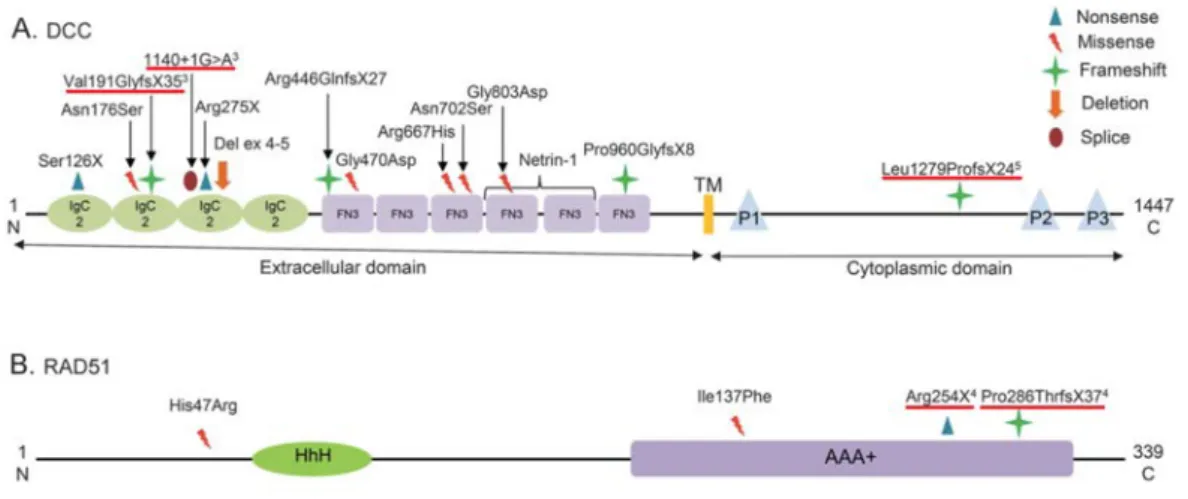
Figure 1. Mutations et variants répertoriés dans les gènes DCC et RAD51 chez des patients atteints de MMC (p. 21)
Figure 2. Phénotype des mutants Dcc-/- chez la souris (p. 23)
Figure 3. Mécanisme de réparation des cassures double brin de l’ADN par recombinaison homologue (p. 25)
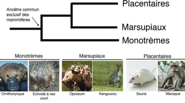
Figure 4. Phylogénie des mammifères (p. 44)
Figure 5. Anatomie comparée des commissures du cerveau antérieur chez les mammifères (p. 46)
Figure 6. Organisation tangentielle, laminaire et topographique du corps calleux (p. 49)
Figure 7. Maturation des connexions calleuses au cours du développement post-natal chez le chat (p. 52)
Figure 8. Les différents types de neurones à projection du néocortex (p. 55) Figure 9. Génération séquentielle des différentes couches néocorticales (p. 58)
Figure 10. Satb2 intervient dans la spécification des neurones calleux chez la souris (p. 61) Figure 11. Molécules impliquées dans la spécification et le guidage des neurones calleux (p. 68)
Figure 12. Différentes anomalies développementales du corps calleux chez l’humain (p. 76) Figure 13. Préparation motrice anormale chez les patients MMC lors de mouvements unimanuels de la main droite (dominante) (p. 121)
Figure 14. Le principe de la stimulation double-pulse : l’inhibition inter-hémisphérique (p. 124)
14 Figure 15. Modulation de l’excitabilité des cortex moteurs primaires lors de la réalisation de mouvements unimanuels chez des sujets sains et chez des patients atteints de MMC (p. 128) Figure 16. Interactions inter-hémisphériques entre PMd et M1 et entre les deux M1 au cours de la préparation d’un mouvement unimanuel (p. 133)
Figure 17. Organisation anatomique et rôle fonctionnel de l’AMS (p. 135)
Résultats
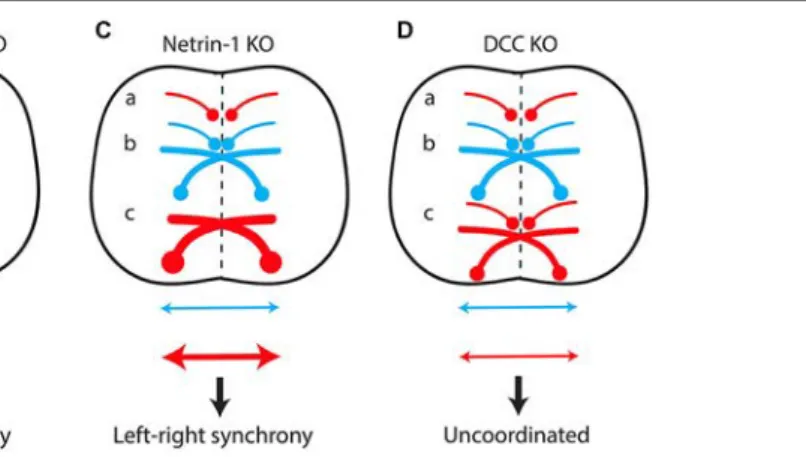
Figure 18. L’anatomie du FCS est anormale chez les souris Shh::cre;Netrinlox/lox (p. 186) Figure 19. Organisation anormale du FCS dans la moelle chez les mutants Shh::cre;Netrin1lox/lox (p. 187)
Figure 20. Netrin-1 est impliquée dans la génération de mouvements asymétriques des membres antérieurs (p. 189)
INTRODUCTION) 15!
15
16) INTRODUCTION!
16
I. Les mouvements en miroir congénitaux : un paradigme pour
l’étude des bases neurophysiologiques de la latéralisation
motrice
A. Préambule : comprendre le normal par le pathologique
Comme l’indique le titre de cette thèse, le travail que j’ai effectué avait pour but d’étudier une situation pathologique, celle de mouvements anormaux appelés « mouvements en miroir », afin d’approfondir nos connaissances sur cette maladie, mais aussi sur le fonctionnement du système moteur dans le cas dit « normal ». Cette démarche s’inscrit dans une tradition datant du XIXe siècle pouvant être simplement résumée : comprendre le normal par le pathologique. Bien que cette approche soit aujourd’hui extrêmement naturelle et qu’il nous semblerait absurde de remettre en cause son bien-fondé scientifique, j’aimerais revenir brièvement sur son histoire afin de mettre en avant les présupposés qu’elle implique.
Dans Le normal et le pathologique (1966), Georges Canguilhem rappelle que cette approche scientifique qui a jeté les bases de la physiologie moderne est principalement due à deux scientifiques français : Auguste Comte et Claude Bernard. Auguste Comte reprend à son compte et généralise un principe énoncé par Broussais, selon lequel il n’existe pas de différence de nature, mais seulement une différence de degré, entre le fonctionnement normal d’un organisme et son état pathologique. Autrement dit, les mécanismes mis en jeux dans des tissus sains et malades sont fondamentalement les mêmes : ils ne diffèrent que d’un point de vue quantitatif, du fait d’un trop plein ou d’un trop peu d’excitation, mais pas d’un point de vue qualitatif. Comte affirme dans la 40e leçon du Cours de philosophie positive (1838) : « […] l’état pathologique ne diffère point radicalement de l’état physiologique, à l’égard duquel il ne saurait constituer, sous un aspect quelconque, qu’un simple prolongement plus ou moins étendu des limites de variation soit supérieures soit inférieures, propres à chaque
INTRODUCTION) 17!
17 phénomène de l’organisme normal, sans pouvoir jamais produire de phénomènes vraiment nouveaux, qui n’auraient point, à un certain degré, leurs analogues purement physiologiques ». Claude Bernard adhère à cette idée que la pathologie ne consiste qu’en une dérégulation des phénomènes normaux, physiologiques. Nous citons ici un extrait des Leçons
sur le diabète et la glycogenèse animale (1877) : « Physiologie et pathologie se confondent et
sont au fond une seule et même chose. […] Toute maladie a une fonction normale correspondante dont elle n’est qu’une expression troublée, exagérée, amoindrie ou annulée. Si nous ne pouvons pas aujourd’hui expliquer tous les phénomènes des maladies, c’est que la physiologie n’est pas encore assez avancée et qu’il y a encore une foule de fonctions normales qui nous sont inconnues ».
C’est en vertu de cette homogénéité, de cette continuité entre le phénomène normal et le phénomène pathologique que nous pouvons prétendre expliquer le premier – en l’occurrence la latéralisation motrice – par l’étude du second – les mouvements en miroir. Cette approche est donc très puissante, mais son présupposé n’est peut être pas si évident : la pathologie est-elle véritablement incapable de « produire des phénomènes vraiment nouveaux » ?
B. Mouvements en miroir congénitaux
Dans cette partie, nous nous intéresserons à la description clinique des mouvements en miroir congénitaux et à leurs causes génétiques. La physiopathologie de ces mouvements anormaux sera abordée dans la section suivante.
1) Définition et description clinique
Les comportements moteurs d’un organisme sont la résultante d’un ensemble de contractions musculaires finement coordonnées qui sont contrôlées par le système nerveux.
18) INTRODUCTION!
18 Les humains possèdent un répertoire de mouvements extrêmement diversifié, notamment en ce qui concerne les mouvements manuels. Nous appellerons mouvement latéralisé (des mains) tout mouvement impliquant une asymétrie de contraction musculaire entre les mains droite et gauche. En ce sens, les mouvements latéralisés comprennent les mouvements strictement unimanuels (impliquant le mouvement d’une seule des deux mains tandis que l’autre est au repos) et les mouvements bimanuels asymétriques. La capacité de réaliser des mouvements latéralisés est un aspect crucial de nos activités quotidiennes. Taper à l’ordinateur, jouer d’un instrument de musique, ouvrir un bocal de confiture, faire de l’escalade, sont autant de gestes qui nécessitent de produire des mouvements asymétriques avec nos deux mains.
Les mouvements en miroir (MM) sont des mouvements involontaires d’une main qui miment et accompagnent les mouvements volontaires de la main opposée. Des MM sont fréquemment observés chez les jeunes enfants de moins de sept ans, et sont considérés comme des manifestations de l’immaturité du système nerveux central, en particulier du corps calleux (Koerte et al., 2010; Beaule et al., 2012). Leur persistance au-delà de cet âge définit leur caractère pathologique (Bonnet et al., 2010). Les MM touchent majoritairement les extrémités distales des membres supérieures (les mains et les doigts). Ils sont généralement d’amplitude inférieure aux mouvements volontaires contralatéraux et sont d’intensité variable d’un patient à l’autre. La sévérité des MM est évaluée avec l’échelle de Woods et Teuber (Woods and Teuber, 1978) comme suit : 0 : pas de MM ; 1 : MM à peine discernables mais répétés ; 2 : MM discrets mais soutenus ; 3 : MM importants, soutenus et répétés ; 4 : MM égaux à ceux qui sont observés sur la main en mouvement volontaire. Les individus atteints ne peuvent donc pas réaliser de mouvements unimanuels ou de mouvements bimanuels asymétriques.
Le syndrome des Mouvements en Miroir Congénitaux (MMC) constitue une pathologie développementale rare, souvent familiale à transmission autosomique dominante,
INTRODUCTION) 19!
19 mais il existe aussi des cas sporadiques (Meneret et al., 1993; Peng and Charron, 2013; Meneret et al., 2014a). Dans ce syndrome, les MM ne sont pas associés à d’autres anomalies neurologiques. Les MMC constituent donc une atteinte extrêmement pure du système de la latéralisation motrice. Dans cette pathologie, les MM apparaissent tôt dans la petite enfance, puis restent stables au cours de la vie. C’est une pathologie très rare, avec une prévalence estimée à moins de 1/1 000 000 (Orphanet #238722), mais la maladie est probablement sous-diagnostiquée.
2) Autres pathologies associées aux mouvements en miroir
Les MM d’origine développementale (par opposition aux formes acquises, qui seront brièvement évoquées à la fin de ce paragraphe) peuvent aussi être associés à d’autres symptômes dans le cadre de syndromes complexes.
Syndrome de Kallmann : le syndrome de Kallmann est caractérisé par l’association d’hypogonadisme hypogonadotrope et d’anosmie. La présence de MM dans ce symptôme est majoritairement associée aux mutations du gène KAL1, mutations responsables de la forme liée à l’X de la pathologie (Quinton et al., 1996; Royal et al., 2002; Dode and Hardelin, 2010). Syndrome de Klippel-Feil : ce syndrome est caractérisé par une fusion des vertèbres cervicales, et inclut un cou court, une mobilité cervicale réduite, une implantation basse des cheveux. Des MM sont fréquemment observés chez ces patients (Bauman, 1932; Baird et al., 1967; Royal et al., 2002).
Syndrome de Joubert : ce syndrome est une entité hétérogène, caractérisée par les malformations du vermis cérébelleux et du tronc cérébral. Les manifestations phénotypiques peuvent inclure faiblesses musculaires, respiration et mouvements oculomoteurs anormaux, maladresse, retards cognitifs, comportement autistique et des dysfonctions variables de
20) INTRODUCTION!
20 différents organes (principalement les reins, la rétine, le foie et le squelette) (Romani et al., 2013). Des MM sont parfois associés à ce tableau clinique (Ferland et al., 2004).
Syndrome de Möbius : ce syndrome inclut une faiblesse faciale non évolutive associée à une abduction limitée des yeux (Verzijl et al., 2003). Une étude récente a décrit le cas de trois patients atteints du syndrome de Möbius ayant des MM (Webb et al., 2014).
Syndrome de Gorlin : ce syndrome est caractérisé par une association de carcinomes, de kystes de la mâchoire et de malformations squelettiques (Gorlin and Goltz, 1960). Le cas d’un patient présentant un syndrome de Gorlin et des MM a été récemment décrit (Sag et al., 2016).
Syndrome de Seckel : ce syndrome associe une microcéphalie primaire à des retards de croissance et une déficience intellectuelle. Un patient atteint de ce syndrome et présentant des mouvements en miroir a été décrit (Thapa and Mukherjee, 2010).
Mouvements en miroir acquis : dans le cas des MM acquis, l’âge de début des MM est bien plus tardif. La présence de MM est souvent associée à d’autres pathologies du mouvement, et ce symptôme peut être retrouvé dans la maladie de Parkinson, le syndrome corticobasal, le tremblement essentiel, la dystonie miroir, le syndrome de Creutzfeldt-Jacob, la maladie de Huntington (Cox et al., 2012). L’apparition de MM a aussi été décrite chez un patient à la suite d’un infarctus touchant l’aire motrice supplémentaire (Chan and Ross, 1988).
3) Génétique des mouvements en miroir congénitaux
Les mouvements en miroir congénitaux sont souvent une pathologie héréditaire à transmission autosomique dominante. Deux gènes responsables des MMC ont été identifiés sans ambiguïté à ce jour : DCC et RAD51 (Figure 1) (Srour et al., 2010; Depienne et al., 2011; Depienne et al., 2012; Peng and Charron, 2013; Meneret et al., 2014a). DNAL4 est potentiellement impliqué, mais des mutations de ce gène n’ont été identifiées que dans ne
! 21! !
! !
Figure 1. Mutations et variants répertoriés dans les gènes DCC et RAD51 chez des patients atteints de MMC. (A) Mutations et variants répertoriés dans le gène DCC. (B)
Mutations et variants répertoriés dans le gène RAD51. Les formes familiales de MMC sont associées à des mutations hétérozygotes tronquantes de RAD51 ou DCC (soulignées en rouge). La première mutation décrite de DCC (A : 1140 + 1G >A) aboutit à une protéine tronquée après l’exon 5, dont la plupart des domaines fonctionnels sont absents et qui ne peut pas interagir avec son ligand NETRIN-1. Les autres mutations aboutissent probablement à une diminution de la quantité de protéine fonctionnelle produite. Adapté de (Meneret et al, 2014).
22) INTRODUCTION!
22 seule famille (Ahmed et al., 2014; Meneret et al., 2014b). RAD51 et DCC à eux seuls ne rendent pas compte de l’ensemble des cas familiaux et MMC, ce qui signifie que d’autres gènes, pour le moment inconnus, sont associés à cette pathologie. Les mutations de RAD51 et
DCC identifiées dans les familles de patients MMC sont hétérozygotes et leur pénétrance est
de 50%. La première mutation décrite de DCC aboutit à la formation d’une protéine tronquée, ne pouvant pas interagir avec son ligand NETRIN-1 (Srour et al., 2010). Il a été proposé que les autres mutations tronquantes soient responsables d’une diminution de la quantité de protéine fonctionnelle produite (ou haploinsuffisance) du fait de la dégradation de l’ARN messager anormal (Depienne et al., 2011; Depienne et al., 2012; Meneret et al., 2014a). Cependant cette hypothèse n’a pas été testée directement, et la possibilité que certaines de ces mutations soient responsables d’effets dominants négatifs ne peut pas être exclue.
DCC assure différentes fonctions au cours du développement. Il joue un rôle important dans la survie cellulaire et dans le contrôle de la prolifération en tant que récepteur à dépendance : en l’absence de son ligand, NETRIN-1, il enclenche un programme de mort cellulaire (Mehlen et al., 1998). En lien avec ce rôle de suppresseur de tumeurs, des mutations de DCC ont été initialement associées à des cancers colorectaux (d’où son nom, DCC signifiant « Deleted in Colorectal Cancer ») (Fearon et al., 1990; Krimpenfort et al., 2012). DCC est aussi impliqué dans le guidage des axones commissuraux (c’est-à-dire des axones qui croisent la ligne médiane du corps) au cours du développement. Dans la moelle épinière, les axones exprimant le récepteur DCC à la membrane de leur cône de croissance sont attirés par leur ligand NETRIN-1, molécule qui est sécrétée au niveau de la ligne médiane par les cellules de la plaque du plancher (Kennedy et al., 1994; Keino-Masu et al., 1996). Cette interaction permet aux axones commissuraux de franchir la ligne médiane, pour ensuite poursuivre leur trajet et contacter leurs cibles dans l’hémi-moelle opposée. Par la suite, DCC s’est révélé être impliqué dans le développement de nombreuses commissures (Figure 2),
! "#! !
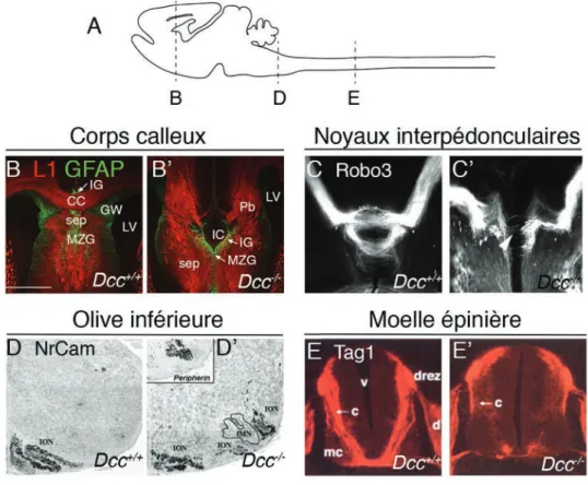
Figure 2. Phénotype des mutants Dcc-/- chez la souris. (A) Schéma représentant le système
nerveux central d’une souris à P0 en vue sagittale et indiquant le niveau des différentes coupes. (B, B’) Coupes coronales du corps calleux (CC) de souris Dcc+/+ et Dcc-/- à P0 avec immuno-marquage anti-L1 (marquage du CC) et anti-GFAP (marquage des populations gliales). CC : corps calleux ; IG : indusium griseum ; GW : glial wedge ; sep : septum ; MZG : midline zipper glia ; IC : kyste inter-hémisphérique ; LV : lateral ventricule. (Données personnelles). (C, C’) Visualisation du fasciculus retroflexus par immuno-marquage anti-ROBO3 combiné à une procédure de clarification 3DISCO chez des souris Dcc+/+ et Dcc-/- à E16 (Extrait de Belle et al, 2014). (D, D’) Coupes coronales de souris Dcc+/+ et Dcc-/- à P0 avec une hybridation in-situ dirigée contre NrCam et contre la peripherin. ION : neurones de l’olive inférieure (Extrait de Marcos et al, 2009). (E, E’) Coupes coronales de moelles épinières d’embryons de souris Dcc+/+ et Dcc-/- à E11.5 avec immuno-marquage anti-Tag1. c : axones commissuraux; mc : colonne motrice ; v : ventricule ; d : ganglion de la racine dorsale ; drez : zone d’entrée du ganglion de la racine dorsale (Extrait de Fazeli et al, 1997).!
24) INTRODUCTION!
24 notamment les commissures du cerveau antérieur (commissure antérieure, commissure hippocampique et corps calleux) (Fazeli et al., 1997; Fothergill et al., 2014), le fasciculus retroflexus (qui relie l’habenula aux noyaux inter-pédonculaires du pont) (Belle et al., 2014), les fibres grimpantes de l’olive inférieure (Marcos et al., 2009), les interneurones commissuraux de la moelle épinière (Rabe Bernhardt et al., 2012) et le faisceau corticospinal (Finger et al., 2002).
RAD51 est une protéine connue essentiellement pour son rôle dans la réparation de l’ADN. Il s’agit de l’homologue du gène RecA, bien connu chez les bactéries. RAD51 assure le maintien de la stabilité génomique au cours de la prolifération cellulaire. En effet, RAD51 intervient dans la réparation des cassures double brin de la molécule d’ADN par un processus de recombinaison homologue (Figure 3) (Thacker, 2005; Jasin and Rothstein, 2013). Alors que des mutations de nombreux partenaires de RAD51 impliqués la voie de réparation de l’ADN sont associées à un risque accru de cancer, aucune mutation dans la séquence codante de RAD51 n’a été associée de manière causale au cancer (Lose et al., 2006; Moynahan and Jasin, 2010). Les seules mutations familiales dans la séquence codante de RAD51 publiées à ce jour sont celles associées au syndrome des mouvements en miroir congénitaux (Depienne et al., 2012; Meneret et al., 2014a; Franz et al., 2015), ce qui peut sembler surprenant à plusieurs égards. Premièrement, RAD51 étant exprimée de façon ubiquitaire dans les cellules progénitrices pendant les phases de proliférations au cours du développement, il est étonnant qu’un déficit de la fonction de réparation d’ADN conduise à un phénotype aussi spécifique que celui des MM. Deuxièmement, RAD51 intervenant dans la réparation des cassures d’ADN par recombinaison homologue, cela signifie qu’elle utilise la chromatide sœur du chromosome lésé pour procéder à la réparation. Ce procédé n’est donc possible que dans les cellules ayant des chromosomes à deux chromatides, autrement dit des cellules dont le cycle cellulaire comprend une phase G2 (post réplication). Ce n’est pas le cas des neurones qui sont
! 25! !
!
!
Figure! 3.! Mécanisme! de! réparation! des! cassures! double! brin! de! l’ADN! par! recombinaison! homologue.! A! la! suite! d’une! cassure! double! brin! de! l’ADN,! RAD51!
forme!un!filament!au!niveau!d’un!simple!brin!d’ADN!de!la!chromatide!lésée.!A!la!suite!de! cette!étape,!L’ADN!double!brin!intact!de!la!chromatide!sœur!est!utilisé!comme!matrice,! permettant!ainsi!la!réparation!de!la!lésion.!!Adapté!de!(Thacker,!2005).!
26) INTRODUCTION!
26 des cellules quiescentes, et présentent donc des chromosomes à une chromatide. Pour ces différentes raisons, il est probable que RAD51 joue un rôle encore inconnu au cours du développement du système nerveux central, et que ce soit la perturbation de cette fonction qui soit responsable du syndrome des MMC.
L’anémie de Fanconi est une pathologie héréditaire caractérisée par une hyper-sensibilité aux lésions de l’ADN, et associée à des symptômes incluant de multiples anomalies congénitales et hématologiques, ainsi qu’une augmentation de la prédisposition aux cancers (Auerbach, 2009). Récemment, deux études ont décrit le cas de patients présentant une anémie de Fanconi associée à une mutation faux-sens hétérozygote du gène RAD51. Ces mutations de novo aboutissaient à la formation d’une forme dominante négative de la protéine qui interfère avec ses fonctions normales (Ameziane et al., 2015; Wang et al., 2015). Dans un des deux cas décrit, l’anémie de Fanconi est une forme atypique associée à des troubles neurologiques (microcéphalie, hydrocéphalie, retard mental) (Ameziane et al., 2015).
Comment les mutations de RAD51 et DCC aboutissent-elles aux MMC ? Tandis que le rôle connu de DCC dans le développement du système moteur chez la souris fournit à première vue une explication satisfaisante pour les MMC causés par des mutations de DCC, le rôle de RAD51 dans le développement du système moteur est complètement inconnu. Dans la partie suivante, la présentation de la physiopathologie des MMC permettra de mieux comprendre les mécanismes nécessaires à la production de mouvements latéralisés. Les liens entre les mutations des gènes DCC et RAD51 et la physiopathologie des MMC ont fait l’objet d’une partie de mon travail de thèse et seront abordés dans le chapitre des résultats.
INTRODUCTION) 27!
27
C.
Revue de la littérature 1 : Applaudir d’une main, la latéralisation du
contrôle moteur
Comprendre le fonctionnement normal d’un organisme par l’étude d’une situation pathologique est une approche scientifique puissante. Dans cette perspective, la compréhension des mécanismes neurophysiologiques sous-tendant la latéralisation motrice a beaucoup à gagner de l’étude de situations pathologiques associées à une perte de la capacité à réaliser des mouvements latéralisés. Dans cette revue, nous nous sommes intéressés aux corrélats neurophysiologiques de deux situations pathologiques qui sont, à notre connaissance, les seules à être associées à l’incapacité de produire des mouvements latéralisés : les mouvements en miroir chez l’humain, et le « hopping gait » chez la souris.
Cette analyse souligne le rôle crucial de commissures situées à différents niveaux du système nerveux central dans la latéralisation motrice. Les commissures, en tant que structures permettant la mise en relation des deux hémicorps, sont en effet les principaux acteurs de la latéralisation du contrôle moteur. Nous identifions trois populations de neurones commissuraux d’une importance primordiale pour la production de mouvements latéralisés :
- les neurones formant le corps calleux (CC), principale commissure assurant la communication entre les deux hémisphères cérébraux
- les neurones formant le faisceau corticospinal (FCS), l’un des principaux effecteurs de la commande motrice volontaire
- les neurones commissuraux de la moelle épinière
Le CC et le FCS sont les deux principales structures impliquées dans la neurophysiologie des MM, ce qui souligne leur rôle dans la production de mouvements asymétriques des mains. Le CC et le FCS semblent n’avoir pas d’impact sur la latéralisation motrice durant la locomotion (quadrupède), ce rôle étant essentiellement assuré par les axones commissuraux de la moelle.
REVIEW published: 02 June 2015 doi: 10.3389/fnana.2015.00075
One hand clapping: lateralization of
motor control
Quentin Welniarz1,2,Isabelle Dusart1,Cécile Gallea2andEmmanuel Roze2,3*
1Neuroscience Paris Seine, CNRS UMR8246, Inserm U1130, Sorbonne Universités, UPMC UM119, Paris, France,2Inserm
U1127, CNRS UMR 7225, Sorbonne Universités, UPMC UMR S1127, Institut du Cerveau et de la Moelle épinière, ICM, Paris, France,3Département des Maladies du Système Nerveux, AP-HP, Hôpital Pitié Salpêtrière, Paris, France
Edited by: Yun-Qing Li, The Fourth Military Medical University, China Reviewed by: José A. Armengol, University Pablo de Olavide, Spain Yu-Qiang Ding, Tongji Unversity, China *Correspondence: Emmanuel Roze, Département des Maladies du Système Nerveux, AP-HP, Hôpital Pitié Salpêtrière, 47–83 boulevard de l’Hôpital, 75013 Paris, France emmanuel.flamand-roze@psl.aphp.fr Received: 31 March 2015 Accepted: 17 May 2015 Published: 02 June 2015 Citation: Welniarz Q, Dusart I, Gallea C and Roze E (2015) One hand clapping: lateralization of motor control. Front. Neuroanat. 9:75. doi: 10.3389/fnana.2015.00075
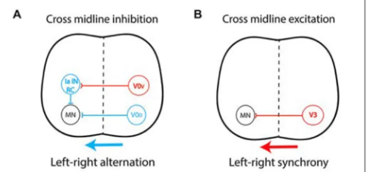
Lateralization of motor control refers to the ability to produce pure unilateral or asymmetric movements. It is required for a variety of coordinated activities, including skilled bimanual tasks and locomotion. Here we discuss the neuroanatomical substrates and pathophysiological underpinnings of lateralized motor outputs. Significant breakthroughs have been made in the past few years by studying the two known conditions characterized by the inability to properly produce unilateral or asymmetric movements, namely human patients with congenital “mirror movements” and model rodents with a “hopping gait”. Whereas mirror movements are associated with altered interhemispheric connectivity and abnormal corticospinal projections, abnormal spinal cord interneurons trajectory is responsible for the “hopping gait”. Proper commissural axon guidance is a critical requirement for these mechanisms. Interestingly, the analysis of these two conditions reveals that the production of asymmetric movements involves similar anatomical and functional requirements but in two different structures: (i) lateralized activation of the brain or spinal cord through contralateral silencing by cross-midline inhibition; and (ii) unilateral transmission of this activation, resulting in lateralized motor output.
Keywords: mirror movement, hopping gait, corticospinal tract, corpus callosum, spinal cord, axon guidance
Introduction
Lateralization of motor control is required for a variety of coordinated movements, including skilled bimanual tasks and locomotion. To our knowledge, only two conditions are associated with the inability to produce asymmetric movements in mammals: human ‘‘mirror movements’’ and rodent ‘‘hopping gait’’.
Mirror movements are involuntary symmetrical movements of one side of the body that mirror voluntary movements of the other side. The affected individuals are unable to perform purely unimanual movements and have difficulties to perform tasks requiring independent actions with the two hands such as holding a cup while filling it with water, opening a jar or playing a musical instrument. During these tasks, the effectors produce different motor outputs that are usually bound together by a shared, object-directed goal.
Quadrupedal locomotion is characterized by coordinated, alternating bilateral activation of limb muscles, in which effectors repeatedly produce similar motor outputs in a specific temporal order. A ‘‘hopping gait’’ is a switch from alternate to synchronous activity of the limbs during locomotion that is observed in rodent mutants with impaired axonal guidance.
Welniarz et al. Lateralization of motor control
Here we discuss the neuroanatomical substrates and pathophysiological underpinnings of lateralized motor output through the study ‘‘mirror movements’’ and ‘‘hopping gait’’. Whereas mirror movements are associated with altered interhemispheric connectivity and abnormal corticospinal projections, abnormal spinal cord interneurons trajectory is responsible for the ‘‘hopping gait’’. Interestingly, the analysis of these two conditions indicates that the production of asymmetric movements involves similar anatomical and functional requirements but in two different structures, the cerebral cortex and the spinal cord, and it emphasizes the importance of proper commissural axon guidance in this process.
The “Mirror Movement” Paradigm: Inability to Produce Asymmetric Skilled Hand
Movements
Humans have a greater ability than other species to produce purposeful handling movements, most of them being asymmetric. With training, we can master highly complex skills ranging from the fluid movements of the virtuoso pianist to the precise life-saving gestures of the heart surgeon. In humans, execution of unimanual movements requires lateralized activation of the primary motor cortex (M1), which then transmits the motor command to the contralateral hand through the crossed corticospinal tract (CST; Figure 1A;Chouinard and Paus, 2010; Galléa et al., 2011).
Loss of this lateralization results in mirror movements (MM), which consist of involuntary symmetrical movements of one side of the body that mirror voluntary movements of the other side. Congenital mirror movement disorder (CMM) is a rare genetic disorder transmitted in autosomal dominant manner in which mirror movements are the only clinical abnormality. These mirror movements predominate in the distal upper limbs, leaving affected individuals unable to perform independent actions with the two hands or to perform purely unimanual movements. They usually have hand clumsiness and pain in the upper limbs during sustained manual activities. The two main culprit genes are Dcc (deleted in colorectal cancer) and Rad51 (Srour et al., 2010; Depienne et al., 2011, 2012; Méneret et al., 2014a). A third gene, Dnal4, might also be involved (Ahmed et al., 2014; Méneret et al., 2014b). Dcc plays a key role in CST midline crossing (Finger et al., 2002), while Rad51 is well known for its role in DNA repair and may also have a major role in motor system development (Depienne et al., 2012; Gallea et al., 2013). In addition to isolated congenital mirror movements caused by Dcc or Rad51 mutations, syndromic forms of MM may be accompanied by numerous other symptoms, in disorders such as Dandy walker syndrome, Joubert’s syndrome, X-linked Kallmann syndrome, Klippel Feil syndrome and congenital hemiparesis (Vulliemoz et al., 2005; Galléa et al., 2011; Peng and Charron, 2013).
CMM provides a unique paradigm for studying the lateralization of motor control (Carson, 2005; Galléa
et al., 2011; Peng and Charron, 2013). Two main non
exclusive mechanisms may account for MM: (i) abnormal
interhemispheric communication resulting in bilateral activation of primary motor areas (Figures 1B,C); and (ii) a corticospinal tract abnormality leading to bilateral downstream transmission of the motor command (Figures 1D,E;Gallea et al., 2013).
Interhemispheric Connectivity and Motor Lateralization
In humans, the default set-up of motor behavior is probably a mirror program (Chan and Ross, 1988; Meyer et al., 1995; Cincotta and Ziemann, 2008). Unilateral and bilateral voluntary movements are preceded by slow negativity on EEG recordings, known as the Bereitschaftpotential (Shibasaki and Hallett, 2006), which starts 2 s before movement onset and is distributed over the two hemispheres. This Bereitschaftpotential may reflect bilateral activation of the supplementary motor areas (SMA) and dorsal premotor cortices (dPMC) during motor planning. Just before movement onset, cortical activity is restricted to the primary motor cortex and dPMC contralateral to the intended movement (Shibasaki and Hallett, 2006). An active mechanism is required to restrict motor activation to one hemisphere during execution of a pure unimanual movement.
Our current understanding of this ‘‘non mirror transformation’’ derives mainly from the study of ‘‘physiological’’ mirror movements. Healthy subjects have a default tendency to produce minimal mirror movements when performing highly complex and effortful unimanual tasks (Koerte et al., 2010; Sehm et al., 2010; Beaulé et al., 2012). Activation of the mirror M1 (ipsilateral to the voluntary movement) is the main explanation for this tendency (Mayston et al., 1999; Cincotta et al., 2004; Zijdewind et al., 2006; Hübers et al., 2008). In order to achieve this ‘‘non mirror transformation’’, the active M1 (contralateral to the intended movement) inhibits the mirror M1 via fibers that pass through the corpus callosum (transcallosal tract, TCT), thereby restricting the motor output to the active M1. This inhibition of one motor cortex by the other is called interhemispheric inhibition (IHI). IHI is thought to rely on transcallosal glutamatergic connections to inhibitory interneurons that in turn innervate pyramidal cells in the receiving hemisphere (Meyer et al., 1995; Reis et al., 2008). Several lines of evidence support the importance of TCT-mediated IHI in the lateralization of motor control. For example, the gradual disappearance of minimal MM frequently observed in young children correlates with the degree of TCT myelination and with the level of IHI (Koerte et al., 2010; Beaulé et al., 2012). Also, experimental modulation of IHI directed from the active M1 to the mirror M1 affects mirror activity: a transient increase in IHI is associated with a decrease in mirror activity, and vice versa (Hübers et al., 2008).
IHI between the two primary motor cortices is modulated differently during the different phases of unimanual movements. IHI is balanced between the two motor cortices at the onset of movement preparation, then shifts towards the ipsilateral M1 (ipsilateral to the voluntary movement) at the end of movement preparation and at movement onset (Murase et al.,
Welniarz et al. Lateralization of motor control
FIGURE 1 | Hypothetical mechanisms of mirror movements. (A) In humans, execution of a unilateral left hand movement requires both lateralized activation of the right primary motor cortex (M1) by interhemispheric inhibition (IHI) and proper motor planning and then transmission of the motor command to the contralateral (left) hand alone, through a crossed corticospinal tract. There are two main mechanisms underlying MM: (i) abnormal IHI(B) or abnormal delivery of the motor plan from the supplementary motor area (SMA) to M1(C), resulting in bilateral activation of the primary motor cortices; and (ii) abnormal
decussation of the CST(D) or abnormal branching of the CST in the spinal cord(E), resulting in bilateral transmission of the motor command to the spinal cord. Mirror movements have not been described in horizontal gaze palsy with progressive scoliosis (HGPPS), despite the absence of CST decussation in these patients(F). This suggests that MM are related to the presence of bilateral spinal cord projections arising from a single primary motor cortex rather than to abnormal decussation of the CST per se. Dark Blue, normal mechanism; Red, abnormal mechanism; Light blue, IHI.
2004; Duque et al., 2007). In parallel, IHI of the contralateral M1 decreases during movement preparation and shifts towards facilitation at movement onset (Murase et al., 2004; Perez and Cohen, 2008). These subtle time-dependent bilateral variations of IHI are necessary to avoid premature execution (Duque and Ivry, 2009), and to prevent mirror activity in the ipsilateral
M1 (Giovannelli et al., 2009). Impairment of IHI may thus
result in bilateral M1 activation and transmission of the motor command to both hands through the two crossed CSTs.
In patients with CMM and X-linked Kallmann syndrome, several studies have revealed abnormal, bilateral M1 activation during voluntary unimanual movements and have confirmed that activation of the mirror M1 is not a sensory consequence of the mirror movement but rather participates actively in the mirroring motor activity (Shibasaki and Nagae, 1984; Cohen et al., 1991; Mayer et al., 1995; Cincotta et al., 1996; Krams et al., 1997; Verstynen et al., 2007). However, studies based on indirect methods have failed to demonstrate consistent impairment of IHI mechanisms in CMM patients (Cincotta et al., 1996, 2002; Papadopoulou et al., 2010). Using dual-site transcranial magnetic stimulation (TMS), a more direct method (Perez and Cohen, 2008), we found that CMM patients with Rad51 mutations had abnormal IHI between the primary motor cortices at rest, together with morphological abnormalities of the TCT (Figure 1B; Gallea et al., 2013). It has been proposed that this impaired IHI is due to an abnormal input of the transcallosal glutamatergic connections onto the inhibitory interneurons in the receiving hemisphere. It is noteworthy that most individuals lacking a corpus
callosum do not exhibit mirror movements, suggesting that the absence of the corpus callosum and interhemispheric connections alone might not be sufficient to generate MM. Finally, a study of a CMM patient with complete agenesis of the corpus callosum concluded that the absence of TCT played little part in the pathophysiology of MM (Lepage et al., 2012).
Interhemispheric pathways are not limited to direct M1-M1 interactions and IHI but also include circuits linking secondary motor areas (SMA and PMd) to contralateral motor areas. These circuits might be involved in restricting the generation of motor output to the active hemisphere during movement preparation. For these reasons it has been proposed that abnormal motor planning and/or abnormal transmission of the motor plan from the secondary motor areas to the primary motor areas might also be involved in MM generation
(Chan and Ross, 1988; Cincotta et al., 2004; Duque et al.,
2010; Galléa et al., 2011; Gallea et al., 2013). Evidence of abnormal motor planning associated with MM was first obtained through studies of two CMM patients and a patient with Kallmann’s syndrome, who showed an abnormal, bilateral (instead of unilateral) distribution of the Bereitschaftpotential during movement preparation (Shibasaki and Nagae, 1984; Cohen et al., 1991). However, two other studies argued against a role of abnormal movement planning in MM: the first showed that movement-related cortical EEG potentials were identical (that is to say, lateralized and not bilateral) in healthy volunteers and in six CMM patients (Mayer et al., 1995), while the second study, a case report, showed normal, unilateral cortical activation during fMRI imaging of imagined movements
Welniarz et al. Lateralization of motor control
closely related to motor planning (Verstynen et al., 2007). More recently, we found that the SMA activation pattern and connectivity are abnormal during both unimanual and bimanual movements in Rad51-mutated CMM patients (Figure 1C;
Gallea et al., 2013) This suggested that cortical activation and connectivity might be modified in CMM patients during movement preparation, resulting in inappropriate delivery of the motor program from the SMA to both primary motor cortices.
Together, these results suggest that interhemispheric connectivity is critical for lateralized activation of the motor cortex when a unilateral movement is intended.
The Corticospinal Tract and Motor Lateralization The CST is a crossed tract that transmits the motor command from one motor cortex to the contralateral spinal cord. The CST first appeared in mammals and was likely critical for the development of voluntary skilled movements through evolution (Vulliemoz et al., 2005). Selective lesions of the CST in humans, non human primates and rodents impair skilled digit movements such as reaching (Schieber, 2007). The CST is massively crossed in humans. About 70–95% of all CST axons cross the midline at the junction between the medulla and the spinal cord, forming the so-called ‘‘pyramidal decussation’’, and establish direct contacts with the motor neurons located in the anterior horn of the spinal cord (Vulliemoz et al., 2005). The approximately 10% of CST axons that do not decussate at the medulla remain ipsilateral, and this ipsilateral tract is mainly located in the ventral part of the spinal cord in both humans and rodents (Brösamle and Schwab, 1997; Vulliemoz et al., 2005). The ipsilateral CST component does not target motor neurons innervating distal limb muscles but rather motor neurons innervating the proximal or axial musculatures (Bawa et al., 2004; Vulliemoz et al., 2005). In humans, cats and rodents, the CST initially establishes strong bilateral projections to the spinal cord. The ipsilateral projections consist of uncrossed CST axons (Joosten et al., 1992; Brösamle and Schwab, 1997; Eyre et al., 2001), and/or of normally crossed CST axons that recross the midline within the spinal cord (Li and Martin, 2000; Rosenzweig et al., 2009). This CST projection pattern is refined during early post-natal development, resulting in the elimination of the majority of the ipsilateral projections (Joosten et al., 1992; Eyre et al., 2000, 2001; Li and Martin, 2000). This refinement of the ipsilateral projections is an activity-dependent process of competition with the crossed CST fibers originating from the contralateral motor cortex (Martin and Lee, 1999; Eyre et al., 2001; Eyre, 2003; Martin et al., 2004; Friel and Martin, 2007; Friel et al., 2014).
Human MM could result from the presence of CST projections to both the ipsilateral and contralateral spinal cord. In patients with CMM, Kallmann syndrome, Klippel-Feil syndrome or congenital hemiparesis, unilateral stimulation of the primary motor cortex hand area at rest by TMS elicits bilateral hand muscle responses with identical latencies, whereas in healthy volunteers the muscle response is strictly contralateral to the stimulated hemisphere (Nass, 1985; Farmer et al., 1990; Benecke et al., 1991; Mayston et al., 1997; Alagona
et al., 2001; Staudt et al., 2002; Cincotta et al., 2003; Bawa et al., 2004; Srour et al., 2010; Depienne et al., 2011; Gallea et al., 2013). This reveals the presence of fast-conducting corticospinal projections from the hand area of one primary motor cortex to both sides of the spinal cord in CMM patients and suggests an anatomic-functional link between anomalies in the CST trajectory and the inability to produce lateralized movements.
Bilateral corticospinal projections to the spinal cord could be due to: (i) abnormal pyramidal decussation resulting in an aberrant uncrossed ipsilateral CST (Figure 1D); or (ii) aberrant branching of crossed CST axons in the spinal cord (Figure 1E). In both cases, the aberrant CST projection pattern could result from abnormal guidance of the CST axons or from an abnormal persistence of the ipsilateral CST projections that are normally eliminated during development. An elegant TMS study of two CMM patients supports the existence of a separate uncrossed ipsilateral CST (Cincotta
et al., 2003). Diffusion tensor imaging (DTI) was used to
study the precise anatomy of the pyramidal decussation in Rad51-mutated patients, confirming abnormal CST decussation (Gallea et al., 2013), although Dcc-mutated CMM patients have yet to be studied. Rad51 expression pattern in the mouse central nervous system (Depienne et al., 2012), and the known role of DCC in commissural axons guidance (Kennedy et al., 1994; Serafini et al., 1994; Keino-Masu et al., 1996; Finger et al., 2002), suggest that abnormal axonal guidance rather than impaired CST maturation is responsible for the bilateral CST projections observed in Rad51- and Dcc-mutated patients. Electrophysiological studies also support the existence of an aberrant uncrossed CST in X-linked Kallmann patients (Mayston et al., 1997; Farmer et al., 2004). In patients with congenital hemiparesis, MM may be explained by an abnormal maturation of the CST due to the unequal activity between the affected and unaffected motor cortices (Eyre et al., 2001; Eyre, 2003; Friel et al., 2014). This would lead to the maintenance and reinforcement of the ipsilateral CST from the unaffected motor cortex, combined with aberrant branching of corticospinal fibers in the spinal cord (Benecke et al., 1991; Alagona et al., 2001; Staudt et al., 2002; Galléa et al., 2011; Friel
et al., 2014). Mirror movements have not been described in
patients with horizontal gaze palsy with progressive scoliosis (HGPPS), despite their lack of CST decussation. HGPPS is linked to mutations in the axon guidance receptor ROBO3 (Jen et al., 2004). The CST is completely uncrossed in HGPPS patients, and each hemisphere thus projects in a strictly ipsilateral manner to the spinal cord (Figure 1F). Together, these findings suggest that MM are related to the presence of bilateral spinal cord projections arising from a single primary motor cortex rather than to abnormal decussation of the CST per se.
Study of MM patients enlightened the critical importance of two mechanisms for the generation of asymmetric movements: (i) lateralized activation of the brain through contralateral silencing by IHI and proper motor planning; and (ii) unilateral transmission of the motor command to the contralateral spinal cord via the CST. Both abnormal interhemispheric connectivity
Welniarz et al. Lateralization of motor control
and an altered CST trajectory could be responsible for MM, but the respective importance of each factor is unclear.
Control of Left-Right Alternation During Locomotion: New Insights from
Genetically Modified Mice with
Developmental Motor System Anomalies
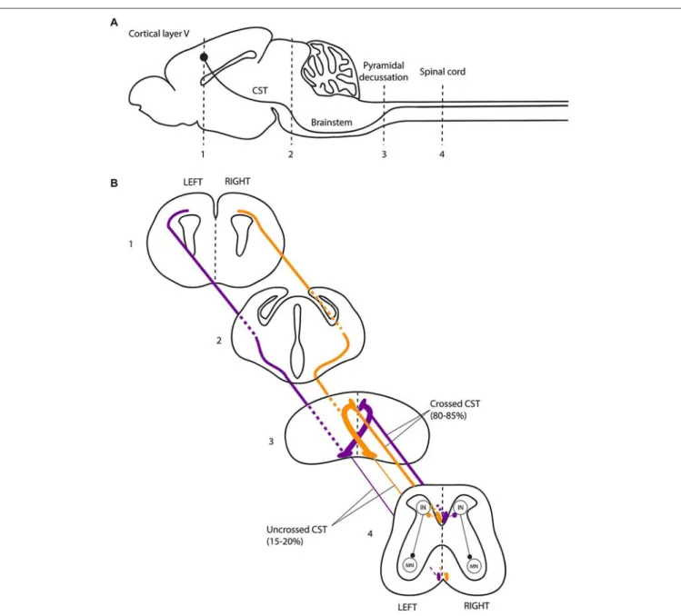
Quadrupedal locomotion requires repeated coordinated activity of each limb in a specific temporal sequence. Alternating left-right activity of the forelimbs and hindlimbs is observed at low locomotor frequencies (walking and trotting), while synchronized activity of the homologous limbs is observed at high locomotor frequencies (galloping) in mice, cats, horses and dogs (Forssberg et al., 1980; Dickinson et al., 2000; Serradj and Jamon, 2009). Lateralized motor output is thus a crucial aspect of locomotion, especially at low motor frequencies. In the past decade, careful analysis of genetically modified mice with a ‘‘hopping gait’’ has shed light on the respective contributions of the corticospinal tract and spinal central pattern generators (CPG) to left-right alternation during mouse locomotion. The Corticospinal Tract and Left-Right Alternation During Locomotion
The CST forms a crossed (lateralized) motor circuit controlling voluntary movements of the four limbs. In rodents, the CST is composed of neurons originating from cortical layer V, projecting mainly to the contralateral side of the spinal cord and eventually connecting to motor neurons via a multisynaptic pathway (Figure 2; Canty and Murphy, 2008). A role of the CST in the control of alternating left-right activity during mouse locomotion was initially suggested by the ‘‘hopping gait’’ described in mice with genetically induced alterations of CST projections (mice with mutations of the EphA4 signaling pathway and kanga mice). EphA4 (a member of the Eph family of tyrosine-kinase receptors) and its ligand ephrinB3 are involved in axonal guidance of the CST during development. Deletion of EphA4, Ephrin-B3 or proteins involved in the EphA4 downstream signaling pathway (a2-chimaerin, Nck, RhoA) results in a hopping-gait phenotype (Dottori et al., 1998; Kullander et al., 2001a,b; Yokoyama et al., 2001; Beg et al., 2007; Fawcett et al., 2007; Iwasato et al., 2007; Mulherkar et al., 2013). In EphA4 and EphrinB3 knockout mice, the CST trajectory is normal from the cortex to the pyramidal decussation. In the spinal cord, CST axons re-cross the midline, resulting in bilateral innervation of the spinal cord by each of the two hemispheres. In wild-type animals, EphA4-expressing CST axons are repelled by ephrin-B3 secreted at the midline, deterring them from re-crossing the midline at the spinal level (Dottori et al., 1998; Kullander et al., 2001a,b; Yokoyama et al., 2001). These findings suggested that the hopping gait might be explained by transmission of motor commands to both sides of the spinal cord through abnormally re-crossed CST axons. Similarly to mice with genetic alterations of the EphA4 signaling pathway, a mutant mouse line carrying a viable mutation of the DCC receptor have a ‘‘kangaroo-like’’ hopping gait phenotype and
are thus named ‘‘kanga’’ (Finger et al., 2002). The DCC ligand Netrin-1 belongs to the netrin family of extracellular guidance molecules. Netrin-1 has an attractive effect on growth cones when it interacts with the DCC receptor (Keino-Masu et al., 1996). This attraction allows commissural axons to approach and cross the midline (Kennedy et al., 1994; Serafini et al., 1994). DCC is expressed within the main forebrain descending tracts during their development (Shu et al., 2000). In kanga mice, the CST fails to cross the midline at the pyramidal decussation and projects exclusively to the ipsilateral side of the spinal cord (Finger et al., 2002).
However, other experimental findings do not support a major contribution of the CST to alternating left-right activity during locomotion. Indeed, abnormal CST midline crossing is not systematically associated with synchronized activity of the limbs during locomotion: mutants for L1 (Cohen et al., 1998; Jakeman et al., 2006), NCAM (Rolf et al., 2002), Sema6A and PlexinA3/PlexinA4 (Faulkner et al., 2008; Runker et al., 2008), exhibit normal locomotion despite having an abnormal CST. In rodents, a lateralized lesion of the cortex or CST, occurring during the first week of life, leads to sprouting of the remaining CST across the midline and thus to bilateral spinal cord projections (Leong and Lund, 1973; Kartje-Tillotson et al., 1987). This results in altered skilled forelimb movements without affecting left-right alternation during locomotion ( Kunkel-Bagden et al., 1992; Whishaw et al., 1993; Whishaw, 2000; Metz and Whishaw, 2002; Tennant and Jones, 2009). Thus, abnormal CST projections do not necessarily induce a hopping gait.
It is important to recall that the genetic alterations induced in EphA4, ephrin-B3 and DCC kanga mutant mice not only impact CST development but also affect commissural cell populations expressing these proteins, such as pre-cerebellar commissural neurons (Hashimoto et al., 2012), and commissural spinal cord interneurons (Kullander et al., 2003; Beg et al., 2007; Iwasato et al., 2007; Rabe Bernhardt et al., 2012). This implies that the hopping gait observed in these mice is not necessarily due to their CST abnormalities. Two recent studies took advantage of the conditional knockout mouse Emx1::cre;EphA4flox/flox in
which genetic deletion of EphA4 is restricted to the forebrain. These mice exhibit normal stereotypical locomotion despite bilateral CST projections to the spinal cord (Borgius et al., 2014; Serradj et al., 2014). Together, these results show that proper CST wiring is not necessary for stereotypic left-right alternation. Supra-spinal control plays a critical role in voluntary movements and adaptive locomotion when sensory-motor integration is required (for example when stepping over an obstacle). Emx1::cre;EphA4flox/flox mice with bilateral CST
projections to the spinal cord exhibit symmetric voluntary movements under conditions when asymmetric limbs movements are normally produced (Borgius et al., 2014; Friel et al., 2014; Serradj et al., 2014). These results emphasize the role of the CST in voluntary asymmetric movements.
In addition to the CST, supra-spinal structures playing an important role in the control of gait are located in the cerebral cortex, the cerebellum and in the brainstem, and constitute an interconnected network. There is no clear evidence implicating a supra-spinal control for left-right alternation and lateralization
Welniarz et al. Lateralization of motor control
FIGURE 2 | The corticospinal tract forms a crossed motor system in mice. (A) Sagittal view of the mouse central nervous system and corticospinal tract (CST).(B) Coronal views of the CST trajectory. The level of each coronal schematic section is indicated in figure A. At the junction between the hindbrain and the spinal cord (pyramidal decussation, level 3), the vast majority (80–85%) of corticospinal tract (CST) axons cross the midline and continue their trajectory through the
most ventral part of the dorsal funiculus within the half of the spinal cord contralateral to their hemisphere of origin. In the spinal cord, the CST undergoes collateral branching principally at the level of the cervical and lumbar enlargement, eventually transmitting motor commands to the forelimb and hindlimb muscles, respectively, via a multisynaptic pathway involving interneurons mainly located in the dorsal horn of the spinal cord. CST, corticospinal tract; IN, interneurons; MN, motor neurons.
of motor control during gait. Among the locomotor centers with direct spinal projections, the mesencephalic locomotor region (MLR) is of particular interest for our purpose. Electrical stimulation of the brainstem in decerebrate cats placed on a treadmill recapitulates normal alternate locomotion without the need of descending commands from the cortex (Shik et al., 1966, 1967). The MLR, which comprises the pedunculopontine (PPN) and cuneiform (CN) nuclei, sends outputs to the basal ganglia, the cerebellar and the cerebral locomotor areas. The MLR plays
a major role in gait initiation and in internal generation of adaptive lower limb movement during the automated gait cycle (Alam et al., 2011; Grabli et al., 2012). The MLR could be involved in the control of gait cadence (Piallat et al., 2009; Karachi et al., 2010), but this involvement is more likely related to higher-order functions during faster gait rather than basic motor control as suggested by rodent models (Winn, 2008). Dysfunction of the MLR and cerebral locomotor centers is observed in patients with Parkinson disease and freezing of