Lasers in Medical Science 1997, 12:123-130
Experimental In Vivo Fenestration of Guinea Pig
Cochlea Using 2,79/ m Laser Radiation
H. PRATISTO a, M. FRENZ a, H.J. ALTERMATT b, A. ARNOLD c, K. EHRENBERGER d, D. FELIX c, H.P. WEBER ~
alnstitute of Applied Physics, University of Bern, Switzerland blnstitute of Pathology, University of Bern, Switzerland CDivision of Neurobiology, University of Bern, Switzerland dFirst ENT Department, University of Vienna, Austria
Correspondence to M. Frenz, Institute of Applied Physics, University of Bern, Sidlerstrasse 5, CH-3012 Bern, Switzerland Received 16 October 1996; accepted pending revision 19 November 1996; accepted in final form 20 December 1996 (Amsterdam)
A b s t r a c t . Erbium-YSGG laser systems are promising tools in ear, nose and throat (ENT) surgery. The high absorption in biological tissues, resulting in precise tissue ablation with minimal thermal tissue damage, and the possibility to guide the radiation through optical fibres make the 2.79 ~m wavelength a favourite for microsurgery.
In order to simulate the fenestration of the human stapes foot plate required for prosthesis implantation when treating otosclerosis, five guinea pig cochleae were irradiated in vivo until perforation was achieved. The laser-induced temperature rise and pressure transients evoke activity in the inner hair cells that was investigated by micro-iontophoresis.
Perforation of the cochlea bone (hole diameter of 350/~m) can be performed with a few laser pulses and high precision with a thermal damage zone of <100~m. The bone ablation rate is 10 + 2 ~m pulse - 1 at a radiant exposure of 12 J c m - 2. The functionality of the afferent inner hair cells in the guinea pig cochlea was verified before and after laser treatment using glutamate receptor agonists A M P A and NMDA. For the above selected laser parameters, the induced 15-min enhanced activity was blockable with the specific reversible A M P A and NMDA antagonists CNQX and AP-7.
Micro-iontophoresis confirms the reversibility of cochlea functionality after its perforation with Er-YSGG laser pulses. A limit of radiant exposure around 12 J cm - 2 is found for safe fenestration. It is demonstrated that the Er-YSGG laser is a precise and safe instrument whilst still using adequate laser parameters. On the other hand, this study demonstrates the potential of uncontrol- lable and unintended induced damage, resulting from vapour channel formation in the perilymph, if a high laser radiant exposure is applied.
INTRODUCTION
For surgical treatments in the field of oto- rhinolaryngology, several laser systems, such as argon, KTP or CO2, have been established during the last two decades (1-5). Middle ear surgery has profited particularly from a laser- assisted operation technique that works with- out physical contact between the instruments and the delicate hearing structures. For treat- ment of otosclerosis with fenestration of the stapes foot plate, lasers provide an attractive alternative to the mechanical drill. Mobiliz- ation of middle ear structures can cause pres-
sure amplitudes in the cochlea, often several magnitudes higher than those usually gener- ated. This implies a high potential risk of hearing loss. Since the inner ear is highly sensitive to vibrational trauma, displace- ments and heat, a good tolerance in the applied energy dose and a high degree of safety is demanded of the laser system. The main laser parameters besides energy that determine the laser-tissue interaction are the wavelength and the duration of irradiation. The wavelength determines the absorption coefficient/~ or the optical penetration depth
5=l/tt~
of the radiation into the irradiatedmedia. For the Er-YSGG laser which emits radiation with a wavelength of 2.79~m, the absorption coefficient in bone is approxi- mately 2000 cm -1, which corresponds to a penetration depth of 5~m. The time duration of irradiation determines, on the one hand, the extent of thermal damage caused by heat diffusion. On the other hand, for short pulse durations, the laser-tissue interaction is char- acterized by an explosive process often con- nected with strong pressure transients if the conditions for thermal and/or stress confine- ment are fulfilled. Thermal confinement means that the pulse duration is shorter than the thermal relaxation time ~th=52/4 K, where 5 is a characteristic length and • is the thermal diffusivity of the tissue (~ = 1.44 10- 3 cm 2 s -1 for water and most tissues) (6, 7). Stress confinement is fulfilled if the pulse duration is shorter than the time "~st
(Tst:~/
Cm) a stress wave needs to propagate out of the irradiated volume, where 5 is again a characteristic length and c m is the speed of sound in the irradiated media (8).Argon and KTP laser radiation (488 and 532nm, respectively) are highly absorbed by chromophors, such as haemoglobin or melanin, but they both show deep penetration depths in unpigmented tissue and, conse- quently, a low bone ablation efficiency. Additionally, an unintentional direct ir- radiation of inner ear structures is possible (9, 10). The high absorption of infra-red laser radiation in biological tissue by virtue of its water content leads to a high bone ablation efficiency of the CO 2 laser wavelength (11). Use of an articulated mirror arm or a free beam, coupled into an operation microscope, con- stricts the freedom of movement in the narrow operation field.
In contrast, the Er-YSGG laser emitting at a wavelength of X=2.79~m seems to have optimal qualities for middle ear surgery: (i) 2.79~m radiation can be transmitted through optical fibres; and (ii) due to the strong absorp- tion of 2.79~m in water, the threshold for bone ablation is lower than 5 J cm -2 (12), and the lateral thermal damage zone around the ablation area is restricted to 10-100~m (although the water content of bone is only 15-20%). The use of an Er-YSGG laser for fenestration of the stapes foot plate was studied experimentally in detail in vitro using an inner ear model (13). Temperature measure- ments indicated a negligible temperature rise in the inner ear during Er-YSGG laser
H. Pratisto, M. Frenz, H.J. Altermatt et al radiation (14). The pressure measurements revealed that a radiant exposure of 10 J c m - 2 pulse -1 generates pressure transients with amplitudes less than 600 mbar during the bone ablation process (recoil pressure), and after collapse of a vapour bubble, formed in the inner ear after complete perforation of the foot plate (13). Acoustic stress of this magnitude in a frequency range between 20 Hz and 20 kHz does not lead to a permanent hearing loss according to the limit graph to avoid the hear- ing defects described by Pfander (15). The present experimental results and the results of other studies have shown the feasibility of using Er-YSGG laser radiation for middle ear surgery (16-19).
Since laser-induced pressure transients have strong frequency components in the MHz region, the aim of this study was to verify, in an in vivo experiment, the potential risk of using Er-YSGG laser radiation for the fen- estration of the stapes foot plate. It is not known whether temperature rise, although small, and pressure transient generation directly coupled into the inner ear, lead to damage of the delicate inner ear structures (eg the hair cells). Electrophysiological methods (20, 21) were used to verify the functionality of the animal's cochlea before and after the laser application. Changes in activities of the afferent inner hair cells in the guinea pig cochlea induced by pulses of an Er-YSGG laser were examined by micro-iontophoresis.
M A T E R I A L S A N D M E T H O D S Preparation of the animals and electrophysiological m e a s u r e m e n t s
Five pigmented adult guinea pigs of both sexes were used for the in vivo examinations. After pre-medication with atropine (0.5ml kg -1) 30 min before surgery, the main anaesthesia was based on a combination of droperidol (Innovar-Vet; 1.3 ml k g - 1; Pitman-Moore, Mudelein, IL, USA) and xylazine (Rompun; 0.4ml k g - 1 ; Bayer, Leverkusen, Germany). The animals were put on an artificial respir- ator via tracheostoma. To ensure a deep level of anaesthesia, doses (0.1 ml) of pentobarbital (Nembutal; Abbot, Chicago, IL, USA) were given in addition when necessary. The left auditory bulla was approached ventrolaterally and opened in order to expose the cochlea. A hole of about 0.5 • I mm was drilled in the
Fenestration of Guinea Pig Cochlea
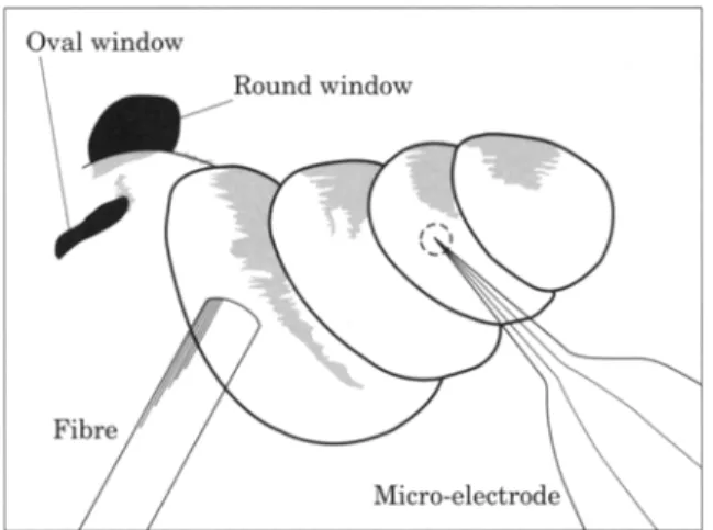
Fig. 1. View of the operation field.
cochlea wall over the pigmented stria vascu- laris and the strial ligaments of the third turn, to gain access to the synaptic field for the electrophysiological evaluations. A schematic drawing of the operation field is shown in Fig. 1.
The animals were fixed in the lateral position and the tip of a five-barrel filament borosilicate glass micro-electrode with a tip diameter of about 2~m was inserted, with a micro-driven arm, into the subsynaptic area of the inner hair cell layer. The extracellular action potentials were recorded with a 2 M NaC1 filled channel of the micro-electrode. The other four micropipette channels were used for micro-iontophoretic application of N-methylD-aspartate (NMDA; Sigma; 0.1M, pH8.5, NaOH adjusted), a-amino-3-hydroxy- 5-methyl-4-isoxazolepropionic acid (AMPA; Tocris; 10mM, pH8.3, NaOH), D-2-amino- p h o s p h o n o h e p t a n o a t e (AP-7; Sandoz; 0.2M, pH 8, NaOH) and 6-cyano-7-nitroquinoxaline- 2,3-dione (CNQX; Tocris; l m M , pH 8.4, NaOH). These substances could be ejected with an appropriate current (30-100nA). Glutamate is known to be the principal excit- atory substance of afferent cochlear neuro- transmission from the inner hair cells (20-23). It is the key substance for different types of receptors that are characterized by specific glutamate agonists. Among these agonists are NMDA and AMPA. Both of these receptors are ion channels: the NMDA type for Ca 2+ and Na+; the A M P A type for Na § alone, resulting in faster action of the latter. Various studies have shown that the afferent synapses of the inner hair cells possess functional properties that are equivalent to those of NMDA and AMPA receptors (24, 25). Specific antagonists for these two receptors have also been found:
125
AP-7 antagonizes NMDA, while CNQX blocks A M P A (26). They provide another means of verifying results as well as providing a phar- macological approach to reset receptor activity imbalances. The changes in electrical potential were recorded on an oscilloscope, printed and additionally audiotaped for later digitalization and off-line computing.
Laser system and radiation delivery applicator
Perforation of the cochlea bone was performed with the radiation of a flashlamp pumped Er-YSGG laser emitting at a wavelength of ~.=2.79/~m. The pulse duration of the laser operating in the free running mode was ~=200~s (full length) and the repetition rate was 2 Hz. The radiation was delivered to the operation site via a 2 m zirconium fluoride (ZrF4) fibre. Due to the hygroscopic and brittle structure of the ZrF 4 fibre, its distal end was protected with a 1 cm low OH quartz piece with a core diameter of 400~m. The pulse energies were either 15 or 5 4 m J pulse 1, resulting in a radiant exposure at the 400~m fibre end of 12 and 43 J c m - 2, respectively.
In vivo experiments
The basal turn of the cochlea was chosen as the target. The thickness of the bone at this location is comparable to that of the human stapes foot plate. The distance between the distal fibre tip and the bone surface of the cochlea was <0.4 mm. Perforation of the coch- lea was performed in two steps. For each of the five guinea pigs, the procedure was the same. As a first step, the cochleae were irradiated with three laser pulses using a radiant expo- sure of 1 2 J cm 2 at the fibre end. In the second step, the cochlea was irradiated at the same location with the same radiation expo- sure of 12 J c m - 2 until total perforation was achieved. Total perforation was recognized if perilymph flowed out of the ablation crater. Response of the neuronal activities after appli- cation of AMPA, NMDA, CNQX and AP-7 was investigated during the laser experiments in order to verify the functionality of the cochlea and the diagnostic set-up.
In a second part of the in vivo experi- ments, the potential risk of further laser
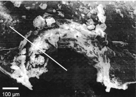
Fig. 2. Scanning electron micrograph picture of half a hole drilled with 17 laser pulses of H=12 J c m - 2 into the cochlea bone. No appreciable melted bone material is visible at the crater edges of the perforation. Histological section was made along the white line.
pulses (principally unnecessary for clinical treatment) delivered into the 'already perfor- ated' cochlea was investigated. One cochlea was irradiated with an additional six laser pulses into the perforation using a radiant exposure of H = 12 J c m - 2 Two cochleae were irradiated with 10 additional laser pulses using a radiant exposure of 43 J c m - e. The extracel- lular action potential was recorded for up to 50 rain after application of the last laser pulse. Subsequently, the animals were killed by an overdose of pentobarbital, and the cochleae were extracted for histology.
RESULTS
Bone morphology and histology
With a radiant exposure of H--12 J cm -2, the perforation of the cochlea with a thickness between 120 and 160~m (evaluated after the experiment by morphometry) was possible with 11-17 laser pulses, resulting in an ablation depth of 10 + 2/lm p u l s e - ~ assuming a con- stant ablation rate for consecutive pulses (27). The diameter of the round hole was typically 350~m. P a r t of the crater wall after perfora- tion with 17 pulses, with a radiant exposure of 12 J c m - 2 , is shown in the scanning electron microscopy figure (Fig. 2). The crystalline bone structure is still recognizable at the
crater wall and at the fragmentation particles distributed on the surface around the hole.
The histology of the laser-treated cochlea (sectioned along the line in Fig. 2) revealed a zone of thermally damaged tissue measuring less than 100~m (see Fig. 3).
Electrophysiological evaluations
A typical sequence of the neuronal activity measured with the micro-electrode tip before and after laser impacts using a radiant exposure of 12 J c m - 2 is presented in Fig. 4. The upper sequence demonstrates the reaction of the normal continuous spontaneous activity by the afferent neurotransmission system after stimulation with glutamate receptor agonists A M P A or NMDA (1-7 min). After this test of the functionality of the guinea pig cochlea and the diagnostic set-up, three laser pulses were applied on the first turn of the cochlea. The continuous spontaneous activity of about 2 s - 1 increased to 11-30 s - 1 with a delay of about i min to the laser impact (beginning at Minute 10), and returned to the normal con- tinuous spontaneous activity after 10-15 min (at Minute 22). This characteristic response is seen after all additional laser pulses with a radiant exposure of H - - 1 2 J cm -2 until perforation. After the enhanced activity had levelled off to the normal continuous spon- taneous activity, the cochlea was exposed to
Fenestration of Guinea Pig Cochlea 127
100 pm
Fig. 3. Histology of the guinea pig cochlea treated with 17 laser pulses of/-/=12 J cm -2. The thermally damaged zone amounts to about 100 #m (white arrows). Laser irradiation was performed in the direction of the black arrow.
the glutamate receptor agonists A M P A or NMDA to verify its functionality (not shown in Fig. 4). This test revealed no change in inner hair cell functionality. With specific reversible antagonists for the two receptors, CNQX for A M P A and AP-7 for NMDA, the functionality of the synaptic transition could be shown dur- ing enhanced activity (28), and is indicated by the coincidence of the activity to 4 s - 1 after their application (Minutes 13 and 18). The cut off in the middle and lower traces in Fig. 4 is due to the range saturation of the recording system.
The electrophysiological evaluations on the cochlea after applying six additional laser pulses with a radiant exposure of 12 J c m - 2 through a pre-existing perforation directly into the perilymph, also revealed an enhanced activity that was blockable with AP-7 and CNQX, but did not normalize until up to 50 min.
A total failure of the functionality test was the result after application of 10 laser pulses with a radiant exposure of H = 4 3 J cm -2 into a pre-existing perforation. At one cochlea, a disruption of the round window was visible.
DISCUSSION
The purpose of this study was to show the suitability of the Er-YSGG laser for middle ear surgery, particularly for stapedotomy. Histology and scanning electron microscopy
revealed that fenestration of the cochlea bone can be performed with high precision and minimal thermal alterations around the ablation site. Due to the high bone ablation efficiency ( ~ 0.064 mm 3 J - 1 , determined by cal- culating the volume of the hole and using the applied energy), a total laser energy of less than 0.3 J is sufficient to produce a perforation into a 150-200 ~m thick stapes foot plate with a diameter of 350~m. This bone ablation emciency is in good agreement with the measurements of other investigations (12, 19). Although thermal confinement (~th=52/ 4 K ~ 4 3 ~ S ) is not fulfilled for the 200~s Er-YSGG laser pulse, temperature-related inner ear damage is unlikely since each indi- vidual laser spike (%p ~ 2-5 ~s) has been shown to contribute to the ablation process. It was demonstrated that the ejection of the tissue is temporally correlated with each single spike of a laser pulse (29, 30). In fact, no temperature rise in the perilymph of an inner ear model was measured during Er-YSGG laser treatment (14). In contrast, continuous wave (cw) laser sources generated a high temperature rise at the ablation site, visualized by carboniz- ation and charring due to their low ablation efficiency (5, 31).
The potential source for inner ear damage associated with pulsed laser systems seems to be more likely correlated with pressure transients than with temperature. During the Er-YSGG laser-assisted fenestration of the stapes footplate, different pressure transients
10 10 10 1 min 2 3 4 5 6 7 8 I J I I I I I I 9min 10 11 12 13 14 15 I I I I I I I I
Three laser pulses
16 min 17 18 19 20 21 22
I [ I I I I I I
[ ] AMPA 9 NMDA [] CNQX [ ] AP-7
Fig. 4. Typical sequence of original recording of inner hair cell transition activity investigated during the in vivo experiment. Enhanced activity after application of AMPA or NMDA, and suppression of the laser-induced activity after application of CNQX and AP-7 is marked with shaded boxes.
are generated. Since stress confinement
['[st----5/
c m ~ l ns, assuming Cbone~3800 m S-1 (32)] is not fulfilled by the parameters used, no strong thermo-elastic pressure t r a n s i e n t is built up, even by a single spike. Therefore, the acoustical sources are the vaporization of water in the bone and, as a consequence, the explosive ejection of bone particles and the collapse of a water vapour bubble generated in the perilymph after perforation. The amplitude and the temporal shape of the pressure tran- sients resulting from the recoil momentum of the ejection are determined by the absorption properties of the tissue and the pulse duration. The amplitude of the recoil pressure transients was found to be below the safety limit of 500 mbar described by Pfander (15) for a radi- ant exposure up to 5 0 J cm -2 (13). It has, however, to be emphasized t h a t hearing loss in h u m a n s from impulse noise is not well studied, and t h a t animal data cannot be reliably trans- ferred to h u m a n s with respect to impulse noise-induced hearing loss.
After the fenestration of the stapes foot plate, a more drastic bang is generated due to the confinement of the liquid perilymph fluid covered with a rigid boundary. Due to the high absorption of 2.79/1m radiation in water, the perilymph acts as a backstop and, therefore, protects the inner ear structures from direct irradiation. The radiation, however, creates a vapour channel in the perilymph. Fast flash video imaging revealed t h a t the vapour channel constricts at the bone-liquid interface, forming a vapour bubble (19). The bubble later collapses and generates a pressure t r a n s i e n t with an amplitude of up to several bars. The amplitude of the collapse pressure t r a n s i e n t depends on the size and geometry of the vapour bubble and, therefore, on the pulse energy and the diameter of the stapes perforation. A laser
- 2
pulse with a r a d i a n t exposure of 40 J cm creates a channel with a maximal size of about 2 mm depth and 0.5 mm diameter (13, 33). The collapse of such a channel generates a pressure t r a n s i e n t with an amplitude of
Fenestration of Guinea Pig Cochlea
about 9 b a r . This high pressure transient and/or the channel dynamics and bubble movement itself resulted in the failure of the electrophysiological functionality test and, in one case, resulted in a disruption of the round window.
A radiant exposure of 12 J cm 2 strongly reduces the size and the dynamics of the vapour bubble and, therefore, the strength of the bubble collapse.
The evoked enhanced activity of the inner hair cells could be the result of a laser-induced temperature rise, a pressure transient genera- tion, an inner ear fluid motion or a combina- tion of these three effects. The duration of the enhanced activities indicates the time needed by the inner ear to regenerate and return to normal spontaneous activity. The typical response of about 15 min enhanced activity is seen for laser pulses of radiant exposure of 12 J c m - 2 applied to the cochlea, as well as for additional laser pulses until fulfilled perfora- tion. The fact that it was possible to block enhanced activity with the specific reversible agonists during distribution was an important indication that the transition of the afferent inner hair cells still worked. The influence and function of AMPA, NMDA, CNQX and AP-7 on the transition of the afferent inner hair cells is well known (22, 24, 25, 28).
After applying additional laser pulses through the hole into the cochlea bone, enhanced activity over a period longer than 50 min was found. Since the enhanced activity was still blockable by the specific agonists, it was assumed that the functionality of the inner ear was still intact and no acute hearing loss was generated. This also indicates that the amplitude of the induced pressure transients seems to be the main parameter for possible tissue damage, and not its frequency spectrum. It has, however, to be mentioned that this research does not address the possibility of a permanent hearing loss associated with mechanical injuries in the inner ear that can occur several hours after the laser treatment.
The electrophysiological methods revealed that total reversibility of the cochlea function- ality and, therefore, safe perforation was poss- ible with radiant exposures of 12 J c m - 2. This value is much lower than the safety limit of 36 J c m - 2 published by J o v a n o v i c et al (34). The discrepancy might be explained by the larger diameter of the perforation (500-600/lm compared to 350ttm in the present study). A larger hole in the foot plate reduces the
129
confinement of the vapour bubble in the perilymph, and prevents the channel from forming a vapour bubble.
CONCLUSION
The suitability of the Er-YSGG laser for pre- cise perforation of the cochlea was demon- strated. The electrophysiological evaluations showed total reversibility of the inner hair cells, transition functionality after irradiation with a radiant exposure of 12 J cm-2. How- ever, vapour channel formation in the inner ear fluid was found to be a serious potential cause of damage mechanism. Due to the incom- pressibility of fluids, the rising pressure might be compensated for by disrupting inner ear structures. This study shows that the Er-YSGG laser is a precise instrument for ENT surgery when using appropriate laser parameters. It is proposed to work in a radiant exposure range of 12 J cm -2 when the operative procedure demands the fenestration of the cochlea. The results of this in vivo study confirm previous in vitro experimental findings (13, 19). For other middle/inner ear surgery procedures, careful determination of laser parameters is essential.
ACKNOWLEDGEMENTS
The a u t h o r s wish to t h a n k A. Friedrich, H. Hutmacher, E. Kr~henbfihl, E. P l a n k and S. Gygax for t h e i r t e c h n i c a l assistance. This work was supported in p a r t by the Swiss Commission of the E n c o u r a g e m e n t of Scientific Research and the 'Stiftung zur FSrderung der wissenschaftlichen F o r s c h u n g a n der Universit~it Bern'.
REFERENCES
1 Kautzky M, T r 5 d h a n A, Susani M e t al. Infrared laser stapedotomy. Eur Arch Otorhinolaringol 1991, 248:449-51
2 Molony T. CO 2 laser stapedotomy. J La State Med Soc 1993, 145:405-8
3 Lesinski S, Palmer A. Laser for otosclerosis: CO2 vs. argon and KT-532. Laryngoscope 1989, 99:1-8
4 Coker N, Ator G, J e n k i n s H et al. Carbondioxide laser stapedotomy: A histology study. Am J Otolaryngol 1986, 7:253-7
5 H~usler R, Messerli A, Romano V e t al. Experimental and clinical results of fiberoptic argon laser stapedo- tomy. Eur Arch Otorinolaryngol 1996, 253:193-200 6 Boulnois JL. Photophysical processes in recent medical
laser developments: a review. Lasers Med Sci 1986, 1:47-66
7 van Gemert MJC, Welch AJ. Time constants in thermal laser medicine. Lasers Surg Med 1989, 9:405-21 8 Jacques SL. Laser-tissue interactions: photochemical,
photothermal and photomechanical. Surg Clin N Am 1992, 72:531-58
9 Gherini S, Horn K, Causse J e t al. Fiberoptic argon laser stapedotoy: Is it safe? Am J Otolaryngol 1993, 14:283 9
10 Thoma J, Unger V, Kastenbauer E. Functional impact of argon-laser on the cochlea of guinea-pig. Laryng Rhinol Otol 1982, 61:473-6
11 Forrer M, Frenz M, Romano V et al. Bone ablation mechanism using CO2 lasers of different wavelenght and pulse duration. Applied Physics B 1993, 56:104-12 12 Nuss R, Fabian R, Sarker R et al. Infrared laser bone
ablation. Lasers Surg Med 1988, 8:381~1
13 Pratisto H, Frenz M, Ith M e t al. Use of 3#m laser radiation in middle ear surgery. Laser Interaction with Hard and Soft Tissue H. SPIE, Bellingham, 1994, Vol. 2323, pp. 179-84
14 Frenz M, Romano V, Pratisto H et al. Stapedotomy: New experimental results of the erbium laser. In: Aktuelle Probleme der Otorhinolaryngologie, Vol. ORL 17. Bern, GSttingen, Toronto, Seattle: Verlag Hans Huber, 1994: pp. 337-45
15 Pfander F. Das Knalltrauma. Berlin: Springer Verlag, 1975
16 Schlenk E, Profeta G, Nelson S et al. Laser assisted fixation of ear prostheses after stapedectomy. Lasers Surg Med 1990, 10:444-7
17 Jovanovic J, Anft D, SchSnfeld U et al. Suitability of pulsed laser systems for stapedotomy--an animal experimental study. Lasermedizin 1995, 11:140-9 18 Pfalz R, Hibst R, Bald N. Suitability of different lasers
for operations ranging from the tympanic membrane to the base of the stapes. Lasers Otorhinolaryngol Head Neck Surg 1995, 49:87 94
19 Pratisto H, Frenz M, Ith M e t al. Temperature and pressure effects during erbium laser stapedotomy. Lasers Surg Med 1996, 18:100-8
20 Felix D, Ehrenberger K. The efferent modulation of mammalian hair cell afferents. Hear Res 1992, 64:1 5 21 Ehrenberger K, Felix D. Glutamate receptors in after-
ent cochlear neurotransmission in guinea pigs. Hear Res 1991, 52:73 80
22 Eybalin M, Pujol R. Cochlear neuroactive substances. Eur Arch Otorhinolaryngol 1989, 246:228-34
H. Pratisto, M. Frenz, H.J. Altermatt et al
23 Klinke R. Neurotransmission in the inner ear. Hear Res 1986, 22:235 43
24 Felix D, Ehrenberger K. N-methyl-D-aspartate-induced oscillations in excitatory afferent neurotransmission in the guinea pig cochlea. Eur Arch Otorhinolaryngol 1991, 248:429-31
25 Puel J, Laedrech R, Chabert R et al. Electrophysiologi- cal evidence for the presence of NMDA receptors in the guinea pig chochlea. Hear Res 1991, 51:255-64
26 Watkins J, Olverman H. Agonists and antagonists for excitatory amino acid receptors. Trends Neurosci 1987, 10:265-72
27 Romano V, Rodriguez R, Altermatt HJ et al. Bone microsurgery with IR-lasers: A comparative study of the thermal action at different wavelengths. Laser Interaction with Hard and Soft Tissue. SPIE, Bellingham, 1993, Vol. 2077, pp. 87 97
28 Arnold A, Ehrenberger K, Frenz M et al. Experimental erbium laser surgery in the guinea pig cochlea: its use in the study of afferent cochlear neurotransmitters. Eur Arch Otorhinolaryngol 1996, 253:460-3
29 Frenz M, Zweig AD, Romano V e t al. Dynamics in laser cutting of soft media. Laser-Tissue Interaction. SPIE, Bellingham, 1990, Vol. 1202, pp. 22-33
30 Frenz M, Pratisto H, Ith M e t al. Transient photoacous- tic effects induced in liquids by pulsed erbium laser. Biomedical Optics, Laser-Tissue-Interaction V. SPIE, Bellingham, 1994, Vol. 2134, pp. 402-12
31 Jovanovic S, SchSnfeld U. Application of the CO2 laser in stapedotomy. Advances Oto-Rhino-Laryngology 1995, 49:95
32 Niemz MH. Laser-Tissue Interactions: Fundamentals and Applications. Berlin, Heidelberg, New York: Springer, 1996
33 Forrer M, Ith M, Frenz M e t al. Mechanism of channel propagation in water by pulsed erbium laser radiation. Laser Interaction with Hard and Soft Tissue. SPIE, Bellingham, 1993, Vol. 2077, pp. 72-7
34 Jovanovic J, Anft D, SchSnfeld U et al. Experimental studies on the suitability of the erbium laser for stape- dotomy in an animal model. Eur Arch Otorhinolaryngol 1995, 252:422 7
Key words: Er-YSGG laser in middle ear surgery; ENT surgery; Micro-iontophoresis