HAL Id: tel-00812776
https://tel.archives-ouvertes.fr/tel-00812776
Submitted on 12 Apr 2013
HAL is a multi-disciplinary open access
archive for the deposit and dissemination of sci-entific research documents, whether they are pub-lished or not. The documents may come from teaching and research institutions in France or abroad, or from public or private research centers.
L’archive ouverte pluridisciplinaire HAL, est destinée au dépôt et à la diffusion de documents scientifiques de niveau recherche, publiés ou non, émanant des établissements d’enseignement et de recherche français ou étrangers, des laboratoires publics ou privés.
Mise au point de vecteurs permettant une expression
fiable de gènes codant pour des ARN interférent et des
microRNA
Ashraf Sawafta
To cite this version:
Ashraf Sawafta. Mise au point de vecteurs permettant une expression fiable de gènes codant pour des ARN interférent et des microRNA. Génétique animale. Université Pierre et Marie Curie - Paris VI, 2008. Français. �NNT : 2008PA066091�. �tel-00812776�
ECOLE DOCTORALE LOGIQUE DU VIVANT
2008
THESIS
In Partial Fulfillment of the Requirements for the degree of
DOCTOR OF SCIENCE
SPECIALIZATION: Biology
Presented by:
Ashraf Sawafta
May 2008
Design of Vector for the Expression of shRNA in Transgenic
Animals
Director of the thesis:
Louis-Marie HOUDEBINE
INRA
The jury composed of:
Michel Raymondjean CNRS Paris 6
President
André Mazabraud
CNRS Université Paris -Sud
Referee
Jean-Luc Vilotte INRA jouy en josas
Referee
Pierre Cherel France Hybrides
Examiner
...
..
DEDICATION
This thesis is dedicated to my wonderful parents,
who have raised me to be the person I am today.
You have been with me every step of the way,
through good times and bad. Thank you for all the
unconditional love, guidance, and support that you
have always given me, helping me to succeed and
instilling in me the confidence that I am capable of
doing anything I put my mind to. Thank you for
everything. I love you! Also this thesis is dedicated
to my brothers, my wonderful sister,also to Dana
the most speacial person in my life, and in loving
memory of my grandparents finally, this thesis is
dedicated to all my friends those who supported me
supervisors Dr. Louis-Marie Houdebine, who accommodated me within his team. I would like to thank him for directed this thesis with such an amount of spirit, for all his support, suggestions and ideas which improve my thesis. I thank him for trusted me, and supplied me with all the means necessary for the realization of this work. I also would like to thank the French Consulate General in Jerusalem for giving me the financial support to finish this thesis. I also express a deep recognition to Dr. Genevieve jolivet without whom this work of thesis would not be what it is today. I would like to thank her for listening, discussing and the active support that she gave to me throughout these three years. I thank also Dominique Thépot for his support, explanation and for teaching me the various techniques of molecular biology.
I extend my gratitude to Nathalie Daniel-Carlier for her daily assistance, her great competence and the quality of here work. I also thank her from my heart for her support and her engagement in this work, Nathalie really thanks for your huge participation in this work. I am also grateful to Mathieu Leroux-Coyau as a special friend and for the helpful and practical advice provided by him. I would like to thank all of them because their motivation did not disappear with the sight of the negative or disappointing results, and they enormously learned to me. For all that and well of other things thank them.I wish to also extend my gratitude to Bruno Passet and Sonia Prince who produced all the transgenic animals. Without their invaluable assistance, their ingeniousness and their engagement, all this work would not have been carried out also quickly and under such good conditions. I thank all the team: Sylvie Rival-Gervier, Caroline Morgenthaler, Céline Viglietta and Sorya saidi, for their assistance throughout my thesis, and for the discussions. I would also like to express my recognition to Michel RAYMONDJEAN to chair the jury of this thesis, also toAndré MAZABRAUD and Jean-Luc VILOTTE, who have both accepted the heavy task of being referee. I also wish to thank Pierre CHEREL andFrancois LEFEVRE, who agreed to judge my thesis work as examiner. Thank you all for your appreciated efforts. I thank all the members of Dr. François lefevre team for their contribution to this work through the virus test and infection. Finally I would like to thank all my friends, Abd el baset al mamo, momen bouziane, Bilal ghareeb, moatz tamimi. For all of them I express my gratitude and appreciation. Last but not least, special thanks for the rest of my family to have been present daily for my support and encouragement.
List of abreviations
Abreviations:
ATP: adenosine 5 -triphosphate -gal: -galactosidase
BAC: bacterial artificial chromosome; biospecific affinity chromatography BLAST: Basic Local Alignment Research Tool
bp: base pair
BSA: bovine serum albumin
cDNA: complementary deoxyribonucleic acid CHO: Chinese hamster ovary (cells)
Da: Dalton
dATP: deoxyadenosine triphosphate ddATP: dideoxyadenosine triphosphate ddNTP: dideoxynucleoside triphosphate ddTTP: dideoxythymidine triphosphate
DMEM: Dulbecco's modified Eagle (or minimum essential) medium DNA: deoxyribonucleic acid
DNase: deoxyribonuclease
dNTP: deoxynucleoside triphosphate DTT: dithiothreitol
EDTA: ethylenediaminetetraacetic acid ELISA: enzyme-linked immunosorbent assay ES: embryonic stem (cells)
GFP: green fluorescent protein HBSS: Hanks' buffered salt solution
hCMV: human cytomegalovirus hGH: human growth hormone
HIV: human immunodeficiency virus HSV: herpes simplex virus
Ig: immunoglobulin
IPTG: isopropyl-1-thio- -D-galactoside IRES: internal ribosomal entry site kb: kilobase
kDa: kilodalton
LTR: long terminal repeat miRNA: microRNA
mRNA: messenger ribonucleic acid nt: nucleotide
5 ' HS4: hypersensitive area in DNAse I in 5 ' of the locus of the globine of chicken Oligo: oligonucleotide, a short, single-stranded DNA or RNA.
Oligo(dT): oligodeoxythymidylic acid ORC: origin recognition complex ORF: open reading frame
Ori: origin of replication PBS: phosphate-buffered saline PCR: polymerase chain reaction RNA: ribonucleic acid
RNAi: RNA interference RNase: ribonuclease
List of abreviations
RT: reverse transcriptase
RT-PCR: reverse transcription/polymerase chain reaction SDS: sodium dodecyl sulfate
siRNA: short interfering RNA ss:single stranded
SSC: sodium chloride/sodium citrate (buffer); side (light) scatter (in flow cytometry) T: thymine or thymidine; one-letter code for threonine
TAE: Tris/acetate (buffer)
Taq:Thermus aquaticus DNA (polymerase) TBE: Tris/borate (buffer)
TE: Tris/EDTA (buffer)
tRNA: transfer ribonucleic acid UTR:untranslated leader region
Sense strand: The coding sequence of mRNA.
Antisense strand: The noncoding strand complementary to the coding sequence of mRNA.
Summary:
DEDICATION... 4 ACKNOWLEDGMENTS: ... 5 Abreviations: ... 6 Summary: ... 9 List of Figures: ... 12 Lits of tables: ... 14 CHAPTER 1 ... 15 BIOLOGY OF RNAi... 151. RNA interference (RNAi) ... 16
2. RNAi mechanism and mode of actions ... 23
2.1. Processing of dsRNA into siRNAs... 23
2.2. MicroRNA processing ... 29
2.3. Degradation of mRNA... 32
2.4 Transcriptional gene silencing (TGS)... 35
2.4.1 Proposed mechanisms for TGS in S. Pombe... 36
2.4.2 Proposed mechanisms for TGS in human. ... 38
A. Model one for siRNA mediated TGS in Human Cells... 38
B. The second model for siRNA mediated TGS in Human Cells. ... 39
3. Natural role of RNAi ... 39
CHAPTER 2 ... 42
THERAPEUTIC GENE SILENCING AND INHIBITION OF PATHOGENS BY RNAi... 42
1. Inhibition of pathogens by RNAi... 43
2. RNAi methodology for the inhibition of virus genes ... 52
CHAPTER 3 ... 54
THE MODEL: PSEUDORABIES VIRUS ... 54
1. Introduction and history ... 55
1.1. Species affected... 55
1.2. Herpesviruses... 55
2. Molecular biology of PRV ... 56
2.1. Genome and gene content... 58
Unknown... 62
2.2. Transcriptional architecture ... 63
2.3. Core genes ... 64
3. Immediate- early gene ... 64
4. Veterinary impact of Pseudorabies (Aujeszky disease) ... 65
CHAPTER 4 ... 66
AIMS OF THE THESIS ... 66
Aim of the thesis ... 67
The following questions were addressed: ... 67
Main steps of the experimental works: ... 68
CHAPTER 5 MATERIALS AND METHODS ... 72
1. Preparation of the shRNA genes ... 73
Introduction of shRNA gene sequence in pBS-U6 vector... 73
Primers used for preparing the shRNA... 73
Summary
Vector preparations and oligonucleotide subcloning:... 75
pBS-U6 digestion and preparation... 75
Digest purification ... 75
Digested vector dephosphorylation (when required)... 76
Subcloning of shRNA into the pBS-U6... 76
PCR amplifications ... 77
Cloning the PCR product in PGEM T Easy... 77
DNA Extraction and purification ... 77
Cloning of U6-ShRNA in M10 plasmid ... 77
BsaBI construct preparation ... 78
ShRNA gene for microRNAs strategy ... 78
Primer design for miRNAs ... 78
Primer for miRNA preparation ... 78
Annealing... 79
PCR reaction ... 79
miRNA cloning in pM10... 79
Sequencing... 80
2. Preparation of the vectors containing the IE mRNA targets ... 80
2.1. The plasmids used as target were the following: ... 80
2.2. IE mRNA targets... 80
2.2.1. Part of IE mRNA sequence (5’UTR and translated region) ... 80
2.2.2. The 3’UTR untranslated region of IE mRNA sequence ... 81
Target plasmid construction... 81
2.3.1. pLM24 IE Luc untranslated region of IE (19 nt) ... 81
Target plasmid ... 82
Preparation of new target containing the complete 3’UTR of IE gene ... 83
RNAi in the intron... 84
Transcription gene silencing (TGS) ... 85
3. Construct validation... 85
3.1.1. Cell lines... 85
3.1.2. Cell cultures ... 85
3.2. Cell tests and in vitro assays... 86
In Vitro bioassays in transiently transfected CHO.K1 cells... 86
Stable clone preparation ... 87
In Vitro Bioassays in Stably Transfected PK15 Cell clones... 87
4. Extraction of RNA from cells or tissues (Chomczynski & Sacchi, 1987) .... 88
4.1. Extraction solutions ... 88
4.2. Procedure... 88
5. Quantification of si/miRNA by real time PCR ... 89
Principle of RNAi quantification: ... 89
Adaptation of the technique for our purpose ... 90
Polyadenalytion step ... 90
Polyadenylation Protocol: ... 91
Condition for Reverse transcription... 91
PCR Quantitative with SYBRGreen ... 91
Transgenic mice ... 92
Mouse strain ... 92
Transgene preparation for microinjection. ... 93
Solution preparation... 93
DNA microinjection to generate transgenic mice ... 95 DNA extraction ... 95 SUR1, 2, 3 primers. ... 95 HGH primer... 95 5’HS4 primer... 95 CHAPTER 6 ... 96 RESULTS ... 96
1. Selection of viral target gene and target sequence. ... 97
2. Designing of vectors expressing the shRNA gene ... 99
Construction and evaluation of the U6-shRNA vector ... 99
3. Preparation of reporter luciferase gene plasmid...101
5. Construction and evaluation of vectors containing the U6-shRNA gene ...103
6. Construction and evaluation of the 5T construct ...106
7. Construction and evaluation of the miRNA construct ...107
8. Measurement of siRNA concentration in transitionally transfected CHO cells ...109
RNAi expression in transient co-transfection of CHO cells ...112
9. Establishment of stable clones expressing specific IE siRNA ...115
10. Generation of transgenic mice harboring the pM10 U6-shRNA constructs and the 5T as well as the miRNA constructs ...117
11. Transcription gene silencing (TGS)...120
12. Infection of transgenic mice with PRV...121
13. Search of new target for siRNA ...125
Identification and study of new shRNAs targeting IE mRNA...125
New constructs targeting 5’ and 3’UTR of the IE gene validation ...127
14. Effect of two different constructs on mRNA of IE gene expression inhibition (cumulative effect)...130
15. New miRNAs constructs...132
CHAPTER 7 ...134
DISCUSSION AND CONCLUSION ...134
REFERENCES ...140
List of Figures
List of Figures:
Figure 1 : RNA silencing pathways in different organisms. ... 20
Figure 2 : Mammalian cells dsRNA pathways. ... 21
Figure 3 : RNAi post-transcriptional gene silencing mechanisms. ... 22
Figure 4 : The mechanism of gene silencing induced by double-stranded RNA... 28
Figure 5 : miRNA biogenesis... 30
Figure 6 : The miRNA biogenesis pathway in vertebrate cells. ... 31
Figure 7 : shRNA mechanism and mode of action... 32
Figure 8: Proposed models for RNAi pathway in Drosophila. ... 34
Figure 9 : Proposed mechanisms for TGS in S. pombe ... 36
Figure 10 : Model for RNA-directed TGS in human cells ... 37
Figure 11 : proposed mechanism for TGS in human. ... 38
Figure 12 : Examples of some viruses that can be targeted by RNAi... 51
Figure 13 : Electron micrograph of Herpesvirus. ... 56
Figure 14 : The herpesvirus virion structure. ... 57
Figure 15 : The linear map of the PRV genome... 59
Figure 16 : Luciferase reporter gene pLM24IELuc... 69
Figure 17 : The basic scheme for the shRNA... 69
Figure 18: Vector of transgenesis pM10. ... 70
Figure 19 : M10 vector includes the multicloning sites. ... 71
Figure 20 : miRNA preparation. ... 79
Figure 21 : 5’UTR and translated region. ... 81
Figure 22 : 3’UTR untranslated region of IE mRNA sequence. ... 81
Figure 23 : Target plasmid pLM24 IE Luc... 82
Figure 24 : pLM24 Luc 3’UTR plasmid... 83
Figure 25 : PRV genomic DNA PCR. ... 83
Figure 26 : pLM24 IE Luc 3’UTR IE plasmid. ... 84
Figure 27 : scheme represent the TGS construct ... 85
Figure 28 : Detection of RNA Interference by real time PCR... 90
Figure 29 : Real time PCR quantification of RNAi by SYBR Green. ... 92
Figure 30 : The IE gene of pseudorabies virus with 5’P and 3’OH UTRs (untranslated region) ... 97
Figure 31 : Structure on the U6 gene promoter plasmid. ... 99
Figure 32: The basic scheme for the shRNA construction. ...100
Figure 33 : Representation of the first shRNA construct. ...101
Figure 34: The target vectors with the reporter luciferase gene. ...102
Figure 35 : Knockdown of the IE mRNA by the U6-shRNA construct in CHO. ...103
Figure 36 : Circular (A) representation of the vector pM10. ...103
Figure 37: Introduction of the U6-shRNA gene in the three sites of pM10 vector, to give the Sh1, Sh2 and Sh3 constructs. ...105
Figure 38 : Knockdown of the IE mRNA by the three vectors containing the pM10 and the U6-shRNA constructs. ...106
Figure 39 : Representation of the 5T construct. ...107
Figure 40 : Knockdown of IE mRNA by the 5T construct. ...107
Figure 42 : Representation of the miRNA construct. ...108
Figure 43 : Representation of the pM10-miRNA construction. ...108
Figure 44 : Knockdown of IE mRNA by the miRNA construct. ...109
Figure 45 : siRNA quantification method using real time PCR. ...110
Figure 46 : Quantitative PCR validation using synthetic siRNA...110
Figure 47 : quantification and sequence analysis of the siRNA produced in CHO cells transfected with different constructs...112
Figure 48 : Concentration of the siRNA in CHO cells transfected by the U6-shRNA. ...113
Figure 49 : Concentration of the siRNA in CHO cells transfected by the sh3, 5T, and miRNA constructs...113
Figure 50 : Relation between the concentration of the siRNA in transfected CHO cells and the knockdown of the IE mRNA. ...115
Figure 51 : Inhibition of IE-luciferase in stable clones prepared from PK15 cells...116
Figure 52 : Concentration of RNAi in PK15 cell stable clones harboring the integrated U6-shRNA construct. ...116
Figure 53 : Recapitulation of the constructs used to generate transgenic mice ...117
Figure 54 : Detection of siRNA produced by the miR30 construct in brain and liver of two transgenic mouse lines (line 15 and line 30). ...119
Figure 55 : miRNA construct harbouring the sequence of a shRNA similar to the promoter region close to the cap site of the IE gene. ...120
Figure 56 : transgenic mice infection by PRV (4 LD50)...122
Figure 57 : Transgenic animal death pattern following infection by PRV. ...122
Figure 58 : The IE gene of pseudorabies virus with 5’P and 3’OH UTRs (untranslated region) ...125
Figure 59 : New shRNA constructs targeting the 3’UTR of IE mRNA ...126
Figure 60: Knockdown of the IE mRNA by the sh3, ASSR1 and SHOLD constructs in CHO. ...127
Figure 61 : Knockdown of the IE mRNA by the sh3, ASSR1 and SHOLD constructs in CHO cells transfected by different quantities of these constructs. ...128
Figure 62 : Concentration of the siRNA in CHO cells transfected by the U6-shRNA, ASSR1, and SHOLD constructs. ...129
Figure 63 : Effect of co-transfection by two different shRNA constructs...131
Figure 64 : Summary of the different target sequences in IE mRNA ...131
Figure 65 : new miRNA constructs. ...133
Lits of Tables
Lits of tables:
Table 1 : Eukaryotic organisms and RNAi phenomena ... 16 Table 2 : Examples of disease-related genes that have been targeted in
mammals using siRNA... 44 Table 3 : Nucleic acid–based antiviral therapeutics that have entered clinical trials... 46 Table 4 : Inhibition of RNA viruses by RNAi as illustrated in (Haasnoot et al., 2003)... 49 Table 5 : Inhibition of DNA viruses by RNAi as illustrated in (Haasnoot et al., 2003)... 50 Table 6 : PRV gene functions... 60 Table 7 : Proposed criteria for choosing the most effective siRNA and shRNA (from Li et al., 2007). ... 98 Table 8 : Sequences in the mouse genome and transcriptome showing
significant homologies with the Sh target sequence. ... 99 Table 9 : Sequences in the mouse genome and transcriptome showing
significant homologies with the TGS target sequence...120 Table 10 : Infection of transgenic animals by PRV virus. ...121 Table 11 : Sequences in the mouse genome showing significant homologies with the AssR1 and Shold target sequences. ...132
CHAPTER 1
BIOLOGY OF
RNAi
CHAPTER I Biology of RNAi
1. RNA interference (RNAi)
In 1998, it was demonstrated that injection of double stranded RNA (dsRNA) into nematodes induces the post-transcriptional silencing of gene encoding homologous mRNA, a process called ‘RNA interference’ (RNAi) (Fire et al., 1998). RNAi was first described in C. elegans in a process in which small double-stranded RNAs induce homology dependant degradation of mRNA (Sharp, 2001). RNAi has been linked to many previously described silencing phenomena such as post-transcriptional gene silencing (PTGS) in plants and quelling in fungi (reviewed by Dykxhoorn et al., 2003). RNAi is an evolutionarily conserved phenomenon (see table 1 for more details). It is a multi-step process that involves generation of active small interfering RNA (siRNA) in vivo through the action of an RNase III endonuclease, named Dicer. The resulting 21- to 23-nt siRNA mediates degradation of the complementary homologous RNA (Sharp, 2001).
Table 1 : Eukaryotic organisms and RNAi phenomena
Examples of eukaryotic organisms exhibiting RNAi- related phenomena from (Agrawal et al., 2003)
Kingdom Species Stage tested Delivery method Protozoans Trypanosoma brucei Procyclic forms Transfection
Plasmodium falcipamm
Blood stage Electroporation and soaking
Toxoplasma gondii Mature forms in fibroblast
Transfection
Paramecium Mature form Transfection and feeding Leishmania donovanii Larval stage
and adult stage
Tried but not working Invertebrates Caenorhabditis
elegans
Adult Transfection, feeding bacteria carrying dsRNA, soaking
Caenorhabditis briggsae
Adult worm Injection Brugia malayi (filarial
worm)
Sporocysts Soaking Schistosoma mansoni Adult Soaking
Hydra Adult Delivered by micropipette Planaria Adult Soaking
Lymnea stagnalis (snail) Cell lines, adult, embryo Injection Drosophila melanogaster Early embryonic stages
Injection for adult and embryonic stages, soaking and transfection for cell lines
Cyclorrphan (fly) Early embryonic stages
Injection Milkweed bug Early
embryonic stages
Injection Beetle Larval stage Injection Cockroach Adult and cell
line
Injection
Spodoptera frugiperda Injection and soaking Vertebrates Zebra fish Embryo Microinjection
Xenopus laevis Embryo Injection Mice Prenatal,
embryonic stages, and adult
Injection
Humans Human cell lines
Transfection
Plants Monocots/dicots Plant Particle bombardment with siRNA/transgenics
Fungi Neurospora crassa Filamentous fungi Transfection Schizosaccharomyces pombe Filamentous fungi Transgene Dictyostelium discoideum Transgene Algae Chlamydomonas reinhardtii Transfection
CHAPTER I Biology of RNAi
RNAi and related phenomena protect the organisms from invasion by both exogenous (eg., viruses) and endogenous (eg., mobile genetic elements) genetic parasites (Bernstein et al., 2001b). Eukaryotic genomes are largely composed of repetitive DNA sequences. Among them, transposable elements have the potential to perform replication cycles involving DNA or RNA intermediates. These repeated and mobile sequences have been found in all living organisms, and can comprise up to 40% of the genome, as is the case in humans. Tight control of these invaders is thus an important feature of their regulation to prevent eukaryotic genomes from their mutational threat. During the last few years, evidence has emerged that the host has developed mechanisms to silence such repeated sequences or destroy any RNA foreign genetic material detected in a cell. One of the main actors of this regulation is the RNAi pathway, which acts as a sequence-specific RNA degradation mechanism (Tabara et al., 1999; Martinez & Tuschl, 2004; Buchon & Vaury, 2006). In addition to a mechanism for degrading transcripts, several observations suggest that RNAi is involved in other active processes. For example, dsRNA delivered by microinjection into the intestine exerts interference effects in tissues throughout both the injected animal and its progeny suggesting the existence of activities that transport and perhaps amplify the interfering agent (Fire et al., 1998).
The process by which specific mRNA are targeted for degradation by complementary short-interfering RNAs (siRNA) has increasingly become a powerful tool for genetic analysis and is likely to become a powerful therapeutic approach for gene silencing and a possible approach to the in vivo inactivation of gene products linked to human disease progression and pathology (Bernstein et al., 2001a; McManus et al., 2002; Heidersbach et al., 2006). For the last 6 years, scientists have learned much about the general mechanisms underlying RNAi, but the detailed mechanism of action of RNAi remains to be elucidated. Consequently, understanding the mechanism of RNAi has become critical for developing the most effective RNAi methodologies for both laboratory and clinical applications (Chiu & Rana, 2003).
Both biochemical and genetic analysis have participated in increasing our understanding of how RNAi works and lead to the recognition of an early step in RNAi mechanism in which the dsRNA is recognized and is targeted for a RNAase-dependent digestion, and a late step that leads to the silencing of the target mRNA. The general mechanism of RNAi involves the cleavage of double-stranded RNA (dsRNA) to short 21-23-nt siRNAs (see Figure 1& Figure 2 for more details). This
processing event is catalyzed by Dicer, a highly conserved, dsRNA-specific endonuclease that is a member of the RNase III family (Hammond et al., 2000; Zamore et al., 2000; Bernstein et al., 2001b; Hamilton et al., 2002; Provost et al., 2002; Zhang & Doudna, 2002). Processing by Dicer results in siRNA duplexes that have 5’-phosphate and 3’-hydroxyl termini, and subsequently, these siRNA are recognized by the RNA-Induced Silencing Complex (RISC) (Hammond et al., 2000). Active RISC complexes promote the unwinding of the siRNA through an ATP-dependant process and the unwind antisense strand guides RISC to the complementary mRNA (Nykanen et al., 2001). The targeted mRNA is then cleaved by RISC at a single site that is defined with regard to where the 5’-end of the antisense is bound to the mRNA target sequence. The cleavage site is located near the center of the region spanned by the guiding siRNA as shown in Figure 3 (Hammond et al., 2000; Elbashir et al., 2001b).
It was first discovered that in plants, RNAi can suppress gene expression via two distinct pathways: post-transcriptional (PTGS) and transcriptional (TGS) gene silencing (Manika Pal-Bhadra et al., 2002). PTGS involves siRNAs targeted to mRNA or pre-mRNA whereas TGS involves siRNAs targeted to gene promoters (i.e. PTGS= mRNA targeting, TGS = DNA targeting) (Kawasaki & Taira, 2004). TGS was only recently reported to be operable in some mammalian cells. The observed TGS in mammalian cells appears to involve both histone and DNA methylation (Morris et al., 2004; Morris & Rossi, 2006).
SiRNA/miRNA duplexes are proceeds from long dsRNA and miRNA precursors by RNase type III enzyme called Dicer. The produced dsRNA then unwound and assembled into RISC, RITS (RNA-induced transcriptional silencing) or miRNP. mRNA-target degradation is mediated by RISC, while target mRNAs translation repression is guided by miRNPs and the RITS complex guides the condensation of heterochromatin. rasiRNA and RITS which was founded in certain groups (Yeast, A. thaliana, D. melanogaster ) then it has been shown that they exist also in mammals. In animals, complementary target RNAs is cleaved by siRNAs, whereas miRNAs mediate translational repression of mRNA targets. Chromatin modifications are guided by rasiRNAs. C. elegans and mammals carry only one Dicer gene. In D. melanogaster and A. thaliana, specialized Dicer or DLC proteins preferentially process long dsRNA or miRNA precursors (see Figure 1).
CHAPTER I Biology of RNAi
Figure 1 : RNA silencing pathways in different organisms.
Long dsRNA and miRNA precursors are processed to siRNA/miRNA duplexes by the RNase-III-like enzyme Dicer. The short dsRNAs are subsequently unwound and assembled into effector complex: RITS (RNA-induced transcriptional silencing) or miRNP. RISC mediates mRNA-target degradation; miRNPs guide translational repression of target mRNAs, and the RITS complex guides the condensation of chromatin. In animals, siRNAs guide cleavage of complementary target RNAs, whereas miRNA mediates translational repression of mRNA targets, rasiRNAs guide chromatin modification. (From Meister & Tuschl, 2004)
Figure 2 : Mammalian cells dsRNA pathways.
Fig.2 shows the two pathways of dsRNA in mammalian cells, the nonspecific as well as the specific pathway are shown.
Mammalian cells have at least two pathways that compete for double-stranded RNA (dsRNA). The RNAi nonspecific pathway (red arrows) is triggered by dsRNA of any sequence larger than 30nt. The nonspecific effect triggers an interferon response which leads to cell death and apoptosis. The nonspecific effects occur because dsRNA activates two enzymes: PKR, which in its active form phosphorylates the translation initiation factor eIF2 to shut down all protein synthesis, and 2', 5' oligoadenylate synthetase (2', 5'-AS), which synthesizes a molecule that activates RNase L, a nonspecific enzyme that targets all mRNAs. The nonspecific pathway represents a
CHAPTER I Biology of RNAi
host response to stress or viral infection (From Bass, 2001). The second pathway is the sequence-specific pathway (green arrows), in which the initiating dsRNA is first broken into short interfering siRNAs. SiRNAs have sense and antisense strands of about 21 nucleotides that form 19 base pairs to leave overhangs of two nucleotides at each 3’ end. SiRNAs are though to provide the sequence information that allows a specific messenger RNA to be targeted for degradation
Figure 3 : RNAi post-transcriptional gene silencing mechanisms.
(A) Short interfering RNAs (siRNAs) are generally between 19 and 27nt in length with the characteristic 2-nt unpaired overhangs and 5’-phosphate and 3’ hydroxyl groups. (B) The siRNA pathway to RNA interference. Long dsRNAs are processed by the RNAse-III-like enzyme Dicer into siRNAs. Processed siRNAs are then to target the available RNA with a complementary sequence. The target RNA will then be cut at the centre of the newly formed duplex between target RNA and the small antisense RNA. (C) The microRNA (miRNA) pathway. Long, imperfect hairpin structures are also processed by Dicer to form single-strand miRNAs that are incorporated in the miRNA-protein complex (miRNP). These miRNAs then pair with partial complementarity to their target mRNAs leading to translational repression. (From Dykxhoorn et al., 2003)
2. RNAi mechanism and mode of actions
A combination of results obtained from several in vivo and in vitro experiments led into a two-step mechanistic model for RNAi/PTGS. The first step, referred to as the RNAi initiating step, involves binding of the RNA nucleases to a large dsRNA and its cleavage into discrete 21- to 25-nucleotide RNA fragments (siRNA). In the second step, these siRNAs join a multinuclease complex, RISC, which degrades the homologous single-stranded mRNAs. At present, little is known about the RNAi intermediates, RNA-protein complexes, and mechanisms of formation of different complexes during RNAi. Although there is still several missing links in the process of RNAi and how RNAi works, the molecular basis of it is not fully known and need extensive research (Agrawal et al., 2003).
2.1. Processing of dsRNA into siRNAs
Hamilton & Baulcombe, (1999) reviewed that studies of PTGS in plants provided the first evidence that small RNA molecules are important intermediates of the RNAi process. This was observed during the course of a research on transgenic petunia flowers that were expected to be more purple (Napoli et al., 1990). Indeed surprisingly, some of the transgenic petunia plants harboring the chsA (chalcone synthase) coding region under the control of a 35S CaMV promoter lost both endogene and transgene chalcone synthase activity, and thus many of the flowers were variegated or developed white sectors (Napoli et al., 1990).
In mammals a collaborative effort of Phil Zamore, Tom Tuschl, Phil Sharp and David Bartel gave the first evidence that RNAi could work in vitro (Zamore et al., 2000). The works that has been done by Tuschl et al., (1999) gave direct evidence that the generation of siRNAs in RNAi occurred in an in vitro cell-free system obtained from a Drosophila syncytial blastoderm embryo. They demonstrated that when dsRNAs radiolabeled within either the sense or the antisense strand were incubated with Drosophila lysate in a standard RNAi reaction, 21- to 23-nucleotide RNAs were generated with high efficiency, then incorporated into the RNA-induced silencing complex (RISC) after being unwound and separated in the siRNP (siRNA-protein complex). Protein components of RISC will use a single strand of the previous siRNA duplex. Single-stranded 32P-labeled RNA of either the sense or antisense strand was
CHAPTER I Biology of RNAi
not efficiently converted to 21- to 23-nucleotide products which indicated that dsRNAs rather than single strand RNAs are responsible in generating the 21-23 nucleotides. The formation of the 21- to 23-nucleotide RNAs did not require the presence of corresponding mRNAs (Tuschl et al., 1999; Sharp & Zamore, 2000). Elbashir et al., (2001a) confirmed the role of the small RNAs in RNAi: in their works they showed that synthetic 21- to 23-nucleotide RNAs, when added to cell-free systems, were able to guide efficient degradation of homologous mRNAs.
The involvement of RNase III-type endonucleases in the degradation of dsRNA to siRNAs was first predicted on the basis of homology with the binding and cleavage properties of E. coli RNase III enzymes (Bass, 2000). The RNase III enzymes work by cutting both strands of dsRNA, leaving a 3' overhang of 2 nucleotides. They chemically analyzed the sequences of the 21- to 23-nucleotide RNAs generated by the processing of dsRNA in the Drosophila cell-free system. They characterized the final product of this process and they showed the presence of 5'-phosphate, 3'-hydroxyl, and a 3' 2-nucleotide overhang and no modification of the sugar-phosphate backbone in the processed 21- to 23-nucleotide RNAs (Hutvagner et al., 2001).
Bernstein et al., (2001a) have performed both biochemical fractionation and candidate gene approaches to identify the enzymes that may play an important role in each step of RNAi. They demonstrated that RISC and the 22- nucleotide sequence generating activity may be separable because they were capable to clear RISC activity from extracts by high-speed centrifugation, whereas the activity that produces 22-nucleotide sequences remained in the supernatant. Ketting et al., (2001) showed that one of these identified genes in Drosophila, Dicer, codes for the RNA processing enzyme that fragments dsRNA into nucleotide fragments in vitro and that this 22-nucleotide was similar to the 22-22-nucleotide which was produced by the RNAi system. They demonstrated by their work that this enzyme was responsible for the initiation of the RNAi, and is capable to digest the dsRNA into uniformly sized small RNAs (siRNAs) (Bernstein et al., 2001a). These types of nuclease are evolutionally conserved in worms, flies, fungi, plants, and mammals (Bernstein et al., 2001a). As reviewed in Agrawal et al., (2003) Dicer consists of four distinct domains, among which are an amino terminal helicase domain, and a PAZ domain (a 110-amino- acid domain present in proteins like Piwi, Argo, and Zwille/pinhead). The direct correspondence in size of these RNAs with those generated from dsRNA by cell extract suggested a role of this protein in dsRNA degradation. The role of Dicer in
RNAi was further confirmed by the fact that the introduction of Dicer dsRNA into Drosophila cells diminished the ability of the transfected cells to carry out RNAi in vitro. Similar experimental studies were carried out with C. elegans extract, and an ortholog of Dicer named DCR1 was identified (Agrawal et al., 2003).
One of the many interesting features of RNA interference is the apparently catalytic nature of the phenomenon in some species. Sijen et al., (2001) have shown that a few molecules of dsRNA are sufficient to degrade a continuously transcribed target mRNA for a long period of time. Although the conversion of long dsRNA into many small siRNAs results in some degree of amplification, it is not sufficient to bring about such continuous mRNA degradation. Evidence that RNA-dependent RNA polymerase (RdRP) play a crucial roles in the RNAi systems comes from the facts that when genes encoding RdRP were mutated, it affected the RNAi system (Lipardi et al., 2001). It was proposed that this type of polymerase might replicate siRNAs as epigenetic agents, permitting their spread throughout plants and between generations in C. elegans. Studies by Lipardi et al., (2001) and Sijen et al., (2001), provided convincing biochemical and genetic evidence that RdRP indeed plays a critical role by amplifying RNAi effects. This amplification occurs in plants and C. elegans only but not in drosophila and vertebrates. This is expected to reduce the efficiency of the RNAi system in higher species.
Figure 4 illustrates the steps of gene silencing induced by double strand RNA (dsRNA) in C. elegans.
CHAPTER I Biology of RNAi
Figure 4 : The mechanism of gene silencing induced by double-stranded RNA.
Illustration of the proposed two-step model for the mechanism of gene silencing induced by double-stranded RNA. In step I, dsRNA is cleaved by the Dicer enzyme to produce siRNAs. A putative kinase seems to maintain 5' phosphorylation at this step. Systemic spread of silencing. Amplification might occur due to the presence of RdRP ( ). In step II, the siRNAs generated in step I bind to the nuclease complex (RISC). A helicase present in the complex might activate RISC by unwinding the siRNAs. The antisense component of siRNA in the RISC guides the complex towards the cognate mRNA (—), resulting in endonucleolytic cleavage ( ) of the mRNA. (RdDM: RNA-dependent DNA methylation). (From Agrawal et al., 2003).
2.2. MicroRNA processing
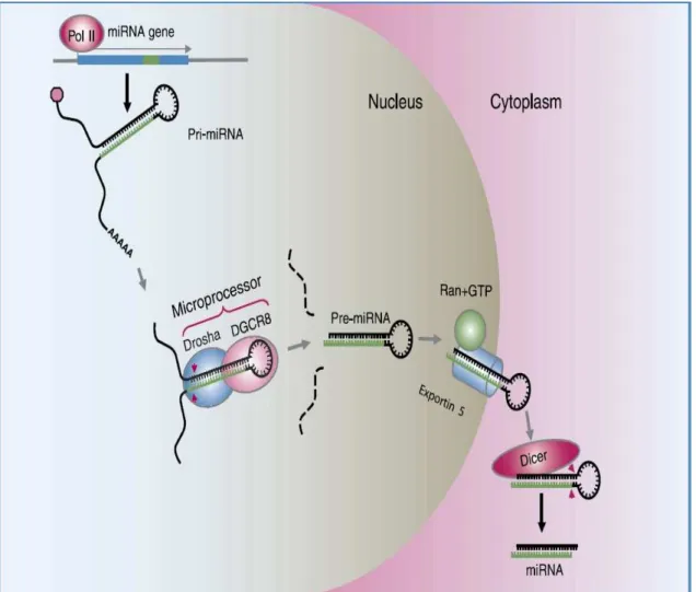
The genome of animals contains at least two hundreds of miRNA genes which encode for short regulatory RNA molecules. miRNAs repress the expression of protein-coding mRNA providing a previously unappreciated regulatory mechanism for gene expression. Upon binding of an individual miRNA, or a combination of several miRNAs to the 3 untranslated region of a target mRNA, either translation repression or mRNA cleavage is induced through activation of the RNA-Induced Silencing Complex (RISC). Studies on multiple vertebrate genomes indicate that miRNA can repress more then a third of all genes. It is impossible to understand the RNAi in vertebrate cells without taking into consideration the biogenesis and function of microRNA (miRNA). Mature miRNAs are noncoding RNAs about 22 nucleotides in length expressed by all metazoan eukaryotes (Bartel, 2004). Gained evidence from several works indicate that the human genome encodes over 300 different miRNA molecules and these miRNAs are believed to play an important role in the post-transcriptional regulation of many aspects of cellular differentiation (Bartel, 2004). As illustrated by Cai et al., (2004) miRNAs are initially transcribed as part of one arm of an RNA stem-loop structure of about 80 nucleotides that in turn forms part of a longer primary miRNA (pri-miRNA) transcript (Figure 5). The first step in miRNA processing occurs in the nucleus. It starts by the recognition of key elements of the secondary structure of the pri-miRNA stem-loop by the RNase III enzyme Drosha and its cofactor DGCR8 (Bohnsack et al., 2004; Denli et al., 2004; Zeng & Cullen, 2006). It is believed that Drosha-DGCR8 cleaves the pri-miRNA stem-loop about 22 nucleotides away from the junction of the stem and the terminal loop, leaving a characteristic two-nucleotide 3' overhang. Then the resulting precursor miRNA (pre-miRNA) hairpin of about 60 nucleotides is bound by the nuclear export factor exportin 5 (Exp5) (Lund et al., 2004) (Figure 5). This step and the recognition are highly dependent on RNA structure and optimally requires an RNA stem of 16 base pairs or more flanked by a short, approximately two-nucleotide 3' overhang (Zeng & Cullen, 2006). Then bound pre-miRNA is transported to the cytoplasm and released there.
CHAPTER I Biology of RNAi
Figure 5 : miRNA biogenesis.
Long primary transcripts (pri-miRNAs) is encoded in the cellular DNA and transcribed in the nucleus, containing one to several miRNAs are generated by polymerase II. Processed by the recently identified microprocessor complex, comprising Drosha (RNase III endonuclease) and DGCR8 (double-stranded RNA binding proteins) recognize the distinct hairpin secondary structure of the pri-miRNA and specifically cleave at the base of the stem loop releasing a 60- to 70-nucleotide pre-miRNA are then transported by Exportin 5 to the cytoplasm where Dicer, a second RNase III endonuclease, cleaves 22-nucleotide from the Drosha cleavage site to yield the mature miRNA. After strand separation, the mature miRNA represses protein production either by blocking translation or causing transcript degradation (Gregory & Shiekhattar, 2005).
In the cytoplasm pre-miRNA is recognized by a heterodimer, consisting of the RNase III enzyme Dicer and its cofactor TRBP10 (see Figure 6 for more details). Once again, structure is important for recognition. It has been demonstrated (Macrae et al., 2006) that RNA stem of 19 base pairs or more and a two-nucleotide 3' overhang are crucial factors in the recognition by Dicer. Then Dicer-TRBP complex binds the base of the pre-miRNA hairpin and cleaves about 22 nucleotides away, leaving another two-nucleotide 3' overhang and removing the terminal loop. Hammond et al., (2000) found that Dicer and TRBP play an important role in facilitating the assembly of one strand of this miRNA duplex intermediate into a protein 'effector complex' called the
RNA-induced silencing complex (RISC). It acts as a 'guide RNA' to direct RISC to homologous mRNA species. This step is similar with what happens with shRNA.
Figure 6 : The miRNA biogenesis pathway in vertebrate cells.
Fig. 6 illustrates the miRNAs biogenesis pathway in vertebrate. Pri-miRNA is first generated by polII, then recognized by Drosha and its cofactor DGCR8 which cleaves this pri-miRNA into pre-miRNA to be exported by exportin 5 from the nucleus into the cytoplasm where it is recognized by Dicer. The processing of pre-miRNA by dicer leads to the production of 21-23nt which will be incorporated to RISC complex.
CHAPTER I Biology of RNAi
2.3. Degradation of mRNA
As previously described the mRNA is cleaved only within the region of sequence identity with the dsRNA (Ngo & Bouck, 1998). Cleavage occurs at sites 21– 23 nucleotides apart, the same interval observed for the dsRNA itself. The 21–23 nucleotide fragments from the dsRNA are guiding mRNA cleavage. Several works that has been done on this mechanism suggested that the double-stranded siRNAs produced in the first step are believed to bind an RNAi-specific protein complex RISC (Elbashir et al., 2001b; Ketting et al., 2001; Cullen, 2006a) (Figure 7). In an ATP dependent manner, this complex might undergo activation which permits for the antisense component of the unwound siRNA to become exposed and allow the RISC to perform the downstream RNAi reaction.(Zamore et al., 2000). It has been found by several works that the antisense siRNAs in the activated RISC pair with cognate mRNAs and the complex cuts this mRNA approximately in the middle of the duplex region (Agrawal et al., 2003).
The importance of RISC complex has been the matter of several investigations. But few independent studies demonstrated the importance of the RISC complex in this part of RNAi reactions (Hammond et al., 2000). The target cleavage site has been mapped to 11 or 12 nucleotides downstream of 5' end of the guide siRNA, and then the cleaved mRNAs are subjected to degradation by exoribonucleases (Hammond et al., 2000).
Three models have been proposed to explain the mechanism by which siRNAs direct target RNA destruction. In the first model, the target destruction is achieved by RNA-dependent RNA polymerase (RdRP). The RdRP is required to convert the target mRNA into dsRNA (Lipardi et al., 2001). As proposed by Tomari et al., (2004), the RdRP is believed to implement single-stranded siRNAs as primers for the target RNA-templated synthesis of complementary RNA (cRNA). Dicer then cleaves the resulting cRNA/target RNA hybrid. This leads to the destruction of the mRNA and to the generation of new siRNAs in the process (Figure 8). This process is ATP-dependent. The second model proposes that single-stranded siRNAs do not act as primers for an RdRP, but they work by assembling along the length of the target RNA and are then ligated together by an RNA ligase to generate cRNA (Lipardi et al., 2001). Dicer destroys the cRNA/target RNA hybrid. Again this model suggests that target recognition and destruction require ATP to catalyze ligation process, and to support Dicer cleavage. Like the first model, Lipardi and coworkers demonstrated that it is necessary for siRNA to have 3’ hydroxyl group for the RNAi. As illustrated in Figure 7, two distinct enzyme complexes act in the RNAi pathway. Dicer generates siRNAs from dsRNA. These siRNAs are then incorporated into a second enzyme complex, the RNA-induced silencing complex (RISC), in an ATP-dependent step or series of steps during which the siRNA duplex is unwound into single strands. One of the resulting single-stranded siRNA is proposed to guide the RISC to recognize and cleave the target RNA. Nowadays it is well known that the third model is the correct pathway.
CHAPTER I Biology of RNAi
Figure 8: Proposed models for RNAi pathway in Drosophila.
In both models, dsRNA is converted to siRNA by an ATP dependant endoribonuclease Dicer. The models differ in the subsequent function of siRNAs. In the left side of the above figure, siRNAs are postulated to function as primers for the target RNA-templated synthesis of cRNA by an RdRP. The final product of this process which is dsRNA is cleaved by Dicer into a new crop of siRNAs, which can prime the conversion of additional target RNAs into dsRNA. In the right, siRNAs are proposed to be incorporated into an endonuclease complex distinct from Dicer, the RISC, and again according to an ATP dependant mechanism, whereas endonucleolytic cleavages of the target RNA appear to require no high energy cofactors.
2.4 Transcriptional gene silencing (TGS)
Transcriptional gene silencing (TGS) is induced by the same molecules that induced post-transcriptional gene silencing (PTGS) but results in activation of the gene for transcription rather than by RNA destruction (Sijen et al, 2001). In plants, double-stranded RNA induces a transcriptional gene silencing accompanied by de novo methylation of a target promoter. This effect can be triggered by a double-stranded RNA containing promoter sequences. The promoter dsRNA is synthesized in the nucleus, is partially cleaved into small RNAs 23 nucleotides in length. Mette et al. (2000) were able to induce transcriptional gene silencing in tobacco and Arabidopsis by using constructs designed to produce double-stranded promoter RNA
Until recently, there was no evidence that a similar pathway operated in mammals. Two new studies suggest that small RNAs can direct DNA methylation and chromatin modification in human cells. Morris et al. (2004) proved that promoter-directed siRNA inhibits transcription of an integrated, proviral, elongation factor 1 alpha (EF1 ) promoter-green fluorescent protein reporter gene and of endogenous EF1 . Silencing was associated with DNA methylation of the target sequence.
CHAPTER I Biology of RNAi
2.4.1 Proposed mechanisms for TGS in S. Pombe
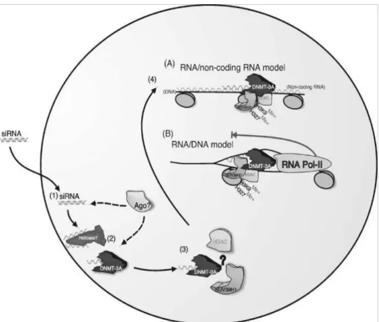
Figure 9 : Proposed mechanisms for TGS in S. pombe
Two models for siRNA mediated TGS have been proposed, either an RNA/RNA or a RNA/DNA mediated mode of silencing. In S. pombe (A) siRNAs may interact with a long non-coding transcript which spans the targeted chromatin (1) subsequently allowing the RITS/RdRP complex to localize to the targeted region (2) resulting in gene silencing (3) reviewed by Agrawal et al, (2003). Alternatively, siRNA mediated silencing may function through an RNA/DNA intermediate. The siRNAs may gain access to the targeted DNA by the effects of RNA Pol-II opening up the targeted region (4) for the siRNA/RITS/RdRP complex to gain access (5) leading to gene silencing (6). (See Fig 9 A&B for more details).
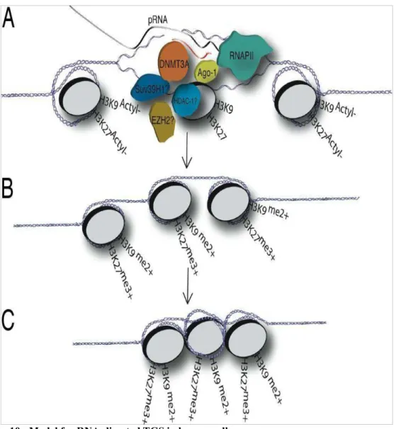
Figure 10 : Model for RNA-directed TGS in human cells
(A) The promoter associated RNA model of RNA-mediated TGS proposes that a variant species of mRNA, a promoter-associated mRNA, essentially containing an extended 5_UTR, is recognized by the antisense strand of siRNAs or possibly endogenous antisense RNAs during RNAPII-mediated transcription of the RNA-targeted promoter. (B) The antisense strand of the siRNA might then guide a putative transcriptional silencing complex (possibly composed of DNMT3A, Ago-1, HDAC-1, and/or EZH2) to the targeted promoter where histone modifications result and the initial gene-silencing event. (C) The initial silencing event or prolonged suppression of the siRNA-targeted promoter may result in heterchromatization of the local siRNA-targeted genomic region and is not, based on these data, thought to be the result of slicing of the low-copy promoter associated RNA but rather due to a recruitment of chromatin remodeling factors or complexes to the targeted promoter that result in the gene silencing (Han et al, 2007).
CHAPTER I Biology of RNAi
Figure 11 : proposed mechanism for TGS in human.
2.4.2 Proposed mechanisms for TGS in human.
A. Model one for siRNA mediated TGS in Human Cells
In a transcriptionally active gene (1) promoters are transcribed by RNA Polymerase II (RNA Pol II) to produce a low copy promoter specific transcript (2). Then the siRNAs may associate with argonaute 1 (Ago-1) and possibly a complex containing DNMT3A (2). The forming complex might interact directly or be bound by HDACs and/or Suv39H1 (3). At this step or prior to this step Ago-1 may also be active to unwind the siRNA, resulting in that the antisense strand from the siRNA probably directs the siRNA/DNMT3a complex with or without the HDACs and/or Suv39H1 or EZH2 to the targeted low copy promoter RNA (3). The promoter RNA corresponds in location to the targeted promoter and the siRNA provides the specificity in targeting a chromatin remodeling complex probably containing HDAC-1 which could deacetylate histone 3 Lysines 9 and/or 27. The deacetylated histones would then permit histone methlytransferases to methylate H3K9 and H3K27 (3). The result of the above mentioned steps would be the initial silencing of transcription at the RNA
Pol II targeted promoter (4). If the silencing is reinforced, the gene may become methylated and permanently silenced (Figure 11B).
B. The second model for siRNA mediated TGS in Human Cells.
In model two a non-coding RNA might be produced by RNA Pol II (1) the produced RNA could be interwoven with the DNA in chromatin (2). This RNA could then act as scaffolding for the antisense strand of promoter specific siRNAs to recognize the corresponding targeted promoter (3). The antisense strand of the promoter specific siRNA is probably associated with a chromatin remodeling complex containing Ago-1, DNMT3A, HDACAgo-1, Suv39HAgo-1, and possibly EZH2 ( as described previously in Model 1). The promoter siRNA targeted complex could then specifically remodel the targeted promoter local histones, i.e. deacetylate histone 3 lysines 9 and/or 27, which would then allow methylation of the promoter associated histones via histone methyltransferases. The result would be the initial silencing of transcription at the RNA Pol II targeted promoter (4). If the silencing is reinforced the gene may become methylated and permanently silenced (Figure 11A).
3. Natural role of RNAi
In 1990, Napoli et al., (1990) was trying to change the color of petunias by inserting a supercharged copy of the gene that controls production of purple pigment. Unexpectedly he got instead white petunias. Nowadays it is clear that Jorgensen had stumbled across a natural mechanism of gene silencing a process which is well known now as RNA interference (RNAi). In this process a double strand RNA is capable of inducing gene turn off (Napoli et al., 1990).
It remained so until 1998 when Fire and coworkers (Fire et al., 1998) showed for the first time that double-stranded RNAs (dsRNAs) were able to trigger sequence-specific gene silencing in a wide variety of organisms, including nematodes, plants, trypanosomes, fruit flies and planaria. For the first time this work opened a promising window towards an effective set of tools to interfere selectively with gene function (Fire et al., 1998). RNA interference (RNAi) is considered to be an effective genome defense mechanism. For the genome defense system to be effective, it meets two main requirements: it needs to be gene-specific and it asks an amplification step to fight off multiplying parasites. RNAi meets these requirements as it specifically recognizes
CHAPTER I Biology of RNAi
dsRNA and sometimes like in plants and c elegans it contains an RNA amplification step.
In plants, RNAi appears to be a defense mechanism against molecular parasites such as viruses and transposons. Motley et al., (2000) and Waterhouse et al.,( 2001) demonstrated that as a consequence of virus infection or transposable elements, plants have developed adaptive mechanism against this infection. In plants, viral genomes can be targeted by the RNAi machinery and probably as a response to it. Viruses on their own have developed strategies to inhibit RNAi, as plant viruses were found to carry silencing-suppresser genes that act to limit the efficacy of RNAi in various ways (Waterhouse & Fusaro, 2006). It is known that RNA viruses replicate via an RNA intermediate of opposite polarity: thus viral genomes and replicative intermediates can form dsRNA which can trigger RNAi formation.
Tabara et al., (1999) proved that C. elegans mutants which are deficient in RNAi were able to activate several transposons. This finding supports the idea that one of the natural roles of RNAi system is transposon silencing. Apparently, transposons are normally silenced in C. elegans (which is a germline-specific process) and this process is dependent on the RNAi pathway. These C. elegans transposons have terminal inverted repeats and transcripts having both terminal inverted repeats can base pair and form dsRNA which could trigger RNAi. Indeed, such dsRNA was detected.
Despite the intense studies of RNAi, there has been no evidence that RNAi work as a system of defense against viral infection in vertebrates, while in plants it is clear that RNAi controls viral infection (Voinnet et al, 1999). Vertebrate viruses recruit several genes to the battle with the host interferon and adaptive immune systems, but still no genes are yet known in vertebrate viruses that antagonize the RNAi system.
Sequencing of the human genome has shown that 45% consists of remnants of transposon as separated sequences and it is conceivable that RNAi plays a role (or played a role in previous time) in defending genome against these molecular parasites, a mechanism probably not strictly necessary in vertebrates.
In worms, parts of the RNAi machinery are important not just for suppressing replication of transposons but also for forming small RNAs—microRNAs (miRNAs). MicroRNA can also function as reverse regulators of disease. So, when certain microRNAs have low levels of expression, their target genes are not suppressed and aggressive. Recently it has been shown that two microRNAs regulate the most
common human leukemia (B-cell lymphocytic). These two microRNAs are miR-29 and miR-181.
RNAi besides its natural function has several and various possible applications. RNAi are being used: (1) to induce virus resistance in transgenic organisms, (2) to improve the quality or production of crops by suppressing unwanted traits (like the softening of tomato fruits or the content in caffeine and nicotine) (3) to eliminate gene transcript (knock-down strategy) acting by reverse genetics strategies known to make it possible (4) to perform certain types of gene-therapy by cleavage of disease-derived transcripts (viral RNAs or mRNAs of cell proliferation genes in cancer cells)
CHAPTER II Therapeutic gene silencing
CHAPTER 2
THERAPEUTIC GENE
SILENCING AND
INHIBITION OF
PATHOGENS BY RNAi
Gene therapy is the process of inserting nucleic acids (e.g. usually DNA/gene) into cells or tissues to correct or prevent a pathological process. Examples include the gene addition for the treatment of genetic disorders as well as therapeutic nucleic acids to stimulate new cell growth for tissue regeneration, demise of cancerous and virus infected cells (as defined by the American society for gene therapy)
(http://www.asgt.org/about_asgt/index.shtml#3).
Nowadays, RNAi is routinely used in laboratories for loss-of-function analyses and increasingly for the rescue of phenotypes caused by dominant acting mutant genes. Scientists have succeeded in applying RNAi in vitro and in vivo to block the effects of disease genes. One of the most important promising features of the RNAi is the ability to generate gene knockdown for studying gene functions by reverse genetics (Haasnoot et al., 2003).
1. Inhibition of pathogens by RNAi
It is now apparent that the mechanisms that mediate RNAi have been evolutionarily conserved in all multicellular eukaryotes, thus indicating that this unique form of homology-dependant gene silencing is a key to one or more aspects of eukaryotic biology. One obvious potential function for RNAi machinery would be to defend cells against viruses that express dsRNA as part of their life cycle.
The role of RNAi during viral infection in mammals is still under investigations. Some reports suggest that mammalian cells have strong-nonspecific responses to viral dsRNA through the direct interaction of dsRNA with the cellular proteins, such as protein kinase R, retinoic acid- inducible gene I (RIG-1) or toll-like receptor (TLR) 3, which is considered to be an important factor triggering signaling pathways that lead to the expression of type one interferon and the activation of non-specific RNases. The expression of interferon leads to the expression of a large number of genes that creates an antiviral state in the host cell as reviewed by Elbashir et al., (2001a). As it was cited by Elbashir et al., (2001a), it has been demonstrated that the introduction of dsRNA longer than 30bp into mammalian cells were responsible for an induction of interferon pathway via dsRNA-dependant protein kinase (PKR) and for apoptosis of infected cells.
Besides, the introduction of chemically synthesized siRNAs or short hairpin RNA (shRNAs) generating siRNA less than 23 bp could be efficient to induce RNAi mediated gene silencing pathway when transfected into cells.
CHAPTER II Therapeutic gene silencing
Moreover, it can be readily demonstrated that the artificial induction of an antiviral RNAi response in mammalian cells can confer strong protection against a wide range of pathogenic viruses (Table 2) (Dykxhoorn et al., 2003; Cullen, 2006b). It has been reviewed by Plaskert (2006) that there are some unanticipated interaction between RNAi machinery and mammalian viruses. The interactions of virus–specific small interfering RNAs (siRNAs) into cell provoke the RNAi system to target viruses, resulting in an effective therapeutic approach to inhibit virus replication in vitro and in animal models. For example Franz et al., (2006) have demonstrated that using RNAi generate Dengue virus resistance in genetically modified Aedes aegypti.
It has been well demonstrated that RNAi is a powerful tool for the inhibition of numerous viruses, including several important human pathogens such as human immunodeficiency virus type 1, hepatitis C virus, hepatitis B virus, dengue virus, poliovirus and influenza virus A (Haasnoot et al., 2003).
Table 2 :Examples of disease-related genes that have been targeted in mammals using siRNA
Gene/mRNA targeted
Type of gene Method Phenotype
p24 HIV-1 capsid protein siRNA transfection; siRNA transfection of in vitro transcribed RNA
Decreased viral protein expression, decreased virus production; inhibition of HIV replication after fusion and before reverse transcription and transcription from integrated provirus
Rev HIV-1 regulatory protein siRNA transfection; plasmid-vector-mediated siRNA expression (tandem U6 promoters)
Decreased viral protein expression, decreased virus production
Vif HIV-1 regulatory protein siRNA transfection; plasmid-vector-mediated siRNA expression
Inhibition of HIV replication, degradation of preintegrated genomic HIV RNA
Tat HIV-1
regulatory protein
siRNA transfection
Decreased viral protein expression, decreased virus production LTR mRNA HIV-1 long
terminal repeat siRNA transfection, in vitro transcribed siRNA
Inhibition of HIV replication after fusion and before reverse transription and transcription from integrated provirus
Poliovirus capsid Capsid structural protein siRNA transfection
Reduced viral titer, clearance of virus from infected cells
Poliovirus RNAP
RNAP siRNA
transfection
Reduced viral titer, clearance of virus from infected cells HPV E6 mRNA Viral transcript E6 siRNA transfection
Selective degradation of E6 mRNA, accumulation of cellular p53, reduced cell growth HPV E7 mRNA Viral transcript E7 siRNA transfection
Selective degradation of E7 mRNA, induced apoptotic cell death RSV P protein Phosphoprotein, smaller subunit of the RNA-dependent RNAP siRNA transfection
Inhibition of P protein expression, reduced amounts of all viral proteins, no syncytia formation
RSV F protein Fusion protein siRNA transfection
No detectable F protein, no effect on other viral proteins, no syncytia formation Hepatitis C virus NS5B Non-structural protein 5B, viral polymerase mRNA Hydrodynamic’ siRNA injection
Decreased levels of the NS5B–luciferase fusion protein in mouse hepatocytes
Ras(V12) Constitutively active oncogenic ras mutant Moloney-based retroviral-vector-mediated siRNA expression
CAPAN-1 cells failed to form colonies in soft agar and failed to form tumours in nude mice when injected
subcutaneously bcr-abl Oncogene, fusion of abl and bcr siRNA transfection
Specifically decreased the bcr–abl mRNA without targeting either the c-abl or c-bcr mRNA, inhibited bcr–abl-dependent cellular proliferation p53 Tumour suppressor gene Plasmid-vector-mediated siRNA expression, Moloney-based retroviral-vector-mediated siRNA expression
Selection of cells stably knocked down in p53 expression; different p53 shRNAs produced different degrees of silencing, which was directly correlated with the severity of Myc-induced
lymphomagenesis; loss of ras-induced senescence, growth in soft agar 53bp1 p53-binding-protein-1, mediator of DNA damage checkpoint siRNA transfection
Decreased p53 accumulation, disruption of G2–M checkpoint arrest, intra-S-phase checkpoint in response to ionizing radiation
p73Dn Tumour
suppressor gene
siRNA transfection
Increased activity of p53-responsive promoter
Fas receptor Proapototic Fas receptor
‘Hydrodynamic’ siRNA injection
Decreased levels of Fas receptor in murine hepatocytes in vivo, increased resistance to Fas-mediated apoptosis CD4 Cell surface receptor, HIV-1 coreceptor siRNA transfection
Decreased HIV-1 infection, decreased free viral titers CCR5 Cell surface receptor; HIV-1 siRNA transfection;
lentiviral-vector-Decreased cell surface expression of receptors, inhibition of CCR5 tropic HIV-1 virus replication
CHAPTER II Therapeutic gene silencing coreceptor mediated siRNA expression CXCR4 Cell surface receptors, HIV-1 coreceptors siRNA transfection
Decreased cell surface expression of receptors, inhibition of CXCR4 tropic HIV-1 virus replication
CD25 IL2 receptor Lentiviral-vector-mediated siRNA
expression
Reduced cell surface expression of CD25, decreased proliferation of T cells when challenged with IL-2
Abreviation : HPV, human papilloma virus; mRNA, messenger RNA; siRNA, short interfering RNA; shRNA, short hairpin RNA; RNAP, RNA polymerase; RSV, respiratory syncytial virus. (From Dykxhoorn et al., 2003).
In recent years, RNAi has therefore been welcomed by the scientific community as a potentially powerful new tool to target diseases. Joost Hassnoot et al., (2007) demonstrated that results from in vitro studies and animal models indicate that RNAi can be highly effective at low dosage. Several RNAi-based antiviral drugs are currently being tested in clinical trials (See Table 3 for more details). The development of RNAi therapeutics is taking place at an unprecedented speed, moving from an obscure phenomenon reported in plant and c. elegans to therapeutic compound in clinical trials in the last few years.
Table 3 : Nucleic acid–based antiviral therapeutics that have entered clinical trials.
Virus Inhibitor (name) Target gene Stage Sponsor CMV Antisense
oligonucleotidea (Vitravene; formivirsen/ISIS 2922
IE2 Approved Isis Pharmaceutials (Carlsbad, CA, USA) HIV-1 Ribozymeb (Rz2, OZ-1) tat Phase 1 complete, Phase 2 ongoing
Johnson & Johnson (New Brunswick, NJ, USA) subsidiary Tibotec Therapeutics (Bridgewater, NJ, USA) 937-nt antisense geneb(VRX496) Phase 1 complete
Gene Shears and Johnson Research env Phase 1 complete VIRxSYS (Gaithersburg, MD, USA) Dominant-negative anti-HIV-1 geneb (RevM10) rev Phase 1/2 ongoing
Systemix (Palo Alto, CA, USA) and National Cancer Institute (Bethesda, MD, USA)
Phase 1 complete
The Saban Research Institute/USC Keck School of Medicine (Los Angeles)
Decoy RNAb RRE Phase 1 complete
Childrens Hospital Los Angeles, University of Southern California School of Medicine (Los Angeles, CA) and Baylor College of Medicine (Houston) Short-hairpin
RNA, ribozyme and RNA decoyb (Triple-R vector)
tat/rev, CCR5, TAR
Phase 1 complete
Colorado State University (Fort Collins, CO, USA) and Beckman Research Institute (Duarte, CA, USA)
Antisense TAR and RevM10b
TAR, rev Phase 1 complete
National Human Genome Research Institute (Bethesda, MD, USA) Antisense oligonucleotidea (Gem92) Gag Phase 2 discontinued
Hybridon (now Idera
Pharmaceuticals, Cambridge, MA, USA) Antisenseb (HGTV43) ND Phase 1/2 ongoing Enzo Biochem(Farmingdale, NY, USA) Peptide nucleic acid (AVR-118) ND Phase 1/2 completed
Advanced Viral Research (Yonkers, NY, USA) RSV Small interfering RNAa (ALN-RSV01) Nucleocapsid Phase 1 ongoing and phase 2 planned for 2008 Alnylam Pharmaceuticals (Cambridge, MA, USA)
HCV Ribozymea (Heptazyme) IRES Phase 2 studies discontinued Ribozyme Pharmaceuticals (Boulder, CO, USA; renamed Sirna, now part of Merck) Antisense oligonucleotidea (AVI-4065) ND Phase 2 studies discontinued
AVI BioPharma (Portland, OR, USA) Antisense oligonucleotidea (ISIS14803) IRES Phase 2 studies discontinued Isis HBV Short-hairpin RNAb(Nuc B1000) Phase 1 completed Isis Pre-gen./pre-C, Pre-Sl, Pre-S2/S, X Phase 1 ongoing
Nucleonics (Horsham, PA, USA) HPVc Antisense oligonucleotidea (MBI1121) El Phase 1 discontinued Migenix (formerly Micrologix Biotech, Vancouver, BC, Canada) Peptide nucleic acid (AVR-118) ND Phase 1 discontinued
Advanced Viral Research (Yonkers, NY, USA)
Abbreviations
a
Chemically synthesized.bGene construct.cHuman papillomavirus. ND, not disclosed. NA, not available
CHAPTER II Therapeutic gene silencing
The RNAi therapy is moving toward clinical applications in humans and animals (Table 2). Its success for some diseases has not yet been definitely met and it needs further investigation. There is huge potential for crossing the obstacles and for using this technology in gene therapy. Yet the off-target effect as well as the delivery system are considered to be the most challenging problems facing this promising technology (Ralph et al., 2005).
Morris, (2006) showed that the in vitro HIV-1 multiplication was suppressed through siRNAs directed against HIV-1 tat and rev transcripts. Other viruses have also been successfully targeted by siRNAs in vitro with some success including Semliki Forest Virus (SFV), poliovirus, dengue virus, influenza virus, and hepatitis C virus and others.
It has been shown by Castanotto et al., (2002) and Lee et al., (2002) that nematodes and insects use RNAi to reduce the infection by flock house virus (FHV), a member of the nodavirus family. This virus is able to cause infection for both insects and vertebrate cells, and the infection of drosophila cells results in the increase of FHV specific siRNAs. It has also been reviewed by Haasnoot et al., (2003) that insect viruses SFV and FHV were strongly inhibited by the introduction of dsRNA.
Several studies were performed to determine whether RNAi targets incoming genomic RNA, the newly synthesized transcripts or both. It has been reported by several groups (Capodici et al., 2002; Jacque et al., 2002; Novina et al., 2002) that the incoming genomic RNA is indeed the target of siRNA leading to its destruction. Other reports by Coburn & Cullen, (2002), Hu et al., (2002) and Verma et al., (2003) showed no or only modest reduction in the level of proviral RNA. From those reports it remains possible that different siRNAs produce different effects. Some incoming RNA is protected from RNAi–mediated degradation by nucleocapsid (Hu et al., 2002). In contrast to RNA viruses, it has been shown that DNA viruses targeted by RNAi can result in degradation of the viral mRNAs (Jia & Sun, 2003).
McCaffrey et al., (2003) were the first to demonstrate that virus inhibition by RNAi in vivo is possible by a co-transfection of Hepatitis B Virus (HBV) DNA and shRNA expressing plasmid targeting the HBV sequence. The HBV multiplication was highly inhibited in mice liver.
There are now several reports on the in vitro inhibition of human and animal viruses (Table 4 and 5 for RNA and DNA viruses respectively).
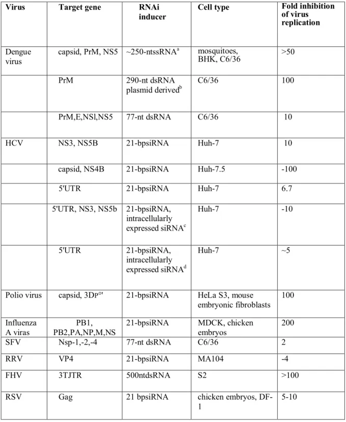
Table 4 : Inhibition of RNA viruses by RNAi as illustrated in (Haasnoot et al., 2003).
Virus Target gene RNAi
inducer
Cell type Fold inhibition
of virus replication
Dengue virus
capsid, PrM, NS5 ~250-ntssRNAa mosquitoes,
BHK, C6/36 >50 PrM 290-nt dsRNA plasmid derivedb C6/36 100 PrM,E,NSl,NS5 77-nt dsRNA C6/36 10 HCV NS3, NS5B 21-bpsiRNA Huh-7 10
capsid, NS4B 21-bpsiRNA Huh-7.5 -100
5'UTR 21-bpsiRNA Huh-7 6.7
5'UTR, NS3, NS5b 21-bpsiRNA, intracellularly expressed siRNAc Huh-7 -10 5'UTR 21-bpsiRNA, intracellularly expressed siRNAd Huh-7 ~5
Polio virus capsid, 3DP°' 21-bpsiRNA HeLa S3, mouse
embryonic fibroblasts 100 Influenza A viras PB1, PB2,PA,NP,M,NS 21-bpsiRNA MDCK, chicken embryos 200 SFV Nsp-1,-2,-4 77-nt dsRNA C6/36 2 RRV VP4 21-bpsiRNA MA104 -4 FHV 3TJTR 500ntdsRNA S2 >100
RSV Gag 21 bpsiRNA chicken embryos,
DF-1
5-10
All dsRNA fragments were transfected in the cells unless indicated otherwise. a Intracellular expressed dengue virus ssRNA using sindbis virus as a vector.b 290-bp hairpin RNA expressed from a transfected plasmid under the control of hsp 70 promoter.
c
siRNAs expressed from a transfected plasmid under the control of two HI promoters for the sense and antisense fragment. d siRNAs expressed from a transfected plasmid under the control of two U6 promoters for the sense and antisense fragment, or as a 19-bp shRNA. The fold inhibition of virus production represents the result obtained with the most efficient siRNA