PERSPECTIVE
Improving the Stability and Activity of Oral Therapeutic
Enzymes
—Recent Advances and Perspectives
Gregor Fuhrmann&Jean-Christophe Leroux
Received: 8 July 2013 / Accepted: 14 October 2013 / Published online: 2 November 2013 # Springer Science+Business Media New York 2013
ABSTRACT Exogenous, orally-administered enzymes are currently in clinical use or under development for the treatment of pathologies, such as celiac disease and phenylketonuria. However, the administration of therapeutic enzymes via the oral route remains challenging due to potential inactivation of these fragile macromolecular entities in the harsh environment of the gastrointestinal tract. Enzymes are particularly sensitive because both proteolysis and unfolding can lead to their inactivation. Current efforts to overcome these shortcomings involve the application of gastro-resistant delivery systems and the modification of enzyme structures by polymer conjugation or protein engineering. This perspective manuscript reviews and critically discusses recent progress in the oral delivery of therapeutic enzymes, whose substrate is localized in the gastrointestinal tract.
KEY WORDS celiac disease . drug therapy . pancreas . PEG
INTRODUCTION
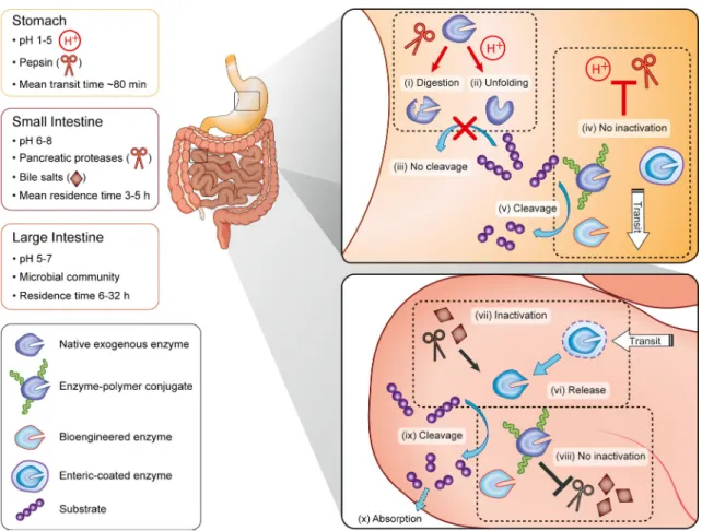
Enzymes are widely used as therapeutic agents and research tools to elucidate molecular pathways (1). As drugs, they are of particular interest because of their high activity and selectivity, and because of the possibility of manipulating these properties through chemical (2) and protein engineering techniques (3). However, due to their complex structure, enzymes are potentially subject to inactivation, especially when administered via the oral route (1). Indeed, it is difficult to stabilize proteins in the gastrointestinal (GI) tract as its primary function is protein digestion (Fig.1). Despite this paramount challenge, the concept of oral enzyme replacement therapy has been around for some time with a certain degree of success
(4), and a number of stabilization strategies have been investigated (functional modifications (2) or formulation approaches (5)). The goal of oral enzyme therapy is to either degrade potentially harmful food components in situ (celiac disease, phenylketonuria) or supplement the organism with digestive enzymes produced in insufficient amounts (exocrine pancreatic insufficiency, disaccharidase deficiencies). This perspective manuscript reviews state-of-the-art oral enzyme therapies against diseases in which activity of the administered enzyme is desired in the GI tract, and not after systemic absorption (e.g., adjuvant therapy in oncology (6)) or when delivered via probiotics (7). Stabilization strategies are highlighted and built upon to shape future research directions.
THE GI TRACT—A HARSH AND DYNAMIC ENVIRONMENT
Modern delivery strategies should aim at stabilizing orally-administered enzymes at different locations in the GI tract (for maximal activity) and at a reasonable cost, since doses of exogenous enzymes are often quite high (e.g ., up to a few g/ day). Stabilization and selective re-activation in different segments of the GI tract can be achieved with various approaches that exploit the intrinsic chemical environments encountered at these locations.
The mammalian digestive system is divided into the stomach, small intestine (duodenum, jejunum, and ileum) and large intestine (cecum, colon, rectum, and anal canal) (Fig.1). Protein digestion begins in the low pH environment of the stomach and involves peptidases (7). In particular, pepsin has a propensity to hydrolyze peptide bonds at hydrophobic residues such as phenylalanine and leucine, while residues such as histidine, lysine, arginine, and proline are left untouched (8). This process is facilitated by protein unfolding at acidic pH (1–2.5) (1). As both mechanisms act synergistically and are very efficient, digestion in the stomach is the main obstacle encountered by oral enzyme formulations. Upon leaving the stomach and G. Fuhrmann
:
J.<C. Leroux (*)Institute of Pharmaceutical Sciences
Department of Chemistry and Applied Biosciences ETH Zurich, Wolfgang-Pauli-Str. 10, HCI H 301 8093 Zurich, Switzerland
e-mail: [email protected] DOI 10.1007/s11095-013-1233-y
entering the small intestine, pH increases to neutral values (7). Although most exogenous enzymes are stable a priori in these environments, they can be degraded by pancreatic peptidases (mainly trypsin and chymotrypsin) (1). Trypsin and chymotrypsin are endopeptidases that preferentially cleave peptide bonds involving arginine, lysine and aromatic amino acids, respectively. In addition, therapeutic enzymes are potentially subject to denaturation by intestinal bile salts (7) (Fig.1).
One approach to overcome digestion in the GI environment is to manipulate the primary sequence of proteins by recombinant technologies to reduce their propensity to unfold in acidic media, and to lower their susceptibility to pepsin-mediated digestion. For instance, replacement of phenylalanine and leucine residues by analogues which are less recognized by pepsin has been explored (9). In addition, to completely avoid digestion in the stomach, gastro-resistant, polymer-coated tablets or microparticles (prepared e.g. with cellulose acetate phthalate or methacrylate copolymers) can be used (10,11). These coatings dissolve in the higher pH environment of the small intestine (see below), thereby releasing therapeutic enzymes. Another way to
improve enzyme GI stability is covalent conjugation to polymers (5). This method provides protein protection mainly through steric shielding from endogenous proteins and has been mostly exploited to stabilize enzymes administered by intravenous injection (e.g., poly(ethylene glycol) (PEG)-L-asparaginase (12) or PEG-recombinant uricase (13)).
Key points associated with the oral delivery of therapeutic enzymes are summarized in Table I. Advantages and drawbacks of current strategies against different GI disorders are discussed in the following sections.
Fig. 1 Current concepts of enzyme stabilization in the GI tract. Upon oral administration, exogenous enzymes encounter different environments in the GI tract (7) (stomach, small, and large intestine). (i) Digestion by stomach peptidases (e.g., pepsin) and/or (ii) pH-induced protein unfolding result in enzyme inactivation. (iii) Inactivated enzymes cannot digest their substrate. (iv) Enzyme inactivation can be overcome by enteric coating, polymer conjugation and protein engineering. (v) Enzyme-polymer conjugates and bioengineered enzymes but not enteric-coated formulations could, in principle, cleave their substrates in the stomach. After transit to the small intestine, (vi) enteric coatings dissolve, releasing enzymes, (vii) which are potentially subject to inactivation by pancreatic peptidases and bile salts. (viii) Such inactivation of enzyme-polymer conjugates and bioengineered enzymes may also be avoided or slowed down. (ix) Enzymes cleave their substrate in the intestine which (x) allows normal absorption of nutrients.
Table 1 Oral Enzyme Therapy: Applications and Challenges Key points
• Orally-administered exogenous enzymes can serve to treat several GI and non-GI diseases
• Enzymes are potentially subject to inactivation in the harsh GI environment • Stability can be enhanced via enteric coating, polymer modification, and
SUCROSE INTOLERANCE: AN EXCEPTION?
Not all oral therapeutic enzymes require specific delivery systems because they show significant intrinsic stability under GI conditions. One example is sacrosidase which is used for the treatment of congenital sucrose-isomaltase intolerance (~1 in 500 white ethnics (14)). Affected patients cannot digest the disaccharide sucrose, leading to its malabsorption. This culminates in considerable abdominal distension, bloating, diarrhea, and failure to thrive (14). Although strict exclusion of sucrose-containing food overcomes these issues, such a diet is difficult to follow, especially in children. Clinical improvement of symptoms is seen after oral administration of yeast extract (14) (preferably on a filled stomach) and treatment with oral sacrosidase solution (15). The sacrosidase solution was found to be exceptionally resistant to acidic environments due to enzyme glycosylation and its use at high concentrations. Moreover, it could escape pepsin digestion in vitro in the presence of “sacrificial” protein substrates (16) that competed for degradation (TableII). It would therefore be advisable to administer sacrosidase with a protein-containing meal, although concomitantly-ingested food may also promote degradation by increasing both residence time in the stomach and the secretion of pancreatic enzymes.
LACTOSE INTOLERANCE
Lactose intolerance (i.e., adult-type hypolactasia) is a common autosomal recessive disease characterized by a deficit in lactase-phlorizin hydrolase, an enzyme hydrolyzing lactose into glucose and galactose (17). The prevalence of this predisposition varies greatly, between 5% in Europe and up to 90% in certain populations from Asia and Africa (18). Its onset often occurs 1–2 years after weaning, and is associated with abdominal bloating and diarrhea but also alterations of bone mineral density due to calcium deficiency (19). Although most people with hypolactasia tolerate moderate amounts of lactose (approximately 12 g/day (20)), the disease can be treated by excluding lactose-containing products from the diet. Such diet is also difficult to maintain due to undersupply with calcium, phosphate and vitamins, but alternatives are available. For example,β-galactosidase can be added to milk prior to consumption, administered directly with a lactose-containing meal (21), or can be consumed as yoghurt (22). Although the concept of enzyme replacement works, oral β-galactosidase application often leads to reduced therapeutic efficiency compared to pre-hydrolyzed milk (23). This effect is presumably attributed to GI inactivation of the enzyme. Comparative studies of different lactase digestive supplements have shown low to moderate in vitro enzyme stability under simulated GI conditions (24) (especially in simulated gastric environments) (TableII). To overcome these limitations,
β-galactosidase was recently modified chemically with branched 40-kDa PEG, a hydrophilic polymer which creates a zone of steric hindrance around the enzyme. Steric hindrance should be controlled to prevent the action of endogenous digestive enzymes while allowing the substrate to reach catalytic sites (25). The conjugate displayed enhanced stability at acidic pH and in simulated gastric fluids containing pepsin (26) (Table II). Alternatively, a bioengineered β-galactosidase mutant (27) and hydrolase isolated from meso-acidophilic fungus (28) have both demonstrated improved stability at low pH (Table II). However, it is still not known if these enzymes would better survive hydrolysis by gastric and pancreatic enzymes.
EXOCRINE PANCREATIC INSUFFICIENCY
Exocrine pancreatic insufficiency, resulting from pancreatic problems, is characterized by a deficiency or absence of certain digestive enzymes (i.e., amylase, proteases, and lipases) (29). The main causes of insufficient pancreatic secretion include cystic fibrosis (30), pancreatic cancer (10), and chronic pancreatitis (reported to be the main cause of pancreatic insufficiency affecting 0.5–4% worldwide (31)). In addition, pancreatic insufficiency is frequent in type 1 (51% prevalence) and type 2 (32% prevalence) diabetes (32). Impairment of pancreatic enzyme secretion is associated with insufficient digestion and, consequently, malabsorption/malnutrition, steatorrhea, and body weight loss. Exocrine pancreatic insufficiency may conceal other GI diseases, making their diagnoses challenging (33). Some studies have also analyzed the potential connection between exocrine pancreatic insufficiency and persistent symptoms in adult celiac (34) and inflammatory bowel disease patients (33). In both cases, supplementation with pancreatic enzymes should be considered as it might improve GI symptoms (35).
The therapy of choice in pancreatic insufficiency is supplementation with bovine and porcine pancreatic enzymes preferably in gastro-resistant (enteric-coated) microspheres (10,11,31). Enteric coating protects enzymes in the stomach and releases them during transit to the duodenum alongside food (11) (Table II). Although oral administration of pancreatic enzymes in enteric formulations has been demonstrated to improve symptoms of malabsorption in patients with pancreatic insufficiency due to human immunodeficiency virus (36), the general therapeutic benefit of these systems remains debatable (4). Comparison between different coated and uncoated microsphere formulations has revealed that bioavailability can fluctuate substantially between products, leading to possible discrepancies in oral activity of the enzymes (37). In addition, malnutrition often persists in patients with pancreatic insufficiency, despite
enzyme supplementation, calling for the development of new methods of clinical evaluation of oral enzyme substitution efficiency (38). In general, depending on the primary cause of pancreatic insufficiency (e.g. , cystic fibrosis), acidic conditions in the intestine can delay enzyme release and thus affect the clinical outcome of the therapy (37). Recently, a mutant of Thermomyces lanuginosus lipase has been suggested as potential enzyme replacement candidate. Although both native enzyme and variant showed comparable resistance towards trypsin (with and without bile salts), the mutant was reported to be more stable at acidic pH and showed increased refolding capacity compared to the native lipase (39) (Table II). The conjugation of synthetic polymers to enzymes could also overcome drawbacks related to substitution therapy by delivering functionally-modified enzymes in the duodenum without relying on prior dissolution of enteric coating. Although pancreatic enzymes (trypsin or chymotrypsin) frequently serve as model enzymes to explore the impact of polymer-protein modifications (40), their systematic analysis after oral administration is still lacking. Ho we ve r, in th is spe cif ic ap plication, p olyme r conjugation might be viewed as being expensive for the formulation of porcine pancreatic enzymes, and should perhaps be restricted to more specialized enzymes (see below).
CELIAC DISEASE
Celiac disease (1 in 100 white ethnics (41)) is a life-long, genetically-inherited disease triggered by the ingestion of cereal proteins (i.e., gluten) from wheat, barley, and rye (42). Insufficient digestion of these proteins induces a T cell-controlled autoimmune reaction precipitated in the small
intestine of genetically-predisposed patients. This inflammation is associated with GI-related (e.g. , diarrhea, malabsorption, abdominal pain) and non-GI-related (e.g. , osteoporosis, anemia, migraine) symptoms (43). Moreover, overall mortality is higher among celiac patients than the general population (44). To date, strict exclusion of gluten from daily nutrition is the only available treatment (45). Such a diet is challenging for affected people and is difficult to follow (43). Different treatment options are currently being explored, among which the oral administration of gluten-degrading enzymes appears to be promising (41,46). The latter approach consists of digesting immunogenic and GI-resistant glutamine-and proline-rich peptides of gluten with proline-specific endopeptidases (PEPs) (47) and barley endoprotease (EP-B2). PEPs, derived from different bacterial and fungal sources, have reached clinical testing (48,49). In clinical phase I trials, PEPs were well tolerated (49,50), and PEP and EP-B2 combination significantly reduced gluten in vivo under fed stomach conditions (50). However, the sensitivity of some PEPs to acidic gastric conditions and their potential degradation by intestinal fluids and bile salts remain problematic (1,51). To address the first issue, PEPs have been incorporated in enteric-coated capsules (52). In vitro, this formulation was efficient in protecting PEPs from inactivation under simulated gastric conditions, but was not tested later in humans. It could be questioned whether enteric formulation of PEPs could be the best option here, since gluten should be preferentially degraded in the upper GI tract before immunogenic peptide sequences reach the jejunum (Table II). Moreover, enteric PEP formulations would not protect them from digestion in intestinal fluids. Protein engineering of PEPs appears to be more promising because it does not impair enzymatic activity in a specific section of the GI tract. A series of PEP mutants with increased gastric enzyme stability in vitro (9) have been obtained with this Table II Strategies to Enhance the GI Stability of Exogenous Enzymes
Disease Oral enzyme Delivery approach Effect References
Sucrose intolerance Sacrosidase None Prolonged activity in the presence of alimentary proteins
Treem et al. (1993) (16) Lactose intolerance Lactase Enteric-coated formulations Improved in vitro stability in
gastric fluids
O’Connell & Walsh (2006) (24) β-galactosidase Polymer conjugation Turner et al. (2011) (26) β-galactosidase Recombinant expression of
mutants
O’Connell & Walsh (2010) (27) Pancreatic insufficiency Pancreatic enzymes Enteric-coated microspheres Improved in vitro stability in
gastric fluids
Domínguez–Muñoz (2011) (11)
Lipase Enzyme variant Wang et al. (2013) (39)
Celiac disease Proline-specific endopeptidases
Enteric-coated capsules Improved in vitro stability in gastric fluids Gass et al. (2005) (52) Protein engineering Enhanced in vitro stability and activity Gordon et al. (2012) (53) Polymer conjugation Enhanced in vivo activity Fuhrmann et al. (2013) (55) Phenylketonuria Phenylalanine hydroxylase Fusion protein Reduction of plasma phenylalanine
levels in mice
Eavri & Lorberboum-Galski (2007) (64)
approach. Interestingly, improvement of stability towards pepsin at low pH was not only attributed to changes in the canonical pepsin specificity pattern (i.e. , replacement of the pepsin substrates phenylalanine and leucine by disfavored substrates histidine, lysine, arginine, or proline (8)) but also mostly to favorable hydrophobic packing that protected the enzyme from attack of pepsin. In a recent computational-based study, an acid resistant but gliadin-unspecific enzyme was mutated to a gliadinase with increased in vitro stability towards GI enzymes. This engineered enzyme was able to degrade 95% of a celiac-toxic peptide at simulated stomach pH (53) (Table II). Additional characterization will clarify whether this new gliadinase also proves useful in vivo. Another approach to stabilize PEPs is to conjugate PEG to the enzyme. PEGylated enzymes show enhanced activity towards gluten peptides in vitro and improved stability under simulated intestinal conditions (54). A recent animal study that examined the activity of PEP-mPEG conjugates by non-invasive real-time fluorescence assay (1) indicated that the modified enzyme displayed enhanced activity compared to native PEPs in the small intestine (55). Very efficient improvement of PEP performance was obtained by conjugating them to a dendronized cationic polymer. PEP-polymer conjugates exhibited prolonged retention and activity (up to 6 and 3 h, respectively) in the stomach (55) (Table II). Conjugation of PEPs to specific polymers in combination with protein engineering methods, to optimize stability but also to control polymer grafting sites, could turn out to be a powerful strategy in the future to produce highly resistant PEPs.
PHENYLKETONURIA
Phenylketonuria (1 in 10,000 white ethnics (56)) is a metabolic disorder caused by a defect in the gene encoding for the enzyme phenylalanine hydroxylase (PAH), which catalyzes the metabolism of phenylalanine to tyrosine. The defect renders PAH non-functional, leading to phenylalanine accumulation and, consequently, to impaired neurophysiological function and reduced cognitive development (57). Current treatment consists of omitting phenylalanine from the diet, which is difficult and does not completely reverse neurological damage (57). Investigated alternatives to this stringent diet include the administration of tetrahydrobiopterin (a natural cofactor of PAH), gene therapy to restore PAH levels, and enzyme replacement with phenylalanine ammonia lyase (PAL) or recombinant PAH (58). PAL, a comparatively robust enzyme requiring no cofactors, converts phenylalanine into the harmless metabolite trans -cinnamate (58). In a proof-of-principle study, PAL was produced recombinantly in yeast and successfully tested after oral administration in a mouse model of human phenylketonuria (59). The concept of
deploying PAL as enzyme replacement therapy was further examined after PEGylation (60) and pre-clinical screening of PEGylated PAL from different species (61) (Table II). After subcutaneous injection, PAL from a cyanobacterium (Anabaena variabilis) was found to be the most therapeutically active enzyme. PAL-mPEG conjugate is currently being evaluated for safety and tolerability in a phase II clinical trial. Recently, the short-term pharmacodynamic profile of PAL-PEG was also assessed after oral administration. PAL conjugation to 5-kDa PEG produced therapeutically-relevant reduction of plasma phenylalanine levels in mice (62) (Table II). Besides PAL, recombinant PAH is being investigated as potential adjuvant therapy. The stability of this enzyme was improved by PEGylation (63) and fusion proteins (64) (Table II). These findings are promising and need to be validated after oral dosing in preclinical and clinical trials.
PERSPECTIVES
Oral administration of enzymes is currently indicated for lactose and sucrose intolerance as well as exocrine pancreatic insufficiency. There are other conditions (e.g., celiac disease (41), phenylketonuria (61)) and medical applications (e.g., treatment of intoxications (65)) where they could be beneficial. One such application could be oral administration of phytase, an enzyme capable of cleaving inositol hexakisphosphate (IP6), an “anti-nutrient” forming insoluble complexes with many mineral ions (e.g., Ca2+, Zn2+and Fe2+) and inducing mineral deficiencies under cereal- and legume-rich nutrition. Degradation of IP6 in the intestine by phytase, which appears to be stable even under gastric conditions, would reduce IP6 concentration and potentially alleviate these deficiencies (66). Functional GI disorders are other states in which oral administration of enzymes could be advantageous. α-Galactosidase can significantly decrease symptoms such as flatulence in affected patients (67). As with other therapeutic proteins, however, enzymes are eventually inactivated in the GI tract, their half-life being determined by their intrinsic molecular structure and surrounding environment (pH, fed vs. fasted state, etc.). Current methods to stabilize proteins mostly rely on enteric coating with debatable clinical outcome. Thus, there is a need for new non-invasive techniques to better evaluate enzyme activity in situ and devise strategies to augment therapeutic efficacy after oral application. Recent developments in protein engineering (68) should foster the design of more potent enzyme formulations (e.g., encapsulation in colloidal carriers to create “nanoreactors” (65)) with improved stability in the GI tract. Preliminary data generated recently with polymer conjugation (55) are encouraging and may stimulate research in this field.
ACKNOWLEDGMENTS AND DISCLOSURES
This work was supported by the Swiss National Science Foundation (310030_135732).
REFERENCES
1. Fuhrmann G, Leroux J-C. In vivo fluorescence imaging of exogenous enzyme activity in the gastrointestinal tract. Proc Natl Acad Sci U S A. 2011;108:9032–7.
2. Harris JM, Chess RB. Effect of pegylation on pharmaceuticals. Nat Rev Drug Discov. 2003;2:214–21.
3. Brannigan JA, Wilkinson AJ. Protein engineering 20 years on. Nat Rev Mol Cell Biol. 2002;3:964–70.
4. Regan PT, Malagelada J-R, DiMagno EP, Glanzman SL, Go VLW. Comparative effects of antacids, cimetidine and enteric coating on the therapeutic response to oral enzymes in severe pancreatic insufficiency. New Engl J Med. 1977;297:854–8.
5. Frokjaer S, Otzen DE. Protein drug stability: a formulation challenge. Nat Rev Drug Discov. 2005;4:298–306.
6. Leipner J, Saller R. Systemic enzyme therapy in oncology—effect and mode of action. Drugs. 2000;59:769–80.
7. Cook MT, Tzortzis G, Charalampopoulos D, Khutoryanskiy VV. Microencapsulation of probiotics for gastrointestinal delivery. J Control Release. 2012;162:56–67.
8. Hamuro Y, Coales SJ, Molnar KS, Tuske SJ, Morrow JA. Specificity of immobilized porcine pepsin in H/D exchange compatible conditions. Rapid Commun Mass Spectrom. 2008;22:1041–6. 9. Ehren J, Govindarajan S, Moron B, Minshull J, Khosla C. Protein
engineering of improved prolyl endopeptidases for celiac sprue therapy. Protein Eng Des Sel. 2008;21:699–707.
10. Imrie CW, Connett G, Hall RI, Charnley RM. Enzyme supplementation in cystic fibrosis, chronic pancreatitis, pancreatic and periampullary cancer. Aliment Pharmacol Ther. 2010;32:1–5. 11. Domínguez–Muñoz JE. Chronic pancreatitis and persistent
steatorrhea: what is the correct dose of enzymes. Clin Gastroenterol Hepatol. 2011;9:541–6.
12. Graham ML. Pegaspargase: a review of clinical studies. Adv Drug Deliv Rev. 2003;55:1293–302.
13. Schlesinger N, Yasothan U, Kirkpatrick P. Pegloticase. Nat Rev Drug Discov. 2011;10:17–8.
14. Harms H-K, Bertele-Harms R-M, Bruer-Kleis D. Enzyme-substitution therapy with the yeast saccharomyces cerevisiae in congenital sucrase-isomaltase deficiency. New Engl J Med. 1987;316:1306–9.
15. Treem WR, McAdams L, Stanford L, Kastoff G, Justinich C, Hyams J. Sacrosidase therapy for congenital sucrase-isomaltase deficiency. J Pediatr Gastroenterol Nutr. 1999;28:137–42.
16. Treem WR, Ahsan N, Sullivan B, Rossi T, Holmes R, Fitzgerald J, et al. Evaluation of liquid yeast-derived sucrase enzyme replacement in patients with sucrase-isomaltase deficiency. Gastroenterology. 1993;105:1061–8.
17. Enattah NS, Sahi T, Savilahti E, Terwilliger JD, Peltonen L, Jarvela I. Identification of a variant associated with adult-type hypolactasia. Nat Genet. 2002;30:233–7.
18. Tishkoff SA, Reed FA, Ranciaro A, Voight BF, Babbitt CC, Silverman JS, et al . Convergent adaptation of human lactase persistence in Africa and Europe. Nat Genet. 2007;39:31–40. 19. Di Stefano M, Veneto G, Malservisi S, Cecchetti L, Minguzzi L,
Strocchi A, et al. Lactose malabsorption and intolerance and peak bone mass. Gastroenterology. 2002;122:1793–9.
20. Suarez FL, Savaiano DA, Levitt MD. A comparison of symptoms after the consumption of milk or lactose-hydrolyzed milk by people
with self-reported severe lactose intolerance. New Engl J Med. 1995;333:1–4.
21. Rosado JL, Solomons NW, Lisker R, Bourges H. Enzyme replacement therapy for primary adult lactase deficiency. Effective reduction of lactose malabsorption and milk intolerance by direct addition of beta-galactosidase to milk at mealtime. Gastroenterology. 1984;87:1072–82. 22. Kolars JC, Levitt MD, Aouji M, Savaiano DA. Yogurt—an
autodigesting source of lactose. New Engl J Med. 1984;310:1–3. 23. Suarez FL, Savaiano DA, Levitt MD. The treatment of lactose
intolerance. Aliment Pharmacol Ther. 1995;9:589–97.
24. O’Connell S, Walsh G. Physicochemical characteristics of commercial lactases relevant to their application in the alleviation of lactose intolerance. Appl Biochem Biotechnol. 2006;134:179–91. 25. Liu M, Tirino P, Radivojevic M, Phillips D, Gibson M, Leroux J-C, et al. Molecular sieving on the surface of a protein provides protection without loss of activity. Adv Funct Mater. 2012;23:2007–15. 26. Turner KM, Pasut G, Veronese FM, Boyce A, Walsh G.
Stabilization of a supplemental digestive enzyme by post-translational engineering using chemically-activated polyethylene glycol. Biotechnol Lett. 2011;33:617–21.
27. O’Connell S, Walsh G. A novel acid-stable, acid-active beta-galactosidase potentially suited to the alleviation of lactose intolerance. Appl Microbiol Biotechnol. 2010;86:517–24.
28. Wang H, Luo H, Bai Y, Wang Y, Yang P, Shi P, et al. An acidophilic beta-galactosidase from bispora sp MEY-1 with high lactose hydrolytic activity under simulated gastric conditions. J Agric Food Chem. 2009;57:5535–41.
29. DiMagno EP, Go VLW, Summerskill WHJ. Relations between pancreatic enzyme outputs and malabsorption in severe pancreatic insufficiency. New Engl J Med. 1973;288:813–5.
30. Rowe SM, Miller S, Sorscher EJ. Cystic fibrosis. New Engl J Med. 2005;352:1992–2001.
31. Fieker A, Philpott J, Armand M. Enzyme replacement therapy for pancreatic insufficiency: present and future. Clin Exp Gastroenterol. 2011;4:55–73.
32. Hardt PD, Ewald N. Exocrine pancreatic insufficiency in diabetes mellitus: a complication of diabetic neuropathy or a different type of diabetes? Exp Diabetes Res. 2011. doi:10.1155/2011/761950. 33. Leeds JS, Oppong K, Sanders DS. The role of fecal elastase-1 in
detecting exocrine pancreatic disease. Nat Rev Gastroenterol Hepatol. 2011;8:405–15.
34. Leeds JS, Hopper AD, Hurlstone DP, Edwards SJ, McAlindon ME, Lobo AJ, et al. Is exocrine pancreatic insufficiency in adult coeliac disease a cause of persisting symptoms? Aliment Pharmacol Ther. 2007;25:265–71.
35. Evans KE, Leeds JS, Morley S, Sanders DS. Pancreatic insufficiency in adult celiac disease: do patients require long-term enzyme supplementation? Dig Dis Sci. 2010;55:2999–3004.
36. Carroccio A, Guarino A, Zuin G, Verghi F, Berni Canani R, Fontana M, et al. Efficacy of oral pancreatic enzyme therapy for the treatment of fat malabsorption in HIV-infected patients. Aliment Pharmacol Ther. 2001;15:1619–25.
37. Aloulou A, Puccinelli D, Sarles J, Laugier R, Leblondt Y, Carriere F. In vitro comparative study of three pancreatic enzyme preparations: dissolution profiles, active enzyme release and acid stability. Aliment Pharmacol Ther. 2008;27:283–92.
38. Domínguez–Muñoz JE, Iglesias–García J, Vilariño–Insua M, Iglesias–Rey M. 13C-mixed triglyceride breath test to assess oral enzyme substitution therapy in patients with chronic pancreatitis. Clin Gastroenterol Hepatol. 2007;5:484–8.
39. Wang H, Hagedorn J, Svendsen A, Borch K, Otzen DE. Variant of the Thermomyces lanuginosus lipase with improved kinetic stability: a candidate for enzyme replacement therapy. Biophys Chem. 2013;172: 43–52.
40. Duncan R, Gilbert HRP, Carbajo RJ, Vicent MJ. Polymer maskedunmasked protein therapy. 1. bioresponsive dextrintrypsin and
-melanocyte stimulating hormone conjugates designed for alpha-amylase activation. Biomacromolecules. 2008;9:1146–54.
41. Pinier M, Fuhrmann G, Verdu E, Leroux J-C. Prevention measures and exploratory pharmacological treatments of celiac disease. Am J Gastroenterol. 2010;105:2551–61.
42. Rashtak S, Murray JA. Coeliac disease, new approaches to therapy. Aliment Pharmacol Ther. 2012;35:768–81.
43. Tack GJ, Verbeek WHM, Schreurs MWJ, Mulder CJJ. The spectrum of celiac disease: epidemiology, clinical aspects and treatment. Nat Rev Gastroenterol Hepatol. 2010;7:204–13. 44. Biagi F, Corazza GR. Mortality in celiac disease. Nat Rev
Gastroenterol Hepatol. 2010;7:158–62.
45. Catassi C, Fasano A. Celiac disease. Curr Opin Gastroenterol. 2008;24:687–91.
46. Pinier M, Fuhrmann G, Galipeau HJ, Rivard N, Murray JA, David CS, et al. The copolymer P(HEMA-co-SS) binds gluten and reduces immune response in gluten-sensitized mice and human tissues. Gastroenterology. 2012;142:316–325.e312.
47. Stoven S, Murray JA, Marietta E. Celiac disease: advances in treatment via gluten modification. Clin Gastroenterol Hepatol. 2012;10:859–62.
48. Tye-Din JA, Anderson RP, Ffrench RA, Brown GJ, Hodsman P, Siegel M, et al. The effects of ALV003 pre-digestion of gluten on immune response and symptoms in celiac disease in vivo . Clin Immunol. 2010;134:289–95.
49. Tack GJ, van de Water JM, Kooy-Winkelaar EM, van Bergen J, Meijer GA, von Blomberg BM, et al . Can prolyl endoprotease enzyme treatment mitigate the toxic effect of gluten in coeliac patients? Gastroenterology. 2010;138:S-54.
50. Siegel M, Garber M, Spencer A, Botwick W, Kumar P, Williams R, et al. Safety, tolerability, and activity of alv003: results from two phase 1 single. Escalating-dose clinical trials. Dig Dis Sci. 2012;57:440–50.
51. Gass J, Vora H, Hofmann AF, Gray GM, Khosla C. Enhancement of dietary protein digestion by conjugated bile acids. Gastroenterology. 2007;133:16–23.
52. Gass J, Ehren J, Strohmeier G, Isaacs I, Khosla C. Fermentation, purification, formulation, and pharmacological evaluation of a prolyl endopeptidase from Myxococcus xanthus: implications for Celiac Sprue therapy. Biotechnol Bioeng. 2005;92:674–84.
53. Gordon SR, Stanley EJ, Wolf S, Toland A, Wu SJ, Hadidi D, et al. Computational design of anα-Gliadin Peptidase. J Am Chem Soc. 2012;134:20513–20.
54. S. Robic. Pegylated glutenase polypeptides. In P. Alvine (ed.), Vol. PCT/US2006/039714, Alvine Pharmaceuticals, USA, 2007. 55. Fuhrmann G, Grotzky A, Lukic R, Matoori S, Luciani P, Yu H, et al.
Sustained gastrointestinal activity of dendronized polymer-enzyme conjugates. Nat Chem. 2013;5:582–9.
56. Bickel H, Bachmann C, Beckers R, Brandt NJ, Clayton BE, Corrado G, et al. Neonatal mass screening for metabolic disorders. Eur J Pediatr. 1981;137:133–9.
57. Bélanger-Quintana A, Burlina A, Harding CO, Muntau AC. Up to date knowledge on different treatment strategies for phenylketonuria. Mol Genet Metab. 2011;104:S19–25.
58. Kim W, Erlandsen H, Surendran S, Stevens RC, Gamez A, Michols-Matalon K, et al. Trends in enzyme therapy for phenylketonuria. Mol Ther. 2004;10:220–4.
59. Sarkissian CN, Shao Z, Blain F, Peevers R, Su H, Heft R, et al. A different approach to treatment of phenylketonuria: Phenylalanine degradation with recombinant phenylalanine ammonia lyase. Proc Natl Acad Sci U S A. 1999;96:2339–44.
60. Gámez A, Wang L, Sarkissian CN, Wendt D, Fitzpatrick P, Lemontt JF, et al . Structure-based epitope and PEGylation sites mapping of phenylalanine ammonia-lyase for enzyme substitution treatment of phenylketonuria. Mol Genet Metab. 2007;91:325–34.
61. Sarkissian CN, Gámez A, Wang L, Charbonneau M, Fitzpatrick P, Lemontt JF, et al . Preclinical evaluation of multiple species of PEGylated recombinant phenylalanine ammonia lyase for the treatment of phenylketonuria. Proc Natl Acad Sci U S A. 2008;105:20894–9.
62. Sarkissian CN, Kang TS, Gamez A, Scriver CR, Stevens RC. Evaluation of orally administered PEGylated phenylalanine ammonia lyase in mice for the treatment of phenylketonuria. Mol Genet Metab. 2011;104:249–54.
63. Gamez A, Wang L, Straub M, Patch MG, Stevens RC. Toward PKU enzyme replacement therapy: PEGylation with activity retention for three forms of recombinant phenylalanine hydroxylase. Mol Ther. 2004;9:124–9.
64. Eavri R, Lorberboum-Galski H. A novel approach for enzyme replacement therapy - The use of phenylalanine hydroxylase-based fusion proteins for the treatment of phenylketonuria. J Biol Chem. 2007;282:23402–9.
65. Leroux J-C. Injectable nanocarriers for biodetoxification. Nat Nanotechnol. 2007;2:679–84.
66. Rao DECS, Rao KV, Reddy TP, Reddy VD. Molecular characterization, physicochemical properties, known and potential applications of phytases: an overview. Crit Rev Biotechnol. 2009;29: 182–98.
67. Di Stefano M, Miceli E, Gotti S, Missanelli A, Mazzocchi S, Corazza GR. The effect of oral α-galactosidase on intestinal gas production and gas-related symptoms. Dig Dis Sci. 2007;52:78–83.
68. Gupta RD, Goldsmith M, Ashani Y, Simo Y, Mullokandov G, Bar H, et al. Directed evolution of hydrolases for prevention of G-type nerve agent intoxication. Nat Chem Biol. 2011;7:120–5.