HAL Id: hal-01188318
https://hal.archives-ouvertes.fr/hal-01188318
Submitted on 28 Aug 2015
HAL is a multi-disciplinary open access
archive for the deposit and dissemination of
sci-entific research documents, whether they are
pub-lished or not. The documents may come from
teaching and research institutions in France or
abroad, or from public or private research centers.
L’archive ouverte pluridisciplinaire HAL, est
destinée au dépôt et à la diffusion de documents
scientifiques de niveau recherche, publiés ou non,
émanant des établissements d’enseignement et de
recherche français ou étrangers, des laboratoires
publics ou privés.
To cite this version:
Elisa Boutet-Robinet, Didier Trouche, Yvan Canitrot. Neutral Comet Assay. protocol ,
Bio-protocol LCC, 2013, 3 (18). �hal-01188318�
Neutral Comet Assay
Elisa Boutet-Robinet1*, Didier Trouche2 and Yvan Canitrot2
1Toxalim, Research Centre in Food Toxicology, INRA, Université de Toulouse, UPS, UMR1331,
Toulouse, France; 2LBCMCP CNRS UMR5088, University of Toulouse UPS, Toulouse, France *For correspondence: [email protected]
[Abstract] The Comet assay (or Single Cell Gel Electrophoresis assay) is a sensitive technique
to detect DNA damage at the level of an individual cell. This technique is based on micro-electrophoresis of cells DNA content. Briefly, cells are embedded in agarose, lysed and submitted to an electric field, before the staining step with a fluorescent DNA binding dye. Damaged DNA (charged DNA) migrates in this field, forming the tail of a “comet”, while undamaged DNA remained in the head of the “comet”. The following document describes the protocol to realize a neutral comet assay. This assay can be applied to different cell types and has been useful for numerous applications in fields of toxicology or DNA damage and repair.
Materials and Reagents
1. Cells to analyze
2. Low Melting Point (LMP) Agarose (Sigma-Aldrich, catalog number: A9414) 3. Seakem® Agarose (Ozyme, catalog number: LON50004)
4. PBS (Ca2+ and Mg2+-free phosphate-buffered saline) 5. 5 N NaOH
6. 0.5 M EDTA disodium salt solution (pH 8) 7. Trisma base
8. Triton X-100
9. N-Lauroylsarcosine (Sigma-Aldrich, catalog number: L5125) 10. Dimethylsulphoxide (DMSO)
11. Absolute ethanol
12. Ethidium bromide (10 mg/ml) 13. Trypsin/EDTA
14. Sodium acetate
15. Lysis solution (see Recipes)
1. Microscope Super Frost plus glass slides 2. Malassez chamber
3. Microscope coverslips (22 x 22 mm) 4. Microscope coverslips (24 x 32 mm) 5. Centrifuges
6. Electrophoresis tank: Econo-Submarine (20 cm x 30 cm) (C.B.S. Scientific, USA)
7. Fluorescence microscope, camera and software (e.g. Nikon Eclipse 50i microscope equipped with a Luca S camera and Komet 6.0 software)
Software
1. Komet 6.0 software (Andor Technology)
Procedure
1. Prepare agarose solution and slides. a. At least 24 h before the experiments:
i. Prepare 0.8% solution of Seakem® Agarose in PBS.
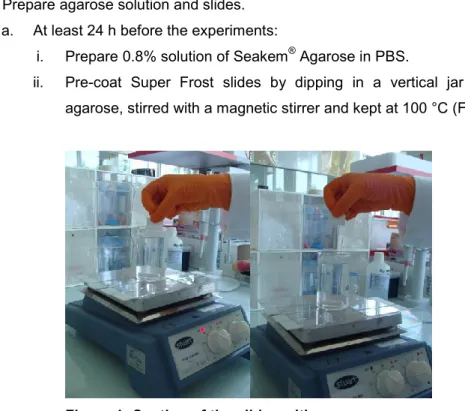
ii. Pre-coat Super Frost slides by dipping in a vertical jar containing melted agarose, stirred with a magnetic stirrer and kept at 100 °C (Figure 1).
Figure 1. Coating of the slides with agarose
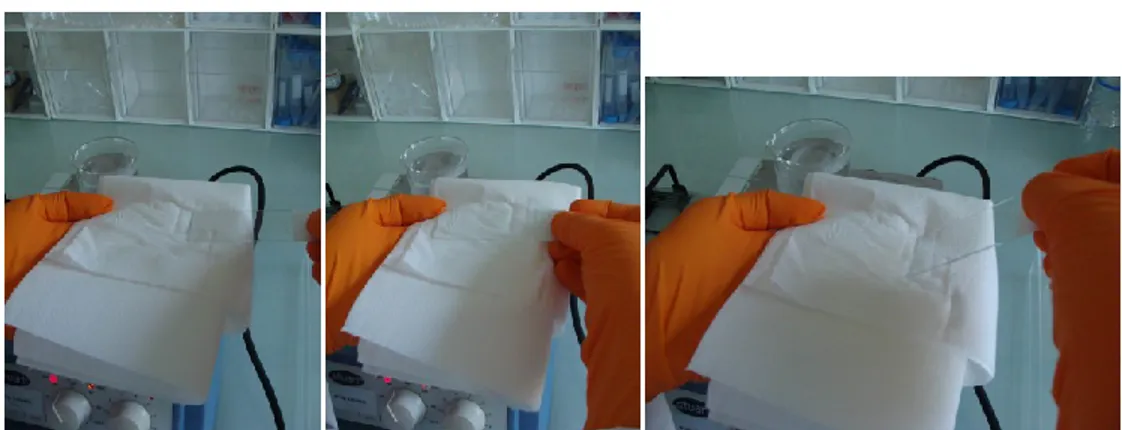
Figure 2. Slides coating with agarose. Removing of the agarose on the back of
the slides.
iv. Let the slides dry and then store at room temperature until use.
b. At least 2 h before the experiments: prepare 0.7% solution of LMP agarose in PBS and place it at 37 °C in a water-bath until use.
2. Prepare cells
The number of harvested cells can be adjusted according to the size of the cells. Cells could be numbered either by an automated cell counter or a counting chamber (e.g. Malassez chamber).
If cells used are adherent, cells must be carefully detached with trypsin/EDTA and isolated before centrifugation and further use.
3. Embed cells in LMP agarose (in a dark room):
a. After cell centrifugation, discard the supernatant and resuspend the pellet of cells (150,000 to 200,000 cells) by gently pipetting with 200 µl of 0.7% LMP agarose. b. Lay 65 µl of agarose containing the cells on each pre-coated glass slide. c. Immediately cover with a 24 x 32 mm coverslip.
d. Put the slide on an ice-pack for solidification during 5-10 minutes. e. Slide off the coverslip to remove it.
f. Finally cover with 80 µl of LMP agarose (top agarose layer) and cover again with a 24 x 32 mm coverslip.
g. Put the slide again on an ice-pack for solidification during 5-10 minutes. h. Remove the coverslip.
4. Lysis and electrophoresis (in a dark room):
a. Place the slides in lysis solution for at least 1 h at 4 °C. b. Wash three times for 5 min with the electrophoresis buffer.
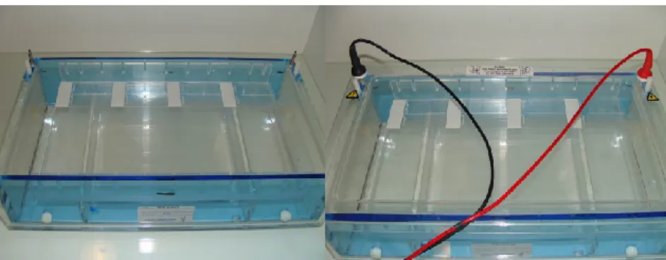
c. Transfer the slides in the electrophoresis tank filled with electrophoresis solution (Figure 3).
Figure 3. Electrophoresis of the slides. Disposition of the slides in the tank. In this
case eight slides are subjected to electrophoresis, four by row.
d. Proceed to electrophoresis at 18 V (0.5 V/cm) during 1 h. e. Wash in PBS for 2 x 5 min.
5. Dehydration, staining and analysis:
a. Fix the cells with 2 x 10 min washes in absolute ethanol, air-dry for at least 2 hours at room temperature.
b. Add 50 µl ethidium bromide (2 µg/ml in water) on the microscope slide and cover with a 22 x 22 mm coverslip for staining.
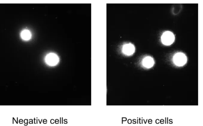
c. Analyze the cells: score 50 cells per slide, 2 slides per condition with the fluorescence microscope equipped with a camera and adapted software (Figures 4 and 5).
Figure 4. Representing analysis of each cell
a = head length b = tail length
% tail DNA = fraction of DNA in the tail Tail moment = % tail DNA x b
Comet tail length can be calculated by different ways depending on the authors. Tail moment is a common parameter used to characterize the comet. For this, the fraction
fluorescence (in the head and in the tail) to be expressed in percentage. Tail moment is the product of % tail DNA and tail length.
Figure 5. Images representing nucleus of undamaged cells, negative for the presence of comet (left panel) and nucleus of damaged cells presenting comet (right panel)
As a positive control, cells irradiated with ionizing radiations at 20 Gy and examined just after irradiation are a good control as shown in the photographs above.
Recipes 1. Lysis solution 2.5 M NaCl 0.1 M EDTA 10 mM Trizma base (pH 10) 1% N-laurylsarcosine 0.5% Triton X-100 10% DMSO final Keep at 4 °C 2. Electrophoresis solution 300 mM sodium acetate 100 mM Tris-HCl (pH 8.3) at 4 °C
This protocol was adapted from previously published papers (Courilleau et al., 2012; Olive et
al., 1990; Ostling and Johanson, 1984; Ostling and Johanson, 1987; Wojewodzka et al.,
2002).
References
1. Courilleau, C., Chailleux, C., Jauneau, A., Grimal, F., Briois, S., Boutet-Robinet, E., Boudsocq, F., Trouche, D. and Canitrot, Y. (2012). The chromatin remodeler p400 ATPase facilitates Rad51-mediated repair of DNA double-strand breaks. J Cell Biol 199(7): 1067-1081.
2. Olive, D. M., Johny, M. and Sethi, S. K. (1990). Use of an alkaline phosphatase-labeled synthetic oligonucleotide probe for detection of Campylobacter jejuni and Campylobacter
coli. J Clin Microbiol 28(7): 1565-1569.
3. Ostling, O. and Johanson, K. J. (1984). Microelectrophoretic study of radiation-induced DNA damages in individual mammalian cells. Biochem Biophys Res Commun 123(1):
291-298.
4. Ostling, O. and Johanson, K. J. (1987). Bleomycin, in contrast to gamma irradiation, induces extreme variation of DNA strand breakage from cell to cell. Int J Radiat Biol Relat
Stud Phys Chem Med 52(5): 683-691.
5. Wojewodzka, M., Buraczewska, I. and Kruszewski, M. (2002). A modified neutral comet assay: elimination of lysis at high temperature and validation of the assay with anti-single-stranded DNA antibody. Mutat Res 518(1): 9-20.