HAL Id: hal-03079334
https://hal.archives-ouvertes.fr/hal-03079334
Submitted on 17 Dec 2020
HAL is a multi-disciplinary open access
archive for the deposit and dissemination of
sci-entific research documents, whether they are
pub-lished or not. The documents may come from
teaching and research institutions in France or
abroad, or from public or private research centers.
L’archive ouverte pluridisciplinaire HAL, est
destinée au dépôt et à la diffusion de documents
scientifiques de niveau recherche, publiés ou non,
émanant des établissements d’enseignement et de
recherche français ou étrangers, des laboratoires
publics ou privés.
Phenotypic analysis of separation-of-function alleles of
MEI-41, Drosophila ATM/ATR.
Anne Laurençon, Amanda Purdy, Jeff Sekelsky, R Scott Hawley, Tin Tin Su
To cite this version:
Anne Laurençon, Amanda Purdy, Jeff Sekelsky, R Scott Hawley, Tin Tin Su. Phenotypic analysis
of separation-of-function alleles of MEI-41, Drosophila ATM/ATR.. Genetics, Genetics Society of
America, 2003, 164 (2), pp.589-601. �hal-03079334�
2003 by the Genetics Society of America
Phenotypic Analysis of Separation-of-Function Alleles of MEI-41,
Drosophila ATM/ATR
Anne Laurenc¸on,*
,1Amanda Purdy,
†Jeff Sekelsky,*
,2R. Scott Hawley*
,3and Tin Tin Su
†,4*Molecular and Cellular Biology Department, University of California, Davis, California 95616 and†MCD Biology,
University of Colorado, Boulder, Colorado 80309-0347
Manuscript received October 23, 2002 Accepted for publication March 11, 2003
ABSTRACT
ATM/ATR kinases act as signal transducers in eukaryotic DNA damage and replication checkpoints. Mutations in ATM/ATR homologs have pleiotropic effects that range from sterility to increased killing by genotoxins in humans, mice, and Drosophila. Here we report the generation of a null allele of
mei-41, Drosophila ATM/ATR homolog, and the use of it to document a semidominant effect on a larval
mitotic checkpoint and methyl methanesulfonate (MMS) sensitivity. We also tested the role of mei-41 in a recently characterized checkpoint that delays metaphase/anaphase transition after DNA damage in cellular embryos. We then compare five existing mei-41 alleles to the null with respect to known phenotypes (female sterility, cell cycle checkpoints, and MMS resistance). We find that not all phenotypes are affected equally by each allele, i.e., the functions of MEI-41 in ensuring fertility, cell cycle regulation, and resistance to genotoxins are genetically separable. We propose that MEI-41 acts not in a single rigid signal transduction pathway, but in multiple molecular contexts to carry out its many functions. Sequence analysis identified mutations, which, for most alleles, fall in the poorly characterized region outside the kinase domain; this allowed us to tentatively identify additional functional domains of MEI-41 that could be subjected to future structure-function studies of this key molecule.
I
N eukaryotes, genome stability is maintained in part In vitro studies have identified severalphosphoryla-tion targets of both ATM and ATR (Kim et al. 1999). by checkpoint pathways that monitor the state of
DNA and regulate the cell division cycle, activate DNA These include other proteins in the checkpoint path-ways such as homologs of CHK1 and CHK2 (Sanchez repair, or promote cell death as required. A central
place in DNA damage and replication checkpoints in et al. 1996; Cliby et al. 1998; Martinho et al. 1998;
Matsuoka et al. 1998, 2000). These studies led to a diverse eukaryotes is occupied by homologs of ATM/
ATR kinases. model in which ATM/ATR homologs act early in the checkpoint pathway to sense the presence of damaged The Schizosaccharomyces pombe homolog, Rad3, was
ini-tially identified in a screen for DNA damage sensitive or incompletely replicated DNA and relay this signal to the remainder of the checkpoint pathway via phosphory-mutants and later found to act in the DNA damage and
replication checkpoints (Al-Khodairy and Carr 1992; lation. As such, the kinase domain, which is at the C terminus, has been shown to be required for function Seaton et al. 1992). The Saccharomyces cerevisiae homolog
Mec1 was isolated as a gene essential for cell cycle pro- of both RAD3 and ATR ( Jimenez et al. 1992; Savitsky
et al. 1995a,b; Siede et al. 1996). For instance, mutations
gression (Kato and Ogawa 1994; Siede et al. 1996).
ATM was identified by studies of the human disease in the catalytic loop of the kinase affect all functions
known for RAD3 and ATR, creating dominant negative ataxia telangiectasia (AT; Savitsky et al. 1995a).
Addi-tional homologs include AtATM and AtRAD3 of Arabi- activities (Bentley et al. 1996; Cliby et al. 1998). A temperature-sensitive mutation in the kinase domain of
dopsis thaliana, Ce-atl-1 of Caenorhabditis elegans and
UVSB of Aspergillus nidulans. All are large proteins of rad3 disrupts the DNA damage checkpoint response
and other functions analyzed (Martinho et al. 1998). ⬎2500 amino acids (aa) that share little similarity
out-Despite its importance, the kinase domain by itself is side the kinase domain.
insufficient for function (Morgan et al. 1997; Chapman
et al. 1999).
Relatively little, however, is known about the function
1Present address: CGMC UMR 5534, Universite´ Claude Bernard
Lyon-1, 69622 Villeurbanne, France. of sequences outside the kinase domain in the PI3K-l
2Present address: Department of Biology, University of North Caro- family. In fission yeast, N-terminal sequences consisting lina, Chapel Hill, NC 27599-3280. of a leucine zipper and a putative protein-protein
inter-3Present address: Stowers Institute for Medical Research, 1000 E. 50th
action site called the P site can confer dominant
nega-St., Kansas City, MO 64110.
tive activity when overexpressed (Morgan et al. 1997;
4Corresponding author: MCD Biology, 347 UCB, University of
Colo-rado, Boulder, CO 80309-0347. E-mail: [email protected]. Chapman et al. 1999). A possible explanation for this
MATERIALS AND METHODS
result is that N-terminal sequences facilitate interaction
of PI3K-l proteins with their partners and therefore com- Stocks: Several mei-41 alleles were tested for lethality: A9 pete with the endogenous protein when overexpressed. and 12-1483 were homozygous lethal. A16, D18, and 12-3616 were semilethal. In the case of two lethal alleles and one
An ATM/ATR homolog in Drosophila is encoded by
semilethal allele (12-3616), we have been able to separate the
mei-41. mei-41 is essential for the DNA damage
check-lethality from the mei-41 locus by recombination. Additionally,
point in larval imaginal discs and neuroblasts and for
all heteroallelic combinations of semilethal alleles were fully
the DNA replication checkpoint in the embryo (Hari viable, suggesting that semilethality in mei-41A16and mei-41D18
et al. 1995; Brodsky et al. 2000; Garner et al. 2001). mutants is also due to mutations at other loci. Other alleles
were fully viable and fell into three classes with regard to
mei-41 also has an essential role during early nuclear
female fertility: A8, A11, D1, D2, D3, D4, D7, D8, D9, D10,
divisions in embryos, where it is required to delay mitosis
D11, and D18 were female sterile, with fertility ranging from
in response to incomplete DNA replication (Sibon et 0 to 5%. The second class showed fertility ranging from 5 to
al. 1999). Consistent with these functions, mei-41 mu- 15% and was composed of alleles A1, A7, A11, A12, A15, A16,
tants are sensitive to hydroxyurea, an inhibitor of DNA A22, A27, A28, A29, 12-3616, D19, RT1, and RT2. The last class of alleles, A10, D5, D9, D12, D13, D14, and D15,
ex-replication, and DNA-damaging agents such as X-ray
pressed fertility ranging from 50 to 98%. The deficiency
and alkylating agents (Boyd et al. 1976; Sibon et al.
Df(1)19 has previously been shown to be mei-41 deficient and
1999). MEI-41 also plays an important role during meio- Dp(1;4)r⫹f⫹, generated by Falk et al. (1984), has been shown
sis, where it is proposed to monitor double-strand-break to contain a wild-type mei-41 gene (Banga et al. 1995). Lethal repair during meiotic crossing over, to regulate the pro- mutations in genes l(1)14Ca, l(1)14Cc, l(1)14Cd, and l(1)14Ce were represented by deficiencies and alleles XR4, XR18, 11b8,
gression of prophase I, and to enforce metaphase I
i19e2, 4d2-5, and 4a47, which were provided by R. Stanewsky
delay observed at the end of oogenesis (Ghabrial and
(Stanewsky et al. 1993; Banga et al. 1995). Genes and
chromo-Schupbach 1999; McKim et al. 2000). somes used in this study but not described in the text can be All existing mei-41 mutant alleles were isolated in found in the Drosophila reference works (Lindsley and Zimm screens for mutants with meiotic defects, female sterility, 1992; FlyBase 2002).
Transposon mobilization:The screen to recover null alleles
or increased killing by genotoxins. Therefore, only
via-was conducted with the two P insertion alleles, mei-41RT1and
ble alleles would have been recovered (Baker and
Car-mei-41RT2 (Figure 2). Introduction of a P-element-encoding
penter 1972; Smith 1973; Boyd et al. 1976; Mohler transposase (denoted TMS) into these genetic backgrounds 1977). One of the strongest of these, mei-41D3, has been
should allow a screen for products of imprecise excision that removes flanking coding sequences. However, because
MEI-described as a null allele on the basis of the absence of
41 is required to repair the damage created by transposon
detectable protein and complete female sterility in these
mobilization, combination of a strong mei-41 mutant and a
mutants (Sibon et al. 1999). Our recent sequence
analy-source of transposase is usually lethal (Banga et al. 1991;
sis (see below), however, sheds doubt on whether it is our unpublished observations). Furthermore, newly induced a true null that would be a valuable tool for analysis of mutations at the mei-41 locus might cause lethality in germ
cells, in which case products of imprecise excision may not
MEI-41 function.
be recovered. To circumvent these difficulties, we used the
Here we report the generation of a null mutant of
duplication Dp(1;4)r⫹f⫹to provide a copy of the mei-41 gene
mei-41, which we find to be fully viable in the absence
on the fourth chromosome. Only mei-41RT1 females survived
of DNA damage and to show a semidominant effect on a the imposed genomic context. That is, Dp(1;4)r⫹f⫹rescued
larval mitotic checkpoint and methyl methanesulfonate the viability of mei-41/Df(1)19; TMS/⫹; Dp(1;4)r⫹f⫹/⫹ females
but not mei-41/Y; TMS/⫹; Dp(1;4)r⫹f⫹/⫹ males, for reasons
(MMS) sensitivity. We use the null allele to document
that remain unclear. Therefore, we performed the screen for
the role of mei-41 in a recently characterized checkpoint
imprecise excision in the female germline (Figure 2). To
in-that delays metaphase/anaphase transition in response
crease the frequency of P-element losses during the
experi-to DNA damage in cellular embryos (Su and Jaklevic ment, we provided a deficiency of the region on the homolo-2001). We then compared five existing mei-41 alleles gous chromosome. Without a sister chromatid as a template, repair of the double-strand breaks created by the transposase
to the null with respect to known phenotypes (female
provided in trans is less accurate (Engels et al. 1990;
Johnson-sterility, G2/M regulation after DNA damage in larvae,
Schlitz and Engels 1993). From the daughters of mei-41/
and sensitivity to MMS). Four of these alleles lack mei- Df(1)19; TMS/⫹; Dp(1;4)r⫹f⫹/⫹ females, a total of 454
chro-otic defects; thus, female sterility can be attributed to mosomes were recovered and analyzed for female sterility over the failure to regulate syncytial divisions where mei-41 mei-41D3allele. Lethal chromosome recovered (78B) was tested
for complementation with lethal mutations in l(1)14Ca,
is required to delay mitosis in response to incomplete
l(1)14Cc, l(1)14Cd, and l(1)14Ce. DNA replication. Interestingly, we find that not all
phe-Measurement of female fertility, recombination, and X
notypes are affected equally by each allele; thus some chromosome loss:During the screen for a null allele of mei-are separation-of-function alleles. Sequence analysis 41, female sterility was checked by allowing 10 females to mate identified mutations that revealed the importance of with 5–10 FM7a,f brothers, and the number of progeny (adult flies) were counted after 10 days. Other fertility
experi-N-terminal sequences and identified putative functional
ments as reported in Table 2 were conducted by crossing 5
domains of MEI-41. Our results support a model in
females to 5–10 wild-type (Canton-S) males. Eggs were
col-which mei-41 interacts with different sets of upstream lected for 24 hr and scored 24 hr later for hatching to deter-and downstream effectors to carry out its many func- mine the percentage of fertility. To estimate recombination
and X chromosome loss frequencies, w mei-41; net ho dp Sp b
pr cn/⫹ virgins were crossed to net ho dp b pr cn males. Crosses of 2⫻ SDS loading dye was added to the sample, which was
then boiled for 10 min before being subjected to electrophore-were scored for net, ho, dp, and b markers for recombination
frequencies and chromosome loss was estimated from the sis on 5% polyacrylamide gels. The proteins were blotted onto PDVF membranes using a semidry apparatus (Pharmacia Bio-number of (w⫹) males. Flies were raised on standard
corn-meal-molasses-agar medium and grown in an incubator at 25⬚. tech, Piscataway, NJ). The blots were blocked in PBT⫹ 5% milk before being probed with the primary antibody diluted
PCR and DNA sequencing:The nucleotide numbers refer
to the sequence of the 10.5-kb fragment containing the mei- (typically 1:300–500) in blocking solution. The membranes were washed in blocking solution and probed with a secondary
41 open reading frame (GenBank accession no. U34925).
Primer sequences (GIBCO BRL, Gaithersburg, MD) and PCR antibody (HRP-conjugated anti-rabbit antibody; Pharmacia Biotech) diluted 1:2000 in blocking solution. The membranes conditions are available upon request. Location of primers
were washed in PBT and processed for enhanced chemilumi-with respect to the mei-41 sequences shown in Figure 1A.
nescence detection according to manufacturer’s instructions Amplification of genomic DNA from mei-41RT1 and mei-41RT2
(Pierce, Rockford, IL). The migration of high-molecular-flies with primers Pout and⌬2-1 generated fragments of 900
weight markers (Amersham, Arlington Heights, IL) was used and 400 bp, respectively. Fragments digested with EcoRI-NotI
in a semilogarithmic plot of mobility vs. molecular weight to for mei-41RT1 and PstI-NotI for mei-41RT2 were cloned and
se-estimate the mass of the putative MEI-41 protein in Figure 5. quenced. P-element insertion creates a direct duplication of
G2/M checkpoint quantification in imaginal discs:Climbing
8 bp at its site of insertion (O’Hare and Rubin 1983). These
larvae were collected and sexed. Males from the mei-41/C(1) direct duplications have been localized at 3370 bp (GTTCA
DX/Y stocks or from the Canton-S control stock were irradi-TAC) and 3870 bp (GGCCAGCT) and share similarity with
ated on petri dishes with X rays from 137Cs source at 5-Gy
the described consensus (O’Hare and Rubin 1983).
dose (Astrophysics Research, Long Beach, CA). Irradiated and For mutant flies recovered from the described screen
(Fig-nonirradiated larvae were kept on grape agar plates for 1–2 ure 2), amplifications were performed with Pout:1964,
hr at 25⬚ and then dissected in phosphate-buffered saline Pout:⌬2-1, and Pout:2549 primer pairs to determine the
pres-(PBS; 0.13 m NaCl, 4 mm Na2HPO4, 3 mm NaH2PO4, pH 7.5).
ence of the P element on the chromosome. With 1964:⌬2-1,
The anterior portions of bisected larvae were inverted to ex-we checked for deletion in the vicinity of the insertion site.
pose the imaginal discs, fixed in 4% paraformaldehyde in PBS The 1964-⌬2-1 amplicon was double digested with SacI and
for 15 min, and washed in PBT (PBS⫹ 0.3% Triton X-100)
EcoRI after purification and cloned. DNA from a 29D/78B
for 1 hr. Discs were blocked for 1 hr in 5% bovine serum hybrid fly did not yield any signal with the 31000A:2549 couple.
albumin (albumin fraction V from United States Biochemical, Sequence matching the primer 31000A is located within the
Cleveland) in PBT and incubated for 2 hr (at room tempera-975 bp that is missing in the 29D allele; therefore the
defi-ture) or 12 hr (at 4⬚) in anti-phosphohistone H3 antibody ciency in 78B chromosome includes at least part of the
mei-(1:1000 in PBT; Upstate Biotechnology, Lake Placid, NY).
41 gene. We sequenced the following alleles in entirety:
mei-After another washing and blocking step, discs were incubated
41D3, mei-41D5, mei-41D9, mei-41D12, mei-41D13, mei-41D14, and mei-41D15.
for 2 hr (at room temperature) or 12 hr (at 4⬚) with HRP-PCR products generated from independent vials were used
conjugated secondary antibody (goat anti-rabbit at 1:500 in in sequencing. For each allele, we were able to identify single
blocking solution; Zymed, S. San Francisco). After another mutations that differ from background; these mutations are
washing step, 3,3⬘diaminobenzidine tetrahydrochloride solu-shown in Tables 2 and 3.
tion in NiCl was applied according to manufacturer’s
recom-MMS treatment:MMS sensitivity was quantified as follows.
mendation. The reaction was stopped by several rapid washes
mei-41/FM7 virgin females were crossed to mei-41 males to
in PBT. Discs were stained with a DNA dye, 6-diamidino-2-determine the sensitivity of homozygotes. mei-41/FM7a,f
vir-phenylindole dihydrochloride at 0.05g/ml. The discs were gin females were crossed to Canton-S to determine the
sensitiv-then further dissected to remove extraneous tissues and ity of heterozygous females. Eggs were collected in vials for
mounted in PBT solution. Slides were observed under visible 24 or 48 hr from 5–10 batches of five females and five males
and UV light on an axioplan Zeiss microscope. Ten or more each. After 24 hr at 25⬚, 250 l of MMS solution was applied
eye discs were scored to estimate the checkpoint defects, and to the food, and the progeny were allowed to develop. Several
data are presented ⫾SE. Chemicals were purchased from hundred flies and all classes of progeny were scored for each
Sigma (St. Louis) unless stated otherwise. dose.
Mitotic checkpoints in the embryo: To detect the G2/M Antibody production and Western blotting:To produce a
delay, embryos were collected for 10 min, aged for 325 min fragment of MEI-41, the largest EcoRI fragment of the mei-41 at 25⬚ to reach interphase of embryonic cycle 16 (Foe et al. genomic DNA inserted into the pCasper vector (predicted to
1993), irradiated, and allowed to rest for 20 min before fixing produce the last 402 aa) was subcloned into the unique EcoRI
in 37% formaldehyde for 5 min. To detect the metaphase/ site of the pET21b expression vector (Novagen, Cambridge,
anaphase delay, embryos were collected for 60 min and aged MA). The plasmid was transformed into Escherichia coli BL21 for 330 min at 25⬚ before fixing (for unirradiated controls) (DE3) strain and the protein was induced with isopropyl
thio-or aged fthio-or 310 min, irradiated, and allowed to rest fthio-or 40 galactoside according to manufacturer’s instructions
(Nova-min before fixing. Embryos were irradiated with 5.7 Gy of X gen). The expressed protein was gel purified and used to
rays in a TORREX X-ray generator (Astrophysics Research) immunize rabbits using a commercial facility (Cocalico
Biolog-set at 115 kV and 5 mA (producing 1.32 Gy/min). Fixed icals, Reamstown, PA). Antisera were purified against the anti- embryos were blocked in PBT containing 3% normal goat genic protein fragment immobilized on PDVF membranes serum and stained with the antiphosphohistone H3 antibody (Immobilon-P; Millipore, Bedford, MA). Plasmid maps and to visualize mitotic cells. Embryos were also counterstained detailed protocols for subcloning and antibody purification with the DNA dye, Hoechst 33258, at 10g/ml in PBT. are available upon request.
For Western blotting, embryos were dechorionated for 2 min in 50% bleach, rinsed in water, and homogenized in
RESULTS HEMG buffer (20 mm HEPES, pH 7.6, 5 mm MgCl2, 1.5 mm
EGTA, 10% glycerol) supplemented with protease and
phos-Generation of null alleles of mei-41 via internal
dele-phatase inhibitors (10 mm-glycerol phosphate, 1 mm
dithi-tions:The mei-41RT1allele contains a single
nonautono-othreitol, 0.2 mm phenylmethylsulfonyl fluoride, 5 g/ml
cod-ing region (Figure 1A; Yamamoto et al. 1990). Followcod-ing the mei-41D3allele, similar to mei-41D3/mei-41RT1control
fe-males. Thirty-three chromosomes were female fertile when mobilization of this transposon (materials and
meth-ods; Figure 2), 454 chromosomes were recovered and heterozygous with the mei-41D3allele and thus may
repre-sent revertants. Twenty-nine exhibited female sterility tested for female sterility when heterozygous with the
mei-41D3 allele. A total of 392 recovered chromosomes when heterozygous with mei-41D3. One of these 29, 78B,
carries a homozygous lethal mutation and was rescued caused reduced female fertility when heterozygous with
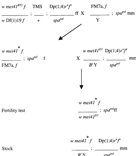
Figure 2.—The mating scheme to recover null alleles of mei-41. The mei-41RT1allele was combined
with a stable transposase source on the third chro-mosome, TMS. These females also carry Df(1)19, uncovering the mei-41 region, and a duplication of the same region on the fourth chromosome,
Dp(1;4)r⫹f⫹. Two females were mated with males carrying the FM7a,f balancer chromosome in 180 crosses. The minute phenotype associated with the Df allowed us to monitor X chromosomes in the progeny and the dominant Sb mutation on TMS, the third chromosome. To rescue any lethal chromosome generated, single virgin f, pol fe-males were mated individually to mei-41D3males
carrying the Dp(1;4)r⫹f⫹chromosome. Each cross is tagged to keep track of cluster events. The steril-ity of 10–20 w, pol daughters was analyzed with their Dp(1;4)r⫹f⫹/pol sisters as a control. From the same brood, males carrying the mutagenized chromosome were saved to raise stocks. Lines showing no pol males were carrying an X lethal mutation. *, mutagenized chromosome; mm, male; ff, female. See materials and methods for stock and genotype information.
by a chromosomal duplication, Dp(1;4)r⫹f⫹, that in- chromosome is missing 975 bp of mei-41 coding se-quence. Moreover, transposon excision was accompa-cludes mei-41. However, 78B fails to complement lethal
mutations at l(1)14Ce locus (alleles 4d25 and 4a27) and nied by the insertion of an extra A, creating a frameshift in the remaining coding sequence (Figure 1B). Conse-therefore is likely to carry a deficiency (materials and
methods). quently, the 40th codon is expected to encode a “stop,” leaving a truncated protein of 39 aa; the native mei-41 Genomic DNA from the above 29 mutants was
ampli-fied by PCR and analyzed. Three mutants, 29D, 99B, gene encodes 2347 aa. The 99B line presents an 8-bp direct duplication with most of the P-element sequence and deficiency-bearing 78B, were found to lack the P
element. Further sequence analysis showed that the 29D deleted, leaving behind 13 bp of 5⬘ inverted repeat (IR)
Figure 1.—Sequence analysis of mei-41 alleles. (A) In the map of the mei-41 locus, the insertion site of the P-element responsible for the mei-41RT1mutation is shown along with the primers used to screen for null alleles (materials and methods). The protein
is highlighted in three shades: white for the N terminus, gray for the rad3 domain, and black for the kinase domain. Asterisks indicate the site of unique mutations affecting D13, D9, D15, D14, D12, and D5. The nucleotide numbers refer to the sequence of the 10.5-kb fragment containing the 41 open reading frame (GenBank accession no. U34925). (B) Comparison of
mei-4129D, mei-4199B, and wild-type sequences. Predicted proteins are depicted at the same scale as full-length MEI-41 protein in A.
Wild-type (WT) nucleotide sequence is shown at the top. The 8-bp target site that was duplicated in 99B is italicized. Boldface nucleotides are filler sequences that became added upon transposon excision. Nucleotide numbers correspond to the genomic clone presented in A. The 29D allele contains up to nucleotide 2401 of the wild-type sequence (encoding the first 28 amino acids), followed by a 975-bp deletion. Wild-type sequence resumes at nucleotide 3376, but insertion of an A (in bold) results in a frameshift that produces a stop codon after 11 additional amino acids. (C) Phylogenic tree as calculated from C-terminal 360 amino acids, which correspond to the kinase domain, of the following proteins (GenBank accession numbers in parentheses): Ce-atl-1 (AB018598), UVSB (AF176575), RAD3 (CAA70297), ATR (U76308), MEI-41 (U34925), CG6535 (AE003708), MEC-1/ ESR1 (Z36005), ATM (U33841), AtATR (BAA92828), AtATM (AJ250248), Tel1S.p. (T41243), and Tel1S.c. (S45416). Maximum parsimony and neighbor-joining methods were used to investigate protein relationships. (D) Mutations in mei-41D9, mei-41D14, and
mei-41D15alleles. Helical wheel of the predicted␣-helix in which these mutations are located is shown. Hydrophobic amino acids
are italicized. (E) Alignment of a part of the rad3/TRAPP homologous domain in which the mei-41D12mutation is located. Amino
and 17 bp of the 3⬘ IR that are separated by nucleotides, CA. This rearrangement creates a stop codon down-stream of the insertion site and is predicted to encode a truncated protein of 340 aa (Figure 1B). 29D and 99B are thus null mutants; we refer to these new alleles as
mei-4129Dand mei-4199B.
Despite the severe nature of lesions, homozygous fe-males and hemizygous fe-males bearing mei-4129D or mei-4199Balleles are fully viable. Thus null alleles of mei-41
do not appear to act as zygotic lethal mutations. Note that the recovery of the 78B deficiency indicates that lethal alleles of mei-41 would have been recovered had they been induced. In mammals, ATR is an essential gene while ATM is not (Barlow et al. 1996; Xu and Baltimore 1996; Brown and Baltimore 2000). The Genome Project identified a second ATM/ATR homo-log in the Drosophila genome (CG6535), which is more closely related to ATM; MEI-41 is more closely related to ATR (Figure 1C). It is therefore interesting that
mei-41 appears to be nonessential while ATR is.
The phenotype of null alleles:Prior to further analy-sis, we first confirmed that newly generated null alleles exhibit phenotypes expected of strong mei-41 alleles. In assays for sensitivity to a genotoxin, MMS, doses as low as 0.01% killed 100% of homozygotes for the mei-4129D
and mei-4199B alleles. Moreover, mei-4129D homozygous
females are semisterile (97.3% of embryos fail to hatch at 25⬚ and 92% at 20⬚) and display meiotic defects such as reduction of recombination (13% of normal between
net and ho markers) and increased chromosome losses
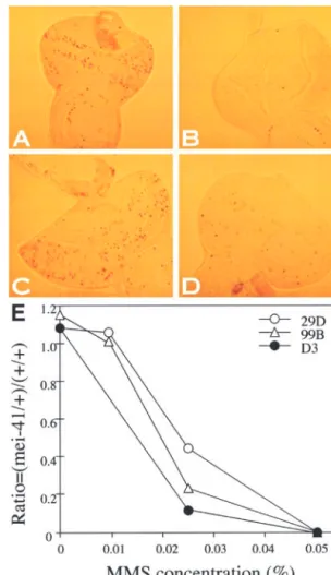
Figure 3.—DNA damage checkpoint and MMS sensitivity
and nondisjunction (10% X chromosome loss; control
in mei-41 mutant larvae. (A–D) Mitotic cells are visualized by
shows⬍1%). Likewise, the ability to delay the entry into
PH3 staining in larval eye discs. In wild-type (Canton-S) larvae,
mitosis upon DNA damage is disrupted in homozygous each disc contains 121 ⫾ 11.1 mitotic cells (A) while this
mei-4129D mutant larvae (Figure 3). These phenotypes
number decreased⬎10-fold to 9.2 ⫾ 1.4 after irradiation (B), indicating a delay in entry to mitosis in the latter. In mei-4129D
are identical to those caused by the strongest mei-41
homozygotes, each eye disc contains 156 ⫾ 10 mitotic cells
alleles isolated to date, namely D1, D3, and 195.
before irradiation (data not shown) and 127⫾ 21.3 mitotic
Boyd et al. (1976) reported a semidominant effect
cells after irradiation (C), indicating that these larvae are
on MMS sensitivity for at least one strong allele of mei- unable to block mitosis after irradiation. In mei-4129D heterozy-41. We find that mei-4129Dand mei-4199Balso exhibited a
gotes, each eye disc contains 170.3⫾ 14.4 mitotic cells before irradiation and this number decreased only 2-fold to 69.3⫾
semidominant effect on larval MMS sensitivity and the
13.3 after irradiation (D), indicating a partial loss of the ability
ability to block mitosis after DNA damage in larval discs
to block mitosis. (E) MMS sensitivity of mei-41 heterozygotes.
(Figure 3). However, female sterility does not appear
The ratio of heterozygous to wild-type females recovered at
to be dose dependent since one copy of the mei-41 different MMS concentrations is shown for three alleles. Ho-genomic DNA can rescue embryonic lethality to wild- mozygous mutants for all three alleles show complete lethality
at all MMS doses shown here except for 0% MMS (data not
type levels (Sibon et al. 1999).
shown).
The role of mei-41 in mitotic metaphase/anaphase delay:We next used mei-4129Dmutants to determine the
role of mei-41 in a recently defined metaphase
check-by about threefold in irradiated embryos (Su and Jak-point in embryos (Su and Jaklevic 2001). In embryonic
levic 2001; Figure 4). Metaphase/anaphase delay ap-cell cycles, DNA damage due to ionizing radiation or
pears to be due to stabilization of a mitotic cyclin, but MMS causes a delay in the entry into mitosis. After this
the role of checkpoint genes in this response has not delay, cells recover but subsequently delay in metaphase/
been addressed. anaphase transition (Su and Jaklevic 2001). This is
We find that the number of mitotic cells in the dorsal seen in live measurement of mitotic timing, where
dura-ectoderm was reduced at 20 min after irradiation in tion of metaphase increased about threefold, as well as
wild type and in homozygous mei-4129D embryos from
in quantification of mitotic phases in fixed embryos; the
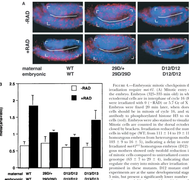
Figure 4.—Embryonic mitotic checkpoints that respond to irradiation require mei-41. (A) Mitotic entry checkpoint in the embryo. Embryos (325–335 min old) in which the dorsal ectodermal cells are in interphase of cycle 16 (Foe et al. 1993) were irradiated with 0 (⫺RAD) or 5.7 Gy of X rays (⫹RAD). Embryos were fixed 20 min later, when dorsal ectodermal cells should be in mitosis of cycle 16, and stained with an antibody to phosphorylated histone H3 to visualize mitotic cells (red). Embryos were also stained to visualize DNA (blue). Mitotic cells are counted in the dorsal ectoderm region, en-closed by brackets. Irradiation reduced the number of mitotic cells in wild type (WT; from 111⫾ 14 to 19 ⫾ 11) and mei4129D
homozygous embryos from heterozygous mothers (29D; from 103⫾ 9 to 16 ⫾ 5), indicating a delay in entry into mitosis. Irradiated mei41D12homozygous embryos (D12) from
homozy-gous mothers showed only twofold reduction in the number of mitotic cells compared to unirradiated controls of the same genotype (63⫾ 7 to 29 ⫾ 4), indicating that the ability to regulate the entry into mitosis after irradiation has been com-promised in these mutants. D12 mutant embryos in these experiments are at the same developmental stage as WT, 355⫾ 5 min, but present a significantly lower number of mitotic cells when untreated. We do not address this issue. (B) Metaphase/ anaphase checkpoint in mei-41 mutants. The ratio of the number of metaphase cells to the sum of anaphase and telophase cells in mitosis 16 in the dorsal ectoderm was quantified from unirradiated (⫺RAD) embryos or embryos that had been irradiated 40 min before fixing (⫹RAD). Cells in wild-type embryos delay metaphase-anaphase transition after irradiation, as indicated by a threefold increase in meta/(ana⫹telo) ratio. This increase is diminished in mei-41 mutants. Data are compiled from at least 3600 mitotic cells from at least 30 embryos for each genotype in two or three experiments. Metaphase/(aNa⫹telo) ratio was also measured at 20 min after irradiation in mei41D12mutants with similar results (not shown). mei-4129Dhomozygous embryos from
heterozygous mothers were identified by the lack of-galactosidase staining from the FM7, ftz:lacZ chromosome. mei-41D12and
mei-41D13embryos are from homozygous mutant mothers.
embryos from homozygous mei-41D12 mothers (Figure More important for this work, at longer times (40 min)
after irradiation when cells have recovered and entered 4A; see figure legend for mitotic indices). There are
several possible explanations for these observations. mitosis, the ratio of metaphase to {anaphase ⫹ telo-phase} increased in wild type; this increase was severely First, homozygous mei-4129Dembryos would have
inher-ited wild-type mei-41 gene products from their heterozy- diminished in mei-4129Dand mei-41D12embryos and
par-tially diminished in mei-41D13embryos (Figure 4B). Thus,
gous mothers, and the maternal product may persist
long enough to enforce the mitotic entry checkpoint; all three mei-41 alleles tested are defective in the meta-phase/anaphase checkpoint, implicating this gene in D12 embryos from homozygous D12 mothers, in
con-trast, would not have inherited any wild-type mei-41 gene the regulation of mitotic progression in response to X-ray damage.
products. It is also possible that mei-41 is dispensable
for the regulation of mitotic entry after X-ray damage mei-4129Dembryos have an intact mitotic entry
point but not an intact metaphase/anaphase check-in embryos or plays a redundant role with a second
TABLE 1
The phenotypes of mei-41 alleles studied
Mutation % loss of G2/M
MMS sensitivity % fertility checkpoint Nucleotide Amino acids
mei-4129D ⫹⫹⫹⫹ 3 100 Deletion at 2401 Stop after 39 aa
mei-41D3 ⫹⫹⫹⫹ 1 98 T3768A A384V mei-41D5 ⫹⫹⫹ 50 74 C8924T P2159L mei-41D9 ⫹⫹ 77 70 C4257T S647F mei-41D12 ⫹ 97 63 C8053T H1869Y mei-41D13 ⫹ 98 35 T2585A W90T mei-41D14 ⫹⫹ 55 15 G4339T L655F mei-41D15 ⫹⫹ 84 6 A4328C T652P
MMS sensitivity, female fertility, and loss of G2/M DNA damage checkpoint are shown for each allele, along
with unique nucleotide and amino acid changes. Data for the null allele, mei-4129D, are given as a reference.
For MMS sensitivity,⫹⫹⫹⫹, ⫹⫹⫹, ⫹⫹, and ⫹ refer to complete lethality at 0.01%, 0.025%, 0.05%, and 0.08%, respectively. Female fertility is expressed as percentage of eggs from homozygous mutant females that hatch into larvae. Loss of G2/M checkpoint represents the average number of mitotic cells per eye disc after
exposure of male hemizygous mutant larvae to 500 R of X rays expressed as percentage of the same number before irradiation. WT values range from 5 to 15%.
sensitive to the level of maternal mei-41 gene products (named in materials and methods) because they be-have as wild type in two standard assays for meiotic that persists or because maternal mei-41 products are
able to substitute for zygotic mei-41 products in the first functions, namely X chromosome nondisjunction fre-quency and meiotic recombination levels (Mason et al. checkpoint but not in the second. Thus, the data
pre-sented here implicate mei-41 in the regulation of meta- 1981, 1989). Thus, we can rule out the contribution of meiotic defects to female sterility. The sixth, D5, was phase/anaphase transition after DNA damage but do
not conclusively address the role of mei-41 in the embry- selected because it affected partially all mei-41 pheno-types described herein (Table 1); mutant females are onic mitotic entry checkpoint. Nonetheless, metaphase/
anaphase regulation is defective in mei-41 mutants that meioticaly impaired, showing 5% of X nondisjunction. Finally, D3 was selected because it behaved as a null are either able (29D) or unable (D12) to regulate the
entry into mitosis. This result excludes the possibility and we thus expected to find a mutation abolishing all
mei-41 functions. Additionally, because all 7 alleles were
that metaphase/anaphase defects are due to variations
in the time of entry into mitosis between wild type and generated during a single EMS mutagenesis, we rea-soned that they will share a common background and mutants.
The phenotype of partial loss-of-function mutants: therefore sequence analysis would reveal mutations af-fecting mei-41 functions. Results of phenotypic compari-ATR homologs act as signal transducers in DNA
replica-tion and damage checkpoint pathways. The presence of son are presented in Figure 4B for metaphase/anaphase delay (in 3 alleles) and summarized in Table 1 for other DNA defects activates ATR homologs, which are kinases,
which in turn carry out their function by phosphoryla- phenotypes.
A comparison of mei-41 alleles leads us to two conclu-tion of downstream substrates. Drosophila ATM/ATR,
MEI-41, when mutated, leads to a number of phenotypes sions. First, not all phenotypes are affected equally by each allele. For instance, D12 and D13 alleles present as described above, ranging from female sterility to MMS
sensitivity. If the same checkpoint pathway and compo- wild-type levels of female fertility but defective meta-phase/anaphase checkpoints in the embryo (Figure 4) nents are at work in processes whose failure generates
these phenotypes, we might expect that an allele that and defective mitotic checkpoints in larval discs (Table 1). Conversely, the larval mitotic checkpoint is as robust affects one phenotype will affect others to a similar
extent. If, on the other hand, the function of mei-41 in D15 as in wild type, even though D15 mutants are MMS sensitive and partially female sterile. Second, an is being executed via different partners/substrates in
different processes, we might expect that an allele that allele that is more defective than another with regard to one phenotype is not necessarily so with regard to affects one phenotype may not necessarily affect
an-other. another phenotype. For instance, the D14 allele is more severe than D9 in female fertility but is less severe than To address these possibilities, we compared the
phe-notypes of 7 previously isolated alleles to each other D9 in the mitotic checkpoint in larval discs. Likewise, D12 shows less severe female fertility and MMS sensitivity and to the null allele we generated. Five (D9, D12, D13,
TABLE 2 TABLE 2
Neutral mutations (Continued)
mei-41 alleles mei-41 alleles Nucleotide D3 D5 D9 D12 D13 D14 D15 Nucleotide D3 D5 D9 D12 D13 D14 D15 2824 G G T T T T G T 2413 T T T C C C T C 3445 C C C T C C C T 3364 C C T T T T C T 3424 C C T T T T C T 3622 C T C T C C T T 4678 C C G C G G C C 3529 G G T T T T G T 3821 C C A A A A C A 4681 T T T T G T T T 4857 G G A G A A G G 3916 G G A A A A G A 4513 A A G G G G A G 5052 A A A A A A G A 5103 G G G G G G T G 4541 T T C C C C T C 4549 T T C C C C T C 5136 G G G A G G G A 5316 A A A T A A A T 4626 G T G G G G T G 5133 T T G G G G T G 5400 G G G A G G G A 5577 C C T C T T C C 5397 C A C C C C A C 6078 G A G G G G A G 5625 T T C T C C T T 5721 A A G A G G A A 6090 G A G G G G A G 6195 T C T T T T C T 5904 C T C T C C T T 7044 G G T T T T T T 6210 A C A A A A C A 6255 T C T T T T C T 7164 A G A G A A G G 7440 G T G G G G G T 6927 T T C C C C T C 7033 T G T T T T G T 7599 G G G G A G G G 7602 T G T T T T T G 7230 T C T T T T C T 7701 T C T T T T C C 7881 G A G G G G G G 7896 C G C C C C G G 7848 C T C C C C T T 7938 T C T T T T C T 8199 C C T C T T C C 8256 C C G C C C G C 8043 T C T T T T C T 8310 T T C T C C T T (continued) 8376 T C C T C C C T 8445 G A A G A A A G 8946 C C T C T T C C 9006 G T T G T T T G
mitotic checkpoint regulation. These observations raise
9066 C T T C T T T C
the possibility that some aspects of the mei-41 phenotype 9162 C T T C T T T C may reflect the specific mutational ablation of domains 9381 A G G A G G G A critical for a specific function. To address this, we deter- 9420 T A A T T T A T
9517 T T C T C C T T
mined the sequence of the mutant alleles.
DNA sequence changes in partial loss-of-function al- Neutral mutations in mei-41 alleles fall into two groups: leles:Sequence analysis revealed several differences be- those in the top half of the table show nucleotide variations tween mutant alleles and the published mei-41 sequence. common to D3 and D14 alleles but not shared by D5, D9, D12, D13, and D15 alleles. All alleles show the following mutations
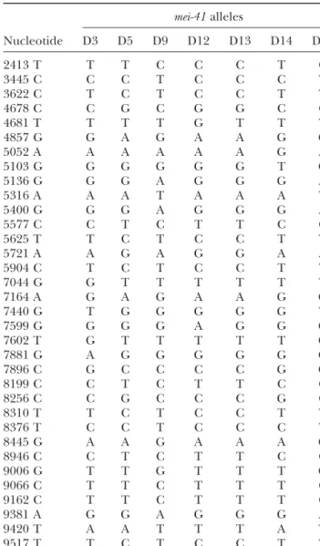
We found 70 silent mutations, 12 of which are shared
compared to the GenBank accession no. U34925 genomic
by all seven mutant alleles (Table 2), and 10 mutations
sequence: 2857, T→ C; 3934, A → T; 4069, G → A; 4261, C →
leading to amino-acid changes, all of which are shared T; 4974, A→ G; 5157, G → T; 5817, A → G; 6735, A → C; by two or more alleles (Table 3). These deviations could 6750, A→ G; 7002, A → G; 7008, T → A; and 7011, A → C. be due to variability originally present in the
mutagen-ized population (as indicated by common mutations within each of two different groups: D3 and D14 in one
group and D5, D9, D12, D13, and D15 in the other; is conserved among all members of the PI3K-L family except for UVSB (which has a T) and Ce-atl-1 (which Table 2) and/or accumulation of mutations in mei-41
mutants. In addition to these common mutations, a has an A). mei-41D5shows a partial defect in all assayed
phenotypes, suggesting that kinase activity of MEI-41 is single unique mutation was found for each allele and
may account for the phenotype. For mei-41D3, a T→ A important for all its functions. Likewise, mutations in
the kinase domain affect all functions known for RAD3 change at nucleotide 3768 is predicted to convert a
nonpolar A to a nonpolar V. We postulate additional and ATR (Bentley et al. 1996; Cliby et al. 1998; Mar-tinho et al. 1998).
mutations in the noncoding region for mei-41D3because
we detect very little protein in these mutants (Sibon et Interestingly, unique mutations in the other five al-leles analyzed here fall outside of the kinase domain.
al. 1999; Figure 5). The mei-41D5 mutation changes
TABLE 3
Mutations that are predicted to cause amino acid changes
mei-41 alleles Nucleotide D3 D5 D9 D12 D13 D14 D15 3096 S→ F S→ F S→ F S→ F S→ F 3210 Q→ R Q→ R Q→ R Q→ R Q→ R 3332 H→ Y H→ Y H→ Y H→ Y H→ Y 3493 E→ D E→ D 7126 L→ V L→ V 2345 L→ I L→ I L→ I 2580 A→ V A→ V A→ V 2585 W→ T 3452 N→ H N→ H N→ H N→ H N→ H N→ H 3768 A→ V 4257 S→ F 4328 T→ P 4339 L→ F 7606 A→ P A→ P 8053 H→ Y 8188 L→ G L→ G L→ G L→ G L→ G 8924 P→ L
Predicted amino acid changes in D3, D5, D9, D12, D13, D14, and D15 alleles are shown. Entries in the top part show variations differentiating D3 and D14 from the other alleles. Single amino acid changes that are unique to each allele are underlined. Mutations common to seven alleles are: 2840, L→ M; 2861, Y → H; 3002, M→ L; 3530, G → S; 3965, A → T; and 4166, L → V. Standard one-letter code for amino acids is used.
DISCUSSION
similarity outside of the kinase domain. The exception is
a computationally defined region ofⵑ500 amino acids, A large body of work indicates that ATM/ATR homo-called the FAT domain that is shared with the TRRAP logs play a central role in DNA checkpoints in eukary-family of proteins (Bosotti et al. 2000). This domain otes. Loss of ATM/ATR function leads to pleiotropic is a part of the rad3 domain, which is conserved only defects, which in humans range from sterility and men-in a subgroup that men-includes Mec1p, Rad3, ATR, and tal retardation to increased killing by ionizing radiation. MEI-41, but not ATM. We find that the mei-41D12
muta-The diversity of these phenotypes is recapitulated in tion affects a conserved histidine within the rad3/FAT model organisms such as Drosophila, which are amena-domain (Figure 1E). mei-41D13 is a mutation in the N
ble to genetic manipulation and provide an opportunity terminus where an algorithm designed to detect DNA- to understand the function of these key molecules. A binding domains detects a putative helix-turn-helix, question of interest is whether the pleiotropic nature I82MWSAVAKWLDMGCMTRQELKR (Network Protein
of mutations in ATM/ATR homologs is due to the par-Sequence Analysis; Dodd and Egan 1990). We find that ticipation of these molecules in different molecular
mei-41D9, mei-41D14, and mei-41D15affect a predicted␣-helix
pathways or their participation in a single signal trans-in the N termtrans-inus (Figure 1D). The mei-41D14
and mei- duction pathway that is important for several cellular
41D15mutations change, respectively, a leucine to a
phe-processes. Our identification of several alleles of mei-41 nylalanine and a threonine to a proline and are likely that affect some phenotypes more severely than others to disorganize the putative ␣-helix (Table 1). mei-41D9
is consistent with the function of mei-41 in several molec-mutants show a more severe defect in larval mitotic ular contexts.
checkpoint than do mei-41D14and mei-41D15mutants; this
ATR homologs act to stall mitosis in response to two suggests that the predicted change of a reactive serine647
types of DNA defects, namely, incompletely replicated to a hydrophobic phenylalanine in mei-41D9may not only
DNA and damaged DNA. In Drosophila, mei-41 is re-destabilize the putative helix but also specifically disrupt quired to stall mitosis when DNA replication is blocked interaction(s) in which this helix participates. In sum, experimentally during embryonic cleavage divisions the sequence analysis of partial loss-of-function alleles (Sibon et al. 1999), which occur in a syncytium and are suggests the importance of N-terminal sequences and driven by maternally supplied gene products. Embryos identifies mutations that presumably affect one pheno- from homozygous mothers of strong mei-41 alleles such as D3 and 29D do not progress beyond syncytial cycles type more severely than another.
tion while retaining nearly wild-type activity for larval mitotic checkpoints. Such alleles should be potentially useful for identifying genes that interact with mei-41 in one context but not another. Additionally, all five alleles (i.e., all except D3 and D5) show a wild-type level of meiotic function as previously described and yet show defects in mitotic cycles during embryogenesis and lar-val development. Thus, all represent separation-of-func-tion alleles that have normal meiotic funcseparation-of-func-tion but are defective for regulation of mitotic proliferation.
Interestingly, mei-41D15 mutants that have a wild-type
level of mitotic checkpoint are more MMS sensitive than
mei-41D12mutants that have a severely defective mitotic
checkpoint. Likewise, mei-41D14and mei-41D15mutants show
significantly different levels of female fertility (thus, pos-sibly different DNA replication checkpoint activity) and yet have similar MMS sensitivity. We propose that defects in cell cycle regulation cannot fully explain the MMS
Figure 5.—Western blot analysis of mei-41D3mutants.
Ex-sensitivity of mei-41 mutants. It is likely that the role of
tracts from 1- to 5-hr-old embryos from homozygous mei-41D3
mutant mothers (D3/D3) and heterozygous mei-41D3/FM7 mu- mei-41 in DNA repair, cell death, and other
yet-to-be-tant mothers (D3/⫹) or 0- to 2-hr-old wild-type Sevelen (WT) characterized processes contributes to MMS sensitivity. embryos were analyzed by Western blotting using an affinity- One key question concerning checkpoint proteins is purified antibody against MEI-41. Equal loading among lanes
whether they are essential for viability in unperturbed
was ensured by controlling for the number of
embryo-equiva-cell cycles or they are essential only in the presence of
lent material loaded (100 per lane). On the basis of the
loca-tion of size markers (materials and methods), the band genetic aberrations. Some PI3K-l proteins are essential
that is present in WT and mei-41D3heterozygotes and absent
for cellular viability, while others are not. mec1 deletion
in mei-41D3homozygotes is calculated to have a MW of 242 kD
mutants of budding yeast are inviable and ATR mutant
(arrowhead) and is presumed to represent the MEI-41 protein
mice die after the blastocyst stage (Kato and Ogawa
(predicted MW of 258 kD).
1994; Brown and Baltimore 2000). However, the S.
pombe rad3⌬ strain, in which rad3 is disrupted, is viable
and the ATM mutations observed to date in mice are (Sibon et al. 1999). Therefore, in the absence of meiotic
defects, female sterility may be attributed to the failure fully viable ( Jimenez et al. 1992; Seaton et al. 1992; Barlow et al. 1996; Elson et al. 1996; Xu and Balti-of syncytial divisions. Syncytial division defects have
been proposed to occur due to a failure to delay mitosis more 1996). We find that null mutants of mei-41 are viable as homozygous mutant flies are produced from in the presence of ongoing DNA replication during
these rapid division cycles (Sibon et al. 1999). heterozygous parents. It is possible, however, that homo-zygous mei-41 mutant progeny survive due to a supply According to the above discussion, mei-41D12and
mei-41D13mutants, which present a wild-type level of syncytial of wild-type MEI-41 deposited into the eggs by
heterozy-gous mothers. Because embryos from homozyheterozy-gous mu-division function (because they are female fertile) but
are unable to regulate metaphase/anaphase in cellu- tant females fail to progress beyond cleavage divisions (Sibon et al. 1999), mei-41 does have an essential role larized embryos or G2/M transition in embryos and
lar-vae following DNA damage, may be responsive to incom- in early embryogenesis.
Interestingly, heterozygotes for the null allele of mei-pletely replicated DNA replication but not to damaged
DNA. This could be because syncytial divisions simply 41 show checkpoint defects and MMS sensitivity.
Hetero-zygous phenotypes have been described for ATM-defi-require less MEI-41 activity than larval and embryonic
checkpoints do. Indeed, 29D heterozygotes, which pre- cient cells of human and mouse, including increased sensitivity to killing by mutagens, defective cell cycle sumably have wild-type MEI-41 but at reduced levels,
have normal female fertility but anⵑ50% loss of larval checkpoints, and chromosome aberrations, among oth-ers (Naeim et al. 1994; Scott et al. 1994; Tchirkov et G2/M checkpoint (Figure 3 legend). If so, however, we
would expect mei-41D14mutants that show a more severe al. 1997; Djuzenova et al. 1999). In mice, heterozygous ATR ES cells did not display increased sensitivity to
DNA-fertility phenotype to be more defective than mei-41D12
or mei-41D13mutants for larval mitotic checkpoint. This damaging agents although other phenotypes such as
cell cycle regulation remain to be assayed (de Klein et is not the case. Therefore, we propose, instead, that
mei-41D12and mei-41D13represent separation-of-function al. 2000). Haplo-insufficiency of ATM/ATR homologs
for checkpoint regulation may explain why heterozy-alleles that retained normal activity for syncytial
divi-sions but not for DNA damage checkpoints. Conversely, gous mutant mice display increased tumor incidence (Brown and Baltimore 2000). This notion remains
func-controversial, however, because recent work suggests point and syncytial divisions. Two other mutations lo-cated in a putative helix-loop-helix in the N terminus that the ATM heterozygotes in question may harbor a
mutant allele that acts in a dominant negative manner and a conserved amino acid in the rad3/FAT domain may be causing defective DNA-damage checkpoint while to inhibit the remaining wild-type allele (Spring et al.
2002). Our null allele, mei-4129D, is predicted to encode sparing syncytial division functions. These possibly
rep-resent separation-of-function alleles of mei-41, which only 39 aa and is therefore unlikely to produce a
domi-nant negative MEI-41. As such, MEI-41 may be truly may be useful in screens for interacting genes. The fact that not all phenotypes are affected equally by the five haplo-insufficient for optimal checkpoint regulation.
We document here a novel role for mei-41 in the alleles we studied is consistent with the idea that mei-41 operates in many different molecular contexts to carry regulation of metazoan mitotic progress. DNA damage
blocks mitosis but the exact mitotic step blocked can out its many functions. There is precedent for this idea because grp, a Drosophila chk1 homolog, that is thought differ from cell type to cell type (Elledge 1996). In
fission yeast, the entry into mitosis is blocked whereas to function downstream of mei-41, appears to do so in regulation of mitosis but not of meiosis (Sibon et al. in budding yeast, chromosome segregation and
meta-phase/anaphase transition are blocked. Drosophila and 1999; McKim et al. 2000). Our data suggest that even in mitotic cycles, signaling networks in which mei-41 human cells, on the other hand, appear capable of
blocking both the entry into mitosis and the metaphase/ participates may not be rigid, but change at different stages in development (syncytial vs. larval) or in re-anaphase transition (Smits et al. 2000; Su et al. 2000;
Su and Jaklevic 2001). The role of mei-41 in blocking sponse to different kinds of DNA defects (DNA damage
vs. incomplete replication).
the entry into mitosis after DNA damage has been
docu-mented before, but this report documents for the first We thank Katherine Hollis and Ginger Elkins for technical assis-time that an ATM/ATR homolog is needed to block tance. This work was supported by grants from the American Cancer Society and the AT Children’s Project to R.S.H. and a grant from the
mitotic progression in metazoa. Moreover, our results
National Institutes of Health (R01 GM66441) and funding from the
rule out the possibility that damaged chromosomes
pres-Cancer League of Colorado to T.T. S.A.P. was supported by a
predoc-ent a physical barrier to their separation and conse- toral training grant from the National Institutes of Health. quently delay anaphase. Rather, the delay of anaphase
is more likely to be an active response since it requires
a checkpoint gene. LITERATURE CITED
We describe here several mutations that fall outside of
Al-Khodairy, F., and A. M. Carr, 1992 DNA repair mutants
defin-the kinase domain but appear to affect mei-41 function ing G2 checkpoint pathways in Schizosaccharomyces pombe.
EMBO J. 11: 1343–1350.
profoundly. Although unique mutations described here
Baker, B. S., and A. T. Carpenter, 1972 Genetic analysis of sex
are most likely culprits for the phenotype of each allele,
chromosomal meiotic mutants in Drosophila melanogaster. Genetics
we cannot rule out the contribution of other mutations 71:255–286.
Banga, S. S., A. Velazquez and J. B. Boyd, 1991 P transposition
present within or without the mei-41 coding region.
Site-in Drosophila provides a new tool for analyzSite-ing postreplication
directed mutagenesis to obliterate domains implicated
repair and double-strand break repair. Mutat. Res. 255: 79–88.
by our data would be valuable in addressing this issue Banga, S. S., A. H. Yamamoto, J. M. Mason and J. B. Boyd, 1995
Molecular cloning of mei-41, a gene that influences both somatic
unequivocally. Nonetheless, in fission yeast, N terminus
and germline chromosome metabolism of Drosophila
melanogas-of RAD3 and, in particular, a putative leucine zipper
ter. Mol. Gen. Genet. 246: 148–155.
and a putative protein-protein interaction domain can Barlow, C., S. Hirotsune, R. Paylor, M. Liyanage, M. Eckhaus et al., 1996 Atm-deficient mice: a paradigm of ataxia
telangiecta-confer dominant functions when overexpressed
(Chap-sia. Cell 86: 159–171.
man et al. 1999). In humans, N-terminal 247 aa of ATM Bentley, N. J., D. A. Holtzman, G. Flaggs, K. S. Keegan, A. Demag-are required for interaction with p53 in vitro (Khanna gio et al., 1996 The Schizosaccharomyces pombe rad3
check-point gene. EMBO J. 15: 6641–6651.
et al. 1998). Thus, N-terminal sequences may contribute
Bosotti, R., A. Isacchi and E. L. Sonnhammer, 2000 FAT: a novel
to the function of ATM/ATR family members via
pro-domain in PIK-related kinases. Trends Biochem. Sci. 25: 225–227.
tein-protein interaction. Interestingly, in two-hybrid assays, Boyd, J. B., M. D. Golino, T. D. Nguyen and M. M. Green, 1976 Isolation and characterization of X-linked mutants of Drosophila
a MEI-41 N-terminal fragment containing this helix
in-melanogaster which are sensitive to mutagens. Genetics 84: 485– teracts with MUS304, a protein needed for larval DNA 506.
damage checkpoint and a homolog of mammalian Brodsky, M. H., J. J. Sekelsky, G. Tsang, R. S. Hawley and G. M. Rubin, 2000 mus304 encodes a novel DNA damage checkpoint
ATRIP proteins (Brodsky et al. 2000; M. Brodsky,
per-protein required during Drosophila development. Genes Dev.
sonal communication). 14:666–678.
In conclusion, this study demonstrates that mei-41 null Brown, E. J., and D. Baltimore, 2000 ATR disruption leads to chromosomal fragmentation and early embryonic lethality.
mutants are viable and show dose-sensitive defects in
Genes Dev. 14: 397–402.
cell cycle checkpoints and MMS sensitivity. Sequence Chapman, C. R., S. T. Evans, A. M. Carr and T. Enoch, 1999 Re-analysis reveals the importance of the kinase domain in quirement of sequences outside the conserved kinase domain of fission yeast Rad3p for checkpoint control. Mol. Biol. Cell 10:
all aspects of MEI-41 function and identifies a putative
3223–3238.
␣-helix in the N terminus, which may be important for Cliby, W. A., C. J. Roberts, K. A. Cimprich, C. M. Stringer, J. R.
Lamb et al., 1998 Overexpression of a kinase-inactive ATR
check-tein causes sensitivity to DNA-damaging agents and defects in McKim, K. S., J. K. Jang, J. J. Sekelsky, A. Laurenc¸on and R. S. Hawley, 2000 mei-41 is required for precocious anaphase in cell cycle checkpoints. EMBO J. 17: 159–169.
de Klein, A., M. Muijtjens, R. Van Os, Y. Verhoeven, B. Smit et Drosophila females. Chromosoma 109: 44–49.
Mohler, J. D., 1977 Developmental genetics of the Drosophila egg.
al., 2000 Targeted disruption of the cell-cycle checkpoint gene
ATR leads to early embryonic lethality in mice. Curr. Biol. 10: I. Identification of 59 sex-linked cistrons with maternal effects on embryonic development. Genetics 85: 259–272.
479–482.
Djuzenova, C. S., D. Schindler, H. Stopper, H. Hoehn, M. Flentje Morgan, S. E., C. Lovly, T. K. Pandita, Y. Shiloh and M. B. Kastan, 1997 Fragments of ATM which have dominant-negative or
com-et al., 1999 Identification of ataxia telangiectasia heterozygotes,
a cancer-prone population, using the single-cell gel electrophore- plementing activity. Mol. Cell. Biol. 17: 2020–2029.
Naeim, A., C. Repinski, Y. Huo, J. H. Hong, L. Chessa et al., 1994 sis (Comet) assay. Lab. Invest. 79: 699–705.
Dodd, I. B., and J. B. Egan, 1990 Improved detection of helix-turn- Ataxia-telangiectasia: flow cytometric cell-cycle analysis of lymph-oblastoid cell lines in G2/M before and after gamma-irradiation. helix DNA-binding motifs in protein sequences. Nucleic Acids
Res. 18: 5019–5026. Mod. Pathol. 7: 587–592.
O’Hare, K., and G. M. Rubin, 1983 Structures of P transposable Elledge, S. J., 1996 Cell cycle checkpoints: preventing an identity
crisis. Science 274: 1664–1672. elements and their sites of insertion and excision in the Drosoph-ila melanogaster genome. Cell 34: 25–35.
Elson, A., Y. Wang, C. J. Daugherty, C. C. Morton, F. Zhou et al.,
1996 Pleiotropic defects in ataxia-telangiectasia protein-defi- Sanchez, Y., B. A. Desany, W. J. Jones, Q. Liu, B. Wang et al., 1996 Regulation of RAD53 by the ATM-like kinases MEC1 and TEL1 cient mice. Proc. Natl. Acad. Sci. USA 93: 13084–13089.
Engels, W. R., D. M. Johnson-Schlitz, W. B. Eggleston and J. Sved, in yeast cell cycle checkpoint pathways. Science 271: 357–360. Savitsky, K., A. Bar-Shira, S. Gilad, G. Rotman, Y. Ziv et al., 1995a 1990 High-frequency P element loss in Drosophila is homolog
dependent. Cell 62: 515–525. A single ataxia telangiectasia gene with a product similar to PI-3 kinase. Science 268: 1749–1753.
Falk, D. R., L. Roselli, S. Curtiss, D. Halladay and C. Klufas,
1984 The characterization of chromosome breaks in Drosophila Savitsky, K., S. Sfez, D. A. Tagle, Y. Ziv, A. Sartiel et al., 1995b The complete sequence of the coding region of the ATM gene melanogaster. I. Mass isolation of deficiencies which have an end
point in the 14A–15A region. Mutat. Res. 126: 25–34. reveals similarity to cell cycle regulators in different species. Hum. Mol. Genet. 4: 2025–2032.
FlyBase, 2002 The FlyBase database of the Drosophila genome
projects and community literature. Nucleic Acids Res. 30: 106– Scott, D., A. R. Spreadborough and S. A. Roberts, 1994 Radia-tion-induced G2 delay and spontaneous chromosome aberrations 108.
Foe, V. E., G. M. Odell and B. A. Edgar, 1993 Mitosis and morpho- in ataxia-telangiectasia homozygotes and heterozygotes. Int. J. Radiat. Biol. 66: S157–S163.
genesis in the Drosophila embryo, pp. 149–300 in The Development
of Drosophila melanogaster, edited by M. Bate and A. Martinez Seaton, B. L., J. Yucel, P. Sunnerhagen and S. Subramani, 1992 Isolation and characterization of the Schizosaccharomyces Arias. Cold Spring Harbor Laboratory Press, Cold Spring
Har-bor, NY. pombe rad3 gene, involved in the DNA damage and DNA synthe-sis checkpoints. Gene 119: 83–89.
Garner, M., S. Van Kreeveld and T. T. Su, 2001 mei-41 and bub1
block mitosis at two distinct steps in response to incomplete DNA Sibon, O. C., A. Laurenc¸on, R. Hawley and W. E. Theurkauf, 1999 The Drosophila ATM homologue Mei-41 has an essential replication in Drosophila embryos. Curr. Biol. 11: 1595–1599.
Ghabrial, A., and T. Schupbach, 1999 Activation of a meiotic checkpoint function at the midblastula transition. Curr. Biol. 9: 302–312.
checkpoint regulates translation of Gurken during Drosophila
oogenesis. Nat. Cell Biol. 1: 354–357. Siede, W., J. B. Allen, S. J. Elledge and E. C. Friedberg, 1996 The Saccharomyces cerevisiae MEC1 gene, which encodes a homolog Hari, K. L., A. Santerre, J. J. Sekelsky, K. S. McKim, J. B. Boyd et
al., 1995 The mei-41 gene of D. melanogaster is a structural of the human ATM gene product, is required for G1 arrest follow-ing radiation treatment. J. Bacteriol. 178: 5841–5843.
and functional homolog of the human ataxia telangiectasia gene.
Cell 82: 815–821. Smith, P. D., 1973 Mutagen sensitivity of Drosophila melanogaster. I. Isolation and preliminary characterization of a methyl methane-Jimenez, G., J. Yucel, R. Rowley and S. Subramani, 1992 The
rad3⫹ gene of Schizosaccharomyces pombe is involved in multi- sulphonate-sensitive strain. Mutat. Res. 20: 215–220.
Smits, V. A., R. Klompmaker, L. Arnaud, G. Rijksen, E. A. Nigg ple checkpoint functions and in DNA repair. Proc. Natl. Acad.
Sci. USA 89: 4952–4956. et al., 2000 Polo-like kinase-1 is a target of the DNA damage checkpoint. Nat. Cell Biol. 2: 672–676.
Johnson-Schlitz, D. M., and W. R. Engels, 1993
P-element-induced interallelic gene conversion of insertions and deletions Spring, K., F. Ahangari, S. P. Scott, P. Waring, D. M. Purdie et
al., 2002 Mice heterozygous for mutation in Atm, the gene in Drosophila melanogaster. Mol. Cell. Biol. 13: 7006–7018.
Kato, R., and H. Ogawa, 1994 An essential gene, ESR1, is required involved in ataxia-telangiectasia, have heightened susceptibility to cancer. Nat. Genet. 32: 185–190.
for mitotic cell growth, DNA repair and meiotic recombination
in Saccharomyces cerevisiae. Nucleic Acids Res. 22: 3104–3112. Stanewsky, R., K. G. Rendahl, M. Dill and H. Saumweber, 1993 Genetic and molecular analysis of the X chromosomal region Khanna, K. K., K. E. Keating, S. Kozlov, S. Scott, M. Gatei et al.,
1998 ATM associates with and phosphorylates p53: mapping 14B17–14C4 in Drosophila melanogaster : loss of function in NONA, the region of interaction. Nat. Genet. 20: 398–400. a nuclear protein common to many cell types, results in specific Kim, S. T., D. S. Lim, C. E. Canman and M. B. Kastan, 1999 Substrate physiological and behavioral defects. Genetics 135: 419–442.
specificities and identification of putative substrates of ATM ki- Su, T. T., and B. Jaklevic, 2001 DNA damage leads to a cyclin nase family members. J. Biol. Chem. 274: 37538–37543. A-dependent delay in metaphase-anaphase transition in the Dro-Lindsley, D. L., and G. G. Zimm, 1992 The Genome of Drosophila sophila gastrula. Curr. Biol. 11: 8–17.
melanogaster. Academic Press, New York. Su, T. T., J. Walker and J. Stumpff, 2000 Activating the DNA dam-Martinho, R. G., H. D. Lindsay, G. Flaggs, A. J. Demaggio, M. F. age checkpoint in a developmental context. Curr. Biol. 10: 119–
Hoekstra et al., 1998 Analysis of Rad3 and Chk1 protein kinases 126.
defines different checkpoint responses. EMBO J. 17: 7239–7249. Tchirkov, A., J. O. Bay, D. Pernin, Y. J. Bignon, P. Rio et al., 1997 Mason, J. M., M. M. Green, K. E. Shaw and J. B. Boyd, 1981 Genetic Detection of heterozygous carriers of the ataxia-telangiectasia analysis of X-linked mutagen-sensitive mutants of Drosophila mel- (ATM) gene by G2 phase chromosomal radiosensitivity of peri-anogaster. Mutat. Res. 81: 329–343. pheral blood lymphocytes. Hum. Genet. 101: 312–316. Mason, J. M., N. N. Scobie and A. H. Yamamoto, 1989 Genetic Xu, Y., and D. Baltimore, 1996 Dual roles of ATM in the cellular
characterization of the mei-41 locus in Drosophila melanogaster. response to radiation and in cell growth control. Genes Dev. 10: Mol. Gen. Genet. 215: 190–199. 2401–2410.
Matsuoka, S., M. Huang and S. J. Elledge, 1998 Linkage of ATM Yamamoto, A. H., R. K. Brodberg, S. S. Banga, J. B. Boyd and to cell cycle regulation by the Chk2 protein kinase. Science 282: J. M. Mason, 1990 Recovery and characterization of hybrid 1893–1897. dysgenesis-induced mei-9 and mei-41 alleles of Drosophila mela-Matsuoka, S., G. Rotman, A. Ogawa, Y. Shiloh, K. Tamai et al., nogaster. Mutat. Res. 229: 17–28.
2000 Ataxia telangiectasia-mutated phosphorylates Chk2 in vivo